Article

Comprehensive Profiling of Klebsiella in Surface Waters from Northern Portugal: Understanding Patterns in Prevalence, Antibiotic Resistance, and Biofilm Formation

by
Sara Araújo
1,2,
Vanessa Silva
1,2,3,4,
Maria de Lurdes Enes Dapkevicius
5,6,*,
José Eduardo Pereira
1,7,8,
Ângela Martins
7,8,
Gilberto Igrejas
2,3,4 and
Patricia Poeta
1,4,7,8,*
1
Microbiology and Antibiotic Resistance Team (MicroART), Department of Veterinary Sciences, University of Trás-os-Montes and Alto Douro (UTAD), 5000-801 Vila Real, Portugal
2
Department of Genetics and Biotechnology, University of Trás-os-Montes and Alto Douro, 5000-801 Vila Real, Portugal
3
Functional Genomics and Proteomics Unit, University of Trás-os-Montes and Alto Douro (UTAD), 5000-801 Vila Real, Portugal
4
Associated Laboratory for Green Chemistry (LAQV-REQUIMTE), University NOVA of Lisboa, 2829-516 Lisboa, Portugal
5
Faculty of Agricultural and Environmental Sciences, University of the Azores, 9500-321 Angra do Heroísmo, Portugal
6
Institute of Agricultural and Environmental Research and Technology (IITAA), University of the Azores, 9500-321 Angra do Heroísmo, Portugal
7
Veterinary and Animal Research Centre, Associate Laboratory for Animal and Veterinary Science (CECAV), University of Trás-os-Montes and Alto Douro (UTAD), 5000-801 Vila Real, Portugal
8
Associate Laboratory for Animal and Veterinary Science (AL4AnimalS), University of Trás-os-Montes and Alto Douro (UTAD), 5000-801 Vila Real, Portugal
*
Authors to whom correspondence should be addressed.
Water 2024, 16(9), 1297; https://doi.org/10.3390/w16091297
Submission received: 15 March 2024 / Revised: 26 April 2024 / Accepted: 27 April 2024 / Published: 2 May 2024

Abstract

:
This study investigates the prevalence of resistance and virulence genes in Klebsiella isolates from surface waters in Northern Portugal, within the broader context of freshwater quality challenges in Southern Europe. The aim of this research is to explain how Klebsiella dynamics, antibiotic resistance, and biofilm formation interact in surface waters. Antimicrobial susceptibility was examined using the Kirby–Bauer disk diffusion method against 11 antibiotics and screening for Extended-Spectrum Beta-Lactamase (ESBL) production using the double-disk synergy. PCR was employed to detect resistance and virulence genes, while biofilm production was assessed using the microplate method. Out of 77 water isolates, 33 Klebsiella (14 Klebsiella spp. and 19 K. pneumoniae strains) were isolated. ESBL production was observed in 36.8% of K. pneumoniae and 28.6% of Klebsiella spp. High resistance rates to blaCTX-U were observed in both. The papC gene was prevalent, signifying potential environmental risks. Biofilm production averaged 81.3% for K. pneumoniae and 86.9% for Klebsiella spp. These findings underscore the intricate interplay between Klebsiella’s dynamics and freshwater quality, with ESBL’s prevalence raising concerns about waterborne dissemination and public health implications. This work supports the need for vigilance of Klebsiella in surface waters in Southern Europe.

Graphical Abstract

1. Introduction

The global rise in antimicrobial-resistant bacteria (ARB) poses a formidable medical challenge, emerging as one of the most concerning issues of our era. It is estimated that by 2050, without sustained efforts, the global mortality attributed to diseases caused by ARB could potentially exceed 10 million, surpassing the mortality rate caused by cancer [1]. The decline in new antibiotic development and the increased prevalence of multidrug-resistant bacteria, some of which are resistant to all antibiotic families, pose a major threat to global public health, potentially leading us back to a pre-antibiotic era [2]. Antimicrobial resistance (AMR) represents an ecological challenge, characterized by intricate interactions among diverse microbial populations that impact human, animal, and environmental health [3]. The assessment of the role of the environment in the development and transmission of AMR is a relatively recent approach, with actions in the environmental sector being the least implemented within the scope of public policies [4]. In recent times, there has been a growing acknowledgment of the environment as a critical source and significant pathway for the dissemination of resistance. The limited understanding of the environment’s role in resistance development presents challenges in mitigating the emergence and spread of mobile resistance factors [5]. Water systems are a major focus of research as they receive high levels of ARBs and antibiotic resistance genes (ARGs) from human and animal waste. The increased concentration of antibiotic residues in wastewater fosters the development of antibiotic resistance in bacteria. Numerous studies have demonstrated that wastewater serves as a reservoir of ARGs, persisting in the effluents of wastewater treatment plants, even after filtration and disinfection [6,7]. The presence of ARB in water is becoming an increasingly pressing concern. Moreover, the presence of antibiotic-resistant bacteria serves as an indicator of antibiotic contamination in the respective aquatic environment. Overall, water quality and safety are paramount for social development and ecological sustainability [8]. The ESKAPE pathogens (Enterococcus faecium, Escherichia coli, Staphylococcus aureus, Klebsiella pneumoniae, Acinetobacter baumannii, Pseudomonas aeruginosa, and some of the Enterobacter species) have been detected in ecosystems influenced by anthropogenic or agricultural factors. Klebsiella spp. is an opportunistic pathogen found in various environments, including surface waters, plants, soil, and wastewater, among others, with its presence dependent on the phylogroup [9,10]. This bacterium demonstrates the ability to thrive in both oxygenated and non-oxygenated environments. This adaptability, coupled with its considerable resistome, poses a risk of transferring genetic determinants of antimicrobial resistance to other bacteria [11]. Numerous species and subspecies of Klebsiella have been identified, with K. pneumoniae regarded as the most clinically significant in both human and animal health, closely followed by K. oxytoca [12,13]. Extended-Spectrum β-Lactamase (ESBL)-producing strains of Klebsiella spp. are a common source of AMR in animals, humans, and the environment. These are bacterial enzymes that degrade antibiotics from the β-lactam class, such as penicillin and third- and fourth-generation cephalosporins [14]. The exploration of surface freshwater and groundwater in the context of AMR is crucial due to the global rise of AMR bacteria. Studies have identified various antibiotics and antibiotic resistance genes in surface water and groundwater, indicating that these water bodies can act as reservoirs and conduits for the spread of AMR [15,16]. Projections of decreased water levels and quality in both surface and groundwater bodies underscore the environmental, societal, political, and economic changes that have potential implications for global health [17]. In lentic water bodies, where the impact of droughts and warmer temperatures can lead to life-threatening events within local communities, the urgency of addressing freshwater quality becomes even more apparent. The potential effects on groundwater quality highlight the critical need for the preservation of freshwater quality as an urgent and crucial issue [18,19]. Thus, with the ultimate goal of contributing to a better understanding of the spread of Klebsiella spp. and K. pneumoniae in surface water, this work’s aim is to investigate the presence and diversity of these pathogens in lotic (streams, rivers, fountains, irrigation ditches, and springs) and lentic (dams, water wells, water tanks, and a water mine) water bodies in Northern Portugal. Moreover, the prevalence of antibiotic-resistant phenotypes, genetic determinants of resistance and virulence, as well as biofilm formation were investigated in all isolates under study.

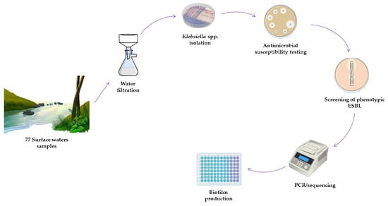
2. Materials and Methods

2.1. Geographical Location and Sample Collection

Seventy-seven locations across the Portuguese region of Trás-os-Montes and Alto Douro (Figure 1) were investigated. All rivers under consideration exclusively pertained to the Douro River Basin, an international hydrographic region spanning an aggregate area of approximately 97,000 km2, of which 18,643 km2 is situated within the confines of Portugal. This hydrological basin is demarcated by the eastern border with Spain and the western boundary adjacent to the Atlantic Ocean.
Samples from 18 rivers, 33 streams, 1 irrigation ditch, 1 dam, 12 fountains, 7 water wells, 2 water tanks, 1 water mine, and 2 springs were collected between October 2022 and April 2023 (Table S1). Water was sampled in 500 mL sterile plastic bottles containing sodium thiosulfate and, subsequently, preserved at 4–8 °C. The filtration of all samples occurred on the day of collection.

2.2. Bacterial Isolation

The water samples collected were filtered for Klebsiella spp. isolation. Approximately 100 mL of the water samples was filtered using a 0.45 µm cellulose nitrate pore membrane filter (Whatman, UK). Subsequently, the filters were immersed into tubes containing 5 mL of BHI (Brain Heart Infusion) broth and incubated at 37 °C for 24 h. After the incubation period, the samples were seeded onto Chromogenic Coliform Agar, supplemented and not supplemented with cefotaxime (CTX). The colonies that were pink in color were picked for the isolation of Klebsiella spp. and were subsequently sown on HiChrome Klebsiella Selective Agar Base. The plates were incubated for 24 h at 37 °C. All isolates that turned purple on HiChrome Klebsiella selective media were considered presumptive Klebsiella spp. The K. pneumoniae species identification of all isolates was carried out by polymerase chain reaction (PCR) to amplify the 428 base pairs (bps) of the khe gene using specific primers, as previously described [20].

2.3. Antimicrobial Susceptibility/Resistance Assessment

The Kirby–Bauer disk diffusion method was employed to evaluate antimicrobial susceptibility according to the EUCAST guidelines, with the exception of ceftazidime, tetracycline, and streptomycin, for which the CLSI guidelines were employed as standards. The following 11 antimicrobials (µg/disc) were used: amoxicillin + clavulanic acid (20 + 10), cefoxitin (30), ceftazidime (30), cefotaxime (30), meropenem (10), tetracycline (30), gentamicin (10), streptomycin (10), tobramycin (10), ciprofloxacin (5), and trimethoprim–sulfamethoxazole (1.25 + 23.75). Screening for phenotypic ESBL production was conducted through the double-disk synergy test utilizing cefotaxime, ceftazidime, and amoxicillin/clavulanic acid disks. Isolates showing resistance to three or more antibiotic classes were considered as multi-resistant (MDR).

2.4. Antimicrobial Resistance Genes and Virulence Factors

For DNA extraction, isolates were seeded on BHI agar and incubated at 37 °C for 18–24 h. After the incubation, genomic DNA from Klebsiella strains was extracted using the “Boiling Method” [21]. The extracted DNA was preserved at −20 °C until further analysis. All isolates were screened for the presence of antimicrobial resistance genes based on their phenotypic resistance profiles. The presence of antimicrobial-resistant genes encoding resistance to cefotaxime and β-lactams (blaCTX-U, blaCTX-M3, blaCTX-M9, blaSHV, blaTEM, ampC), tetracyclines (tetA, and tetB), gentamicin (aac(3)-II, aac(3)-IV, and ant(2)), streptomycin (strA, strB, aadA1, and aadA5), trimethoprim–sulfamethoxazole (sul1, sul2, and sul3), carbapenems (blaOXA, blaOXA-48, blaVIM, blaIMP, and blaKPC), ciprofloxacin (parC), and colistin (mcr-1) was investigated by PCR, as previously reported. The presence of virulence genes, including Pap pili (papC), pilus associated with pyelonephritis G allele III (papG-III), cytotoxic necrotizing factor 1 (cnf1), aerolysin gene (aer), and bundle-forming pili (bfp), was also tested using PCR. Positive and negative controls used in all experiments were derived from the strain collection of the University of Trás-Os-Montes and Alto Douro [22,23]. The specific primer sequences used in this study and the amplified product size are shown in Table S2.

2.5. Biofilm Production

Biofilm production was conducted using the microtiter assay, following a previously described protocol with certain modifications [24]. In short, each Klebsiella isolate was streaked on BHI agar plates and incubated at 37 °C for 24 h. After the incubation period, a few colonies were transferred to tubes containing 3 mL of Tryptic Soy Broth (TSB, Oxoid Ltd., Basingstoke, UK) and incubated at 37 °C for 16 ± 1 h, with continuous shaking at 120 rpm (ES-80 Shaker-incubator, Grant Instruments, Cambridge, UK). Then, the bacterial suspension was adjusted to an optical density of 1 × 10⁶ colony forming units, and 200 µL of bacterial suspension was added to each well of the 96-well flat-bottom microplate. To standardize the results, the biofilm formation of each isolate was given as percentage from the results obtained for the positive control strain, Klebsiella spp. ATCC® 13883. Isolates were characterized as strong, moderate, or weak biofilm producers when the percentages obtained were, respectively, >100%, 70–100%, or <70%. TSB without bacterial inoculum was used as a negative control. The plates were incubated at 37 °C for 24 h without shaking under aerobic conditions. All experiments were performed in duplicate and had 7 technical replicates.
To quantify biofilm biomass, the Crystal Violet (CV) staining method was used, following the procedure established by Peeters et al., with some adaptations [25]. After incubation, the medium was carefully removed from each well, and the plates were washed twice with distilled water to remove non-attached bacterial cells. The plates were allowed to dry at room temperature. To fix the biofilms, 100 µL of methanol (VWR International) was added to each well and incubated for 10 min. Methanol was then removed, the plates were air-dried at room temperature for 15 min, and 100 µL of CV at 1% (v/v) was added to each well for 15 min. Then, the CV was removed, and the plates were washed twice with distilled water to remove the excess dye. Next, 150 µL of acetic acid 33% (v/v) was added to solubilize the CV, and the absorbance was measured at 570 nm using a microplate reader BioTek ELx808U (BioTek, Winooski, VT, USA).

2.6. Statistical Analysis

Statistical analyses were performed using GraphPad Prism Version 8.0.2. (GraphPAD Software Inc., San Diego, CA, USA) to compare the biofilm formation capacity of surface waters and MDR isolates. Results were expressed as mean values and standard deviation. The level of significance was determined using the Student t-test. Moreover, a principal components analysis (PCA) was carried out using the JMP®, Version 17 (SAS Institute Inc., Cary, NC, USA, 1989–2023) between source and genotype and between source and virulence genes.

3. Results

3.1. Distribution of Klebsiella in Surface Waters

Bacterial growth was observed in nearly all analyzed water samples, although only 33 (70.2%) out of the 77 samples were positive for Klebsiella. The distribution of Klebsiella spp. and K. pneumoniae among the different sources is shown in Table 1. Klebsiella was isolated from 54 lotic (streams, rivers, fountains, irrigation ditches, and springs) and 23 lentic (dams, water wells, water tanks, and water mines) water samples. Klebsiella spp. was detected in 14 (18.1%) of the 77 water samples, whereas K. pneumoniae was found in 19 (24.7%) samples.

3.2. Antimicrobial Resistance Phenotype in Klebsiella spp. and K. pneumoniae Strains

Seven (36.8%) K. pneumoniae isolates were ESBL producers. Most K. pneumoniae isolates were resistant to amoxicillin-clavulanic acid (n = 11; 57.9%), trimethoprim–sulfamethoxazole (n = 10; 52.6%), cefoxitin (n = 7; 36.8%), cefotaxime (n = 8; 42.1%), and streptomycin (n = 8; 42.1%). Moreover, the MDR phenotype was observed in seven (36.8%) of the K. pneumoniae isolates. However, resistance to meropenem and tetracycline was detected in only one isolate. Regarding the 14 Klebsiella spp. isolates, 28.6% (n = 4) were ESBL producers. A high rate of antibiotic resistance was found in these isolates for amoxicillin + clavulanic acid (n = 4; 28.6%), trimethoprim/sulfamethoxazole (n = 4; 28.6%), and cefoxitin (n = 3; 21.4%). Nevertheless, only one (7.1%) Klebsiella spp. isolate was categorized as MDR. Table 2 details the antimicrobial resistance profiles of the isolates under study.

3.3. Characterization of Resistance Genes and Virulence Factors

Data on the resistance and virulence phenotypes and genetic determinants are given in Table 2. A high diversity of resistance genes was detected among the 19 K. pneumoniae isolates, namely, blaCTX-U (n = 11), blaCTX-M9 (n = 12), blaTEM (n = 7), aac(3)-II (n = 3), aac(3)-IV (n = 3), strA (n = 7), aadA1 (n = 5), aadA5 (n = 2), sul2 (n = 10), and parC (n = 1). The majority of Klebsiella spp. isolates with phenotypic resistance to β-lactams and cefotaxime (n = 5; 100%) harbored the blaCTX-M9 gene. Furthermore, the diversity of other resistance genes was detected among these isolates, namely, blaCTX-U (n = 6) and strA (n = 1). The most frequently found virulence gene in K. pneumoniae isolates was papC (n = 6; 30%), while in Klebsiella spp. Isolates, a high prevalence of papC (n = 3; 21.4%) and aer (n = 3; 21.4%) was observed. A low prevalence of papG-III and bfp virulence genes was detected in both K. pneumoniae and Klebsiella spp.

3.4. Biofilm Production

Biofilm production was measured by a microtiter plate assay for the Klebsiella spp. and K. pneumoniae isolates from surface waters. The results were standardized using Klebsiella spp. ATCC® 13883 (biofilm producer) to enhance the consistency of result comparisons. Figure 2 shows the percentage of biofilm production by each of the isolates in this study.
Isolates of Klebsiella spp. and K. pneumoniae produced a moderate amount of biofilm biomass, with very similar averages of biofilm formation percentages (83.1% and 86.9%, respectively). Among K. pneumoniae strains, 13 (65%) were confirmed as weak producers, 2 (10%) were moderate, and 5 (25%) were strong biofilm producers. Similarly, regarding Klebsiella spp., five isolates (35.7%) were weak producers, eight (57.1%) were moderate, and the remaining isolate (7.1%) was a strong biofilm producer. The weakest biofilm producer (60.2%) was one K. pneumoniae isolate from the river Rio Corgo (Vila Real), while the strongest biofilm producer (118.6%) was isolated from a stream located in Valpaços. For a more complete analysis of the prevalence of biofilm-producing species, we compared the formation of biofilms in multi-resistant and non-multi-resistant species. Among MDR isolates, one (14.3%) was a strong biofilm producer and six (85.7%) were weak biofilm producers. Additionally, of the 26 non-MDR Klebsiella isolates, 5 (19.2%) had a high capacity to form biofilms, 9 (34.6%) were moderate biofilm producers, and 12 (46.1%) were weak biofilm producers. Comparing the results obtained in the MDR and non-MDR isolates under study, the difference between the means was not statistically significant, since the isolates produced approximately the same amount of biofilm (Figure 3). In addition, among 11 ESBL-producing Klebsiella strains, all were confirmed as biofilm producers; 8 isolates (72.7%) were weak producers, 2 (18.2%) were moderate, and 1 (9.1%) was a high biofilm producer. On the other hand, of the 21 non-ESBL-producing Klebsiella strains, 9 isolates (42.9%) were weak producers, 7 (33.3%) were moderate biofilm producers, and the remaining 5 (23.8%) were high biofilm producers.

3.5. Principal Component Analysis (PCA)

Regarding the relationship between source and genotypes, the first two main components do not explain most of the variability in the data (component 1—9.34%; component 2—10.2%). On the other hand, regarding the relationship between source and virulence, the first two main components explain around 37% of the total variability (component 1—19%; component 2—18%) (Figure 4).

4. Discussion

The environment, particularly the aquatic environment, is recognized as a reservoir for antimicrobial resistance (AMR) and antimicrobial resistance genes (ARGs), even within highly confined habitats, like drinking water sources. Surface waters constitute one of the main sources of potable water for human and animal consumption. Therefore, when contaminated, they can become an important contributor to the dissemination of antimicrobial-resistant Klebsiella and its resistance and virulence factors [7]. However, data on the prevalence of antimicrobial resistance in Klebsiella from surface waters are scant. Therefore, studies on Klebsiella’s prevalence, antibiotic resistance, and virulence are of the utmost importance for evaluating the potential roles of different aquatic environments in the spread of these pathogens, as well as their impact on human and animal exposure to these pathogens.
Klebsiella spp. Is a frequent member of the environmental microbiota, including that of surface water bodies. The presence of Klebsiella spp. in freshwater systems, such as drinking water, rivers, lakes, and streams, as well as in seawater, has been demonstrated in several studies [26,27,28,29,30]. In this study, a total of 77 surface water samples were collected, and 33 Klebsiella strains were isolated (42.9%). When comparing different surface water sources, it became evident that rivers exhibited a higher prevalence of both Klebsiella spp. and K. pneumoniae. Specifically, among the 18 isolates from rivers, 33.3% were identified as Klebsiella spp., while 50% were characterized as K. pneumoniae. These findings are in line with several other studies, in which a higher prevalence of both Klebsiella spp. and K. pneumoniae in rivers was noted, with K. pneumoniae being the most prevalent [29,31].
K. pneumoniae shows resistance to a wide array of antibiotics as well as the production of β-Lactamase enzymes and the capacity to form biofilms [32]. Given that many β-Lactamase genes are plasmid-borne, or located in other mobile genetic elements, resistant strains can spread fast, leading to increased illness, death rates, and healthcare expenses. In this study, most isolates were resistant to the tested antibiotics, including amoxicillin-clavulanic acid, cefoxitin, cefotaxime, ceftazidime, streptomycin, and trimethoprim–sulfamethoxazole, belonging, respectively, to the penicillin, cephalosporin, aminoglycoside, and sulfonamide antibiotic classes. However, among these isolates, meropenem (carbapenems) was the antibiotic with the lowest resistance rate, followed by tetracycline (tetracyclines) and gentamicin (aminoglycosides). The penicillin antibiotic class exhibited the highest prevalence of resistance (44.1%) among the studied surface water isolates. Notably, isolates of Klebsiella spp. displayed lower resistance rates to ciprofloxacin (fluoroquinolones) and meropenem (carbapenems), with no instances of resistance observed. However, antibiotics with higher resistance rates were consistent across both taxonomic groups. Numerous reports on penicillin resistance in Klebsiella species from surface waters in Portugal [33] and elsewhere [30,34] align with the elevated resistance rates found in this study. Of particular concern is the alignment of these results regarding ceftazidime resistance and those of Teixeira et al. (2020), who also reported a high number of ceftazidime-resistant Klebsiella spp. isolates from a Portuguese river [33]. These results raise concerns, both from the environmental and public health perspectives. Ceftazidime is a third-generation cephalosporin that should be more effective against Gram-negative bacteria than both the first and second generations of antibiotics in this family. Third-generation cephalosporins are also more active against bacteria that may be resistant to previous generations of cephalosporins. These are important antibiotics, mostly used in hospital settings to treat severe infections involving multidrug-resistant Gram-negative pathogens, such as Klebsiella spp. and K. pneumoniae [35]. Moreover, third-generation oral cephalosporins, such as ceftazidime, can be combined with amoxicillin × clavulanic acid to tackle urinary tract infections involving ESBL-producing Klebsiella spp. [36], further enhancing the concerns these findings raise. Regarding the prevalence of Klebsiella spp. with the MDR phenotype, these findings differ from the results of other studies carried out in surface waters, in which high prevalences of Klebsiella spp. with the MDR phenotype were reported [37,38]. On the basis of antibiotic susceptibility results, it is evident that Klebsiella spp. and K. pneumoniae present a significant therapeutic challenge. The slightly higher resistance rates observed in the environment raise concerns, especially regarding the potential spread of multidrug-resistant bacteria responsible for infections like pneumonia and meningitis to both humans and animals. Notably, the β-Lactam and carbapenem classes of antibiotics have garnered increased interest and concern due to the observed high rates of resistance in isolated Klebsiella spp. [31], which this study also shows.
Less than 50% of the isolates were ESBL producers. These results agree with the findings of Caltagirone et al., in which only 7/33 (21.2%) K. pneumoniae isolated from surface waters were ESBL producers [30]. However, Falgenhaeur et al. reported a higher prevalence (83.3%) of ESBL-producing Klebsiella spp. in surface waters, contrary to what was found in this study [39]. Several reports on the prevalence of ESBLs in surface waters report that they are frequently associated with MDR [40]; however, in this study, there was a higher prevalence of ESBL-producing Klebsiella in non-MDR isolates (n = 8; 24.2%) than in MDR isolates (n = 3; 9.1%). Furthermore, the most common resistance gene, both in K. pneumoniae and Klebsiella spp., was blaCTX-M9, followed by sul2. However, in K. pneumoniae, a high prevalence of blaTEM, blaSHV, ampC, and strA was also observed. These findings diverge from those of several studies, where a high prevalence of blaCTX-U, blaKPC, tet(A), and sul2 genes was reported. In this study, the most prevalent gene was blaCTX-M9, which was not reported in other studies from surface waters. However, the prevalence of sul2 in those studies aligns with the prevalences reported in this study [33,41,42]. In studies carried out by Caltagirone et al., Muller et al., Teixeira et al., and Hoffman et al., in Italy, Germany, and Portugal, respectively, a high prevalence of blaKPC and blaOXA-48 genes was detected [30,33,43,44]. However, in this study, none of the isolates from surface waters possessed these genes.
As previously mentioned, papC—an operon that is an outer membrane protein, essential for the regulation of P fimbriae biogenesis—was the most prevalent virulence gene in both Klebsiella spp. and K. pneumoniae. This result may be related to the virulence of the isolated strains, since adhesion is the most important determinant of pathogenicity in the Klebsiella genus [45]. These results are quite concerning, since this genetic virulence determinant may increase the pathogenicity in Klebsiella strains, and we demonstrated its high prevalence in the environment. To our knowledge, the presence of these virulence genes has not been previously reported in environmental samples, including surface waters. This study represents a pioneering effort in this regard, and it highlights the importance of water as a potential reservoir of virulence genes.
The high environmental prevalence of papC we found makes it important to check whether a high prevalence of this gene has also been reported in clinical samples. The results obtained in studies on hospital patients, in which the presence of these genes was investigated in isolates of Klebsiella spp. of diverse clinical samples, were divergent from those in the present study. In the reports of Liu et al. and Düzgün et al., papC was the gene with the lowest prevalence in Klebsiella spp. isolates [46,47]. Nevertheless, they showed a high prevalence of aer, which aligns with what was observed in the Klebsiella spp. isolates from surface waters in this study. Moreover, in the aforementioned studies, the bfp and papG-III genes demonstrated the lowest prevalence among hospital patients, mirroring these results. Contrastingly, Hassan et al. described a notable prevalence of the papC gene in K. pneumoniae isolates obtained from clinical infection specimens [48], a finding that concurs with the outcomes observed in this study. The alignment of these results on the prevalence of aer and, in some cases, papC may suggest a plausible transfer of these genes between environmental sources, such as water bodies and human hosts.
Biofilm formation represents a crucial virulence trait in Klebsiella and serves as an adaptative response to diverse stressors, such as alterations in the physical environment and exposure to drugs (particularly antibiotics) [49]. In addition, bacteria present in surface waters can also produce biofilms, promoting an ideal environment for horizontal gene transfer that can lead to the accumulation of genetic mobile elements. The accumulation of biofilm-producing bacteria in ecosystems may pose environmental and public health concerns [50]. Changes in water levels have been demonstrated as among the most relevant stressors affecting the structure and function of biofilms [51]. As biofilms play a crucial role in aquatic environments, studying the prevalence of biofilm production isolates is essential for preventing the spread of pathogens in the environment and ensuring the biosafety of drinking water. To the best of our knowledge, no studies have yet been conducted on environmental samples (including surface waters) on the biofilm production ability of Klebsiella strains. In this study, all Klebsiella isolates were confirmed as biofilm producers, similar to the very high (99%) rates of biofilm production reported by Türkel et al. in clinical samples. In addition, in the same study, a higher prevalence of strong biofilm producers was found in ciprofloxacin-susceptible isolates [52], similar to these results, in which 6/30 (20%) of the ciprofloxacin-susceptible Klebsiella had a high capacity to produce biofilms. Nevertheless, in contrast with these results, other studies have reported a correlation between multidrug resistance (MDR) and biofilm production, indicating a higher prevalence of high biofilm producers among MDR isolates [53], a concerning trend when the results on water bodies are considered.
In this study, considerable rates of weak biofilm-producing isolates were found, both among ESBL-producing (72.7%) and non-ESBL-producing Klebsiella isolates (43.5%). While the majority of studies suggest that β-Lactamase-producing strains are usually high biofilm producers [54], others have observed no correlation between the capacity to form biofilms and ESBL production [55].
PCA showed that the environmental variables (type of water body) under study only account for a low extent of the observed gene distribution patterns. However, in spite of the low eigenvalues observed (Table S3), the presence of sul2 correlated positively with “fountain” and “spring” and negatively with “water well”, while blaCTX-U and blaCTX-M9 correlated positively with “water well” but negatively with “fountain” and “spring”. Regarding virulence determinants, papC correlated positively with “water well” and “stream” but negatively with “dam”. On the other hand, papC and bfp correlated positively with “dam” but negatively with “water well” and “stream”. Furthermore, aer correlated positively with “fountain” but negatively with “water well” and “stream”. Moreover, it was observed that some virulence genes exhibited positive and negative associations with lotic and lentic waters. This was the case with papC, which exhibited positive associations with “streams” and negative associations with “fountains”, both lotic waters. Additionally, aer presented positive associations with “fountains” and negative associations with “streams”, both lotic waters. Furthermore, the papC and bfp genes also demonstrated positive associations with “dam” and negative associations with “water well”, both lentic waters.
The distinctive patterns of antibiotic resistance, coupled with the incidence of virulence factors and biofilm-forming capacity found in this study, may raise concerns about the dissemination potential of pathogenic Klebsiella strains through surface waters. A further cause for concern is that individuals, both animal and human, engaging in recreational activities or using these waters for potable purposes, may belong to risk groups with increased susceptibility to Klebsiella infections or colonization. However, it must be taken into account that the available information on the connection between Klebsiella-contaminated waters and the onset of infections in humans is still scarce.
In the present context of the critical importance of surface freshwater, these findings underscore the urgent need for integrated research on freshwater quality. The unexpectedly high prevalence of Klebsiella spp. and K. pneumoniae in surface waters, coupled with the concerning presence of resistance and virulence genes, highlights the complex interplay between microbial dynamics and environmental factors. To address such challenges, the One Health perspective is paramount, emphasizing the interconnectedness of human, animal, and environmental health. This study supports the urgency for collaborative, multidisciplinary solutions to tackle freshwater monitoring, especially in Southern Europe, where severe water quality issues are expected to increase under extreme precipitation and droughts derived mainly from climate change. Comprehensive research on freshwater ecology, toxicity, hydrochemistry, and monitoring approaches is essential.

5. Conclusions

This work demonstrates that surface waters may act as reservoirs of Klebsiella, with higher prevalences of K. pneumoniae in river samples than in the other types of surface waters tested. In terms of phenotypic resistance to a wide range of antibiotic classes, ceftazidime (a third-generation cephalosporin), the high prevalence of blaCTX-M9, and biofilm production are causes for concern. This study constitutes, to our knowledge, the first report on the presence of the papC virulence determinant in Klebsiella isolates from surface waters. When compared to the findings of studies on hospital isolates, these results may suggest a plausible transfer of genetic determinants of antibiotic resistance (particularly aer but, also, in some cases, papC) between surface waters and the human host. Thus, they highlight both the need for including these sources of pathogens under the One Health effort and the need for vigilance of Klebsiella in lentic and lotic water bodies to assess their distribution and dissemination in these habitats.

Supplementary Materials

The following supporting information can be downloaded at: https://www.mdpi.com/article/10.3390/w16091297/s1, Table S1: Location, source, and coordinates of the surface water samples in this study, and the identity of the respective isolates; Table S2: Primer pairs used for molecular typing, detection of antimicrobial resistance and virulence genes in Klebsiella strains; Table S3: Principal component analysis. (I) Source versus genotype, Eigenvalues for surface water PCA; (II) source versus virulence, Eigenvalues for surface water PCA. References [56,57,58,59,60,61,62,63,64,65,66,67,68,69] are cited in the Supplementary Materials.

Author Contributions

Conceptualization, S.A.; methodology, S.A., V.S., G.I. and P.P.; software, V.S. and Â.M.; validation, V.S., M.d.L.E.D., J.E.P. and P.P.; formal analysis, S.A., V.S. and Â.M.; investigation, S.A.; resources, S.A., V.S. and J.E.P.; data curation, V.S.; writing—original draft preparation, S.A.; writing—review and editing, S.A., V.S. and M.d.L.E.D.; visualization, S.A., V.S., M.d.L.E.D. and P.P; supervision, V.S., G.I. and P.P. All authors have read and agreed to the published version of the manuscript.

Funding

This work was supported by the project UIDB/00772/2020 (Doi:10.54499/UIDB/00772/2020) funded by the Portuguese Foundation for Science and Technology (FCT). This work was supported by the Associate Laboratory for Green Chemistry-LAQV, which is financed by national funds from FCT/MCTES (UIDB/50006/2020, UIDP/50006/2020). MD acknowledges the funding from the Regional Government of the Azores (Portugal), reference M1.1.A/FUNC.UI&D/001/2021-2024, measure 01.1.a.Apoio UI&D.2022 (SRCCTD/DRCTD).

Data Availability Statement

All the data supporting our findings are contained within the article and Supplementary Materials.

Conflicts of Interest

The authors declare no conflicts of interest.

References

  1. Antimicrobial Resistance: Tackling a Crisis for the Health and Wealth of Nations. Available online: https://iiif.wellcomecollection.org/file/b28552179_AMR%20Tackling%20a%20crisis%20for%20the%20health%20and%20wealth%20of%20nations.pdf (accessed on 19 February 2024).
  2. Varaldo, P.E.; Facinelli, B.; Bagnarelli, P.; Menzo, S.; Mingoia, M.; Brenciani, A.; Giacometti, A.; Barchiesi, F.; Brescini, L.; Cirioni, O.; et al. Antimicrobial Resistance: A Challenge for the Future. In The First Outstanding 50 Years of “Universita Politecnica delle Marche”: Research Achievements in Life Sciences; Springer: Cham, Switzerland, 2020; pp. 13–29. [Google Scholar] [CrossRef]
  3. Collignon, P.J.; McEwen, S.A. One Health—Its Importance in Helping to Better Control Antimicrobial Resistance. Trop. Med. Infect. Dis. 2019, 4, 22. [Google Scholar] [CrossRef] [PubMed]
  4. Haenni, M.; Dagot, C.; Chesneau, O.; Bibbal, D.; Labanowski, J.; Vialette, M.; Bouchard, D.; Martin-Laurent, F.; Calsat, L.; Nazaret, S.; et al. Environmental Contamination in a High-Income Country (France) by Antibiotics, Antibiotic-Resistant Bacteria, and Antibiotic Resistance Genes: Status and Possible Causes. Environ. Int. 2022, 159, 107047. [Google Scholar] [CrossRef] [PubMed]
  5. Berendonk, T.U.; Manaia, C.M.; Merlin, C.; Fatta-Kassinos, D.; Cytryn, E.; Walsh, F.; Bürgmann, H.; Sørum, H.; Norström, M.; Pons, M.N.; et al. Tackling Antibiotic Resistance: The Environmental Framework. Nat. Rev. Microbiol. 2015, 13, 310–317. [Google Scholar] [CrossRef] [PubMed]
  6. Kumar, A.; Pal, D. Antibiotic Resistance and Wastewater: Correlation, Impact and Critical Human Health Challenges. J. Environ. Chem. Eng. 2018, 6, 52–58. [Google Scholar] [CrossRef]
  7. Silva, V.; Caniça, M.; Capelo, J.L.; Igrejas, G.; Poeta, P. Diversity and Genetic Lineages of Environmental Staphylococci: A Surface Water Overview. FEMS Microbiol. Ecol. 2020, 96, fiaa191. [Google Scholar] [CrossRef] [PubMed]
  8. Zeng, Y.; Chang, F.; Liu, Q.; Duan, L.; Li, D.; Zhang, H. Review Article Recent Advances and Perspectives on the Sources and Detection of Antibiotics in Aquatic Environments. J. Anal. Methods Chem. 2022, 2022, 5091181. [Google Scholar] [CrossRef] [PubMed]
  9. Barati, A.; Ghaderpour, A.; Chew, L.L.; Bong, C.W.; Thong, K.L.; Chong, V.C.; Chai, L.C. Isolation and Characterization of Aquatic-Borne Klebsiella pneumoniae from Tropical Estuaries in Malaysia. Int. J. Environ. Res. Public Health 2016, 13, 426. [Google Scholar] [CrossRef] [PubMed]
  10. Hu, Y.; Anes, J.; Devineau, S.; Fanning, S. Klebsiella pneumoniae: Prevalence, Reservoirs, Antimicrobial Resistance, Pathogenicity, and Infection: A Hitherto Unrecognized Zoonotic Bacterium. Foodborne Pathog. Dis. 2021, 18, 63–84. [Google Scholar] [CrossRef] [PubMed]
  11. Kowalczyk, J.; Czokajło, I.; Gańko, M.; Śmiałek, M.; Koncicki, A. Identification and Antimicrobial Resistance in Klebsiella spp. Isolates from Turkeys in Poland between 2019 and 2022. Animals 2022, 12, 3157. [Google Scholar] [CrossRef]
  12. Gómez, M.; Valverde, A.; Del Campo, R.; Rodríguez, J.M.; Maldonado-Barragán, A. Phenotypic and Molecular Characterization of Commensal, Community-Acquired and Nosocomial Klebsiella spp. Microorganisms 2021, 9, 2344. [Google Scholar] [CrossRef] [PubMed]
  13. Rajkumari, J.; Paikhomba Singha, L.; Pandey, P. Genomic Insights of Aromatic Hydrocarbon Degrading Klebsiella pneumoniae AWD5 with Plant Growth Promoting Attributes: A Paradigm of Soil Isolate with Elements of Biodegradation. 3 Biotech 2018, 8, 118. [Google Scholar] [CrossRef] [PubMed]
  14. Kimera, Z.I.; Mgaya, F.X.; Mshana, S.E.; Karimuribo, E.D.; Matee, M.I.N. Occurrence of Extended Spectrum Beta Lactamase (ESBL) Producers, Quinolone and Carbapenem Resistant Enterobacteriaceae Isolated from Environmental Samples along Msimbazi River Basin Ecosystem in Tanzania. Int. J. Environ. Res. Public Health 2021, 18, 8264. [Google Scholar] [CrossRef] [PubMed]
  15. Guan, X.; He, R.; Zhang, B.; Gao, C.; Liu, F. Seasonal Variations of Microbial Community Structure, Assembly Processes, and Influencing Factors in Karst River. Front. Microbiol. 2023, 14, 1133938. [Google Scholar] [CrossRef]
  16. Beal, C.; Matsubae, K.; Ylä-Mella, J.; Elagroudy, S.; Campanale, C.; Losacco, D.; Triozzi, M.; Massarelli, C.; Uricchio, V.F. An Overall Perspective for the Study of Emerging Contaminants in Karst Aquifers. Resources 2022, 11, 105. [Google Scholar] [CrossRef]
  17. Cuadrado-Quesada, G.; Holley, C.; Gupta, J. Groundwater Governance in the Anthropocene: A Close Look at Costa Rica. Water Policy 2018, 20, 475–489. [Google Scholar] [CrossRef]
  18. Arroita, M.; Flores, L.; Larrañaga, A.; Chauvet, E.; Elosegi, A. Hydrological Contingency: Drying History Affects Aquatic Microbial Decomposition. Aquat. Sci. 2018, 80, 31. [Google Scholar] [CrossRef]
  19. Giri, S.; Mishra, A.; Zhang, Z.; Lathrop, R.G.; Alnahit, A.O. Meteorological and Hydrological Drought Analysis and Its Impact on Water Quality and Stream Integrity. Sustainability 2021, 13, 8175. [Google Scholar] [CrossRef]
  20. He, Y.; Guo, X.; Xiang, S.; Li, J.; Li, X.; Xiang, H.; He, J.; Chen, D.; Chen, J. Comparative Analyses of Phenotypic Methods and 16S RRNA, Khe, RpoB Genes Sequencing for Identification of Clinical Isolates of Klebsiella pneumoniae. Antonie Van Leeuwenhoek Int. J. Gen. Mol. Microbiol. 2016, 109, 1029–1040. [Google Scholar] [CrossRef] [PubMed]
  21. Solberg, O.D.; Ajiboye, R.M.; Riley, L.W. Origin of Class 1 and 2 Integrons and Gene Cassettes in a Population-Based Sample of Uropathogenic Escherichia coli. J. Clin. Microbiol. 2006, 44, 1347–1351. [Google Scholar] [CrossRef] [PubMed]
  22. Silva, A.; Silva, V.; De Lurdes, M.; Dapkevicius, E.; Azevedo, M.; Cordeiro, R.; Pereira, J.E.; Valentão, P.; Falco, V.; Igrejas, G.; et al. Unveiling Antibiotic Resistance, Clonal Diversity, and Biofilm Formation in E. Coli Isolated from Healthy Swine in Portugal. Pathogens 2024, 13, 305. [Google Scholar] [CrossRef] [PubMed]
  23. Carvalho, I.; Cunha, R.; Martins, C.; Martínez-Álvarez, S.; Chenouf, N.S.; Pimenta, P.; Pereira, A.R.; Ramos, S.; Sadi, M.; Martins, Â.; et al. Antimicrobial Resistance Genes and Diversity of Clones among Faecal ESBL-Producing Escherichia coli Isolated from Healthy and Sick Dogs Living in Portugal. Antibiotics 2021, 10, 1013. [Google Scholar] [CrossRef]
  24. Oniciuc, E.A.; Cerca, N.; Nicolau, A.I. Compositional Analysis of Biofilms Formed by Staphylococcus Aureus Isolated from Food Sources. Front. Microbiol. 2016, 7, 184310. [Google Scholar] [CrossRef]
  25. Peeters, E.; Nelis, H.J.; Coenye, T. Comparison of Multiple Methods for Quantification of Microbial Biofilms Grown in Microtiter Plates. J. Microbiol. Methods 2008, 72, 157–165. [Google Scholar] [CrossRef] [PubMed]
  26. Hartinger, S.M.; Medina-Pizzali, M.L.; Salmon-Mulanovich, G.; Larson, A.J.; Pinedo-Bardales, M.; Verastegui, H.; Riberos, M.; Mäusezahl, D. Antimicrobial Resistance in Humans, Animals, Water and Household Environs in Rural Andean Peru: Exploring Dissemination Pathways through the One Health Lens. Int. J. Environ. Res. Public Health 2021, 18, 4604. [Google Scholar] [CrossRef] [PubMed]
  27. Mondal, A.H.; Siddiqui, M.T.; Sultan, I.; Haq, Q.M.R. Prevalence and Diversity of BlaTEM, BlaSHV and BlaCTX-M Variants among Multidrug Resistant Klebsiella spp. from an Urban Riverine Environment in India. Int. J. Environ. Health Res. 2018, 29, 117–129. [Google Scholar] [CrossRef] [PubMed]
  28. Altayb, H.N.; Elbadawi, H.S.; Alzahrani, F.A.; Baothman, O.; Kazmi, I.; Nadeem, M.S.; Hosawi, S.; Chaieb, K. Co-Occurrence of β-Lactam and Aminoglycoside Resistance Determinants among Clinical and Environmental Isolates of Klebsiella pneumoniae and Escherichia coli: A Genomic Approach. Pharmaceuticals 2022, 15, 1011. [Google Scholar] [CrossRef] [PubMed]
  29. Hooban, B.; Fitzhenry, K.; Cahill, N.; Joyce, A.; O’ Connor, L.; Bray, J.E.; Brisse, S.; Passet, V.; Abbas Syed, R.; Cormican, M.; et al. A Point Prevalence Survey of Antibiotic Resistance in the Irish Environment, 2018–2019. Environ. Int. 2021, 152, 106466. [Google Scholar] [CrossRef] [PubMed]
  30. Caltagirone, M.; Nucleo, E.; Spalla, M.; Zara, F.; Novazzi, F.; Marchetti, V.M.; Piazza, A.; Bitar, I.; De Cicco, M.; Paolucci, S.; et al. Occurrence of Extended Spectrum β-Lactamases, KPC-Type, and MCR-1.2-Producing Enterobacteriaceae from Wells, River Water, and Wastewater Treatment Plants in Oltrepò Pavese Area, Northern Italy. Front. Microbiol. 2017, 8, 272364. [Google Scholar] [CrossRef]
  31. Hassen, B.; Abbassi, M.S.; Benlabidi, S.; Ruiz-Ripa, L.; Mama, O.M.; Ibrahim, C.; Hassen, A.; Hammami, S.; Torres, C. Genetic Characterization of ESBL-Producing Escherichia coli and Klebsiella pneumoniae Isolated from Wastewater and River Water in Tunisia: Predominance of CTX-M-15 and High Genetic Diversity. Environ. Sci. Pollut. Res. 2020, 27, 44368–44377. [Google Scholar] [CrossRef]
  32. Nirwati, H.; Sinanjung, K.; Fahrunissa, F.; Wijaya, F.; Napitupulu, S.; Hati, V.P.; Hakim, M.S.; Meliala, A.; Aman, A.T.; Nuryastuti, T. Biofilm Formation and Antibiotic Resistance of Klebsiella pneumoniae Isolated from Clinical Samples in a Tertiary Care Hospital, Klaten, Indonesia. BMC Proc. 2019, 13, 20. [Google Scholar] [CrossRef]
  33. Teixeira, P.; Tacão, M.; Pureza, L.; Gonçalves, J.; Silva, A.; Cruz-Schneider, M.P.; Henriques, I. Occurrence of Carbapenemase-Producing Enterobacteriaceae in a Portuguese River: BlaNDM, BlaKPC and BlaGES among the Detected Genes. Environ. Pollut. 2020, 260, 113913. [Google Scholar] [CrossRef] [PubMed]
  34. Mahato, S.; Mahato, A.; Pokharel, E.; Tamrakar, A. Detection of Extended-Spectrum Beta-Lactamase-Producing E. coli and Klebsiella spp. in Effluents of Different Hospitals Sewage in Biratnagar, Nepal. BMC Res. Notes 2019, 12, 641. [Google Scholar] [CrossRef] [PubMed]
  35. van Duin, D.; Bonomo, R.A. Ceftazidime/Avibactam and Ceftolozane/Tazobactam: Second-Generation β-Lactam/β-Lactamase Inhibitor Combinations. Clin. Infect. Dis. 2016, 63, 234–241. [Google Scholar] [CrossRef]
  36. Paterson, D.L. Recommendation for Treatment of Severe Infections Caused by Enterobacteriaceae Producing Extended-Spectrum β-Lactamases (ESBLs). Clin. Microbiol. Infect. 2000, 6, 460–463. [Google Scholar] [CrossRef] [PubMed]
  37. Khan, F.A.; Hellmark, B.; Ehricht, R.; Söderquist, B.; Jass, J. Related Carbapenemase-Producing Klebsiella Isolates Detected in Both a Hospital and Associated Aquatic Environment in Sweden. Eur. J. Clin. Microbiol. Infect. Dis. 2018, 37, 2241–2251. [Google Scholar] [CrossRef] [PubMed]
  38. Shrestha, P.; Prasai Joshi, T.; Nhemhaphuki, S.; Sitoula, K.; Maharjan, J.; Ranjit, R.; Shrestha, P.; Joshi, D.R. Occurrence of Antibiotic-Resistant Bacteria and Their Genes in Bagmati River, Nepal. Water Air Soil Pollut. 2023, 234, 475. [Google Scholar] [CrossRef]
  39. Falgenhauer, L.; Schwengers, O.; Schmiedel, J.; Baars, C.; Lambrecht, O.; Heß, S.; Berendonk, T.U.; Falgenhauer, J.; Chakraborty, T.; Imirzalioglu, C. Multidrug-Resistant and Clinically Relevant Gram-Negative Bacteria Are Present in German Surface Waters. Front. Microbiol. 2019, 10, 482269. [Google Scholar] [CrossRef]
  40. Aguilar-Salazar, A.; Martínez-Vázquez, A.V.; Aguilera-Arreola, G.; de Jesus de Luna-Santillana, E.; Cruz-Hernández, M.A.; Escobedo-Bonilla, C.M.; Lara-Ramírez, E.; Sánchez-Sánchez, M.; Guerrero, A.; Rivera, G.; et al. Prevalence of ESKAPE Bacteria in Surface Water and Wastewater Sources: Multidrug Resistance and Molecular Characterization, an Updated Review. Water 2023, 15, 3200. [Google Scholar] [CrossRef]
  41. Fadare, F.T.; Okoh, A.I. Distribution and Molecular Characterization of ESBL, PAmpC β-Lactamases, and Non-β-Lactam Encoding Genes in Enterobacteriaceae Isolated from Hospital Wastewater in Eastern Cape Province, South Africa. PLoS ONE 2021, 16, e0254753. [Google Scholar] [CrossRef]
  42. Nascimento, T.; Cantamessa, R.; Melo, L.; Lincopan, N.; Fernandes, M.R.; Cerdeira, L.; Fraga, E.; Dropa, M.; Sato, M.I.Z. International High-Risk Clones of Klebsiella pneumoniae KPC-2/CC258 and Escherichia coli CTX-M-15/CC10 in Urban Lake Waters. Sci. Total Environ. 2017, 598, 910–915. [Google Scholar] [CrossRef] [PubMed]
  43. Hoffmann, M.; Fischer, M.A.; Neumann, B.; Kiesewetter, K.; Hoffmann, I.; Werner, G.; Pfeifer, Y.; Lübbert, C. Carbapenemase-Producing Gram-Negative Bacteria in Hospital Wastewater, Wastewater Treatment Plants and Surface Waters in a Metropolitan Area in Germany, 2020. Sci. Total. Environ. 2023, 890, 164179. [Google Scholar] [CrossRef] [PubMed]
  44. Müller, H.; Sib, E.; Gajdiss, M.; Klanke, U.; Lenz-Plet, F.; Barabasch, V.; Albert, C.; Schallenberg, A.; Timm, C.; Zacharias, N.; et al. Dissemination of Multi-Resistant Gram-Negative Bacteria into German Wastewater and Surface Waters. FEMS Microbiol. Ecol. 2018, 94, 5. [Google Scholar] [CrossRef] [PubMed]
  45. López-Banda, D.A.; Carrillo-Casas, E.M.; Leyva-Leyva, M.; Orozco-Hoyuela, G.; Manjarrez-Hernández, Á.H.; Arroyo-Escalante, S.; Moncada-Barrón, D.; Villanueva-Recillas, S.; Xicohtencatl-Cortes, J.; Hernández-Castro, R. Identification of Virulence Factors Genes in Escherichia coli Isolates from Women with Urinary Tract Infection in Mexico. Biomed. Res. Int. 2014, 2014, 959206. [Google Scholar] [CrossRef] [PubMed]
  46. Liu, J.; Xu, Z.; Li, H.; Chen, F.; Han, K.; Hu, X.; Fang, Y.; Chen, D. Metagenomic Approaches Reveal Strain Profiling and Genotyping of Klebsiella Pneumoniae from Hospitalized Patients in China. Microbiol. Spectr. 2022, 10, e0219021. [Google Scholar] [CrossRef]
  47. Özad Düzgün, A.; Yüksel, G. Detection of Virulence Factor Genes, Antibiotic Resistance Genes and Biofilm Formation in Clinical Gram-Negative Bacteria and First Report from Türkiye of K. oxytoca Carrying Both blaOXA-23 and blaOXA-51 Genes. Biologia 2023, 78, 2245–2251. [Google Scholar] [CrossRef]
  48. Hassan, R.; El Naggar, W.; El Sawy, E.; El Mahdy, A. Characterization of Some Virulence Factors Associated with Enterbacteriaceae Isolated from Urinary Tract Infections in Mansoura Hospitals. Egypt. J. Med. Microbiol. 2011, 20, 9–18. [Google Scholar]
  49. Li, Y.; Ni, M. Regulation of Biofilm Formation in Klebsiella pneumoniae. Front. Microbiol. 2023, 14, 1238482. [Google Scholar] [CrossRef] [PubMed]
  50. Silva, V.; Pereira, J.E.; Maltez, L.; Poeta, P.; Igrejas, G. Influence of Environmental Factors on Biofilm Formation of Staphylococci Isolated from Wastewater and Surface Water. Pathogens 2022, 11, 1069. [Google Scholar] [CrossRef]
  51. Romero, F.; Acuña, V.; Font, C.; Freixa, A.; Sabater, S. Effects of Multiple Stressors on River Biofilms Depend on the Time Scale. Sci. Rep. 2019, 9, 15810. [Google Scholar] [CrossRef]
  52. Türkel, İ.; Yıldırım, T.; Yazgan, B.; Bilgin, M.; Başbulut, E. Relationship between Antibiotic Resistance, Efflux Pumps, and Biofilm Formation in Extended-Spectrum β-Lactamase Producing Klebsiella pneumoniae. J. Chemother. 2018, 30, 354–363. [Google Scholar] [CrossRef] [PubMed]
  53. Shadkam, S.; Goli, H.R.; Mirzaei, B.; Gholami, M.; Ahanjan, M. Correlation between Antimicrobial Resistance and Biofilm Formation Capability among Klebsiella pneumoniae Strains Isolated from Hospitalized Patients in Iran. Ann. Clin. Microbiol. Antimicrob. 2021, 20, 13. [Google Scholar] [CrossRef]
  54. Sanchez, C.J.; Mende, K.; Beckius, M.L.; Akers, K.S.; Romano, D.R.; Wenke, J.C.; Murray, C.K. Biofilm Formation by Clinical Isolates and the Implications in Chronic Infections. BMC Infect. Dis. 2013, 13, 47. [Google Scholar] [CrossRef]
  55. Hasan, M.E.; Shahriar, A.; Shams, F.; Nath, A.K.; Emran, T. Bin Correlation between Biofilm Formation and Antimicrobial Susceptibility Pattern toward Extended Spectrum β-Lactamase (ESBL)- and Non-ESBL-Producing Uropathogenic Bacteria. J. Basic Clin. Physiol. Pharmacol. 2021, 32, 20190296. [Google Scholar] [CrossRef]
  56. Zong, Z.; Partridge, S.R.; Thomas, L.; Iredell, J.R. Dominance of BlaCTX-M within an Australian Extended-Spectrum β-Lactamase Gene Pool. Antimicrob. Agents Chemother. 2008, 52, 4198–4202. [Google Scholar] [CrossRef] [PubMed]
  57. Maynard, C.; Bekal, S.; Sanschagrin, F.; Levesque, R.C.; Brousseau, R.; Masson, L.; Larivière, S.; Harel, J. Heterogeneity among Virulence and Antimicrobial Resistance Gene Profiles of Extraintestinal Escherichia coli Isolates of Animal and Human Origin. J. Clin. Microbiol. 2004, 42, 5444–5452. [Google Scholar] [CrossRef] [PubMed]
  58. Carvalho, I.; Chenouf, N.S.; Cunha, R.; Martins, C.; Pimenta, P.; Pereira, A.R.; Martínez-álvarez, S.; Ramos, S.; Silva, V.; Igrejas, G.; et al. Antimicrobial Resistance Genes and Diversity of Clones among ESBL- and Acquired AmpC-Producing Escherichia coli Isolated from Fecal Samples of Healthy and Sick Cats in Portugal. Antibiotics 2021, 10, 262. [Google Scholar] [CrossRef]
  59. Garcês, A.; Correia, S.; Amorim, F.; Pereira, J.E.; Igrejas, G.; Poeta, P. First Report on Extended-Spectrum Beta-Lactamase (ESBL) Producing Escherichia coli from European Free-Tailed Bats (Tadarida teniotis) in Portugal: A One-Health Approach of a Hidden Contamination Problem. J. Hazard. Mater. 2019, 370, 219–224. [Google Scholar] [CrossRef] [PubMed]
  60. Caroff, N.; Espaze, E.; Berard, I.; Richet, H.; Reynaud, A. Mutations in the AmpC Promoter of Escherichia coli Isolates Resistant to Oxyiminocephalosporins without Extended Spectrum β-Lactamase Production. FEMS Microbiol. Lett. 1999, 173, 459–465. [Google Scholar] [CrossRef] [PubMed]
  61. Vanhoof, R.; Content, J.; Van Bossuyt, E.; Dewit, L.; Hannecart-pokorni, E. Identification of the AadB Gene Coding for the Aminoglycoside-2″-O-Nucleotidyltraiisferase, ANT(2″), by Means of the Polymerase Chain Reaction. J. Antimicrob. Chemother. 1992, 29, 365–374. [Google Scholar] [CrossRef] [PubMed]
  62. Sáenz, Y.; Briñas, L.; Domínguez, E.; Ruiz, J.; Zarazaga, M.; Vila, J.; Torres, C. Mechanisms of Resistance in Multiple-Antibiotic-Resistant Escherichia coli Strains of Human, Animal, and Food Origins. Antimicrob. Agents Chemother. 2004, 48, 3996–4001. [Google Scholar] [CrossRef] [PubMed]
  63. Wei, Q.; Jiang, X.; Yang, Z.; Chen, N.; Chen, X.; Li, G.; Lu, Y. DfrA27, a New Integron-Associated Trimethoprim Resistance Gene from Escherichia coli. J. Antimicrob. Chemother. 2009, 63, 405–406. [Google Scholar] [CrossRef] [PubMed]
  64. Mazel, D.; Dychinco, B.; Webb, V.A.; Davies, J. Antibiotic Resistance in the ECOR Collection: Integrons and Identification of a Novel Aad Gene. Antimicrob. Agents Chemother. 2000, 44, 1568–1574. [Google Scholar] [CrossRef] [PubMed]
  65. Aarestrup, F.M.; Agerso, Y.; Gerner-Smidt, P.; Madsen, M.; Jensen, L.B. Comparison of Antimicrobial Resistance Phenotypes and Resistance Genes in Enterococcus faecalis and Enterococcus faecium from Humans in the Community, Broilers, and Pigs in Denmark. Diagn. Microbiol. Infect. Dis. 2000, 37, 127–137. [Google Scholar] [CrossRef] [PubMed]
  66. Neyestanaki, D.K.; Mirsalehian, A.; Rezagholizadeh, F.; Jabalameli, F.; Taherikalani, M.; Emaneini, M. Determination of Extended Spectrum Beta-Lactamases, Metallo-Beta-Lactamases and AmpC-Beta-Lactamases among Carbapenem Resistant Pseudomonas aeruginosa Isolated from Burn Patients. Burns 2014, 40, 1556–1561. [Google Scholar] [CrossRef] [PubMed]
  67. Vila, J.; Ruiz, J.; Goñi, P.; Jimenez De Anta, M.T. Detection of Mutations in ParC in Quinolone-Resistant Clinical Isolates of Escherichia coli. Antimicrob. Agents Chemother. 1996, 40, 491–493. [Google Scholar] [CrossRef]
  68. Rita Rebelo, A.; Bortolaia, V.; Kjeldgaard, J.S.; Pedersen, S.K.; Leekitcharoenphon, P.; Hansen, I.M.; Guerra, B.; Malorny, B.; Borowiak, M.; Andre Hammerl, J.; et al. Multiplex PCR for Detection of Plasmid-Mediated Colistin Resistance Determinants, Mcr-1, Mcr-2, Mcr-3, Mcr-4 and Mcr-5 for Surveillance Purposes. Eurosurveillance 2018, 23, 17–00672. [Google Scholar] [CrossRef] [PubMed]
  69. Bardiau, M.; Labrozzo, S.; Mainil, J.G. Study of Polymorphisms in Tir, Eae and TccP2 Genes in Enterohaemorrhagic and Enteropathogenic Escherichia coli of Serogroup O26. BMC Microbiol. 2011, 11, 124. [Google Scholar] [CrossRef]
Figure 1. Geographical location of surface water sampling points in Trás-os-Montes and Alto Douro (Portugal). Each number corresponds to a specific number of water sample described in Table S1.
Water 16 01297 g001
Figure 2. Ability of K. pneumoniae and Klebsiella spp. strains from surface waters to form biofilm, expressed as percentage of the positive control strain (Klebsiella spp. ATCC® 13883). The symbols (•) represent the average biomass of the biofilm formed in independent tests of each individual isolate tested for each surface water sample. The red lines represent the average of biofilm biomass formed by all isolates of each surface water sample. Statistical significance was determined using the Student t-test.
Water 16 01297 g002
Figure 3. Ability of MDR and non-MDR K. pneumoniae and Klebsiella spp. strains from surface waters to form biofilm. Statistical significance was determined using Student t-test (MDR (multidrug resistance); non-MDR (non-multidrug resistance)).
Water 16 01297 g003
Figure 4. Principal component analysis. (A) Source versus genotype, biplot for surface waters PCA; (B) source versus virulence, biplot for surface waters PCA.
Water 16 01297 g004
Table 1. Prevalence of Klebsiella spp. and K. pneumoniae water samples collected from different sources.
SourceNumber of SamplesKlebsiella spp.K. pneumoniae
Rivers1878
Streams3336
Irrigation ditches1--
Dams1-1
Fountains1123
Water wells711
Water tanks2--
Water mines1--
Springs21-
Total:771419
Table 2. Characteristics of the isolates recovered from surface waters from Northern Portugal.
IsolateSpeciesESBL ProductionAntimicrobial ResistanceVirulence Genes
PhenotypeGenotype
VS3296K. pneumoniaeNCN, S, TOB, CIP, SXTaac(3)-IV, aac(3)-II, sul2, strA, aadA1, aadA5-
VS3297K. pneumoniaeNAUG, FOX, CAZ, CTX, MRP, CN, S, SXTaac(3)-IV, aac(3)-II, blaCTX-U, blaCTX-M9, blaSHV, sul2, strA, ampC, aadA1, parC, blaTEM-
VS3298K. pneumoniaePAUG, CAZ, CTX, CN, S, TOB, CIP, SXTaac(3)-IV, aac(3)-II, blaCTX-U, blaCTX-M9, blaSHV, sul2, strA, ampC, blaTEM-
VS3299K. pneumoniaeNAUG, CAZ, CTX, TE, SXTtetA, blaSHV, sul2, ampC, blaTEMpapC
VS3300K. pneumoniaeNSXTsul2papG-III, papC
VS3301K. pneumoniaePAUG, CTX, S, SXTblaCTX-U, blaCTX-M9, blaSHV, sul2, strA, ampC, aadA1-
VS3302K. pneumoniaeNSXTsul2-
VS3303K. pneumoniaeNAUG, FOXblaCTX-U, blaCTX-M9papC
VS3304K. pneumoniaeNAUGblaCTX-U, blaCTX-M9-
VS3305K. pneumoniaeNCIP-papG-III
VS3306K. pneumoniaePCAZ, CTX, S, SXTblaCTX-U, blaCTX-M9, blaSHV, sul2, strA, aadA1, aadA5, blaTEM-
VS3307K. pneumoniaePAUG, FOX, CAZ, CTXblaCTX-U, blaCTX-M9, ampC-
VS3308K. pneumoniaePFOX, CAZ, CTXblaCTX-U, blaCTX-M9, ampC-
VS3309K. pneumoniaeNAUG, FOXblaCTX-U, blaCTX-M9, ampC-
VS3310K. pneumoniaeN--papC
VS3311K. pneumoniaeN---
VS3312K. pneumoniaePCTX, S, SXTblaCTX-U, blaCTX-M9, blaSHV, sul2, strA, aadA1, blaTEM-
VS3313K. pneumoniaePCAZ, CTX, S, SXTblaCTX-U, blaCTX-M9, blaSHV, sul2, strA, aadA1, blaTEMpapC, bfp
VS3314K. pneumoniaeN--bfp
VS3315Klebsiella spp.NTE, S, SXTsul2, strA, aadA1papC
VS3316Klebsiella spp.PCTXblaCTX-U, blaCTX-M9-
VS3317Klebsiella spp.NCAZ--
VS3318Klebsiella spp.NCTX, SXTblaCTX-U, blaCTX-M9, sul2aer
VS3319Klebsiella spp.NAUG, FOX, CAZ, CTXblaCTX-U, blaCTX-M9-
VS3320Klebsiella spp.N--papC
VS3321Klebsiella spp.N---
VS3322Klebsiella spp.NAUG, FOX, CTXblaCTX-U, blaCTX-M9papC, bfp
VS3323Klebsiella spp.NSXTsul2-
VS3324Klebsiella spp.NAUG--
VS3325Klebsiella spp.P---
VS3326Klebsiella spp.NS, CIP, SXTsul2aer
VS3327Klebsiella spp.PAUG, CTXblaCTX-U, blaCTX-M9-
VS3328Klebsiella spp.PCTXblaCTX-U, blaCTX-M9aer
Notes: Trimethoprim–sulfamethoxazole (SXT), Ciprofloxacin (CIP), Tobramycin (TOB), Streptomycin (S), Gentamicin (CN), Tetracycline (TE), Meropenem (MRP), Cefotaxime (CTX), Ceftazidime (CAZ), Cefoxitin (FOX), and Amoxicillin-clavulanic acid (AUG).
Disclaimer/Publisher’s Note: The statements, opinions and data contained in all publications are solely those of the individual author(s) and contributor(s) and not of MDPI and/or the editor(s). MDPI and/or the editor(s) disclaim responsibility for any injury to people or property resulting from any ideas, methods, instructions or products referred to in the content.

