Article

Role of Extracts Obtained from Rainbow Trout and Sole Side Streams by Accelerated Solvent Extraction and Pulsed Electric Fields on Modulating Bacterial and Anti-Inflammatory Activities

1
Nutrition and Food Science Area, Preventive Medicine and Public Health, Food Science, Toxicology and Forensic Medicine Department, Faculty of Pharmacy, Universitat de València, Avda. Vicent Andrés Estellés, s/n, 46100 Burjassot, Spain
2
Department of Biotechnology, Institute of Agrochemistry and Food Technology—National Research Council (IATA-CSIC), Agustin Escardino 7, 46980 Paterna, Spain
3
Department of Food Technology, Islamic University of Science and Technology, Awantipora 192122, India
*
Author to whom correspondence should be addressed.
Separations 2021, 8(10), 187; https://doi.org/10.3390/separations8100187
Submission received: 27 August 2021 / Revised: 1 October 2021 / Accepted: 8 October 2021 / Published: 14 October 2021

Abstract

:
In this study, accelerated solvent extraction (ASE) and pulsed electric field (PEF) were used as innovative approaches to recover extracts from rainbow trout and sole side streams rich in high-added-value compounds. Then, after aseptic filtration, the impact of the obtained extracts on bacterial growth and anti-inflammatory potential was evaluated. Moreover, the protein content and the total antioxidant capacity of the samples were determined. The results showed that some extracts could inhibit the growth of pathogenic bacteria, including the ASE rainbow trout skin and the PEF sole viscera extracts, which showed significant antibacterial activity on Staphylococcus aureus. The PEF sole viscera extract also showed an inhibitory effect on the growth of Salmonella. In addition, some extracts promoted probiotic bacteria growth. For example, the PEF rainbow trout head and skin extracts promoted Lactobacillus casei growth, while the ASE rainbow trout head and skin extracts promoted Bifidobacterium lactis growth. In addition, some samples, such as the ASE rainbow trout viscera and the PEF sole skin extracts had interesting anti-inflammatory properties. Therefore, the use of ASE and PEF can be considered as useful strategies to recover antimicrobial, prebiotic and anti-inflammatory extracts from rainbow trout and sole side streams, although it is necessary to evaluate each specific side stream.

Graphical Abstract

1. Introduction

Over the last decades, growing attention has been paid to the development of natural and alternative antibiotics, especially due to the large use of the traditional ones, which has led to an increase in drug-resistant bacteria [1]. At the same time, food safety problems caused by food-borne pathogens are also of concern for consumers, which has led researchers to urgently seek new natural antibacterial compounds from food and side streams [2]. There are thousands of naturally active compounds or foods that are thought to have antibacterial potential. Marine species in the oceans are an interesting potential source of these antimicrobial compounds [3,4].
For instance, around 70% of the earth’s surface is covered by water. Marine species represent ≈ 50% of global biodiversity, among which fish resources occupy an important position. As the annual output of fish products increases, some by-products from processing side streams are produced. In the industrial processing of fish, each ton of fish processed produces ≈ 350–600 kg of waste, including head, viscera, bones and so on [5,6]. These side stream by-products may be used as fertilizers, livestock feeds or directly discarded. The high-value-added bioactive compounds in the side streams have not been utilized very well, causing a waste of resources [7].
These side streams contain several biologically active ingredients, such as protein, fish oil, gelatin, etc., which have high application value. For example, fish protein can be used as an important source of high-quality protein, in addition to containing a large amount of collagen. Moreover, it is an important source of bioactive peptides with antioxidant properties [8]. In addition, previous studies have also shown that some components in fish and their side streams show interesting antibacterial and antiviral capacities. For example, Beaulieu et al. [9] confirmed that the enzymatic hydrolysates of mackerel by-products show antibacterial effects on Listeria and Escherichia coli; Fuochi et al. [10] also found that the skin mucus of the Dasyatis pastinaca (Linnaeus, 1758) showed antibacterial and anti-fungal effects. So, at this stage of development, there is a growing interest in the valorization of these side streams as potential sources of high-added-value compounds for the development of antioxidants, antimicrobials or antiviral compounds.
Traditionally, heat treatment and/or organic solvent extraction, etc., have been used as conventional extraction methodologies to recover valuable compounds from the food side streams. However, these techniques are not in full correspondence with the green extraction concept as they use large amounts of solvents, which in some cases are toxic, have long extraction times and can have negative effects on thermolabile valuable compounds due to the high temperatures used, among other drawbacks [11,12].
In this study, two innovative non-thermal approaches, such as pulsed electric fields (PEF) and accelerated solvent extraction (ASE) were applied to improve the extraction rate according to the green extraction concept. As a short-time pulse effect, PEF has been widely studied in non-thermal food processing. The application of PEF disintegrates the biological cell membrane of the food matrix and forms temporary or permanent membrane pores, which can retain the nutritional and health characteristics of the food to a large extent, ensure the taste and improve the extraction rate [13]. The use of PEF to pretreat fish and algae to extract bioactive compounds has been reported [14,15]. ASE is also a green and efficient extraction method which works in a high-pressure environment and can increase the extraction rate of the samples through the accumulation of heat and pressure. Due to its environmentally friendly and safe characteristics, ASE has been widely used in the extraction of a variety of high-added-value compounds [16,17] and has recently been shown to be a useful technique to recover bioactive peptides with antioxidant and antimicrobial properties from salmon side streams [18].
Therefore, in the present work based on a previous study [19], the fish side streams of two fish species (rainbow trout and sole) with high nutritional values were selected as the target matrices to recover high-value-added compounds with potential antioxidant, antimicrobial and prebiotic activities. For this purpose, PEF and ASE were used to recover the bioactive compounds from fish side streams (head, skin and viscera), then the protein content and the total antioxidant capacity of the recovered compounds were evaluated. Afterwards, the effect of these recovered compounds on bacterial growth (pathogenic and probiotic) and anti-inflammatory activity was explored.

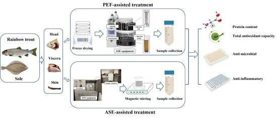
2. Materials and Methods

2.1. Chemicals and Reagents

Sodium carbonate (Na2CO3) was purchased from VWR (Saint-Prix, France). Trolox (6-hydroxy-2,5,7,8-tetramethylchroman-2-carboxylic acid), AAPH (2,2′-azobis-2-methyl-propanimidamide), ABTS (2,2′-azinobis (3-ethylbenzothiazoline 6-sulfonic acid)), fluorescein sodium salt and potassium persulfate (K2S2O8) were purchased from Sigma-Aldrich (Steinheim, Baden-Württemberg, Germany). Ethanol (99%) was acquired from Baker (Deventer, Overijssel, The Netherlands). Potassium dihydrogen phosphate and sodium phosphate dibasic were purchased from VWR International Eurolab S.L. (Barcelona, Spain). Deionized water was obtained by a Milli-Q SP Reagent Water System (Millipore Corporation, Bedford, MA, USA).

2.2. Sample Preparation

The rainbow trout (Oncorhynchus mykiss) and sole (Dover sole) samples used in the experiments were purchased from a local market in Valencia (Spain). The whole fish was separated in the laboratory to obtain the different fish side streams, including the fish head, skin and viscera for extraction. For PEF treatments, fresh samples were used, while for the ASE experiments, samples were pretreated and stored at −20 °C for more than 12 h, then freeze-dried at −48 °C for 72 h. The freeze-dried samples were stored at −20 °C until needed.

2.3. Extraction Conditions

2.3.1. PEF-Assisted Extraction

A PEF-Cellcrack III (German Institute of Food Technologies (DIL)) equipment (ELEA, Quakenbrück, Osnabrück, Germany) was used for the extractions. Specifically, the fish side streams were placed in the treatment chamber and tap water was added. Then, the conductivity was measured to be between 1000 and 2000 µs/cm. The samples were pretreated according to the best conditions previously obtained in the laboratory (a in Table 1). Samples were transferred to a beaker and were kept under agitation using a magnetic stirrer for a certain period at room temperature. Then, the supernatant was filtered through a 0.22 µm sterile filter membrane to obtain the samples. The control group was obtained under the same conditions but without the application of PEF pretreatment.

2.3.2. ASE-Assisted Extraction

Similarly, the selection of ASE conditions was also based on the optimal conditions obtained previously in the laboratory [20]. An ASE-200 accelerated solvent extractor (ASE 200 Dionex, Sunnyvale, CA, USA) was used in this study. According to the different samples, it was modified as the ratio of diatomaceous earth: the sample utilized was comprised of 1.0:2.0 g/g, 1.5:3.0 g/g and 2.0:2.0 g/g for the head, skin and viscera, respectively. The samples and diatomaceous earth were mixed in a mortar and transferred to the extraction tank. The standard parameters used for ASE extraction are preheating time (1 min), heating time (5 min), flushing volume (60%), nitrogen scanning (60 s) and pumping pressure (103.4 bar). The other conditions are shown in (b) of Table 1. The samples processed by ASE were filtered through a 0.22 µm sterile filter membrane and the control groups were also prepared.

2.4. Chemical Analyses

2.4.1. Protein Content

The BCA (Bicinchoninic acid) assay was used to determine the protein content of the extracts [21]. The working solution was prepared according to the BCA kit. Bovine serum albumin (0–2000 mg/L) was used as a standard to prepare the standard curve. Ten microliters of sample/standard and 200 µL of BCA working solution were added to the microplate, then the mixture was mixed well and incubated at 37 °C for 30 min. The absorbance of the samples was measured at 562 nm.

2.4.2. Total Antioxidant Capacity

Oxygen Radical Absorbance Capacity Assay (ORAC)

The determination of the ORAC values was carried out according to a previously described method [18,22]. A phosphate buffer (pH 7.0–7.4) was used as the blank group and 1 mM Trolox solution was the standard. Fifty microliters of the sample and the 50 µL fluorescein sodium salt were added, respectively, to a 96-well plate, then the 25 µL AAPH was added and the plate was kept under 37 °C for 10 min. Wavelengths of emission at 520 nm and excitation at 480 nm were established to record the results within 60 min. Then, the antioxidant capacity of the sample was calculated according to the formula:
ORAC   trolox   unit   =   A s a m p l e A b l a n k A t r o l o x A b l a n k
A measured ORAC value of 1 unit indicates that the antioxidant capacity of the sample solution is equivalent to 100 µM Trolox solution.

Trolox Equivalent Antioxidant Capacity Assay (TEAC)

The TEAC assay was used to determine the ABTS free radical scavenging capacity of the extracts. According to De la Fuente’s method and with some modifications, 25 mL ABTS (7 mM) and 440 µL K2S2O8 (140 mM) were mixed to obtain an ABTS+ working solution, which was stored at room temperature in the dark for about 12–16 h for use [18]. The ABTS+ working solution was diluted with 96% (v/v) ethanol to maintain the absorbance of 0.700 ± 0.020 at 734 nm. During the test, the samples were diluted to obtain a 50% free radical inhibition rate. The absorbance of 2 mL of the working solution was recorded as the initial value, then 100 µL of the correct dilution of the samples was added and the absorbance was recorded after 3 min of reaction. Different concentrations of Trolox (0–250 µM) were used as the standard to prepare the standard curve and to calculate the total antioxidant capacity of the samples.

2.4.3. Impact of the Extracts on Bacterial Growth

The impact of the different extracts obtained on several pathogenic and probiotic bacteria was investigated. The culture conditions of the different bacteria are shown in Table 2.
In this study, four common pathogenic and two probiotic bacteria that modulate human health were selected to evaluate growth patterns in the presence or absence of the extracts. Bacterial cultures were collected by centrifugation and inoculated in the corresponding medium at a final optical density at 595 nm of 0.05. The culture medium (200 μL) and sample (20 μL) were added to 96-well microplates and incubated in POLARstar (BMG, Labtech, Offenburg, Germany) equipment at 37 °C for 20 h and the optical density was recorded. The Gompertz equation was used to describe the bacterial growth rate and the maximum optical density:
y   =   K + A   exp exp μ max e A + 1
where y is the extent of growth at time t (h), K is an initial cell number, A is the change in the number of cells between the inoculum and the stationary phase, µmax is the maximum growth rate (the variation in the number of cells per unit of time), λ is the length of the lag phase (h) and e is a constant (2.7182).

2.4.4. Anti-Inflammatory Analysis

Cell Culture

To investigate the anti-inflammatory potential of the extracts, a reporter gene assay to analyze the activation of the pro-inflammatory transcription factor NF-κB was performed. Therefore, the human colon tumorigenic cell line HT-29 was previously stably transfected with the plasmid pNiFty2-SEAP (Invivogen, San Diego, CA, USA) containing a secreted alkaline phosphatase (SEAP) reporter gene [23]. The cell line was routinely cultured in a DMEM high glucose medium, supplemented with 10% fetal bovine serum, 1 mM sodium pyruvate, 100 U/mL penicillin, 100 μg/mL streptomycin and 150 µg/mL zeocin. Cells were cultured at 37 °C and 5% CO2 under a humidified atmosphere.

Analysis of NF-κB Activation

In the experiment, 65,000 cells/well were seeded into 96-well plates and grown for 24 h. Then, to investigate the activation of NF-κB, 10 µL of pro-inflammatory cytokine TNF-α was added to achieve a final concentration of 10 ng/mL and 10 µL of the extracts was added to achieve a total volume of 100 µL in each well. After 24 h of stimulation, the supernatant was collected and the cells were lysed in PBS containing 1% Triton, 1 mM PMSF (phenylmethylsulfonyl fluorid) and 1 mM EDTA (ethylenediaminetetraacetic acid). The protein content of each well was determined using the Bradford Protein Assay (Biorad). SEAP activity in the supernatant was measured using p-nitrophenyl phosphate as the phosphatase substrate and was normalized to the protein content of each well. The absorbance at 414 nm was measured with a microplate analyzer and the activity of NF-κB induced by TNF-α was recognized to be 100%.

2.5. Statistical Analysis

Significant differences between the results were analysed by analysis of variance (ANOVA). A Tukey’s Multiple Comparison test was used to indicate the significant differences in the means. All statistical analyses were performed using the software Statgraphics Centurion XVI.I software (Statpoint Technologies, Inc., The Plains, VA, USA).

3. Results and Discussion

3.1. Protein Content

Although the antimicrobial, prebiotic and anti-inflammatory activities were the main focus of this study, first of all, the protein content was evaluated. Figure 1 shows the effect of the two selected alternative technologies (PEF and ASE) to assist the recovery of protein from the rainbow trout and sole side streams. As can be seen in the table, when ASE-assisted extraction was used, the protein content of the fish head extracts was significantly increased (p < 0.05) and the content reached values that were almost 2-fold higher compared to the control group. ASE also significantly (p < 0.05) increased the protein content of the fish skin and viscera extracts. Moreover, the impact of the ASE extraction conditions on the recovery of protein from the rainbow trout was even stronger than the pattern found for the sole samples. Conversely, the effect on the sole viscera was stronger than that of rainbow trout, which may be related to the different composition and structure of the fish side streams according to the species evaluated.
Compared with ASE, PEF-assisted extraction had less effect on the protein content of fish side streams extracts. For rainbow trout, PEF-assisted treatment had no significant (p > 0.05) effect on the increase in the protein content compared with the control group; the protein content of the fish head and skin extracts was slightly lower than that observed for the control group, while no significant effect was observed for the viscera extracts (p > 0.05). The PEF-assisted treatment significantly increased (p < 0.05) the protein content of the sole head extract by three times compared to the control group. However, it had a negative impact on the protein content of the sole skin extracts, while there was no significant difference in the viscera; the results were similar to those found for the rainbow trout.
Fish side streams contain a large amount of protein and other bioactive compounds with high-added value, which can be used as sources of high-quality amino acids. In recent years, in order to reduce the waste of fish side streams, a variety of technologies have been used for the recovery of protein from fish side streams. In this line, Álvarez et al. [24] used ultrasound-assisted acid/alkaline isoelectric solubilization precipitation to recover the protein in mackerel. The results showed that compared with traditional methods, ultrasound-assisted acid/alkaline isoelectric solubilization precipitation can significantly increase the protein recovery rate from 50–64% to 94%. Similarly, Al-Khawli et al. [25] also used ultrasound to assist the extraction of protein from the sea bass side stream. Under ultrasound-assisted treatment, the protein content of the fish side stream extracts was significantly increased, with the highest modifications observed for the viscera samples. ASE and PEF have also recently been used as green processing technologies to assist the recovery of nutrients and bioactive compounds from the side streams of different species. For instance, De la Fuente et al. [26] used pressurized liquid-extraction (PLE)-assisted technology to obtain protein with antioxidant activity from the sea bream side stream and showed that the protein recovery rate could reach 1.2–4.5 times that of the control group. During PLE processing, high pressures and temperature increase the solubility and diffusion rate of high-added-value compounds, thereby improving their extraction efficiency.

3.2. Total Antioxidant Capacity

Figure 2 shows the antioxidant capacity of different extracts. Oxygen-free radical absorbance capacity (ORAC) and trolox equivalent antioxidant capacity (TEAC) were used to evaluate the antioxidant capacity of the extracts. For the rainbow trout side stream extracts, both the ASE and PEF treatments improved the ORAC values of the extracts, showing significant (p < 0.05) differences, except for the PEF-assisted skin sample. Among the different side stream extracts, the skin extract obtained under ASE-assisted treatment showed the most obvious difference, which was about three times higher than that observed for the control group. The TEAC values also showed the same trend after the application of ASE, with a significant increase observed for all the extracts independently of the target side streams evaluated. For instance, compared with the control group, the TEAC values increased by about 1.4–3.3 times. On the contrary, after PEF-assisted treatment, the TEAC values of the PEF fish head extracts were slightly lower than those of the control samples. Moreover, no significant effect on the TEAC values was observed after applying PEF to the skin compared to the control samples (p > 0.05). However, interestingly, PEF enhanced the TEAC values of the viscera extracts to a certain extent.
Regarding the sole side streams, the behaviour of the ORAC values of the extracts was similar to that found for rainbow trout and thus showed a considerable improvement in the ORAC values compared to control samples. However, no significant (p > 0.05) differences in the ORAC values were observed for ASE sole skin compared to the control group. The PEF treatment also contributed to an enhancement of the ORAC values of the sole side stream extracts; however, no significant differences (p > 0.05) in the ORAC values of the extracts were found for the PEF skin and viscera compared to the control group.
ASE also significantly increased the TEAC values of the extracts, which was about 1.7–2.0 times higher than that of the control group (without ASE). However, the impact of PEF on the TEAC values of the sole extracts was different from that found after applying ASE. For instance, PEF increased (p > 0.05) the TEAC values of the head extracts without any significant differences being observed. Two different methods have shown different effects on the antioxidant capacity of the extracts. Previously, the antioxidant capacity of the extracts without sterile membrane filtration was also measured [19]. Comparing the results before and after filtration, it was found that filtration had relatively little effect on the antioxidant capacity of the extracts, meaning the nutrients and antioxidant compounds in the extracts were well retained, which provides a basis for further experiments.

3.3. Impact of Fish Side Stream Extracts on Bacterial Growth

3.3.1. Antibacterial Activity against Pathogenic Bacteria

The effects of the different extracts on the growth of four common pathogens are presented in Table 3 and Table 4. The growth rate of different bacteria in 20 h and the optical density of bacterial strains were obtained by fitting the Gompertz equation [27]. As can be depicted from Table 3, the extracts obtained from the rainbow trout side streams after applying PEF and ASE induced the growth of Listeria and E. coli, with the viscera extracts showing the most obvious effect. In addition, the viscera extract also significantly increased the optical density of bacteria. When the effect of PEF was evaluated, it was observed that the extracts obtained from the head and skin had a significant effect on the growth of E. coli (p < 0.05), but there was not a significant effect on the optical density (p > 0.05) independently of the PEF treatment. Compared to the control group, the ASE-assisted extracts reduced the optimal density of the viscera extract, with no significant differences observed with the blank group (without the ASE extract).
Moreover, for the PEF extracts, no significant effect was found on the growth of S. aureus. For instance, the extracts increased the optical density of bacterial growth, showing a significant difference for the viscera extracts (p < 0.05). Unlike PEF, the head and skin extracts obtained by ASE reduced the growth rate of S. aureus (p < 0.05), but the viscera extract showed an inducing effect on the growth of S. aureus. The addition of the extracts did not show any inhibitory effect on the growth of Salmonella and had no significant effect on the optical density of the bacteria. In summary, among the four pathogens, the rainbow trout head and the skin extracts had an inhibitory impact on S. aureus, which was significant (p < 0.05) when ASE was used.
As is depicted in Table 4, the viscera extracts promoted the growth rate of Listeria and increased the optical density, while the sole head and skin extracts did not have a significant (p > 0.05) effect on the growth of Listeria. Independently of the treatment applied (PEF or ASE), no significant effect was observed. The addition of the extracts induced the growth of E. coli and the extracts from the head and viscera had a significant effect on the growth of bacteria (p < 0.05). However, the addition of the head extracts reduced the optical density of E. coli, while no effect was observed for the PEF extracts on the growth of E. coli. When ASE was used, similar to PEF, the extracts showed a promoting effect on E. coli growth and the application of ASE did not show any significant (p > 0.05) effect on the results.
For the extracts obtained after the application of PEF, the head and skin extracts did not have any significant effect on the growth and optical density of S. aureus, but the addition of viscera extracts reduced the growth rate of S. aureus. Meanwhile, compared with the control group, the PEF-assisted viscera extracts showed significant differences (p < 0.05). In contrast to the PEF samples, the ASE viscera extracts did not have a significant effect on the growth rate of S. aureus but increased the optical density. The head and skin extracts showed the opposite trend on the growth rate of S. aureus. For instance, compared to the control group, the ASE head extracts significantly reduced the growth rate of S. aureus (p < 0.05), while the ASE skin extract had a weaker effect on the S. aureus growth rate than that of the extracts without ASE treatment. Except for the ASE head and skin extracts, the other extracts did not show any significant effect on the optical density of S. aureus (p > 0.05).
Since fish side streams contain a large amount of high-added-value compounds, many people have explored their antibacterial properties over the last years to further expand their applications in food and health. For instance, Robert et al. [28] evaluated the in vitro antibacterial activity of the tilapia by-product hydrolysate. The peptides produced by hydrolysis showed important antibacterial activity against Yersinia ruckeri. Moreover, they also observed the resistance of these hydrolysates against Edwardsiella tarda and Bacillus megaterium, thus indicating that tilapia by-products have important antimicrobial activity. In another study, Ennaas et al. [29] used different proteases to hydrolyse the by-products of Atlantic mackerel skin and the antibacterial properties of hydrolyzed collagen were evaluated. The results showed that the crude hydrolysates of mackerel had an inhibitory effect on Listeria and E. coli, while the inhibition rate varied according to the different hydrolysates used. In addition to the properties against the different pathogens, the impact on the antibacterial properties varied with the type of fish. Previous studies have shown that fish by-products with a large number of low-molecular-weight peptides had a higher activity [30]. In this study, two kinds of fish showed inhibitory effects on S. aureus and could be considered as potential sources of new antibacterial products.

3.3.2. Effect on the Growth of Probiotic Bacteria

Table 5 shows the effect of different extracts on the growth of two probiotics. By analysing the effect of rainbow trout on the growth of probiotics, it can be seen that the addition of the head and skin extracts in the PEF group promoted the growth of Lactobacillus casei with significant differences (p < 0.05), while no significant differences were observed between PEF and the control group (p > 0.05). Moreover, these extracts also increased the optical density of Lactobacillus casei. Compared to the control group, the optical density of the PEF extracts was lower than that of the control group, with no significant differences observed between the two skin extracts (p > 0.05). In addition, the viscera extracts did not have any significant effect on the growth rate and optical density of the Lactobacillus casei. Studying the effect of the PEF extracts on Bifidobacterium lactis, it was observed that the addition of the extracts did not have any significant effect on the growth rate of Bifidobacterium lactis.
On the other hand, the addition of the ASE extracts reduced the growth rate of Lactobacillus casei. Compared with the control group, the head and viscera extracts obtained with the ASE-assisted treatment did not show any significant difference on the Lactobacillus casei growth rate. The ASE-assisted skin extract had a weaker effect on the growth rate of Lactobacillus casei than the control group (skin-control) and this difference was significant (p < 0.05). At the same time, it was also seen that the head and skin extracts increased the optical density of the Lactobacillus casei, showing a significant difference compared to the control group, but there was not a significant effect of ASE-assisted extracts (p > 0.05). Compared to the control group, the ASE-assisted extracts increased the growth rate of Bifidobacterium lactis, but no significant differences were observed. For example, the addition of the ASE-assisted viscera extracts did not have any effect on the growth rate of Bifidobacterium lactis. On the other hand, the head and skin extracts increased the optical density of Bifidobacterium lactis and the optical density of the ASE-assisted extracts was significantly higher than that of the control group.
The effect of the sole side stream extracts on probiotic growth was also studied in this work. As can be seen from Table 5, the PEF-assisted head extract increased the growth rate of Lactobacillus casei, which was significantly different from the control group (without PEF) and blank group. The viscera extract inhibited the growth of Lactobacillus casei and the addition of the other side streams did not have any significant effect on the growth of Lactobacillus casei. The addition of the PEF-assisted extracts did not have a significant effect on the optical density of the Lactobacillus casei. At the same time, under the PEF-assisted treatment, the addition of different extracts did not have a significant effect on the growth rate of Bifidobacterium lactis, but the skin and viscera extracts increased the optical density of Bifidobacterium lactis and the optical density of the control group was higher than that of the PEF-assisted extraction.
Exploring the effect of ASE, it was observed that the skin extract significantly (p < 0.05) reduced the growth rate of Lactobacillus casei, thus ASE had an obvious impact on the results. The ASE-assisted viscera and head extracts did not have a significant effect on the growth rate of Lactobacillus casei, while the viscera extract without ASE-assisted treatment reduced the growth rate of Lactobacillus casei. In addition, all the extracts increased the optical density, but there was not a significant difference between them (p > 0.05). At the same time, the addition of the head extract decreased the growth rate of Bifidobacterium lactis, but ASE did not have any significant effect on the results. Moreover, the skin and viscera extracts did not show any significant effect on the growth rate of Bifidobacterium lactis. Compared to the control group, the ASE-assisted head and skin extracts increased the optical density of Bifidobacterium lactis and the viscera extract also increased the optical density, but no impact of ASE was observed (p > 0.05).
In recent years, many studies have shown that bioactive compounds and hydrolysates derived from fish side streams have antibacterial activity, which can inhibit the growth of pathogenic bacteria to a certain extent, although there are only a few studies on the growth of probiotics. Probiotics are used in the fermentation and preservation of food to help maintain food quality and improve nutrition. In the study of Safari et al. [31], two different peptones were obtained from the yellowfin tuna (Thunnus albacares) head by enzymatic hydrolysis and their effects on the growth of a variety of bacteria including pathogens and probiotics were explored. The results obtained by these authors showed that the proteins obtained by hydrolysis promoted the growth of Lactobacillus plantarum and Lactobacillus bulgaricus, making their growth rate higher than that of Lactobacillus sakei and others. It has been speculated that the main reason for this may be that the growth of Lactobacillus from different sources has different requirements for the types of amino acids, while the matching degree of the peptides produced by enzymatic hydrolysis to different types of Lactobacillus is also different. Combined with the effect of the extract on the pathogenic bacteria, it can be seen that the type of peptide in the extract will affect the growth of bacteria, while peptides of appropriate molecular weight can make probiotics show greater growth activity. In addition, the difference in the form and concentration of the samples during PEF and ASE-assisted extraction is also one of the reasons for the different results.

3.4. Anti-Inflammatory Activity

The anti-inflammatory potential of the rainbow trout and sole fish side streams are shown in Figure 3. For rainbow trout, the PEF extracts did not show any significant anti-inflammatory potential, while the ASE extracts had a significant inhibitory effect on NF-κB activity, which could inhibit ≈40–45% of TNF-α-induced NF-κB activity in the viscera extracts. On the other hand, for sole side streams, the PEF skin extracts showed a significant anti-inflammatory potential, inhibiting the TNF-α activity by ≈35%. The sole head extract without ASE treatment showed significant anti-inflammatory potential, while the other extracts did not show any significant difference. Interestingly, in the PEF group of rainbow trout extracts, the extracts from the skin and viscera (without PEF and ASE treatments) enhanced the TNF-induced NF-κB activity to levels of 150% and 126%, suggesting that the PEF treatment could alter some components of these extracts and reduce their intrinsic pro-inflammatory potential. A similar response in SEAP activity was observed for sole extracts in PEF treated skin vs. skin-control extracts.
Inflammation is the immune system’s response that can effectively protect our body from injury and infection; however, the excessive release of inflammatory mediators can become chronic and lead to many inflammatory diseases. In the intestinal tract, proinflammatory stimulants activate in intestinal epithelial cells NF-κB, a master regulator of inflammatory processes among several others, which upregulates cytokines and chemokines [32,33]. Studies have shown that protein polypeptides are anti-inflammatory, anti-hypertensive, etc. For instance, Gao et al. [34] obtained synthetic peptides from sturgeon muscle and found that they can effectively reduce the release of inflammatory mediators and cytokines. It can be speculated that the anti-inflammatory potential of some extracts may be related to the bioactive peptide in extracts.

4. Conclusions

From the results obtained in this study, it can be concluded that PEF and ASE may be used as useful alternative approaches in recovering extracts with antimicrobial, prebiotic and anti-inflammatory properties. Some extracts showed antibacterial and anti-inflammatory effects, including those obtained by ASE rainbow trout and sole head and skin extracts that promoted inhibitory effects on the growth of S. aureus and Salmonella. When PEF was studied, the PEF rainbow trout head and skin extracts also showed an inhibitory effect on the growth of S. aureus. In addition, they also enhanced the growth of Lactobacillus casei. It was also found that some extracts showed anti-inflammatory potential, including those obtained from the ASE and non-ASE rainbow trout viscera, the PEF sole skin and the non-PEF sole head. This may be because the bioactive peptides in them play a vital role. In general, these extracts can be considered as potentially valuable functional substances to further study their beneficial effects on humans.

Author Contributions

Conceptualization, F.J.B., M.C.C., M.W. and J.Z.; methodology, M.W., J.Z., C.B. and N.P.; formal analysis, M.W. and J.Z.; software, M.W., J.Z. and B.N.D.; investigation, M.W., J.Z., N.P., M.C.C., C.B., B.N.D. and F.J.B.; resources, F.J.B. and M.C.C.; data curation, M.W., J.Z. and B.N.D.; writing—original draft preparation, M.W., J.Z. and F.J.B.; writing—review and editing, M.W., J.Z., N.P., M.C.C., B.N.D. and F.J.B.; supervision, F.J.B. and M.C.C.; funding acquisition, F.J.B. and M.C.C. All authors have read and agreed to the published version of the manuscript.

Funding

This research was funded by BBI-JU through the H2020 Project AQUABIOPRO-FIT “Aquaculture and agriculture biomass side stream proteins and bioactives for feed, fitness and health promoting nutritional supplements” (Grant number 790956).

Institutional Review Board Statement

Not applicable.

Informed Consent Statement

Not applicable.

Data Availability Statement

Not applicable.

Acknowledgments

M.W. was supported by a PhD fellowship from the China Scholarship Council (CSC) (No. 201908420245). J.Z. would also like to thank the China Scholarship Council (CSC) for her PhD fellowship (No. 201908420246). Authors would also like to thank Generalitat Valenciana for the financial support (IDIFEDER/2018/046- Procesos innovadores de extracción y conservación: pulsos eléctricos y fluidos supercríticos) through European Union ERDF funds (European Regional Development Fund).

Conflicts of Interest

The authors declare no conflict of interest.

References

  1. Lewis, K. New approaches to antimicrobial discovery. Biochem. Pharmacol. 2017, 134, 87–98. [Google Scholar] [CrossRef] [PubMed]
  2. Negi, P.S. Plant extracts for the control of bacterial growth: Efficacy, stability and safety issues for food application. Int. J. Food Microbiol. 2012, 156, 7–17. [Google Scholar] [CrossRef]
  3. Enan, G. Nature and phenotypic characterization of plantaricin UG1 resistance in Listeria monocytogenes LMG 10470. J. Food Agric. Environ. 2006, 4, 105–108. [Google Scholar]
  4. Tong, J.; Zhang, Z.; Wu, Q.; Huang, Z.; Malakar, P.K.; Chen, L.; Liu, H.; Pan, Y.; Zhao, Y. Antibacterial peptides from seafood: A promising weapon to combat bacterial hazards in food. Food Control 2021, 125, 108004. [Google Scholar] [CrossRef]
  5. Pérez-Gálvez, R.; Espejo-Carpio, F.J.; Morales-Medina, R.; García-Moreno, P.J.; Guadix-Escobar, A.; Guadix-Escobar, E. Fish Residues as Source of Health-Promoting Biopeptides; Elsevier Inc.: Amsterdam, The Netherlands, 2018; ISBN 9780128114469. [Google Scholar]
  6. Stevens, J.R.; Newton, R.W.; Tlusty, M.; Little, D.C. The rise of aquaculture by-products: Increasing food production, value, and sustainability through strategic utilisation. Mar. Policy 2018, 90, 115–124. [Google Scholar] [CrossRef] [Green Version]
  7. Ananey-Obiri, D.; Matthews, L.G.; Tahergorabi, R. Proteins from Fish. Processing By-Products; Elsevier Inc.: Amsterdam, The Netherlands, 2019; ISBN 9780128166956. [Google Scholar]
  8. Rivero-Pino, F.; Espejo-Carpio, F.J.; Guadix, E.M. Evaluation of the bioactive potential of foods fortified with fish protein hydrolysates. Food Res. Int. 2020, 137, 109572. [Google Scholar] [CrossRef]
  9. Beaulieu, L.; Thibodeau, J.; Bryl, P.; Carbonneau, M.É. Proteolytic processing of Atlantic mackerel (Scomber scombrus) and biochemical characterisation of hydrolysates. Int. J. Food Sci. Technol. 2009, 44, 1609–1618. [Google Scholar] [CrossRef]
  10. Fuochi, V.; Li Volti, G.; Camiolo, G.; Tiralongo, F.; Giallongo, C.; Distefano, A.; Petronio, G.P.; Barbagallo, I.; Viola, M.; Furneri, P.M.; et al. Antimicrobial and anti-proliferative effects of skin mucus derived from Dasyatis pastinaca (Linnaeus, 1758). Mar. Drugs 2017, 15, 342. [Google Scholar] [CrossRef] [Green Version]
  11. Cheng, J.; Sun, J.; Huang, Y.; Feng, J.; Zhou, J.; Cen, K. Dynamic microstructures and fractal characterization of cell wall disruption for microwave irradiation-assisted lipid extraction from wet microalgae. Bioresour. Technol. 2013, 150, 67–72. [Google Scholar] [CrossRef]
  12. Chemat, F.; Abert Vian, M.; Fabiano-Tixier, A.S.; Nutrizio, M.; Režek Jambrak, A.; Munekata, P.E.S.; Lorenzo, J.M.; Barba, F.J.; Binello, A.; Cravotto, G. A review of sustainable and intensified techniques for extraction of food and natural products. Green Chem. 2020, 22, 2325–2353. [Google Scholar] [CrossRef] [Green Version]
  13. Barba, F.J.; Jäger, H.; Meneses, N.; Esteve, M.J.; Frígola, A.; Knorr, D. Evaluation of quality changes of blueberry juice during refrigerated storage after high-pressure and pulsed electric fields processing. Innov. Food Sci. Emerg. Technol. 2012, 14, 18–24. [Google Scholar] [CrossRef]
  14. Luo, Q.; Hamid, N.; Oey, I.; Leong, S.Y.; Kantono, K.; Alfaro, A.; Lu, J. Physicochemical changes in New Zealand abalone (Haliotis iris) with pulsed electric field (PEF) processing and heat treatments. LWT 2019, 115, 108438. [Google Scholar] [CrossRef]
  15. Scherer, D.; Krust, D.; Frey, W.; Mueller, G.; Nick, P.; Gusbeth, C. Pulsed electric field (PEF)-assisted protein recovery from Chlorella vulgaris is mediated by an enzymatic process after cell death. Algal Res. 2019, 41, 101536. [Google Scholar] [CrossRef]
  16. Ahmad, R.; Ahmad, N.; Al-Anaki, W.S.; Ismail, F.A.; Al-Jishi, F. Solvent and temperature effect of accelerated solvent extraction (ASE) coupled with ultra-high-pressure liquid chromatography (UHPLC-PDA) for the determination of methyl xanthines in commercial tea and coffee. Food Chem. 2020, 311, 126021. [Google Scholar] [CrossRef]
  17. Ahmad, R.; Ahmad, N.; Alkhars, S.; Alkhars, A.; Alyousif, M.; Bukhamseen, A.; Abuthayn, S.; Aqeel, M.; Aljamea, A. Green accelerated solvent extraction (ASE) with solvent and temperature effect and green UHPLC-DAD analysis of phenolics in pepper fruit (Capsicum annum L.). J. Food Compos. Anal. 2021, 97, 103766. [Google Scholar] [CrossRef]
  18. De Fuente, B.; Pallarés, N.; Berrada, H.; Barba, F.J. Salmon (Salmo salar) side streams as a bioresource to obtain potential antioxidant peptides after applying pressurized liquid extraction (PLE). Mar. Drugs 2021, 19, 323. [Google Scholar] [CrossRef]
  19. Wang, M.; Zhou, J.; Collado, M.C.; Barba, F.J. Accelerated solvent extraction and pulsed electric fields for valorization of rainbow trout (Oncorhynchus mykiss) and Sole (Dover sole) by-products: Protein content, molecular weight distribution and antioxidant potential of the extracts. Mar. Drugs 2021, 19, 207. [Google Scholar] [CrossRef] [PubMed]
  20. De la Fuente, B.; Pallarés, N.; Barba, F.J.; Berrada, H. An integrated approach for the valorization of sea bass (Dicentrarchus labrax) side streams: Evaluation of contaminants and development of antioxidant protein extracts by pressurized liquid extraction. Foods 2021, 10, 546. [Google Scholar] [CrossRef]
  21. Parniakov, O.; Barba, F.J.; Grimi, N.; Marchal, L.; Jubeau, S.; Lebovka, N.; Vorobiev, E. Pulsed electric field assisted extraction of nutritionally valuable compounds from microalgae Nannochloropsis spp. using the binary mixture of organic solvents and water. Innov. Food Sci. Emerg. Technol. 2015, 27, 79–85. [Google Scholar] [CrossRef]
  22. Cao, G.; Alessio, H.M.; Cutler, R.G. Oxygen-radical absorbance capacity assay for antioxidants. Free Radic. Biol. Med. 1993, 14, 303–311. [Google Scholar] [CrossRef] [Green Version]
  23. Rocchetti, G.; Alcántara, C.; Bäuerl, C.; García-Pérez, J.V.; Lorenzo, J.M.; Lucini, L.; Collado, M.C.; Barba, F.J. Bacterial growth and biological properties of Cymbopogon schoenanthus and Ziziphus lotus are modulated by extraction conditions. Food Res. Int. 2020, 136, 109534. [Google Scholar] [CrossRef] [PubMed]
  24. Álvarez, C.; Lélu, P.; Lynch, S.A.; Tiwari, B.K. Optimised protein recovery from mackerel whole fish by using sequential acid/alkaline isoelectric solubilization precipitation (ISP) extraction assisted by ultrasound. LWT-Food Sci Technol. 2018, 88, 210–216. [Google Scholar] [CrossRef] [Green Version]
  25. Khawli, F.A.; Pallarés, N.; Martí-Quijal, F.J.; Ferrer, E.; Barba, F.J. Sea bass side streams valorization assisted by ultrasound. LC-MS/MS-it determination of mycotoxins and evaluation of protein yield, molecular size distribution and antioxidant recovery. Appl. Sci. 2021, 11, 2160. [Google Scholar] [CrossRef]
  26. De Fuente, B.; Pallar, N.; Berrada, H.; Barba, F.J. Development of antioxidant protein extracts from gilthead seabream (Sparus aurata) side streams assisted by pressurized liquid extraction (PLE). Mar. Drugs 2021, 19, 199. [Google Scholar] [CrossRef] [PubMed]
  27. Zwietering, M.H.; Jongenburger, I.; Rombouts, F.M.; van’t Riet, K. Modeling of the bacterial growth curve. Appl. Environ. Microbiol. 1990, 56, 1875–1881. [Google Scholar] [CrossRef] [Green Version]
  28. Robert, M.; Zatylny-Gaudin, C.; Fournier, V.; Corre, E.; Le, G.; Bernay, B.; Henry, J. Molecular characterization of peptide fractions of a Tilapia (Oreochromis niloticus) by-product hydrolysate and in vitro evaluation of antibacterial activity. Process. Biochem. 2015, 50, 487–492. [Google Scholar] [CrossRef]
  29. Ennaas, N.; Hammami, R.; Beaulieu, L.; Fliss, I. Production of antibacterial fraction from Atlantic mackerel (Scomber scombrus) and its processing by-products using commercial enzymes. Food Bioprod. Process. 2015, 96, 145–153. [Google Scholar] [CrossRef]
  30. He, S.; Franco, C.; Wei, Z. Functions, applications and production of protein hydrolysates from fish processing co-products (FPCP). Food Res. Int. 2013, 50, 289–297. [Google Scholar] [CrossRef]
  31. Safari, R.; Motamedzadegan, A.; Ovissipour, M.; Regenstein, J.M.; Gildberg, A.; Rasco, B. Use of hydrolysates from Yellowfin Tuna (Thunnus albacares) heads as a complex nitrogen source for lactic acid bacteria. Food Bioprocess. Technol. 2012, 5, 73–79. [Google Scholar] [CrossRef]
  32. Ávila-Román, J.; Talero, E.; de los Reyes, C.; García-Mauriño, S.; Motilva, V. Microalgae-derived oxylipins decrease inflammatory mediators by regulating the subcellular location of NF-κB and PPAR-γ. Pharmacol. Res. 2018, 128, 220–230. [Google Scholar] [CrossRef]
  33. Peng, Y.; Gan, R.; Li, H.; Yang, M.; McClements, D.J.; Gao, R.; Sun, Q. Absorption, metabolism, and bioactivity of vitexin: Recent advances in understanding the efficacy of an important nutraceutical. Crit. Rev. Food Sci. Nutr. 2021, 61, 1049–1064. [Google Scholar] [CrossRef] [PubMed]
  34. Gao, R.; Shu, W.; Shen, Y.; Sun, Q.; Bai, F.; Wang, J.; Li, D.; Li, Y.; Jin, W.; Yuan, L. Sturgeon protein-derived peptides exert anti-inflammatory effects in LPS-stimulated RAW264.7 macrophages via the MAPK pathway. J. Funct. Foods 2020, 72, 104044. [Google Scholar] [CrossRef]
Figure 1. Protein content in the control and optimal PEF/ASE assisted extracts from fish by-products. (A) rainbow trout and (B) sole; PEF: pulsed electric fields; ASE: accelerated solvent extraction; Different lowercase letters (a–i) in the figure indicate statistically significant differences (p < 0.05).
Separations 08 00187 g001
Figure 2. Total antioxidant capacity (ORAC and TEAC) in the control and optimal PEF/ASE-assisted extracts from fish by-products. (A) rainbow trout and (B) sole; PEF: pulsed electric fields; ASE: accelerated solvent extraction; Different lowercase letters (a–g) in the figure indicate statistically significant differences (p < 0.05). ORAC: oxygen radical absorbance capacity; ABTS: ABTS+ scavenging ability.
Separations 08 00187 g002
Figure 3. The NF-κB activation induced by TNF-α and the effect of fish by-product extracts using different treatment technology (PEF/ASE) were determined. (A) rainbow trout and (B) sole; the SEAP activity induced by TNF-α was considered to be 100%. PEF: pulsed electric fields; ASE: accelerated solvent extraction; Different lowercase (a–c) letters in the figure indicate statistically significant differences (p < 0.05).
Separations 08 00187 g003
Table 1. (a) Pulsed electric fields (PEF)-assisted extraction experimental conditions, (b) Accelerated solvent extraction (ASE) experimental conditions.
(a)
SampleWeight (g)Field Strength (kV/cm)H2O (mL)Specific Energy (kJ/kg)Time (h) 1
Head100.251.001500219.7621.33
Skin45.303.00675300.0024.00
Viscera45.303.00675123.7515.17
(b)
SampleT (°C)Time (min)pHPressure
Head55155.2103.4
Skin45156.5103.4
Viscera50156.8103.4
1 time of supplementary extraction.
Table 2. The culture conditions of the bacteria used in this study.
BacterialCollection NumberCulture MediumCulture Conditions
Listeria innocua(CECT 910)BHI 137 °C, 24 h, aerobic
Escherichia coli(CECT 99)
Staphylococcus aureus(CECT 86)
Salmonella enterica(CECT 4138)
Lactobacillus casei(BB 12)MRS37 °C, 48 h, anaerobic
Bifidobacterium lactis(NCC 2818)MRS + 0.05% L-cys 2
1 BHI: Brain heart infusion medium. 2 MRS: Man rogosa sharpe medium; L-cys: L-cysteine hydrochloride; CECT: Spanish National Culture Collection (www.cect.org).
Table 3. Effect of rainbow trout extracts on the growth rate and maximal optical density of four pathogenic bacteria strains.
SamplePEF 1ASE 2
Growth Rate (μmax·h −1)*MODGrowth Rate (μmax·h −1)*MOD
Listeria
Bacteria-control0.442 ± 0.027 a1.558 ± 0.039 a0.435 ± 0.017 a1.524 ± 0.015 a
Head0.472 ± 0.004 a1.538 ± 0.046 a0.479 ± 0.025 a1.511 ± 0.019 a
Head-control0.461 ± 0.001 a1.543 ± 0.022 a0.454 ± 0.016 a1.525 ± 0.031 a
Skin0.464 ± 0.001 a1.503 ± 0.001 a0.467 ± 0.014 a1.588 ± 0.017 b
Skin-control0.475 ± 0.006 a1.498 ± 0.026 a0.435 ± 0.011 a1.486 ± 0.020 a
Viscera0.599 ± 0.007 c1.692 ± 0.023 b0.576 ± 0.007 b1.666 ± 0.026 c
Viscera-control0.526 ± 0.031 b1.686 ± 0.015 b0.614 ± 0.037 b1.635 ± 0.028 b,c
E. coli
Bacteria-control0.176 ± 0.009 a2.346 ± 0.009 a0.176 ± 0.009 a,b2.346 ± 0.009 a,b
Head0.208 ± 0.002 b2.300 ± 0.044 a0.180 ± 0.002 b2.307 ± 0.061 a,b
Head-control0.201 ± 0.001 b2.284 ± 0.030 a0.193 ± 0.003 c2.203 ± 0.034 a
Skin0.194 ± 0.009 b2.318 ± 0.033 a0.185 ± 0.002 b2.472 ± 0.103 b
Skin-control0.195 ± 0.011 b2.292 ± 0.006 a0.168 ± 0.003 a2.338 ± 0.120 a,b
Viscera0.172 ± 0.000 a2.644 ± 0.046 b0.275 ± 0.002 d2.188 ± 0.038 a
Viscera-control0.178 ± 0.001 a2.730 ± 0.064 b0.167 ± 0.008 a2.757 ± 0.135 c
S. aureus
Bacteria-control0.591 ± 0.039 b2.216 ± 0.215 a0.524 ± 0.056 c,d2.401 ± 0.047 a
Head0.560 ± 0.054 a,b2.309 ± 0.142 a0.441 ± 0.041 a,b2.559 ± 0.018 a,b
Head-control0.559 ± 0.003 a,b2.309 ± 0.124 a0.448 ± 0.026 b2.455 ± 0.053 a
Skin0.505 ± 0.043 a,b2.545 ± 0.126 a,b0.404 ± 0.008 a2.796 ± 0.034 c
Skin-control0.496 ± 0.036 a2.533 ± 0.135 a,b0.482 ± 0.003 b,c2.492 ± 0.048 a
Viscera0.550 ± 0.026 a,b2.751 ± 0.067 b0.579 ± 0.037 d2.724 ± 0.034 b,c
Viscera-control0.596 ± 0.041 b2.579 ± 0.077 a,b0.578 ± 0.049 d2.642 ± 0.053 b,c
Salmonella
Bacteria-control0.335 ± 0.026 a1.838 ± 0.0650.335 ± 0.026 a1.838 ± 0.065
Head0.353 ± 0.030 a,b1.831 ± 0.1640.308 ± 0.002 a1.714 ± 0.151
Head-control0.361 ± 0.025 a,b1.756 ± 0.1510.308 ± 0.005 a1.655 ± 0.055
Skin0.323 ± 0.007 a1.859 ± 0.0430.315 ± 0.024 a1.766 ± 0.157
Skin-control0.346 ± 0.022 a,b1.810 ± 0.1710.302 ± 0.025 a1.798 ± 0.049
Viscera0.308 ± 0.021 a1.863 ± 0.2140.418 ± 0.021 b1.687 ± 0.049
Viscera-contro0.390 ± 0.005 b1.678 ± 0.1320.447 ± 0.031 b1.675 ± 0.004
1 PEF: pulsed electric fields. 2 ASE: accelerated solvent extraction. Results are expressed as mean ± standard deviation. Different superscripts (a–d) in the same column (in group with the same bacteria) indicate statistically significant differences (p < 0.05) and mean values without any superscript indicate statistically non-significant difference (p > 0.05). *MOD: maximal optical density measured at 595 nm (difference between initial and final optical density); Bacteria-control: bacterial growth without fish by-products extracts; Head-control/Skin-control/Viscera-control: Head/Skin/Viscera extracts without PEF/ASE-assisted treatment.
Table 4. Effect of the sole extracts on the growth rate and maximal optical density of four pathogenic bacteria strains.
SamplePEF 1ASE 2
Growth Rate (μmax·h −1)*MODGrowth Rate (μmax·h −1)*MOD
Listeria
Bacteria-control0.442 ± 0.027 a,b1.558 ± 0.039 a,b0.435 ± 0.017 a,b1.524 ± 0.015 a
Head0.427 ± 0.032 a,b1.518 ± 0.022 a0.454 ± 0.009 a,b1.603 ± 0.008 b,c
Head-control0.426 ± 0.034 a,b1.563 ± 0.025 a,b0.442 ± 0.019 a,b1.554 ± 0.041 a,b
Skin0.395 ± 0.014 a1.498 ± 0.040 a0.412 ± 0.036 a1.503 ± 0.037 a
Skin-control0.396 ± 0.041 a1.551 ± 0.005 a0.456 ± 0.016 a,b1.512 ± 0.016 a
Viscera0.468 ± 0.000 b1.634 ± 0.021 c0.502 ± 0.009 c1.613 ± 0.015 c
Viscera-control0.473 ± 0.017 b1.622 ± 0.017 bc0.479 ± 0.016 b,c1.616 ± 0.005 c
E. coli
Bacteria-control0.176 ± 0.009 a2.346 ± 0.009 b,c0.176 ± 0.009 a2.346 ± 0.009 c
Head0.208 ± 0.005 c2.182 ± 0.070 a0.207 ± 0.008 c,d2.274 ± 0.039 b
Head-control0.213 ± 0.007 c2.218 ± 0.041 a0.191 ± 0.002 a,b2.256 ± 0.029 b
Skin0.182 ± 0.005 a,b2.343 ± 0.004 b,c0.200 ± 0.003 b,c2.202 ± 0.106 a,b
Skin-control0.200 ± 0.009 b,c2.273 ± 0.044 a,b0.204 ± 0.000 b,c,d2.159 ± 0.059 a
Viscera0.212 ± 0.013 c2.391 ± 0.038 c0.216 ± 0.006 d2.299 ± 0.003 b,c
Viscera-control0.211 ± 0.003 c2.337 ± 0.022 b0.215 ± 0.003 d2.243 ± 0.022 a,b
S. aureus
Bacteria-control0.591 ± 0.039 b,c2.401 ± 0.047 b,c0.524 ± 0.056 c2.401 ± 0.047 a
Head0.533 ± 0.046 b,c2.546 ± 0.178 c,d0.452 ± 0.026 b2.721 ± 0.009 c
Head-control0.616 ± 0.025 c2.216 ± 0.086 a0.474 ± 0.004 b,c2.483 ± 0.056 a,b
Skin0.623 ± 0.008 c2.300 ± 0.067 a,b0.425 ± 0.024 a,b2.434 ± 0.104 a
Skin-control0.533 ± 0.060 b,c2.530 ± 0.123 b,c,d0.378 ± 0.032 a2.385 ± 0.078 a
Viscera0.458 ± 0.046 a2.726 ± 0.007 d0.523 ± 0.043 c2.634 ± 0.076 b,c
Viscera-control0.513 ± 0.013 a,b2.650 ± 0.115 d0.531 ± 0.018 c2.542 ± 0.101 a,b
Salmonella
Bacteria-control0.335 ± 0.026 b1.838 ± 0.0650.335 ± 0.0261.838 ± 0.065
Head0.300 ± 0.007 a,b1.676 ± 0.0370.284 ± 0.0201.656 ± 0.071
Head-control0.280 ± 0.016 a1.715 ± 0.0180.303 ± 0.0111.658 ± 0.035
Skin0.335 ± 0.029 b1.817 ± 0.1910.323 ± 0.0291.696 ± 0.108
Skin-control0.338 ± 0.020 b1.771 ± 0.1460.339 ± 0.0221.686 ± 0.145
Viscera0.287 ± 0.018 a1.779 ± 0.1460.282 ± 0.0031.799 ± 0.138
Viscera-control0.276 ± 0.001 a1.784 ± 0.1510.308 ± 0.0181.796 ± 0.145
1 PEF: pulsed electric fields. 2 ASE: accelerated solvent extraction. Results are expressed as mean ± standard deviation. Different superscripts (a–d) in the same column (in group with the same bacteria) indicate statistically significant differences (p < 0.05) and mean values without any superscript indicate the statistically non-significant difference (p > 0.05). *MOD: maximal optical density measured at 595 nm (difference between initial and final optical density). Bacteria-control: bacterial growth without fish by-products extracts; Head-control/Skin-control/Viscera-control: Head/Skin/Viscera extracts without PEF/ASE-assisted treatment.
Table 5. Effect of the rainbow trout and sole extracts on the growth rate and maximal optical density of two probiotic bacteria strains.
FishSamplePEF 1ASE 2
Growth Rate (μmax·h −1)*MOD Growth Rate (μmax·h −1)*MOD
RainbowtroutLactobacillus casei
Bacteria-control0.349 ± 0.008 a,b3.597 ± 0.011 a,b0.360 ± 0.012 c1.524 ± 0.015 a
Head0.382 ± 0.011 c,d3.681 ± 0.038 b0.349 ± 0.007 b,c1.603 ± 0.008 b,c
Head-control0.374 ± 0.004 c,d3.836 ± 0.053 c0.337 ± 0.009 b1.554 ± 0.041 ab
Skin0.369 ± 0.002 bc,d3.719 ± 0.062 b,c0.334 ± 0.007 b1.503 ± 0.037 a
Skin-control0.390 ± 0.017 d3.683 ± 0.069 b0.288 ± 0.011 a1.512 ± 0.016 a
Viscera0.360 ± 0.000 b,c3.533 ± 0.036 a0.283 ± 0.006 a1.613 ± 0.015 c,d
Viscera-control0.336 ± 0.012 a3.595 ± 0.071 a,b0.271 ± 0.005 a1.616 ± 0.005 d
Bifidobacterium lactis
Bacteria-control0.536 ± 0.0273.597 ± 0.011 b0.536 ± 0.027 a,b2.346 ± 0.009 a,b,c
Head0.542 ± 0.0353.681 ± 0.006 c0.557 ± 0.019 b2.274 ± 0.039 a,b
Head-control0.544 ± 0.0273.836 ± 0.006 d0.536 ± 0.024 a,b2.256 ± 0.029 a,b
Skin0.508 ± 0.0283.719 ± 0.000 d0.547 ± 0.014 b2.202 ± 0.106 ab
Skin-control0.561 ± 0.0143.683 ± 0.010 c0.498 ± 0.008 a2.159 ± 0.059 a
Viscera0.550 ± 0.0213.533 ± 0.007 a0.529 ± 0.010 a,b2.299 ± 0.003 a,b,c
Viscera-control0.532 ± 0.0233.595 ± 0.009 a0.530 ± 0.007 a,b2.243 ± 0.022 a,b
SoleLactobacillus casei
Bacteria-control0.349 ± 0.008 b,c3.597 ± 0.020 a,b0.360 ± 0.012 c2.401 ± 0.047 a
Head0.405 ± 0.003 d3.565 ± 0.053 a0.355 ± 0.010 c2.721 ± 0.009 c
Head-control0.365 ± 0.007 c3.572 ± 0.041 a0.357 ± 0.002 c2.483 ± 0.056 a,b
Skin0.338 ± 0.000 b3.637 ± 0.076 a,b0.273 ± 0.001 a2.434 ± 0.104 a
Skin-control0.345 ± 0.025 b,c3.662 ± 0.011 a,b0.324 ± 0.016 b2.385 ± 0.078 a
Viscera0.308 ± 0.004 a3.663 ± 0.014 ab0.363 ± 0.001 c2.634 ± 0.076 b,c
Viscera-control0.305 ± 0.002 a3.695 ± 0.027 b0.328 ± 0.007 b2.542 ± 0.101 a,b,c
Bifidobacterium lactis
Bacteria-control0.536 ± 0.0273.597 ± 0.011 a0.536 ± 0.027 b,c1.838 ± 0.065
Head0.526 ± 0.0453.565 ± 0.003 a0.499 ± 0.010 a1.656 ± 0.071
Head-control0.517 ± 0.0163.572 ± 0.008 a0.483 ± 0.003 a1.658 ± 0.035
Skin0.537 ± 0.0013.637 ± 0.001 b0.504 ± 0.009 a,b1.696 ± 0.108
Skin-control0.545 ± 0.0153.662 ± 0.004 b0.553 ± 0.033 d1.686 ± 0.145
Viscera0.541 ± 0.0343.663 ± 0.029 b0.544 ± 0.027 c,d1.799 ± 0.138
Viscera-control0.527 ± 0.0083.695 ± 0.036 c0.530 ± 0.018 b,c1.796 ± 0.145
1 PEF: pulsed electric fields. 2 ASE: accelerated solvent extraction. Results are expressed as mean ± standard deviation. Different superscripts (a–d) in the same column (in group with the same bacteria) indicate statistically significant differences (p < 0.05) and mean values without any superscript indicate statistically non-significant difference (p > 0.05). *MOD: maximal optical density measured at 595 nm (difference between initial and final optical density). Bacteria-control: bacterial growth without fish by-products extracts; Head-control/Skin-control/Viscera-control: Head/Skin/Viscera extracts without PEF/ASE-assisted treatment.
Publisher’s Note: MDPI stays neutral with regard to jurisdictional claims in published maps and institutional affiliations.

