Review

Cyclodextrin Inclusion Complexes for Improved Drug Bioavailability and Activity: Synthetic and Analytical Aspects

by
Álvaro Sarabia-Vallejo
1,
María del Mar Caja
2,
Ana I. Olives
2,*,
M. Antonia Martín
2,* and
J. Carlos Menéndez
1,*
1
Unidad de Química Orgánica y Farmacéutica, Departamento de Química en Ciencias Farmacéuticas, Facultad de Farmacia, Universidad Complutense, 28040 Madrid, Spain
2
Unidad de Química Analítica, Departamento de Química en Ciencias Farmacéuticas, Facultad de Farmacia, Universidad Complutense, 28040 Madrid, Spain
*
Authors to whom correspondence should be addressed.
Pharmaceutics 2023, 15(9), 2345; https://doi.org/10.3390/pharmaceutics15092345
Submission received: 31 August 2023 / Revised: 13 September 2023 / Accepted: 17 September 2023 / Published: 19 September 2023
(This article belongs to the Special Issue Development of Chitosan/Cyclodextrins in Drug Delivery Field)

Abstract

:
Many active pharmaceutical ingredients show low oral bioavailability due to factors such as poor solubility and physical and chemical instability. The formation of inclusion complexes with cyclodextrins, as well as cyclodextrin-based polymers, nanosponges, and nanofibers, is a valuable tool to improve the oral bioavailability of many drugs. The microencapsulation process modifies key properties of the included drugs including volatility, dissolution rate, bioavailability, and bioactivity. In this context, we present relevant examples of the stabilization of labile drugs through the encapsulation in cyclodextrins. The formation of inclusion complexes with drugs belonging to class IV in the biopharmaceutical classification system as an effective solution to increase their bioavailability is also discussed. The stabilization and improvement in nutraceuticals used as food supplements, which often have low intestinal absorption due to their poor solubility, is also considered. Cyclodextrin-based nanofibers, which are polymer-free and can be generated using environmentally friendly technologies, lead to dramatic bioavailability enhancements. The synthesis of chemically modified cyclodextrins, polymers, and nanosponges based on cyclodextrins is discussed. Analytical techniques that allow the characterization and verification of the formation of true inclusion complexes are also considered, taking into account the differences in the procedures for the formation of inclusion complexes in solution and in the solid state.

Graphical Abstract

1. Introduction

Cyclodextrins (CDs) are cyclic oligosaccharides bearing several glucopyranose residues connected by α-1,4 glycosidic bonds. Natural cyclodextrins contain six, seven, or eight glucopyranose units (α-, β- and γ-cyclodextrin, respectively) and are natural products, biodegradable, and generally lacking in toxicity. Cyclodextrins have toroidal shapes, with the smaller opening of the toroid (primary rim) corresponding to the C6-OH primary hydroxyls and the larger opening (secondary rim) to the C2-OH and C3-OH secondary hydroxyls. The inner part of the toroid provides a hydrophobic cavity that can include organic molecules (Figure 1 and Table 1). Thus, the physical and chemical properties of cyclodextrins [1,2,3] make them natural cavitands that have attracted an extraordinary amount of attention in analytical chemistry, organic synthesis and catalysis, and drug delivery, with a large number of applications in the pharmaceutical and food industries [4,5,6,7,8,9,10]. Moreover, their applications have been extended to the recovery of soil and water owing to their capacity to capture pollutants in their cavities [11,12].
The 1987 Nobel Prize in Chemistry was awarded to discoveries on “molecules with structure-specific interactions of high selectivity”. Since then, the development of these structures and their applications in the field of supramolecular chemistry has grown exponentially in all areas, and the development and use of cyclodextrins as drug nanocarriers has become particularly significant, since one of the challenges facing pharmaceutical formulation in the 21st century continues to be increasing the solubility of drugs and improving their chemical, thermal, and photochemical stability. We have witnessed and, at the same time, been involved in the exciting adventure of including pharmaceuticals and nutrients in natural cyclodextrins to improve their organoleptic and biopharmaceutical properties and, later on, the use of chemically modified cyclodextrins custom-designed for the desired vehicle and purpose in a specific pharmaceutical dosage form [13,14,15,16]. In today’s era of personalized medicine, and with therapies that preferably involve oral administration and aim at maintaining therapeutic effects over a prolonged period of time, cyclodextrin-based polymers and specifically nanosponges containing drug–cyclodextrin inclusion complexes [17] constitute a field of research of undoubted importance.
The present review focuses on the improvement in the bioavailability of active pharmaceutical ingredients through the formation of inclusion complexes with cyclodextrins. Under this perspective, the importance of the bioavailability of antitumor drugs is illustrated, as well-established examples of drugs belonging to class IV of the biopharmaceutical classification, owing to the inclusion in the cavities of the CDs, often see their biological activity increased. We have also tried to illustrate the role of cyclodextrins in the resolution of problems related to pediatrics and ophthalmic formulations and to the chemical stability of antibiotics. In this connection, the chemical modifications of the structure of natural cyclodextrins and the simplest and most efficient synthesis routes have been reviewed. Likewise, the synthetic routes for the production of polymers based on cyclodextrins and their inclusion complexes with active principles has also been reviewed. Taking into account that in order to verify the improvement in the bioavailability of drugs it is essential to have sensitive and selective analytical techniques that differentiate the included drugs from the free forms, the analytical techniques used in the characterization of inclusion complexes have been reviewed and summarized in tables, given the high number of articles dealing with this subject.

2. Cyclodextrins for Increased Bioavailability

The therapeutic efficiency of an active pharmaceutical ingredient depends to a large extent on the excipients accompanying it in the particular pharmaceutical dosage form. Thus, the same quantity of an active substance may achieve a greater, faster or, in other cases, a longer (sustained) effect depending on the excipients accompanying it inside the pharmaceutical dosage form in which it is formulated. Bioavailability can be defined as the fraction of the active substance that, when administered via a route other than intravenous administration, reaches the bloodstream without having undergone transformation. Thus, it is assumed that an active pharmaceutical ingredient administered intravenously has a bioavailability of 100%, but by any other route of administration its bioavailability will be significantly reduced [18] due to many causes that include the physicochemical properties of active pharmaceutical ingredients in the solid state (crystalline and amorphous, polymorphic forms) and their aqueous solubility and dissolution rate in biological fluids. Chemical causes affecting bioavailability include chemical reactivity at physiological pH (hydrolysis, oxidation, metal chelation, and photochemical transformations). Physiological factors affecting bioavailability include the ability to cross cell and tissue membranes as well as structural modifications due to the metabolism of the active substances (Figure 2).
The Biopharmaceutical Classification System (BCS), established in the 1990s [19,20], classifies drugs into four groups: Class I, II, III, and IV, differentiating them by their solubility and membrane penetration capacity (Figure 3). The BCS can be used with predictive value for in vivo and in vitro assays, and good extrapolations are obtained by relating drug aqueous solubility to permeability across the gastrointestinal membrane [21,22].
The formation of inclusion complexes between drugs and natural or modified cyclodextrins increases aqueous solubility and improves the bioavailability of drugs [23,24,25,26]. On the other hand, cyclodextrin polymers [27] and protein-encapsulated cyclodextrins provide considerable improvements in bioavailability by facilitating membrane penetration and chemical stability [28]. The increase in therapeutic efficacy, in many cases, is a consequence of a modification of the pharmacokinetics of the active pharmaceutical ingredients by increasing the percentage of drug release [29,30,31] and also involves achieving a sustained concentration over time (Figure 4).
Similarly, the increase in the half-life of the active pharmaceutical ingredients to improve their distribution and therapeutic activity is shown in Figure 5. Thus, the formation of complexes with CDs (natural or chemically modified) or with polymers [27] based on CDs increases the plasma concentration of the active pharmaceutical ingredients [32,33] and also increases the percentage of released drug with respect to the corresponding free drugs. Controlled drug release, as well as improved pharmacokinetic properties and bioavailability can be achieved using CD-based nanomaterials [34] (nanosponges, polymeric nanoparticles, graphene nanoparticles, and silica nanoparticles). These nanomaterials achieve an increase in the stability of many active pharmaceutical ingredients. These advantageous properties must be examined in relation to the chemical characteristics of these nanomaterials because although cyclodextrins are biodegradable and show low toxicity [6], some of these nanomaterials do not have these advantages.

3. Cyclodextrins for Improved Drug Solubility

The increased solubility [26,35] of drugs following their encapsulation in the cavities of CDs has gained them inclusion in pharmacopoeias as excipients and adjuvants. By increasing the solubility of class IV (BCS) bioactive compounds, significant positive changes in the bioavailability of drugs are achieved.
Gallic acid (Figure 6), a natural polyphenol used in traditional Chinese medicine, has interesting antibacterial properties. The formation of inclusion complexes with HPβ-CD makes it possible to increase its activity against Gram-negative bacteria owing to an improvement in solubility of more than 100 times with respect to the drug alone [36].
Ellagic acid, which can be viewed as a dimer of gallic acid (Figure 6), is another polyphenol present in many vegetables that has relevant antioxidant and anti-inflammatory properties, acting as a hepatoprotective agent. It has a planar structure with four rings, which makes it highly lipophilic, and it is also unstable due to the presence of two lactone moieties. For these reasons it has low water solubility and very low oral bioavailability. The formulation of this active pharmaceutical ingredient in β-CD-based nanosponges with dimethyl carbonate as the cross-linking agent has been proposed. This delivery system not only improves the water solubility but also enables the controlled and sustained release of the active ingredient over 24 h [37].
Fungal infections often require local, topical treatment to allow transdermal penetration of antifungal agents. Amphotericin B (Figure 6) is a broad-spectrum antifungal drug, which due to its chemical structure has an amphiphilic character, hence its name. Amphotericin B has low water solubility and therefore very poor bioavailability. The formation of inclusion complexes with CDs that interpose in a stable oil emulsion [38] has led to a significant increase in solubility with encouraging results in its pharmacokinetics and bioavailability.
Another example of a very widely used active substance with low water solubility is carbamazepine (Figure 6), which is used in the treatment of certain neurological disorders such as epilepsy and neuropathic pain. These conditions affect a significant percentage of the population, and therefore problems related to its poor bioavailability due to its slow dissolution remain a challenge. A significant improvement in both the percentage of active ingredient released and the plasma concentration of carbamazepine was achieved by means of hydroxypropyl-β-cyclodextrin (HP-βCD) inclusion complexes and is clearly dependent on the way in which the inclusion complexes were obtained [39].
The case of anti-cancer drugs is also relevant, as most of the cytotoxic compounds have very poor solubility and also poor penetrability in aqueous fluids, with most of them falling into BCS class IV, while also showing toxic effects even at low concentrations. Therefore, the role of natural, modified cyclodextrins and cyclodextrin-based polymers in their administration has been the subject of much research [40,41,42,43]. Moreover, many anti-tumor drugs are highly lipophilic and, therefore, even when administered intravenously, it is sometimes difficult to achieve the concentration required to achieve the desired effect. Phenoxodiol (Figure 6) is an anti-tumor flavone derivative whose activity is significantly reduced due to its poor water solubility [44]. The formation of inclusion complexes with β-CD increases its cytotoxic activity against breast cancer and neuroblastoma cell lines and it also reduces its side effects more than five-fold.

4. Cyclodextrins for Improved Drug Stability

In some cases, drugs have stability problems associated with their chemical structure that can be alleviated by cyclodextrin inclusion. Thus, the compartmentalization of isolated molecules in the cavities of cyclodextrins improves their stability by preventing the formation of dimers or protecting against hydrolysis under physiological pH conditions and, in some cases, increases bioavailability by slowing down metabolization. These effects have been reviewed [45] and the effects on the stability of penicillin antibiotics [46], the above-mentioned antifungal compound amphotericin B [38,47], and antidepressant drugs [48] have also been described.
One particularly challenging case was that of camptothecin (Figure 7), an anticancer topoisomerase inhibitor characterized not only by its lipophilicity but also its low chemical stability, due to the presence of a lactone ring in its structure that is readily hydrolyzed at physiological pH. The stabilization of camptothecin within cyclodextrin cavities reduces the hydrolysis of the lactone ring of camptothecin from 40% to 3.4% in the case of β-CD complexes and to 6.7% in the case of HP-β-CD, leading to a significant increase in antitumor activity in several tumor cell lines [49]. Alternatively, the formulation of the active substance in the form of nanocapsules with chemically modified cyclodextrins (heptakis(6-O-hexanoyl)cyclomaltoheptose) and coated with a cationic polymer based on chitosan has been shown to increase the stability of camptothecin and its in vitro effectiveness [50], and a clear enhancement in the stability of camptothecin was shown when camptothecin was formulated as cyclodextrin-based polymeric nanoparticles prepared from poly(lactide-co-glycolide) and poly-ε-caprolactone as biocompatible polymers [51].
Topotecan (TPT, Figure 7) is a synthetic analogue of camptothecin designed to improve its aqueous solubility and widely used for treating refractory lung cancer. As in the case of camptothecin, its active form is a lactone that is hydrolyzed in physiological fluids, resulting in a low bioavailability. TPT-loaded crosslinked cyclodextrin metal–organic frameworks have been shown to protect topotecan from hydrolysis and improve its bioavailability [52].
Irinotecan (Figure 7) is another member of the camptothecin family, acting as a carbamate prodrug, whose stabilization by the formation of inclusion complexes with α-, β-, and γ-CD has been studied, leading to the conclusion that stability improvement is greater in the case of β-CD complexes, which show a higher loading capacity [53].

5. Cyclodextrins for Solving Formulation Problems

Additional challenges that the use of CDs or their polymeric derivatives has been able to solve are linked to the poor bioavailability of some active pharmaceutical ingredients in certain pharmaceutical dosage forms, such as pediatric and ophthalmic preparations.
Pediatric formulations are conditioned by multiple factors due to the peculiarities of the target population segment. These factors include the weight of the infant/child, which changes constantly during the first years of life, and the difficulty of ingesting oral pharmaceutical forms frequently used by the adult population. Most commonly, pediatric formulations are extemporaneously prepared liquid presentations, which allow for the correct dosage of active pharmaceutical ingredients according to the weight of the child. Many of the active pharmaceutical ingredients are not water-soluble as required for pediatric oral liquid presentations and therefore the formation of inclusion complexes with cyclodextrins solves one of the problems posed by these presentations. A four- to six-fold improvement in the bioavailability of a pediatric formulation of the antifungal drug griseofulvin [54] (Figure 8) has been demonstrated following its incorporation into β-CD- and β-CD-based nanosponges. This improvement in bioavailability is a consequence of the increase in solubility with respect to the active substance.
Hydrochlorothiazide (Figure 8) shows very low water solubility (class IV) which also poses problems for pediatric formulations. Sulfobutyl-ether-β-CD and HPβ-CD inclusion complexes of this drug have been formulated and incorporated into nanoparticles of surfactant-containing solid lipid dispersed systems. The bioavailability after 120 min is shown to be improved by 3–4 times. Importantly, this improvement in bioavailability over the commercial hydrochlorothiazide suspension is significantly greater for the HPβ-CD than for the sulfobutyl-ether-β-CD nanoparticles, for which little increase in bioavailability is obtained [55].
The ocular mucosa has unique characteristics as it is a barrier to prevent the passage of micro-organisms and other elements and at the same time is a good route for drug administration. The drugs have to cross several lipophilic barriers separated by hydrophilic fluids, and cyclodextrins provide the right hydrophilicity–lipophilicity balance to overcome these barriers [56,57]. Among the infectious pathologies affecting the ophthalmic mucosa, keratitis of fungal and bacterial origin is a serious problem that can become chronic. Azoles, such as terconazole (Figure 8), are highly effective topical antifungals that are widely employed in the treatment of ocular infections due to their good activity and low toxicity. However, their poor solubility is a drawback toward maintaining a prolonged and sustained antifungal action. The formulation of terconazole in silica/chitosan nanoparticles, using CDs as carriers, enables a sustained release of this drug. Thus, the use of dimethyl β-CD (DMβ-CD) as a carrier increases the drug concentration in the ocular mucosa by 5–7-fold compared with the terconazole suspension, thus achieving a significant improvement in its bioavailability [58].
After ophthalmic surgery, corticosteroids are often required to reduce the inflammation associated with the surgical process. Loteprednol etabonate (Figure 8) is a glycocorticoid that displays 1.5–2 times the anti-inflammatory activity of dexamethasone. It also has fewer side effects than classic glycocorticoids and it is therefore frequently used for the treatment of allergy-related inflammatory problems. One of the advantages of loteprednol treatment is that it can be prolonged for more than a month without intraocular pressure problems. The formation of inclusion complexes with β-CD and HPβ-CD and the additional incorporation of hydroxypropyl methylcellulose, methylcellulose, and sodium alginate as gel-forming agents improves the solubility and, therefore, the bioavailability of this active pharmaceutical ingredient. This enhancement is even greater in the case of inclusion complexes with HPβ-CD [59].

6. Cyclodextrins for Targeted Drug Delivery

Cyclodextrins and related polymeric nanostructures have found broad application in the selective delivery of drug molecules to specific tissues [60].

6.1. Introduction: Cyclodextrin-Derived Nanoparticles and the EPR Effect

The use of cyclodextrin inclusion complexes encapsulated/embedded in liposome/niosome-like entities allows for targeted delivery. On the other hand, polymers based on CDs and polyethylene glycol (PEG), which are non-toxic and display a low antigenic capacity, improve bioavailability by increasing the solubility and chemical stability of some drugs. These nanostructures take advantage of the EPR (Enhanced Permeability Retention) effect, which is related to the fact that certain molecules and macromolecules with a particular size (nanoparticles, nanosponges, liposomes, niosomes, as well as drugs and prodrugs linked to polymers and antibodies) tend to accumulate in tissues with high vascular permeability. Cancerous tissues with increased metabolism and high inflammation are characterized by high vascular permeability and insufficient lymphatic clearance due to the accumulation of a large amount of debris cells. In addition, in many cases the lymphatic vessels are unable to fulfil their mission due to the pressure exerted by the tumor mass. Due to the insufficient work of the lymphatic vessels, there is an accumulation of this type of supramolecular entity in the tumor tissues, effectively contributing to enhance the activity of antitumor drugs in this mode of delivery (Figure 9) [40,42,61].
The use of CDs as components of these drug nano-carriers has the additional advantage of facilitating selective tumor uptake due to the high consumption of glucose by the neoplastic cells.
Paclitaxel is often used in the treatment of metastatic breast cancer, but its oral bioavailability is very low and it also presents problems in its formulation for endovenous administration due to its low water solubility. Therefore, different ways to increase its bioavailability have been tested. Inclusion in native β-CD, HPβ-CD, and aminoβ-CD, followed by the formation of nanoparticles with poly(anhydride), results in improved oral bioavailability. However, significant variations in pharmacokinetics and bioavailability are observed depending on the CD chemistry, with the best results being obtained with HPβ-CD which produces plasma concentrations of paclitaxel four times higher than those obtained with aminoβ-CD [62].
Irinotecan CD complexes were functionalized through conjugation with nanoparticles based on iron oxides and graphene, a strategy that significantly increases the biological activity in human colon adenocarcinoma cells and improves the stability of the active substance [54].

6.2. Cyclodextrin-Based Nanoparticles for Combined Photothermal/Chemotherapy

CD-pendent polymers can serve as hosts to encapsulate a large number of drug molecules that cannot be readily included by free cyclodextrins. In one example of the application of this strategy, polymeric nanoparticles were prepared by a host–guest interaction between a prodrug of the anticancer drugs camptothecin (CPT) and naphthalimide (NAP) containing an adamantane molecule (AD) and a CD-pendent hyaluronic acid (HA) polymer, which endows the assembly with colloidal stability and biocompatibility. The IR825 photothermal agent, a near-infrared-absorbing dye able to transfer the absorbed light into local heat, was also loaded into the hydrophobic core during the self-assembly process. After entering cancer cells, the disulfide bond in the prodrug is cleaved, leading to the release of CPT and naphthalimide via an intramolecular nucleophilic substitution (Figure 10). Besides the anticancer activity of camptothecin and naphthalimide, upon 808 nm laser irradiation, these nanoparticles absorbed light and transferred the energy to heat efficiently, leading to a temperature of 53 °C at the surface of tumors [63].

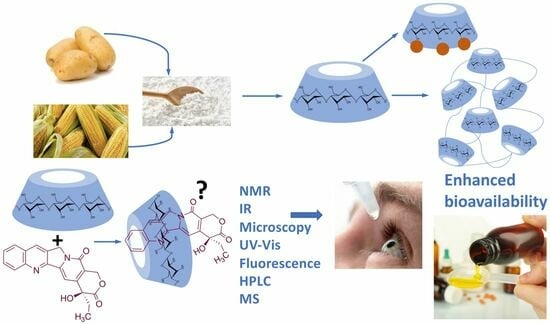
7. Cyclodextrins for Increasing the Bioavailability of Diet Components

This section will consider some essential compounds that are present in foodstuffs and for which the inclusion in the cavities of CDs represents a considerable improvement from the bioavailability point of view. There is a growing interest in natural compounds (nutraceuticals) used as food supplements with the aim of improving the general state of the organism. Among these compounds, those with antioxidant capacity stand out, due to their potential beneficial effect on cellular/physiological aging. The beneficial effects of cyclodextrins on the bioavailability of various nutraceuticals has been the subject of a great deal of research [64,65].
Many polyphenolic compounds, flavones and isoflavones, as well as terpenes, have beneficial pharmacological properties but have very low water solubility and therefore low bioavailability. Such is the case of curcumin [66], which is the main component of turmeric and, in addition to its antioxidant properties, has anti-inflammatory and anticancer activities. The formation of curcumin/HPβ-CD inclusion complexes increases the plasma concentration of curcumin by two-fold, thus improving its bioavailability compared with free curcumin. In addition, the inclusion complexes show more sustained plasma levels over time and, consequently, the beneficial effects of curcumin are prolonged.
Another interesting example of a compound that is present in many fruits is naringenin (Figure 11). Naringenin is a flavonoid with antioxidant activity and also has hypolipidemic and hypoglycemic activity. Naringenin [67] has very low water solubility, and thus its beneficial effects are limited by its poor bioavailability. The formation of naringenin/HPβ-CD inclusion complexes increases its water solubility by hundreds of times and increases its penetrability 11-fold in the Caco-2 cell membrane transport model. The naringenin/HPβ-CD inclusion complexes also lower plasma glucose levels due to a two-fold increase in glucose clearance compared with free naringenin.
Some additional examples of increased bioavailability via CD inclusion are summarized in Table 2.

8. Cyclodextrin Nanofibers

CDs are capable of self-assembling into nanotube-like structures due to intermolecular interactions through hydrogen bonding between the primary and secondary hydroxyl groups on the rims of CD units, connecting each CD unit with the next one. This phenomenon can take place both in solution and in the solid state. CD nanofibers can be produced from either native CDs or chemically modified CDs and have important advantages over CD-based polymeric materials. Thus, CD nanofibers are free of other chemical additives and only CDs are involved, leading to low toxicity in comparison with CD-based polymers. Furthermore, nanofibers based on CDs present high specific surface (surface area to volume ratio) and thus facilitate the solubility and dispersion of many active pharmaceutical ingredients. Nanofibers also present low density and high porosity compared with other conventional fibers [92].
One of the problems associated with the formation of nanofibers is the difficulty in obtaining reproducible degrees of porosity, morphology, and size (diameter and length of the filaments). A successful and reproducible way to achieve the formation of nanofibers that maintain the desired porosity characteristics and dimensions is electrospinning, which is based on the application of a high-voltage electrostatic field to a homogeneous system (solution) or a heterogeneous system (liquid or solid dispersion). The electric field generates a charged jet and charged droplets which are continuously ejected toward a collector device. In this process, solvent is eliminated and thus a solidified nanofiber is originated on the plate surface [93]. In the specific case of nanofibers involving CDs, as well as their inclusion complexes, the energy provided by electrospinning [94] is sufficient to effectively establish the interactions between the CD units and, at the same time, to maintain and elongate the nanofibers and achieve the formation of stable chains, both in the solid state (melts) and in solution. In this way, nanofibers with highly reproducible characteristics (morphology and size distribution) are achieved under fixed experimental conditions (electrical field, flow rate, and tip to collector distance). The use of this technique in the preparation of polymer-free cyclodextrin-based nanofibers and its application in drug delivery through the formation of inclusion complexes with cyclodextrins have been comprehensively reviewed [95,96]. One additional advantage of the use of electrospinning in the production of nanofibers is that it can be considered a sustainable methodology due to the products and reagents used and the almost zero use of solvents, which, together with the use of cyclodextrins, make nanofibers green materials.
Due to the higher specific surface area of the CD nanofibers, an improvement in aqueous solubility is generally observed with a considerable improvement in the bioavailability of many active pharmaceutical ingredients. Thus, a considerable improvement in drug release concentration is observed when comparing the released concentration of powdered sulfisoxazole (Figure 12) with nanofibers produced by electrospinning of the sulfisoxazole inclusion complex with HPβ-CD and hydroxypropyl cellulose. It is significant that in the case of the CD-based nanofibers, a more sustained action over time was achieved [97].
Acyclovir (Figure 12) is an antiviral widely used in the treatment of the herpes simplex virus, but it has the disadvantage of a very low hydrosolubility and therefore low oral bioavailability. The formation of inclusion complexes with HPβ-CD and the subsequent generation of nanofibers via electrospinning provides an improvement in solubility of the drug for oral administration [98].
Ciprofloxacin and dexamethasone (Figure 12) are a frequent combination of an antibiotic and a steroidal drug employed in the treatment of otitis externa. Both active ingredients belong to class IV (BCS) with low hydrosolubility and permeability. The production of nanofibers based on inclusion complexes with β-CD and polycaprolactone and gelatin as polymeric materials has enhanced drug release and pharmacological activity [99]. Significant improvements in the solubility of hydrocortisone [100] and piroxicam [101] have been reported due to the high degree of wetting of the nanofibers obtained via electrospinning from the inclusion complexes with HPβ-CD, with the advantage of not using additional polymeric materials.
Perillaldehyde (Figure 12) is a natural product with antioxidant and antibacterial activity. Besides being volatile, it has a very low water solubility and therefore very poor bioavailability. The formation of inclusion complexes with HPγ-CD and the formation of nanofibers free of other polymeric materials leads to a substantial increase in water solubility. In addition, it has been shown that nanofibers containing the perillaldehyde/HPγ-CD inclusion complex have a higher antioxidant capacity, improved thermal stability, and increased antibacterial activity [102].
The prolonged and repeated use of fungicides and other agrochemicals and the fact that some of them have very low water solubility means that they can accumulate both in soils and in the atmosphere. The formation of inclusion complexes with CDs can be applied to remedy these negative effects. It has recently been described that the solubility of pentachloronitrobenzene (Figure 12), an antifungal compound used in agriculture to combat numerous insect pests, is increased by approximately three times owing to the formation of an inclusion complex with HPβ-CD and the formation of nanofibers via electrospinning [103]. The formation of nanofibers based on inclusion complexes of the agrochemical thiram (Figure 12) and HPβ-CD is very effective in the release of this pesticide and conforms to the principles of safety and green and sustainable agriculture [104]. In the search for sustainable formulations, it has been proposed that pesticide inclusion complexes should be formulated to have a higher water solubility and be environmentally friendly. Thus, the formulation of thiabendazole/HPβ-CD inclusion complexes (Figure 12) and the formation of nanofibers free of additional polymers and generated via electrospinning is an attractive approach. The results obtained are successful in the context of green technology and this methodology has been proposed with the aim of reaching a higher level of environmental protection [105]. Similar results have been described for the fungicide difenoconazole (Figure 12) [106].

9. Functionalization of Cyclodextrins: Synthetic Aspects

9.1. Introduction

The functionalization of natural cyclodextrins is very important for pharmaceutical applications. Although natural CDs are hydrophilic, their aqueous solubility is relatively low due to their tendency to generate aggregates by intermolecular hydrogen bonding. This may cause some nephrotoxicity, especially in the case of β-CD, due to the renal accumulation of insoluble cyclodextrin crystals or cyclodextrin–cholesterol complexes. Thus, much effort has been devoted to the chemical modification of natural cyclodextrins in order to improve their solubility and complexation capacity. Over 11,000 cyclodextrin derivatives have been prepared by incorporation of various moieties such as amines, amino acids, peptides, and aromatic rings. Moreover, more sophisticated supramolecular systems such as polymers, metal–organic frameworks, hydrogels, and other supramolecular assemblies have also been assembled from cyclodextrins [107].
The chemical modification of cyclodextrins is carried out via reactions in nucleophilic hydroxyl groups. However, selective cyclodextrin functionalization, as opposed to random substitution, is a challenging task because their hydroxyl groups exhibit only slight differences in their reactivity. There are, nevertheless, some characteristic properties for each position that may allow for selective modification if moderately reactive reagents are employed: the hydroxyl group at position 2 is the most acidic, the hydroxyl at position 6 is the most nucleophilic, and the hydroxyl residue at position 3 is the least accessible due to steric hindrance and hydrogen bonding, and hence the least reactive [108]. On the other hand, cyclodextrins can form complexes with reagents in different ways depending on the size of the cavity and the solvent, modifying their reactivity and sometimes making their performance unpredictable [109] (Figure 13A). To add an additional layer of complexity, many different functionalization patterns can be obtained for the cyclodextrins resulting from a functionalization reaction (Figure 13B).
Three main strategies can be employed for the preparation of functionalized cyclodextrins [110]:
(a)
Straightforward selective modification of the desired positions. This is chemically challenging due to the competition between the hydroxyl groups at positions 2, 3, and 6 of the glucopyranose.
(b)
Protection–deprotection strategies which involve taking advantage of the different reactivity of the hydroxyl groups toward protection and deprotection conditions. Deprotected hydroxyl groups can then undergo the desired modifications.
(c)
Non-selective modification of hydroxyl groups to provide a mixture of derivatives that is then purified to isolate the desired one. This method generates a large number of undesired by-products, requires tedious purifications, and is not chemically efficient.

9.2. Selective Modification of the Primary Rim

9.2.1. Monosubstitution at C6-OH

Primary hydroxyl groups are more nucleophilic than their secondary counterparts, making modification of the primary rim of cyclodextrins relatively straightforward. Electrophiles react with the hydroxyl groups at C-6, yielding the desired product and an acid, which is neutralized by a base added to the reaction medium in order to avoid decomposition of acid-sensitive cyclodextrins. Common strong electrophiles suitable for nucleophilic attack by C6-OH include alkyl, sulfonyl, phosphoryl, silyl, and acyl chlorides [64,109]. Some representative transformations are summarized in Figure 14, and will be discussed below.
Monosubstituted O-sulfonylcyclodextrins are commonly used as substrates for the attack of nucleophiles to introduce functional groups at position 6. Mono-6-tosylcyclodextrin, for instance, is prepared via the reaction of the starting cyclodextrin with tosyl chloride in an aqueous basic environment [111,112] and can be further attacked by nucleophiles to yield 6-monofunctionalized cyclodextrins after chromatographic purification. Preactivation of the tosylate with imidazole improves selectivity toward 6-O-monotosylation [113]. The reaction with p-toluenesulfonic anhydride, on the other hand, does not require purification via chromatography since unreacted anhydride can be simply removed using filtration [114]. Strong bases need to be avoided because they can deprotonate the more acidic hydroxyl group in position 3, generating an alkoxide that displaces the leaving group at C-6 yielding a bicyclic structure.
Aldehydes are versatile groups for further functionalization. α, β, and γ-cyclodextrins can be converted to their mono-6-formyl derivatives in a straightforward manner using the Dess–Martin periodinane reagent [115]. An alternative, indirect way to obtain 6-formylcyclodextrins involves the reaction of the corresponding O-tosyl derivatives with DMSO and collidine [112,116].
The direct introduction of a single aromatic thioether is achieved via a thio-Mitsunobu reaction where β-cyclodextrin is mixed with thiophenol and diisopropyl azodicarboxylate in dimethylacetamide to yield the mono-6-phenylthio derivative with relatively good selectivity over the di- and trisubstituted by-products. The relative amounts of differently functionalized cyclodextrins could be tuned by modification of the reaction conditions [117].
A solid phase method provides mono-6-functionalized α and β-cyclodextrins bearing a variety of complex substituents bridged through a phosphodiester link from the unprotected native cyclodextrins. The Novagel amino solid support was derivatized with 4-hydroxy-3-nitrophenylacetic acid, which was then functionalized with a suitable phosphoramidite derivative containing the structural fragment to be attached to the cyclodextrin (e.g., a saccharide). Cyclodextrins were added to this support in the presence of 1-(mesitylene-2-sulfonyl)-3-nitro-1,2,4-triazole (MSNT) as a coupling reagent, and this was followed by treatment with aqueous ammonia [118].
Mono-6-azidocyclodextrins are accessible through a reaction of α, β, or γ-cyclodextrins with lithium azide, triphenylphosphine, and carbon tetrabromide [119]. However, di- and triazido cyclodextrins are formed as well, making a purification step necessary. Azido cyclodextrins can be readily reduced to the corresponding amino derivatives using the Staudinger reaction [120] or hydride donors [121] and are good 1,3-dipoles for click chemistry.
Together with tosylation, halogenation is the most common starting point for cyclodextrin functionalization. The most convenient and general route to these halo derivatives uses N-halosuccinimides as the halogenating reagent [122]. These halides (and also the tosylates) can be readily transformed by nucleophilic substitution. For instance, their reaction with an excess of sodium or lithium azide quantitatively provides an alternative route to mono-6-deoxy-6-azido-cyclodextrins.
Selective mono-6-esterification of β-cyclodextrin has been achieved through its reaction with maleic acid or itaconic acid to selectively yield the corresponding monoesters using phosphate as a catalyst in a semi-dry process [123].
In another approach, selective protection of the secondary rim makes the primary rim readily available for modifications. Taking advantage of their higher basicity, the secondary hydroxyl groups at position 2 of β-cyclodextrins can be selectively protected with t-butyldimethylsilyl chloride using NaH as base. The introduction of these bulky groups allows for the primary hydroxyls to undergo reactions with electrophiles without competition from the C-2 or C-3 hydroxyls. The mild removal of silyl protective groups from the secondary rim is then accomplished using tetrabutylammonium fluoride [124].

9.2.2. Disubstitution at C6-OH

Compared with monosubstitution, the synthesis of disubstituted cyclodextrins entails an additional issue: the formation of regioisomers, which need to be separated using chromatographic techniques. However, in some cases it is possible to guide the reaction toward the formation of a specific regioisomer.
Some of the first cyclodextrin capping strategies involved the introduction of aromatic residues from aromatic disulfonyl chloride reagents. Highly regioselective capping was found in the case of benzophenone-3,3′-disulfonylchloride, which mainly resulted in the AC regioisomer, while biphenyl-4,4′-sulfonyl chloride mainly afforded the AD capping product (Figure 15) [125].
Diiodide derivatives obtained from disulfonyl cyclodextrins can also serve as substrates for further modifications through nucleophilic attack. Sulfonyl groups are displaced via reaction with iodide ion to yield the corresponding diiodo cyclodextrins [126]. Similarly, diazido cyclodextrins can be readily obtained from sulfonyl derivatives [127].
The selective synthesis of AD diols at the primary side is achieved through a DIBAL-H-mediated regioselective deprotection of the primary hydroxyl groups of perbenzylated α, β, and γ cyclodextrins [128]. For steric reasons, this bis-debenzylation takes place at the opposite glucose rings and involves two independent Lewis acid-assisted debenzylations (Figure 16). It was possible to stop the deprotection after the first debenzylation and isolate the monohydroxy cyclodextrin [129].
Perbenzylated α-cyclodextrins can be functionalized using an iterative process that predictably directs the debenzylation steps to access cyclodextrins with two, three, and five points of attachment for one, two, or three different new functions [130]. In a refinement of the method, through five consecutive site-directed debenzylations mediated by diisobutylaluminium hydride (DIBAL-H), a 2,3-tetradecabenzylated cyclodextrin hexa-hetero-functionalized with six different substituents at the primary hydroxyls was accessed [131].

9.2.3. Other C6-OH Substitution Patterns

Trisubstitution of cyclodextrins is a difficult goal for the variable substitution degree the reactions provide as well as for the generation of numerous positional isomers. However, some methods have successfully introduced three substituents in the primary side of cyclodextrins. Thus, complex di- and trifunctionalization patterns on the primary side of α-cyclodextrins are readily accessible owing to selective, iterative deprotections of perbenzylated α-cyclodextrin, where the first steps guide the selectivity of the subsequent ones. These modifications allow to selectively deprotect certain positions and also introduce deoxymethyl and deoxyvinyl residues at C-6 [131]. Similarly, perbenzylated α-cyclodextrins were selectively deprotected to yield a variety of primary-rim-functionalized compounds such as an ABD trihydroxy perbenzylated cyclodextrin and an AD diol, CF O-diTBS-protected perbenzylated cyclodextrin containing three pairs of functionalities on its primary rim [132].
Regarding the complete functionalization of primary alcohols at the primary rim of cyclodextrins, it has, in principle, the advantage that no positional isomers are generated. Nevertheless, factors such as steric crowding and complex formation decrease the yield as the degree of substitution increases. Sulfonylation with tosyl chloride is a good approach to perfunctionalize cyclodextrins, and has been applied to α- [133], β- [134], and γ- [135] cyclodextrins. Persulfonylated cyclodextrins are generally used as intermediates toward other functionalized CD derivatives.
Perhalogenated cyclodextrins also represent a good intermediate for further functionalization, with a higher stability than sulfonyl derivatives but with compromised solubility in non-polar solvents. Direct per-6-iodination of α and β-cyclodextrins can be achieved through reaction with iodine and triphenylphosphine in DMF. Similarly, the reaction of β-cyclodextrin with triphenylphosphine and bromine yields the corresponding per-6-brominated β-cyclodextrin [136]. α, β, and γ-cyclodextrins react with chloromethylenemorpholinium chloride and bromomethylenemorpholinium bromide in DMF to furnish the per-6-chlorinated and per-6-brominated cyclodextrins, respectively [137]. These per-6-halo cyclodextrins are very useful starting materials for additional functionalization processes such as the synthesis of per-amino derivatives and amino acid derivatives [122]. The use of per-6- iodo-per-6-deoxy-β- and γ-cyclodextrins for the synthesis of azido or mercapto derivatives has been performed under solvent-free conditions in a planetary ball mill. This mechanochemical protocol simplified the isolation and purification processes, while also allowing an efficient scale-up [138].
Per-6-alkylation of cyclodextrins is not a straightforward transformation and requires the adoption of a protection–deprotection strategy. An example with α-cyclodextrin involves, in this order, selective protection of the primary hydroxyls as silyl ethers, benzylation of the secondary hydroxyls, selective desilylation of the primary side, reaction of the primary hydroxyls with methyl iodide under strongly basic conditions (sodium hydride), and a final hydrogenolysis of the benzyl ethers [139].
The selective introduction of azido groups at all primary hydroxyls is achieved by the reaction of β-cyclodextrin with triphenylphosphine, lithium azide, and carbon tetrabromide in DMF with further treatment with acetic anhydride to yield heptakis(6-azido-6-deoxy)-β-cyclodextrin-tetradeca(2,3)acetate [140]. Per-azido cyclodextrins have been investigated as substrates for copper-catalyzed azide–alkyne cycloadditions [141].

9.3. Selective Modification of the Secondary Rim

In general, chemical modification of this side of cyclodextrins is not as straightforward as at the primary rim. This side is more crowded and hydroxyl groups at positions 2 and 3 establish hydrogen bonds making it more rigid. Furthermore, 2 and 3 hydroxyls differ in their reactivity: while the hydroxyl group C-2 is the most acidic of the glucopyranose and is selectively deprotonated with strong bases such as NaH, the C-3 hydroxyl is the least reactive.

9.3.1. Monosubstitution at C2-OH

β-cyclodextrin can be transformed into the corresponding 2-monotosyl-β-cyclodextrin via reaction with m-nitrophenyl tosylate [142]. The ability of cyclodextrins to form complexes with chemical reagents through the hydrophobic cavity modifies their reactivity, and this feature is used here to selectively direct the reaction to the secondary rim of the cyclodextrin instead of the more reactive primary rim. This complex-formation-based approach was optimized with the design of a more efficient tosyl donor, 1-(p-tolylsulfonyl)-1,2,4-triazole [143]. An additional method to selectively synthesize monotosyl α and β-cyclodextrins involves their reaction with N-tosylimidazol in DMF and molecular sieves [144] or under solid-state conditions, using mechanochemical activation [145]. 2-Monotosyl cyclodextrins are key intermediates for the functionalization of not only C-2 but also the C-3 position through the formation of an epoxide.
Direct alkylation at position 2 of β-cyclodextrin is achieved through its reaction with dialkyl sulfates in dilute aqueous alkali [146]. An indirect method to reach position 2, as well as position 3, is the regioselective bis-O-demethylation of 2,3,6-permethylated α and β-cyclodextrins with DIBAL-H in toluene to yield 2A,3B-diol permethylated cyclodextrins as major products, allowing for selective functionalization of a single 2 hydroxyl and/or a single 3 hydroxyl [147,148].

9.3.2. Disubstitution at C2-OH

The reaction in DMF of γ-cyclodextrin with benzophenone-3,3-disulfonylimidazole, a rigid disulfonyl precursor, selectively yields 2AB-disulfonylated γ-cyclodextrin [149]. A related rigid disulfonyl imidazole reagent, 1,4-dibenzoyl benzene-3′,3″-disulfonyl imidazole, selectively orients the sulfonylation toward the positional isomer 2AC in α, β, and γ-cyclodextrins [150]. The design of a longer disulfonyl imidazole bridge yielded the positional isomer 2AD in α, β, and γ-cyclodextrins, with a better selectivity for β [151]. These 2-disulfonyl cyclodextrins can serve as versatile building blocks for further modifications at position 2 as well as position 3 through the formation of an intermediate epoxide or by treatment with ammonia to yield the corresponding amine.

9.3.3. Persubstitution at C2-OH

Persubstitution at position 2 is favored by the use of strong bases that deprotonate C2-hydroxyls. However, the successive introduction of electrophiles makes the secondary rim increasingly crowded, making subsequent substitutions more difficult and shifting the reactivity toward the primary hydroxyls. To avoid this, per-2-tosylations and per-2-alkylations are often carried out after the protection of primary hydroxyls with silyl groups, which can be deprotected afterward [152]. An exception is the selective per-2-methylation of β-cyclodextrin with methyl iodide, sodium hydride, and DMSO [153], or using N-tosylimidazole and a catalytic amount of Cs2CO3 in DMF [154].
A different approach for the selective per-2-alkylation of cyclodextrins starts with the selective per-2,6-silylation of β-cyclodextrin with tert-butyldimethylsilyl chloride and pyridine. Then, during the reaction with methyl iodide and sodium hydride, the silyl groups at the C-2 hydroxyls migrate to the C3-OH groups, and the deprotected position 2 is selectively methylated. A final desilylation affords the per-2-methylated cyclodextrin [155]. This C2-OH to C3-OH migration is also observed in other cases where a more complex substitution pattern is present [156,157].

9.3.4. Monosubstitution at C3-OH

C-3 in cyclodextrins are the least reactive hydroxyls in the glucopyranose ring due to their low accessibility. A common strategy to functionalize position 3 consists of the tosylation at C2-OH and treatment with a base to generate an epoxide, followed by the introduction of nucleophiles [158]. This provides a mixture of 2- and 3-substituted products that can be separated with chromatographic techniques, with substitution at position 3 being highly favored in most cases. The combination of N-benzoylimidazole and carbonate buffer in DMF gave mainly acylation C-3 hydroxyl groups of β-CD, which was ascribed to acylation at both C2-OH and C3-OH accompanied by C2-OH to C3-OH acyl migration [159]. In other transformations, reagent complexation in the cyclodextrin cavity may direct the reaction to C3-OH. For instance, the 3-sulfonylation of α-cyclodextrins is achieved by direct reaction with β-naphtalenesulfonyl chloride [160]. A particular case of alkylation is the selective introduction of an alkyl group at just one 3 position of β-cyclodextrins through the reaction of per-6-terbutyldimethylsilylated β-cyclodextrin with N-methyl-4-chloromethyl-2-nitroaniline in the presence of lutidine [161].

9.3.5. Di- and Trisubstitution at C3-OH

In a similar way to that of monosubstitutions at position 3, the manno-2,3-epoxycyclodextrin is a common intermediate used for the introduction of nucleophiles. The positional isomers obtained depend on the initial 2-ditosylate involved in the route. Also, 3-sulfonylation of α-cyclodextrins is achieved via reaction with β-naphtalenesulfonyl chloride and further isolation of the 3-disulfonylated derivatives, particularly a mixture of positional isomers 3AC and 3AD disulfonyls that are obtained due to an oriented synthesis owing to complex formation between the β-cyclodextrin and sulfonyl chloride [162]. The mixture of regioisomers is easily resolved with reverse-phase column chromatography.
The sulfonylation of β-cyclodextrin with β-naphtalenelsulfonyl chloride in basic aqueous acetonitrile was selectively found to yield the 3ACE-trisulfonylated isomer [163].

9.3.6. Persubstitution at C3-OH

Perfunctionalization at position 3 of cyclodextrins is a challenging task because 3-hydroxyls are the least reactive and are involved in hydrogen bonding with 2-hydroxyls. Furthermore, they are located in a crowded side of the cyclodextrin which becomes even more crowded after protection of 2-hydroxyls, hindering further reactions.
Despite all these setbacks, perbenzylated α-cyclodextrin can be selectively deprotected at position 3 with Et3SiH and I2 to provide a 2A–F,6A–F-dodeca-O-benzyl α-cyclodextrin. This strategy works as well starting from a 2A–F,3A-F,6A-E-heptadec-O-benzyl α-cyclodextrin and a 2A–F,3A-F,6BCEF-hexadec-O-benzyl α-cyclodextrin to yield the corresponding 3-deprotected derivatives with one or two deprotected hydroxyls in the primary side, respectively. These derivatives stand out as useful starting materials for the selective perfunctionalization of the relatively unreactive position 3 [164].

9.4. Persubstitution of the Primary and Secondary Rims

Although steric hindrance of persubstituted cyclodextrins make the introduction of substituents increasingly difficult, the absence of regioisomers is a major advantage in the synthesis of 2,3,6-perfunctionalized cyclodextrins. Perbenzylation of α and β-cyclodextrins is performed in excellent yield (>90%) by treatment with benzyl chloride and sodium hydride [130]. Similarly, peralkylation of β-cyclodextrin is achieved in 74% yield via reaction with methyl iodide and sodium hydride in DMSO [165].

9.5. Cyclodextrin-Based Polymers

CD-based polymers are the basis for new CD-based materials for different types of application fields for pharmaceutical and biomedical applications, but are also useful in cosmetic and food science [166,167,168].

9.5.1. Radical Polymerization Reactions

Atom transfer radical polymerization (ATRP) is a type of radical polymerization having as the key step a transition metal-catalyzed C-C bond generation. Generally, an alkyl halide R-X is used as the initiator and a complex formed by a transition metal (e.g., CuI) and a chelating agent is used to generate radicals via a one-electron transfer process, while simultaneously the transition metal is oxidized to a higher oxidation state (Figure 17).
α-Haloester moieties attached to cyclodextrin-based monomers have been widely employed for performing ATRP processes. These moieties can be introduced by direct per-acylation of β-CD with 2-bromoisobutyryl anhydride (Figure 18A) [169]. Alternatively, the primary rim can be selectively functionalized starting from the corresponding mercaptocyclodextrin using a radical thiol–ene reaction (Figure 18B) [170]. As an example of one of these polymerizations, we summarize in Figure 18C the synthesis of a 21-arm star polymer via a core-based approach, with the core being a molecule of β-cyclodextrin. To this end, heptakis [2,3,6-tri-O-(2-bromo-2-methylpropionyl]-β-cyclodextrin, prepared via the peracylation method described above, was treated with methyl methacrylate in the presence of copper bromide and n-propyl-2-pyridylmethanimine as a bidentate ligand for copper complexation.
In a reverse approach, the acrylate moiety required for the polymerization process can be introduced first into the cyclodextrin. Thus, treatment of 6-monopiperazino-β-cyclodextrin, obtained from the corresponding 6-monotosyl derivative, with glycidyl methacrylate afforded a mono-methacrylate substituted cyclodextrin via epoxide opening (Figure 19A) [171]. A similar type of starting material can be obtained by means of copper-mediated azide–alkyne couplings (CuAAC) of azido-cyclodextrins (Figure 19B) [172].
Mono-methacrylate cyclodextrins are useful as starting materials for radical polymerization reactions. Thus, poly(ethyleneglycol)-β-poly(cyclodextrin) (PEG-β-PCD), can be easily synthesized via atom transfer radical polymerization using a poly(ethylene glycol) macroinitiator (Figure 19C). This well-defined, hydrophilic, CD-pendant copolymer was able to include a variety of guest molecules and self-assembled into advanced nanostructures due to the synergistic effect of CD moieties [172].
Reversible addition–fragmentation chain transfer (RAFT) polymerization is another relevant modality of radical-induced polymerization that has proved to be an excellent tool to access a range of well-defined macromolecular architectures. This technique involves the use of a chain-transfer agent, usually a thiocarbonylthio compound, which helps to achieve control over the generated molecular weight and polydispersity during free-radical polymerization (Figure 20) [173].
Thiocarbonylthio functional groups can be installed onto cyclodextrin molecules by a simple acylation process using benzyl (3-chloro-3-oxopropyl) carbonotrithioate, an acid chloride [174], or the corresponding acid in the presence of dicyclohexylcarbodiimide (DCC) and hydroxybenzotriazole (HOBT) as a coupling reagent [175]. RAFT polymerization of this starting material in the presence of 2-hydroxyethyl acrylate (HEA), using 2,2′-azobisiobutyronitrile (AIBN) as initiator, afforded CD-based polymers with seven-arm star architectures [175] (Figure 21).

9.5.2. Ring-Opening Polymerization (ROP)

Cyclodextrins can serve as core structures for anionic ring-opening polymerization (ROP) processes, which involve CD derivatization using cyclic esters [176]. Thus, treatment of β-cyclodextrin acetylated at its secondary rim with ε-caprolactone in the presence of Sn(Oct)2 as a catalyst resulted in polymers with narrow dispersity by esterification of the primary rim. The polymer arms were then coupled with DCC to carboxyl-terminated poly(ethylene glycol) to form amphiphilic copolymers that were able to undergo micellization (Figure 22) [177]. The ring opening polymerization of ε-caprolactone can also be carried out in the presence of wet β-cyclodextrin and has been performed in pressurized (12–13 MPa) batch reactors [178]. Lactide (3,6-dimethyl-1,4-dioxane-2,5-dione) is also widely employed to generate cyclodextrin-based polymers and, in the presence of 4-dimethylaminopyridine as an organocatalyst, has allowed the quantitative functionalization of α-, β-, γ-, and 2,3-dimethyl-β-cyclodextrins, with narrow molecular weight distributions (dispersity < 1.1) [179].

9.5.3. CuAAC-Based Polymerization

Although ATRP is a suitable tool for the core-first synthesis of multiarmed polymers, it has some limitations because the polymerization of many arms from one core can lead to steric hindrance and a high local concentration of free radicals that facilitates star–star coupling and other termination events. This can be improved by use of the so-called grafting-to approach, where a functional core is coupled to a functional polymer, where the most extended reaction for such coupling is the copper-catalyzed azide–alkyne cycloaddition (CuAAC) [180], which is frequently associated with ATRP polymerization. To this end, a polymer containing an alkyne moiety joined to poly(N-isopropylacrylamide) (PNIPAM) is prepared via the ATRP polymerization technique (Figure 23A) and this precursor is then coupled to an azido-CD, which can be prepared via nucleophilic displacement of halogens at the primary rim of cyclodextrins (Figure 23B). In order to involve all cyclodextrin hydroxy groups, the primary rim can be modified via the CuAAC reaction and the secondary rim is used as the substrate of an ATRP or ROP polymerization. Alternatively, per-acylation with 2-bromopropionic bromide followed by bromide nucleophilic displacement with sodium azide affords a per-azidocyclodextrin, which is then submitted to a CuAAC reaction with the alkyne-PNIPAM polymer to furnish a 21-star polymer (Figure 23C) [181].

9.6. Cyclodextrin-Embedded Covalently Crosslinked Networks

Hydrogels are three-dimensional crosslinked polymeric networks [182,183]. These biomaterials show unique characteristics such as their biocompatibility, water retention ability, and morphological similarity to bodily tissues that make them very useful for many biomedical applications. The incorporation of cyclodextrins, combining hydrophilic exteriors with hydrophobic inner pockets able to form inclusion complexes, provides an interesting approach to the design of hydrogels. The main crosslinking methods employed to prepare CD-based materials are summarized in Figure 24 and include radical polymerization, CuAAC-based polymerization, and polymerization processes based on nucleophilic addition or substitution reactions.
One interesting example of cyclodextrin-based networks are nanosponges, which can be described as colloidal structures that contain solid, three-dimensional, hyper-reticulated nanoporous structures that protect the entrapped molecules from degradation and help to improve the solubility of lipophilic compounds [184]. Cyclodextrin-based nanosponges (CDNSs), in particular, can be efficiently engineered for drug delivery purposes, especially in cancer therapy [185,186,187]. Indeed, due to the existence of lipophilic cavities in the cyclodextrin building blocks and the possibility to install a hydrophilic network in the nanosponge itself, depending on the nature of the crosslinker, these materials are ideal candidates to increase the stability of sensitive and volatile compounds, as well as the solubility of both lipophilic and lipophobic compounds [188,189].
The synthesis of these nanostructures is achieved following the general scheme shown in Figure 25, where a suitable difunctional cross-linking reagent generates covalent bridges between cyclodextrin molecules by reacting with their hydroxyl groups to generate carbamate, carbonate, diester, or diether links. These transformations can be promoted under a number of experimental conditions, including, among others, solution conditions in conventional or non-conventional solvents, the joint melting of the CD and the linker [190], and ultrasound-assisted [191], microwave-assisted [192], and mechanochemical [193] conditions.

10. Analytical Aspects

10.1. Introduction

The characterization of inclusion complexes is usually carried out by combining several analytical techniques [194] that allow differentiating the included drugs from their free forms. For this purpose, it is important to study the possible changes observed in the experimental data obtained with the presumed inclusion complexes with respect to those obtained with pure guests, CDs, whose characterization has been reviewed by Szente et al. [195], or guest-CD physical mixtures. These changes in the measured instrumental magnitudes are either the result of alterations in the physical properties of the included compound or CDs, or result from the occurrence of chemical interactions (mainly hydrogen bonds, van der Waal forces, dipole–dipole electrostatic bonds, and hydrophobic interactions) between the guest and the host entities. Therefore, it is essential to apply sensitive and selective analytical techniques. Generally, techniques such as nuclear magnetic resonance or those performed on the complexes in the solid state present a lower sensitivity than that shown by techniques such as mass spectrometry or fluorimetry.
For studying the inclusion complexes, it is necessary to take into account the state in which they are obtained (solution or in solid state), with the procedure used for their preparation being critical [196], which strongly influences the properties of the presumed inclusion complex. Thus, Logadekar et al. [197] achieved the optimal inclusion complex of niclosamide-HP-β-CD by freeze drying compared with kneading or solvent evaporation procedures, while Araujo-Filho et al. [198] and Heimfarth et al. [199] found that the slurry complex method was the best one for the complexation of D-limonene with β-CD and of myrtenol with β-CD, respectively. Moreover, Shende et al. [200] obtained better results with respect to increased solubility and wettability of meloxicam when forming the inclusion complex with β-CD-based nanosponges instead of β-CD.

10.2. Analytical Techniques for Studying Inclusion Complexes in the Solid State

The solid CD inclusion complexes can be prepared [201,202] by removing the solvent from a solution of guest and CDs through co-evaporation, co-lyophilization, or spray drying. They can also be obtained by kneading a slurry of a physical mixture of the compound and CDs in the presence of a volatile solvent or a small volume of water that later is evaporated, or alternatively co-grinding the drug and CDs under microwave irradiation, sealed-heating using supercritical fluid, ultrasound, magnetic stirring, or high energy ball milling.
The main analytical techniques that can be used for the characterization of solid-state inclusion complexes include thermal analytical techniques, X-ray diffraction, electron microscopy, and spectroscopic methods [203]. Among them, the most commonly used are Fourier-transform infrared spectroscopy (FTIR), differential scanning calorimetry (DSC), scanning electron microscopy (SEM), and powder X-ray diffraction (PXRD) [202,203,204]. Nuclear magnetic resonance spectrometry (NMR) may be employed both for studying solid state inclusion complexes and isolated solid inclusion complexes and then solved in adequate conditions for NMR determination.

10.2.1. X-ray Diffraction

This technique is suitable for characterization and identification of solid compounds, either crystalline or powder solids. It is a non-destructive technique that allows the sample to be used in further analyses via other analytical techniques. If the guest-CD inclusion complex crystallizes in stable single crystals of dimensions between c.a. 80 and 250 μm, these complexes can be studied via single-crystal X-ray diffraction (SCXRD), as in the case of the resveratrol/trimethyl-α-CD inclusion complex [205].
Inclusion complexes with these characteristics are difficult to obtain, and hence powder X-ray diffraction (PXRD) is used in most cases. PXRD does not require any sample pre-treatment since it is performed on a finely ground and homogenized sample and so the sample does not undergo any physical or chemical change during analysis. The diffractograms for the free guest, the single CD, the physical mixture, and the presumed inclusion complex are recorded and compared with each other. New peaks or variations in the intensity of the characteristic peaks of the pure compound may appear in the diffractogram of the presumed inclusion complex, showing the formation of a new crystalline species [202], as can be observed in the case of the (-)-borneol/β-CD inclusion complex [206], or a decrease in the crystallinity of the guest, such as in the case of the nifedipine/β-CD inclusion complex [86], which means a change in the properties of the drug due to the occurrence of interactions between the drug and the CDs. In other cases, in the diffractogram of the presumed inclusion complex, the loss of the crystalline structure of the pure compound and its transformation into an amorphous structure are observed, while the diffractogram of the physical mixture usually retains the representative peaks of the single CD and the free drug, although with less intensity. These observations can be interpreted as evidence of the formation of a true inclusion complex as observed for the niclosamide/HP-β-CD inclusion complex [198], meloxicam/β-CD-based nanosponges [201], and genistein/amino acid appended β-CD [79]. These results were corroborated by the experimental data obtained using other analytical techniques, especially DSC.

10.2.2. Thermal Analytical Techniques

These are a set of techniques that, by subjecting the sample to controlled heating, allow the study of processes that involve a change in enthalpy such as crystallization, melting, or vaporization, or a change in weight like dehydration and volatilization, as well as their thermal stability (degradation or decomposition). Differential scanning calorimetry (DSC) and thermogravimetric analysis (TGA) [207] provide relevant information for the characterization of drug-CD inclusion complexes. For this purpose, it would be ideal for the guest drug to be a crystalline solid with a melting or boiling point below the thermal degradation range of CD or to be volatile between 60 and 250 °C to allow the evaluation of the formation of a real inclusion complex with CDs. Thermograms of the CDs show two clearly differentiated zones, the first one between 90–130 °C corresponding to the loss of water molecules housed in the CD cavity, and the second one at temperatures above 300 °C, corresponding to their thermal decomposition [202].
In the cases of compounds whose melting points are in the dehydration range of the CDs, these events overlap in the DSC thermograms and cannot be distinguished. A complementary technique such as hot-stage microscopy (HSM) can be of assistance, since with this technique it is possible to observe the appearance of bubbles corresponding to the evaporation of water within the molten solid. This technique also allows to observe phase changes/recrystallizations, which are hardly visible in DSC thermograms. HSM was employed in conjunction with DSC for the study of the resveratrol/trimethyl-α-CD inclusion complex [206]. It is possible to evaluate the formation of inclusion complexes between CDs and amorphous solids if there is an increase in the decomposition temperature in the thermogram of the inclusion complex with respect to that observed in the thermogram of the pure drug or the physical mixture because complex formation provides an increased thermal stability to the included drug molecule [202].
Differential scanning calorimetry (DSC) is the most widely used of all thermal methods and one of the most powerful analytical tools employed in the solid-state characterization of compounds. DSC is a calorimetric method in which differences in energy (heat flow) are measured under a controlled-temperature program. The thermogram of the free drug, if it is a crystalline solid, will show a well-defined sharp endothermic peak corresponding to its melting point, and if it is a volatile compound it will show a peak representing its volatilization. In the case that it also presents polymorphism, exothermic peaks corresponding to recrystallization processes would be observed. Finally, a descending zone will appear showing the thermal decomposition stage. All these events allow the accurate characterization of the drug. The thermogram of the physical mixture presents peaks due to both the drug and the CD clearly showing the peak corresponding to the melting point of the drug, as well as the dehydration stage of the CD. The complete loss of the melting point peak of the drug in the thermogram of the presumed inclusion complex would be evidence for the formation of a true inclusion complex [198,201]. Other changes that can be observed in this thermogram are a shift to lower temperatures and/or lower intensity in the dehydration zone of the CD and a shift to higher temperatures of the thermal decomposition of the included drug [87]. Thus, in the case of the usnic acid/β-CD inclusion complex, the disappearance of the melting point peak is accompanied by a decrease in the intensity of the β-CD dehydration band and an increase in the thermal stability of the usnic acid [208].
In the DSC study of 17-α-methyltestosterone and β-CD inclusion complexes, it was observed that when the inclusion complex was prepared by lyophilization the peak corresponding to the melting point of the free drug disappeared completely, whereas when prepared by kneading, the peak was observed but with a much-decreased intensity with respect to the free hormone. In addition, the dehydration zone of the β-CD shifted to lower temperatures along with a decrease in its intensity, indicating the loss of water molecules upon inclusion of 17-α-methyltestosterone into the CD cavity [83]. The same behavior was observed for the binary and ternary inclusion complexes of dapsone with HP-β-CD (melting point peak of the free drug is missing) and β-CD (melting point peak decreases in its intensity) [209].
In thermogravimetric analysis (TGA), the loss of mass of the sample during a controlled heating program under a controlled atmosphere is recorded, arising as a consequence of processes such as dehydration, volatilization, or degradation. It is a useful technique for monitoring the process of inclusion of a drug into the CD cavity since it is possible to observe whether the dehydration stage of the CD or the volatilization of a volatile drug is modified, or if the thermal stability of both the CD and the drug is modified. Thus, Dalmolin et al. [210] studied the thermal stability of β-CD and modified β-CD with hexanoic anhydride using this technique, finding that the latter increases its stability since the degradation temperature is shifted from 325 °C in β-CD to 354 °C in chemically modified β-CD. Moreover, in the case of the modified CD, dehydration is not observed, indicating that this CD presents higher hydrophobicity. Trindade et al. [211] formed inclusion complexes between carvacrol (a volatile monoterpene) and β-CD via two different procedures: freeze drying and ultrasound. The formation of these inclusion complexes was evidenced using several analytical techniques, including thermal analytical techniques, such as DSC, TGA, and DTG (differential thermogravimetry). In the thermograms of the inclusion complexes obtained by both procedures, the disappearance of the broad peak corresponding to the volatilization of carvacrol (32–169 °C) was observed. Cerutti et al. complemented the studies of the nifedipine/β-CD inclusion complex with and without aspartic acid using DSC and TGA, with the data collected being in agreement with those obtained via PXRD [86]. It was observed that aspartic acid contributed to the formation/stabilization of the inclusion complex since nifedipine decreased its crystallinity by around 50% in multicomponent systems.

10.2.3. Electron Microscopy

Morphology and surface characterization of the solid particles of the inclusion complexes can be mainly carried out by means of scanning tunneling microscopy (STM) and scanning electron microscopy (SEM); the latter being the most commonly used for this purpose. Atomic force microscopy (AFM) provides extensive surface information that other electron microscopy techniques cannot reach.
Although scanning electron microscopy (SEM) is not suitable to confirm the formation of a true inclusion complex, it nevertheless provides morphologic and topographic information about the surface of a solid, complementing the information obtained via X-ray or thermal analysis. The samples must be coated with gold or another metal element under vacuum prior to analysis and this fact could alter the solid, therefore being a disadvantage of this technique. The use of SEM enables the determination of the size, shape, and homogeneity of the particles of the solids studied, helping to find the most optimal procedure to obtain the inclusion complex [86,201] or revealing the micro-morphologic changes suffered by the guest and the host when the inclusion complex is formed. Dapsone changed from irregular orthorhombic crystals to well-defined spherical particles when it interacted with HP-β-CD [210]. Genistein appeared as large cuboidal particles with claviform crystals of different size and amino-appended β-CD as hollow spherical particles; however, their inclusion complex appeared as an irregular massive aggregate, which was very different from the physical mixture [79]. Scanning probe microscopy, AFM and STM, make it possible to visualize surface details of solids at the atomic level, achieving an average resolution of 20 Å [212].
Tapping-mode AFM is adequate for distinguishing molecules with similar morphological features but different local mechanical properties, such as CD inclusion complexes and their hosts. Thus, we observed [213] that, as a consequence of complexation, retinal/β-CD nanotubes showed a higher local rigidity than those coming from β-CD. Desai and Prabhakar studied the surface topography of cilostazol/HP-β-CD inclusion complexes, observing a significant change in the morphology of the surface: from rough in the case of the free cilostazol to smooth in the case of the inclusion complex due to the guest–host interactions [214].

10.2.4. Spectroscopic Techniques

Spectroscopic techniques, such as Fourier-transform infrared spectroscopy (FT-IR), attenuated total reflectance (ATR)-FT-IR spectroscopy and Raman spectroscopy, are valuable analytical techniques to evidence and characterize solid state compounds.
Fourier-transform infrared spectroscopy (FT-IR) is largely employed for qualitative organic analysis and structure determination due to its high degree of structural information and intermolecular hydrogen bonding association. Each organic compound presents characteristic absorption bands in the IR spectrum. Therefore, changes in the spectrum of the drug such as an increase or decrease in the intensity of the bands, broadening or shifts in the bands, or the disappearance of some bands may be indicative of the formation of the inclusion complex, suggesting which bonds or functional groups of the drug may be involved in the interaction with the CD cavities. Despite the fact that the main characteristic bands of the free drug are often overlapped or masked by the bands of the CDs, there are certain bands corresponding to carbonyls, carboxyl, nitrile, and amine groups that can be observed and shifted when the inclusion occurs; these shifts and shape changes are not observed in the corresponding physical mixture [202,205]. Thus, the FT-IR spectrum corresponding to meloxicam/β-CD-based nanosponges inclusion complex showed shifts in the aromatic peaks together with an intense broad peak at 3429.7 cm−1 that indicate that the aromatic ring of meloxicam penetrates into the hydrophobic cavity of the β-CD, and then forms hydrogen bonds with the nanosponges [201]. In the case of sulfamethoxazole/HP-β-CD, the absence of SO2 stretching bands is indicative of the interaction of this moiety with the CD cavity [215].
Attenuated total reflectance Fourier-transform infrared spectroscopy (ATR-FT-IR) combines the selectivity and sensitivity provided via FT-IR with the reproducibility, simplicity, and speed of ATR, in addition to being a non-destructive technique. The spectra show the attenuation of the evanescent wave produced by the absorption of this REM by the sample, which is independent of the sample thickness. These spectra are similar to those obtained via FT-IR, but the relative intensity among the peaks or bands are different. The sample handing is very easy, just it is placed and pressed on opposite sides of a transparent crystalline material of high refractive index, thus avoiding the disadvantages derived from the preparation of KBr tablets for conventional FT-IR measurements.
ATR-FT-IR is very useful for studying intra- and inter-molecular hydrogen bonds, allowing to probe the formation of the inclusion complex and showing the specific functional groups implicated in the guest–host interaction [202]. Thus, comparing the spectral changes observed in the O-H stretching band between the physical mixture and the idebenone/HP-β-CD inclusion complex, Venuti et al. [216] demonstrated the involvement of OH groups of idebenone in new guest–host interactions through the formation of a drug-CD hydrogen bond. The complexation was also proven by the relevant changes in the C=O and C=C stretching bands associated with the breakdown of intermolecular hydrogen bonds of the idebenone. ATR-FT-IR has been employed to study different CD-based nanosponges loaded with different drugs, such as repaglinide or dexamethasone [213].
Raman spectroscopy is a complementary technique to FT-IR that measures the inelastically scattered radiation by the molecules of the compound under study. Raman spectra also have functional group and fingerprint regions that permit the identification of specific compounds. In these spectra, the appearance of a peak usually corresponds to a mode of vibration accompanied by a change in the polarizability of the molecule. This technique is useful to study drug-CD inclusion complexes because the inclusion of the guest molecule in the CD cavity leads to changes in the polarizability together with changes in the ring size or/and a decrease in the vibrational relaxation time of the included functional groups, which results in changes in the Raman peaks of the drug and the CD.
As in FT-IR, the peaks from CD Raman spectra overlap the guest Raman peaks. However, there is a zone between 1500–1750 cm−1, corresponding to stretching vibrations of C=O and C=C of the aromatic rings, where CDs present no peaks, which enables observation of the changes in the intensity and mainly the shifts in the wavenumber produced in the characteristic peaks of the drug molecules after inclusion. This region of the Raman spectrum was used to evidence the formation of inclusion complexes of sulfamethoxazole with HP-β-CD and β-CD [216] and the inclusion complex of idebenone with HP-β-CD [217].

10.3. Analytical Techniques for Studying Inclusion Complexes in Solution

In the case of the inclusion complexes in solution, there is a dynamic equilibrium between the free drug and the host. This makes it possible to carry out tests of entrapment efficiency, solubility, stability, or monitoring of reactions that may be altered by the interaction of the drug with the CDs, such as the photo-isomerization of retinoids [13], the hydrolysis of the camptothecin lactone ring [50], and acid–base equilibrium in norharmane [217].
The main analytical techniques that can be used for the characterization of inclusion complexes in solution include spectroscopic, electroanalytical, and separation techniques. Among them, the most commonly used being ultraviolet/visible (UV-Vis) absorption spectroscopy, fluorescence spectroscopy, high-performance liquid chromatography (HPLC) and, above all, nuclear magnetic resonance spectrometry (NMR). Other techniques can also be used for this purpose, such as mass spectrometry, circular dichroism, polarimetry, isothermal titration calorimetry, or dynamic light scattering [204,218]. Molecular modeling techniques help spectroscopic techniques in the elucidation of the three-dimensional structure of the complex, establishing the points of interaction between the drug and the CD, as well as the stability of the inclusion complex. The experimental characterization of the complex stability is concluded by determining its association constant and stoichiometry. Kfoury et al. summarized the main analytical tools employed for the determination of association constants and stoichiometry for CD inclusion complexes [2,219]. It is important to note that the analytical method chosen for the calculation of the drug/CD association constants, as well as the concentration range used in the assay, influences the value found [220].

10.3.1. UV-Vis Absorption Spectroscopy

This technique is widely used for the calculation of association constants and stoichiometry of inclusion complexes, as well as for solubility assays or dissolution tests, drug release studies, and encapsulation efficiency. Although UV-Vis absorption spectrophotometry has limited application for qualitative analysis, it is useful for detecting the presence of certain functional groups that act as chromophores. Therefore, it helps to evidence the formation of the inclusion complex—whether the chromophore group of the drug molecule interacts or is included in the CD cavity—passing into an environment of higher hydrophobicity and thus producing an effect similar to the solvatochromic effect produced by the change in the polarity of the solvents.
Upon interaction of the drug with the CDs, changes are observed in the absorption spectrum of the free drug, which can be in the intensity of the peaks, shifts of the absorption maxima, or appearance of new peaks that were not usually observed under the experimental conditions used. Thus, we studied the formation of inclusion complexes of β-carboline alkaloids harmane and harmine with β-CD and chemically modified β-CDs via UV-Vis absorption spectrophotometry in conjunction with spectrofluorimetry and NMR [14]. The β-carboline ring possess an acidic nitrogen, which loses its proton in alkaline media. In the UV-Vis absorption spectra of harmane and harmine in aqueous solution (pH 7.2–7.9, close to their pKa values), only the band corresponding to the cationic form is observed as a consequence of the rapid proton transfer with the solvent. However, in their UV-Vis absorption spectra in ethanol, on the contrary, only the band corresponding to the neutral form is observed, as it corresponds with a lower polarity environment than water. UV-Vis absorption spectra of the complexes evidence that the inclusion of these alkaloids in the CD cavities hampers the proton transfer and decreases the formation of the cationic form of these alkaloids. The environment provided by CD is partly reminiscent of that provided by ethanol. In the case of the niclosamide inclusion complex with HP-β-CD, only a hyperchromic effect is observed at its absorption maximum with respect to the UV-Vis absorption spectrum of the free drug [198].

10.3.2. Fluorescence Spectroscopy

This technique is characterized by its good selectivity and sensitivity, being a potent tool to verify the efficiency of the formation of the drug/CD inclusion complex. It is also employed for the calculation of association constants and stoichiometry of inclusion complexes, as in the case of the inclusion complexes of camptothecin and luotonin A with β-CD and HP-β-CD [50] and the inclusion complexes of harmane and harmine with β-CD and chemically modified β-CDs [14].
Generally, it is observed that the formation of the inclusion complexes produces an increase in the fluorescence emission intensity and/or in the fluorescence quantum yield of the drug due to the fact that the radiationless deactivation processes are notably decreased, resulting in enhancement of the fluorescence emission. It may also be accompanied by shifts in the fluorescence emission maxima or the appearance of new peaks that were not usually observed for the free drug under the experimental conditions employed.
The formation of inclusion complexes of the antitumor alkaloids camptothecin and luotonin A with β-CD and HP-β-CD was verified spectrofluorimetrically via titration of these alkaloids with increasing amounts of both CDs in aqueous solution at strongly acidic pH values [50]. The fluorescence emission spectra of both alkaloids in aqueous solution at pH 1 show the characteristic maxima of the cationic forms and a shoulder corresponding to the neutral forms, while the corresponding spectra of the inclusion complexes with the studied CDs show, in addition to the maxima of the cationic forms whose intensity hardly varies, the appearance of the maxima of the neutral forms whose intensity increases as the amount of added CD increases. Thus, the more lipophilic neutral form is included in the CD cavity, allowing it to be observed in strongly acidic media.
Furthermore, inclusion complexes of sulfamethoxazole with β-CD and HP-β-CD were also studied via fluorescence spectroscopy [216]. This drug presents an aniline moiety and an isoxazole ring localized at the end of its structure, which can act as an electron donor-acceptor system. The fluorescence emission spectrum of free sulfamethoxazole in aqueous solution shows only one intense peak, centered around 350 nm. The addition of increasing amounts of HP-β-CD to an aqueous solution of sulfamethoxazole leads to the appearance of an additional broad band (centered at 425 nm) in the fluorescence emission spectrum due to the intramolecular charge transfer process that is favored by the inclusion of the drug inside the cavity, in addition to the main emission band of the sulfamethoxazole, the intensity of which increases with the amount of added CD.

10.3.3. Nuclear Magnetic Resonance Spectrometry (NMR)

NMR is one of the most valuable analytical tools for the characterization of inclusion complexes, as it provides insight into the interactions that occur between the drug and the CD, the structure of the inclusion complex, and the orientation of the molecule within the cavity of the CD. This is due to the fact that the proximity between groups and the interactions among the hydroxyl groups of CDs and the drug molecules cause changes in the chemical shifts as well as in the shape of the signals. These changes are more pronounced in the parts of the molecule that are inside the CD cavity.
NMR spectrometric techniques suitable for the study of CD complexes include, among others, the well-known 1H NMR and 13C NMR, diffusion-ordered spectroscopy (DOSY), nuclear Overhauser effect spectroscopy (NOESY), and rotational Overhauser effect spectroscopy (ROESY).
1H NMR and 13C NMR are very powerful techniques to study the interaction between drug molecules and cyclodextrins because the chemical and electronic environments of the protons or the carbons, respectively, are affected by CD complexation, which causes changes in the chemical shifts registered in the inclusion complex spectrum with respect to the free drug and cyclodextrin spectra.
DOSY involves the measurement of diffusion coefficients and it is a technique suitable to study inclusion complexes and show binding between molecules of very different sizes. When a drug is included inside the host cavity, the diffusion coefficients for both drug and host decrease. Nuclear Overhauser effect-based techniques are very useful to explore the existence of intra- and inter-molecular interactions, if the atoms are in spatial vicinity (3–5 Å), helping to clarify the three-dimensional structure of the inclusion complex and providing information about inter-molecular distances [219]. Due to the molecular weight of cyclodextrins, ROESY experiments are usually more sensitive than NOESY.
By simple comparison of the 1H NMR spectra of CDs and the presumed inclusion complex, changes in the chemical shifts of the H-3 and H-5 protons of the CDs (located inside the cavity) prove that the drug molecule has been included inside the CD cavity, as it occurs in the inclusion complex of methyltestosterone and β-CD [83]. If the change in chemical shift occurs only at the H-3 proton, it means that the cavity inclusion is shallow. This was observed in the case of the inclusion complex of cilostazol with HP-β-CD [215]. In addition, in this case, the differences observed between 1H NMR spectra of free cilostazol and the inclusion complex revealed the partial inclusion of the cyclohexane ring and tetrazole moiety of this drug in the HP-β-CD cavity, which was also corroborated via 2D-ROESY and molecular modeling experiments.
For the study of inclusion complexes of camptothecin [50] with β-CD and HP-β-CD, in addition to 1H RMN, 13C RMN was used. Comparing the 13C RMN spectra for the free camptothecin and its inclusion complexes with these CDs, the more intense changes were observed in the signals corresponding to the carbons of the quinoline ring of the camptothecin, which is the most lipophilic part of the molecule, supporting that the quinoline moiety is placed inside the CD cavity. 1H RMN, docking, and molecular dynamics computational studies confirm the full insertion of this part of the molecule in the CD.
β-Carboline alkaloids norharmane, harmane, and harmine inclusion complexes with β-CD and HP-β-CD were characterized using 1H NMR, 13C NMR, and 2D ROESY [15]. From the study of the 1H NMR spectra of the inclusion complexes, it can be observed that the signal corresponding to the water molecules included in the CD cavity disappears and that the signals due to the protons of the -OH at the rims of the CDs are shifted, indicating that the inclusion of these compounds takes place in the CD cavity. The 13C NMR spectra study allowed to distinguish the formation of 1:1 and 1:2 β-carboline/CD inclusion complexes and to propose that the benzene rings of these drugs enter the CD cavity first since they are the most lipophilic part of these molecules. A study of the 2D-ROESY spectra revealed that, in the case of norharmane, the benzene and pyridine rings can be included in the CD cavity with similar probability since it lacks the methyl group on the pyridine ring as harmane and harmine.
Venuti et al. [217] employed 1H NMR, 2D-ROESY, and DOSY to characterize the idebenone/HP-β-CD inclusion complex. The 2D-ROESY assay revealed that the inclusion occurs through the quinone ring of the molecule. DOSY experiments pointed out that the diffusion coefficients for the drug and the HP-β-CD decreased when the inclusion complex was formed, thereby supporting the results obtained via both 1H NMR and 2D-ROESY.
NMR can also be employed for the calculation of association constants and stoichiometry of inclusion complexes. In the case of the association constants, however, discrepancies can be found in the values obtained via NMR with those obtained using other techniques due to the concentration range or the solvent employed for obtaining the NMR spectra. Thus, in the methyltestosterone/β-CD inclusion complex [83], the association constant was calculated using DOSY (in D2O/DMSO) and solubility isotherm (in water via UV-Vis spectrophotometry), obtaining values of 2846 L mol−1 and 8045 L mol−1, respectively.

10.3.4. Separation Techniques

Gas chromatography (GC), capillary electrophoresis (CE), and, mainly, high performance liquid chromatography (HPLC) are widely used for the calculation of association constants and stoichiometry of inclusion complexes, as well as for solubility assays or dissolution tests, encapsulation efficiency, and stability studies [195,197,219,220]. HPLC with evaporative light scattering detection is useful to evaluate the formation of CD aggregates [221] and to quantify CDs as excipients in pharmaceutical formulations [222].
In the case of volatile compounds/CD inclusion complexes, GC can be employed for their quantification and calculation of their association constants using static headspace gas chromatography (SH-GC), in which the volatile compound is in equilibrium between the gas phase and the condensed liquid (or solid) phase in a closed vial [2]. SH-GC achieves improved sensitivity and LODs in the determination of these compounds that exhibit low solubility and stability. Lima et al. [203] reviewed the preparation, characterization, and pharmacological characteristics of terpenes/CD inclusion complexes.
HPLC is the most widespread technique for analytical separations in many different fields, including pharmaceutical and food industries, due to its sensitivity, its accuracy quantitative determination of both volatile and non-volatile compounds, its versatility, and its ability to separate members of a homologous series and, under certain experimental conditions, isomers of chiral substances.
Shende et al. [201] carried out the study of solubility and encapsulation efficiency of meloxicam inclusion complexes with β-CD and β-CD-based nanosponges via HPLC-UV-Vis detection, using a C18 column as the stationary phase and a mobile phase composed of acetonitrile and phosphate buffer 60:40 v/v (pH = 3.4). From the results obtained, it can be deduced that encapsulation with the nanosponges produces a significant improvement in the solubility and dissolution rate of meloxicam. Therefore, nanosponges are promising and interesting carriers for the controlled release of this drug to increase its pharmacology activity.
An HPLC-UV-Vis detection (C18 column as stationary phase; mobile phase composed of water/acetonitrile 25:75 v/v) method has been validated for the quantitation of idebenone and its inclusion complex with HP-β-CD, the study of solubility, and the determination of the inclusion complex association constant. The encapsulation of this drug inside the CD cavity achieves an important increase in its solubility in water, demonstrating that HPβ-CD could be a good delivery system for idebenone [217].
In the cases described above, the solutions analyzed were prepared from drug solutions at a fixed concentration to which different amounts of CD were added to prepare inclusion complex solutions with different molar ratios. Therefore, these analytical methods are more laborious and time-consuming.
There are other strategies to approach the study of drug/CD inclusion complexes via HPLC, which involve using the CDs in the stationary phase or adding them to the mobile phase acting as mobile phase additives (MPAs) [223,224]. When CDs are used as MPAs, an increase in the selectivity of the separation is achieved as a consequence of a secondary chemical equilibrium between the drug and the CDs associated with the chromatographic separation equilibrium. In addition, by improving the solubility of the analytes and modifying their retention properties, CDs as MPAs also improve efficiency and reduce retention factors. Another advantage of their use as MPAs is that they allow the use of a higher proportion of water in the mobile phases and the substitution of acetonitrile by less toxic organic solvents such as ethanol or methanol. This leads to a more sustainable and environmentally friendly HPLC method [225].
It is important to identify the association constants between the CDs and the analytes to be separated in order to evaluate the best experimental conditions to achieve good efficiency and resolution in the chromatographic separation. For the separation of three chemically and structurally related compounds (norharmane, harmane, and harmine) via RP-HPLC with fluorometric detection using TMβ-CD and DMβ-CD as MPAs, the association constants of the inclusion complexes were calculated. Two types of stationary phases (C1 and C18) and two hydroalcoholic mobile phases (ethanolic and methanolic) were also studied. With these MPAs, an organic solvent decrease of up to 50% was achieved with good resolution and efficiency [226]. The analysis of these beta-carbolines in human serum samples via RP-HPLC with fluorometric detection was developed and validated, employing a C18 column and native β-CD and HPβ-CD as MPAs [15]. Alternatively, employing a C1 column as the stationary phase and hydroalcoholic mobile phases (ethanolic and methanolic) with native β-CD and HPβ-CD as MPAs [227], a significant reduction in organic solvent (50–70%) was achieved.
Moreover, CDs are frequently employed as chiral selectors in capillary electrophoresis (CE) and their potential and analytical possibilities have been recently reviewed [228,229,230]. The enantioseparation is achieved by the partial or total inclusion of the chiral compound in the CD cavity and the establishment of secondary interactions with hydroxyl groups on the rim of the CD. Discrimination between enantiomers will occur if they exhibit different electrophoretic mobility as a result of their different inclusion complex association constants. A greater difference in electrophoretic mobility will contribute to higher resolution.
An important requirement for the CE separation is that either the analyte, the CD, or both must be charged and, thus, possess different electrophoretic mobility. Thus, a quantitative determination for lercanidipine enantiomers [231] via CE-UV-Vis detection in pharmaceutical formulations was developed and validated. Since this drug has a basic character, it was worked at pH = 4 so that it would present a positive charge and move toward the cathode. The best separation was obtained by adding uncharged CDs, specifically 10 mM TMβ-CD, to 200 mM of sodium acetate buffer as the background electrolyte. In this way, a green method was achieved by not using toxic organic solvents or pollutants, with good efficiency and resolution, and analysis times of less than 15 min. Szabó et al. [232] developed and validated a CE-UV-Vis-based method for the enantioseparation of asenapine maleate using 7 mM β-CD as the chiral selector and 160 mM of TRIS-acetate buffer as background carrier at pH 3.5 in order to maintain the drug in a charged state. However, in the case of enantiomeric separation of four benzodiazepines [233] via CE with UV-Vis detection, sulfated CDs (anionic CDs) were used as chiral selectors because the benzodiazepines are neutral in the pH range 2,9–11,6. The best resolution was achieved with 5% heptakis-6-sulfate-β-CD and methanol as the organic modifier added to borate buffer (pH = 9). In these conditions, a very high efficiency is obtained due to the stacking induced by the opposite direction of the electrophoretic mobility of the sulfated-CD to the electro-osmotic flow and the neutral drugs that move with the buffer. When the benzodiazepine/CD inclusion complex is formed, the electrophoretic mobility of the drug decreased, allowing better resolution and efficiency in the chiral separation. The benzodiazepine/CD inclusion complex association constants were also calculated, revealing differences between the enantiomers of the benzodiazepines studied and leading to different mobilities between the free drug and its inclusion complex.

10.3.5. Other Techniques

Circular dichroism and polarimetry are two attractive analytical techniques that allow distinguishing and quantifying chiral substances due to their optical activity of deviating the plane of polarized light. In the case of circular dichroism, it is possible to take advantage of the chirality of CDs to also analyze non-chiral substances since when the inclusion complex is formed, it induces this property of optical activity (induced Cotton effect), with the peak being observed instead of in the characteristic zone of the CD (below 220 nm) but at the wavelength of the UV-Vis absorption maximum of the included compound. In addition, depending on the positive or negative sign of the circular dichroism signal, it is possible to determine whether the compound inclusion has occurred parallel (positive Cotton effect) or transverse (negative Cotton effect) to the longitudinal axis of the CD, providing complementary information on the configuration of the inclusion complex. Moreover, this technique enables the calculation of association constants and stoichiometry of inclusion complexes [219,234]. The Cilostazol/HPβ-CD inclusion complex was studied using circular dichroism, taking advantage of the Cotton effect induced by the CD, which revealed a positive band at 260 nm (drug absorption maximum) in the circular dichroism spectrum. The positive signal indicated that the inclusion of the compound was parallel to the longitudinal axis of the CD through its tetrazole moiety [215].
Mass Spectrometry (MS) is one of the most widespread employed analytical techniques because it allows structure determination leading to an exact identification of the studied compounds together with a very sensitive quantitation. It is frequently used in tandem with separation techniques, GC-MS, HPLC-MS, and CE-MS, thus increasing the performance of both techniques. One of their applications is the study of inclusion complexes to determine the stoichiometry of the complex and to carry out solubility tests since it is able to differentiate between the free drug and the included drug. In spite of there being many different modes/techniques for compound ionization, it is necessary to select the “soft” ionization modes in order to not alter the supramolecular entity drug-CD. For that reason, in the study of CD inclusion complexes, the most efficient soft ionization mode is electrospray ionization (ESI) due to the fact that the interactions between the drug and CD are non-covalent [204]. ESI-MS has been applied to study inclusion complexes with sulfamethoxazole [216] and β-CD or HPβ-CD, and HPβ-CD inclusion complexes with luotonin A and camptothecin [16], asenapine/β-CD [233], curcumin/β-CD, baicalein/β-CD, and ferulic acid/HPβ-CD [195].
Isothermal titration calorimetry is a thermal analytical technique that enables recording of the thermal changes produced by the drug/CD interaction when aliquots of CDs are added to the aqueous solution of the drug, providing in a single experiment the determination of entropy and enthalpy changes and the possibility of the determination of inclusion complex association constants and stoichiometry. Some applications of this technique for the study of CD inclusion complexes with drugs such as procaine hydrochloride, ibuprofen, valsartan, and flurbiprofen have been described [204,208].
Dynamic light scattering (DLS) is the technique of choice for particle size measurement. It is also frequently used for drug release studies and for the control of the manufacture of CD-based nanofibers. Doxorubicin/β-CD and hydrocortisone/γ-CD have been analyzed by this technique [204]. DLS allows to evidence the formation of CD aggregates and to calculate the critical aggregation concentration, which is important information in the preparation of pharmaceutical dosage forms [222].
Another group of attractive techniques for the characterization and quantification of CD inclusion complexes with electroactive drugs is electroanalytical techniques, among which the most widely applied in this field are polarography and voltammetry. These two techniques are affordable, sensitive, and selective and the latter can be coupled with HPLC. Moreover, in voltammetry, the amount of analyte required for the analysis is minimal and current measurements are made as a function of the applied potential under conditions that increase the polarization of the working microelectrode. Applications of voltammetry, polarography, potentiometry, and conductimetry for the study of drug/CD inclusion complexes and the determination of their association constants are discussed by Mura [219]. The drugs analyzed include nifedipine, lumazin, bupivacaine, or ibuprofen.
Table 3 summarizes the preparation methods as well as the analytical techniques used for the characterization of different drug/inclusion complexes, with emphasis on whether the complexes are in solid state or in aqueous solution. In addition, the technique used for the calculation of the association constants as well as for the stoichiometry determination is indicated.

10.4. Analytical Techniques for Determining Drug Loading and Encapsulation Efficiency

The formation of drug–cyclodextrin or drug–polymer complexes based on cyclodextrin requires different analytical methodologies that provide different but complementary information on the existence of these supramolecular aggregates. The multiple experimental methodologies for the formation of drug-CD complexes make it necessary to select the appropriate analytical techniques to characterize them. Thus, once the complexes have been obtained, they can be precipitated and isolated in the solid state or kept in solution, and the existence of these drug-CD complexes can be demonstrated by means of characterization techniques in solution.
While increasing the stability or solubility of drugs contributes to improving their bioavailability, it is important that this relevant property is enhanced by the physical nature and size of the particles that make up the active pharmaceutical ingredient encapsulated in CDs. Therefore, microscopy techniques are frequently used to determine the particle size, or aspects related to the granulometry and porosity measurement of the drug-CD aggregates. Different analytical techniques have been detailed above for characterization of complexes in solution
However, it is important to remark that an increase or improvement in bioavailability involves comparing the plasma concentrations of the dosage form in use with the drug-CD aggregates. This implies that for comparison purposes, the exact “load” of the drug that has been trapped by the CD or CD polymer must be known. This aspect must also be considered since the analytical technique employed for the quantification of the released drug as well as the extraction procedure are very important.
To determine the entrapment/encapsulation efficiency, the inclusion complex has to be dissociated using an organic solvent (usually methanol, acetonitrile, or isopropyl alcohol) and kept in sonication for a time that can vary from a few minutes to one or two hours; it is then centrifuged and the fraction of drug in the organic solvent is quantified. Quantification can be performed via UV-Vis absorption spectrophotometry [59,69,271] or via high-performance liquid chromatography HPLC [32,48,85], which are the most commonly used techniques for this purpose.
The percentage of drug in the organic solvent is then established and quantified by means of spectrophotometry or HPLC. This can be calculated as drug loading (DL) or entrapment efficiency (EE) according to the following equations:
DL   ( % ) = Drug   content Weight   of   complex × 100
EE   ( % ) = D T   D S D T × 100 ; D T = total   drug   amount ,   D S = total   drug   determined   in   the   organic   solvent

11. Conclusions

Cyclodextrins (CDs) are cyclic oligosaccharides that behave as natural cavitands that are biodegradable and lacking in toxicity, with a plethora of possibilities in pharmaceutical and food industry. Compartmentalization of isolated molecules in the cavities of cyclodextrins improves their stability by preventing undesired reactions at physiological pH and achieves an increase in the half-life of the active pharmaceutical ingredients, improving their distribution and therapeutic activity. CD complexation is also able to solve bioavailability issues of active pharmaceutical ingredients in certain pharmaceutical dosage forms, such as pediatric and ophthalmic preparations. The beneficial effects of cyclodextrins on the bioavailability of various nutraceuticals has been also considered. Cyclodextrin-related polymeric nanostructures have found broad application in the selective delivery of drug molecules to treat specific tissues. CD-pendent polymers can serve as hosts to encapsulate a large number of drug molecules that cannot be readily included via free cyclodextrins. The synthetic routes for the production of polymers based on cyclodextrins and their inclusion complexes with active principles have been reviewed. Taking into account that in order to verify the improvement in the bioavailability of drugs it is essential to have sensitive and selective analytical techniques that allow differentiating the included drugs from their free forms, the analytical techniques used in the characterization of inclusion complexes have been reviewed and summarized.
In today’s era of personalized medicine, the possibility to use natural, non-toxic cyclodextrins and their chemically modified and custom-designed derivatives, including polymeric nanomaterials, enables sustained delivery and improved therapeutic effects. In this context, the present review aims at providing readers with a critical perspective of recent developments in the use of cyclodextrins and related nanomaterials to improve the bioavailability of drug molecules and other bioactive compounds, including agrochemicals and nutraceuticals. We hope that the combination of these developments with synthetic and analytical aspects will stimulate the interest of researchers in the fascinating world of the supramolecular chemistry of cyclodextrins.

Author Contributions

Conceptualization, A.I.O., J.C.M. and M.A.M.; investigation, Á.S.-V., M.d.M.C., A.I.O., J.C.M. and M.A.M.; writing—original draft preparation, Á.S.-V., M.d.M.C., A.I.O., J.C.M. and M.A.M.; writing—review and editing, Á.S.-V., M.d.M.C., A.I.O., J.C.M. and M.A.M.; funding acquisition, J.C.M. All authors have read and agreed to the published version of the manuscript.

Funding

This research was funded by Ministerio de Ciencia e Innovación, Spain (MICINN), grants TED2021-129408B-I00 and PID2021-124983OB-I00. The APC was not funded.

Institutional Review Board Statement

Not applicable.

Data Availability Statement

Not applicable.

Conflicts of Interest

The authors declare no conflict of interest.

References

  1. Mazurek, A.H.; Szeleszczuk, Ł. Current status of quantum chemical studies of cyclodextrin host–guest complexes. Molecules 2022, 27, 3874. [Google Scholar] [CrossRef] [PubMed]
  2. Kfoury, M.; Landy, D.; Fourmentin, S. Characterization of cyclodextrin/volatile inclusion complexes: A review. Molecules 2018, 23, 1204. [Google Scholar] [CrossRef] [PubMed]
  3. Loftsson, T.; Sigurdsson, H.H.; Jansook, P. Anomalous properties of cyclodextrins and their complexes in aqueous solutions. Materials 2023, 16, 2223. [Google Scholar] [CrossRef]
  4. Duchêne, D. (Ed.) New Trends in Cyclodextrins and Derivatives; Editions de Santé: Paris, France, 1991. [Google Scholar]
  5. Duchêne, D.; Bochot, A. Thirty years with cyclodextrins. Int. J. Pharmaceut. 2016, 514, 58–72. [Google Scholar] [CrossRef] [PubMed]
  6. Szejtli, J.; Osa, T. Cyclodextrins. In Comprehensive Supramolecular Chemistry; Atwood, J.L., Davies, J.E.D., MacNicol, D.D., Vögtle, F., Eds.; Pergamon: Exeter, UK, 1996; Volume 3. [Google Scholar]
  7. Szejtli, J. Introduction and general overview of cyclodextrin chemistry. Chem. Rev. 1998, 98, 1743–1753. [Google Scholar] [CrossRef] [PubMed]
  8. Fourmentin, S.; Crini, G.; Lichtfouse, E. (Eds.) Cyclodextrin Fundamentals, Reactivity and Analysis; Springer: Berlin/Heidelberg, Germany, 2018. [Google Scholar]
  9. Kurkov, S.V.; Loftsson, T. Cyclodextrins. Int. J. Pharmaceut. 2013, 453, 167–180. [Google Scholar] [CrossRef] [PubMed]
  10. Santos-Braga, S. Cyclodextrin superstructures for drug delivery. J. Drug. Deliv. Sci. Technol. 2022, 75, 103650. [Google Scholar] [CrossRef]
  11. Kawano, S.; Kida, T.; Miyawaki, K.; Noguchi, Y.; Kato, E.; Nakano, T.; Akashi, M. Cyclodextrin polymers as highly effective adsorbents for removal and recovery of polychlorobiphenyl (PCB) contaminants in insulating oil. Environ. Sci. Technol. 2014, 48, 8094–8100. [Google Scholar] [CrossRef]
  12. Zhou, Y.; Cheng, G.; Chen, K.; Lu, J.; Lei, J.; Pu, S. Adsorptive removal of bisphenol A, chloroxylenol, and carbamazepine from water using a novel β-cyclodextrin polymer. Ecotoxicol. Environ. Saf. 2019, 170, 278–285. [Google Scholar] [CrossRef]
  13. Muñoz-Botella, S.; Martín, M.A.; del Castillo, B.; Lerner, D.A.; Menéndez, J.C. Differentiating geometrical isomers of retinoids and controlling their photo-isomerization by complexation with cyclodextrins. Anal. Chim. Acta 2002, 468, 161–170. [Google Scholar] [CrossRef]
  14. Martín, L.; León, A.; Olives, A.I.; del Castillo, B.; Martín, M.A. Spectrofluorimetric determination of stoichiometry and association constants of the complexes of harmane and harmine with β-cyclodextrin and chemically modified β-cyclodextrins. Talanta 2003, 60, 493–503. [Google Scholar] [CrossRef] [PubMed]
  15. González-Ruiz, V.; León, A.G.; Olives, A.I.; Martín, M.A.; Menéndez, J.C. Eco-friendly liquid chromatographic separations based on the use of cyclodextrins as mobile phase additives. Green Chem. 2011, 13, 115–126. [Google Scholar] [CrossRef]
  16. González-Ruiz, V.; León, A.G.; Olives, A.I.; Martín, M.A. A down-scaled fluorimetric determination of the solubility properties of drugs to minimize waste generation. Green Chem. 2013, 15, 2558–2565. [Google Scholar] [CrossRef]
  17. Hoti, G.; Appleton, S.L.; Pedrazzo, A.R.; Cecone, C.; Matencio, A.; Trotta, F.; Caldera, F. Strategies to develop cyclodextrin-based nanosponges for smart drug delivery. In Smart Drug Delivery; Ahmad, U., Haider, M.F., Akhtar, J., Eds.; Intech Open: London, UK, 2021; pp. 1–22. [Google Scholar]
  18. Danish, K.A.; Lubhan, S. Various techniques of bioavailability enhancement: A review. J. Drug Deliv. Ther. 2016, 6, 34–41. [Google Scholar]
  19. Chavda, H.V.; Patel, C.N.; Anand, I.S. Biopharmaceutics classification system. Sys. Rev. Pharm. 2010, 1, 62–69. [Google Scholar] [CrossRef]
  20. Yasir, M.; Asif, M.; Kumar, A.; Aggarval, A. Biopharmaceutical classification system: An account. Int. J. PharmaTech. Res. 2010, 2, 1681–1690. [Google Scholar]
  21. Cook, J.; Addicks, W.; Wu, Y.H. Application of the biopharmaceutical classification system in clinical drug development—An industrial view. AAPS J. 2008, 10, 306–310. [Google Scholar] [CrossRef] [PubMed]
  22. Ku, M.S. Use of the biopharmaceutical classification system in early drug development. AAPS J. 2008, 10, 208–212. [Google Scholar] [CrossRef]
  23. Vyas, A.; Saraf, S.; Saraf, S. Cyclodextrin based novel drug delivery systems. J. Incl. Phenom. Macrocycl. Chem. 2008, 62, 23–42. [Google Scholar] [CrossRef]
  24. Arun, R.; Ashok-Kumar, C.K.; Sravanthi, V.V.N.S.S. Cyclodextrins as drug carrier molecules: A review. Sci. Pharm. 2008, 76, 567–598. [Google Scholar] [CrossRef]
  25. Vikas, Y.; Sandeep, K.; Braham, D.; Manjusha, C.; Budhwar, V. Cyclodextrin Complexes: An approach to improve the physicochemical properties of drugs and applications of cyclodextrin complexes. Asian J. Pharm. 2018, 12, S394–S409. [Google Scholar]
  26. Kim, D.-H.; Lee, S.-E.; Pyo, Y.-C.; Tran, P.; Park, J.-S. Solubility enhancement and application of cyclodextrins in local drug delivery. J. Pharm. Inv. 2020, 50, 17–27. [Google Scholar] [CrossRef]
  27. Topuz, F.; Uyar, T. Advances in the development of cyclodextrin-based nanogels/microgels for biomedical applications: Drug delivery and beyond. Carbohydr. Polym. 2022, 297, 120033. [Google Scholar] [CrossRef] [PubMed]
  28. Jansook, P.; Ogawa, N.; Loftsson, T. Cyclodextrins: Structure, physicochemical properties and pharmaceutical Applications. Int. J. Pharmaceut. 2018, 535, 272–284. [Google Scholar] [CrossRef]
  29. Morina, D.; Sessevmez, M.; Sinani, G.; Mülazımoğlu, L.; Cevher, E. Oral tablet formulations containing cyclodextrin complexes of poorly water soluble cefdinir to enhance its bioavailability. J. Drug Deliv. Sci. Technol. 2020, 57, 101742. [Google Scholar] [CrossRef]
  30. Amruta, T.; Nancy, P.; Prashant, K.; Niteshkumar, S. Encapsulation of boswellic acid with β- and hydroxypropyl-β-cyclodextrin: Synthesis, characterization, in vitro drug release and molecular modelling studies. J. Mol. Struct. 2018, 1154, 504–510. [Google Scholar]
  31. Liao, Y.; Zhang, X.; Li, C.; Huang, Y.; Lei, M.; Yan, M.M.; Zhou, Y.; Zhao, C. Inclusion complexes of HP-β-cyclodextrin with agomelatine: Preparation, characterization, mechanism study and in vivo evaluation. Carbohydr. Polym. 2016, 147, 415–425. [Google Scholar] [CrossRef]
  32. Gabra, M.M.; Mortada, S.M.; Sallam, M.A. Carboxylate cross-linked cyclodextrin: A nanoporous scaffold for enhancement of rosuvastatin oral bioavailability. Eur. J. Pharm. Sci. 2018, 111, 1–12. [Google Scholar] [CrossRef]
  33. Lahiani-Skib, M.; Hallouard, F.; Bounoure, F.; Milon, N.; Karrout, Y.; Skiba, M. Enhanced dissolution and oral bioavailability of cyclosporine A: Microspheres based on αβ-cyclodextrins polymers. Pharmaceutics 2018, 10, 285. [Google Scholar] [CrossRef]
  34. Real, D.A.; Bolaños, K.; Priotti, J.; Yutronic, N.; Kogan, M.J.; Sierpe, R.; Donoso-González, O. Cyclodextrin-modified nanomaterials for drug delivery: Classification and advances in controlled release and bioavailability. Pharmaceutics 2021, 13, 2131. [Google Scholar] [CrossRef]
  35. Saokham, P.; Muankaew, C.; Jansook, P.; Loftsson, T. Solubility of cyclodextrins and drug/cyclodextrin complexes. Molecules 2018, 23, 1161. [Google Scholar] [CrossRef] [PubMed]
  36. Fan, G.; Yu, Z.; Tang, J.; Dai, R.; Xu, Z. Preparation of gallic acid-hydroxypropyl-β-cyclodextrin inclusion compound and study on its effect mechanism on Escherichia coli in vitro. Mater. Express 2021, 11, 655–662. [Google Scholar] [CrossRef]
  37. Mady, F.M.; Mohamed Ibrahim, S.R. Cyclodextrin-based nanosponge for improvement of solubility and oral bioavailability of ellagic acid. Pak. J. Pharm. Sci. 2018, 31, 2069–2076. [Google Scholar] [PubMed]
  38. Maw, P.D.; Jansook, P. Cyclodextrin-based Pickering nanoemulsions containing amphotericin B: Part I. Evaluation of oil/cyclodextrin and amphotericin B/cyclodextrin inclusion complexes. J. Drug Deliv. Sci. Technol. 2022, 68, 103118. [Google Scholar] [CrossRef]
  39. Manne, A.S.N.; Hegde, A.R.; Raut, S.Y.; Rao, R.R.; Kulkarni, V.I.; Mutalik, S. Hot liquid extrusion assisted drug-cyclodextrin complexation: A novel continuous manufacturing method for solubility and bioavailability enhancement of drugs. Drug Deliv. Transl. Res. 2021, 11, 1273–1287. [Google Scholar] [CrossRef]
  40. Gidwani, B.; Vyas, A. A comprehensive review on cyclodextrin-based carriers for delivery of chemotherapeutic cytotoxic anticancer drugs. Biomed. Res. Int. 2015, 2015, 198268. [Google Scholar] [CrossRef]
  41. Yang, J.; Jia, L.; He, Z.; Wang, Y. Recent advances in SN-38 drug delivery system. Int. J. Pharmaceut. 2023, 637, 122886. [Google Scholar] [CrossRef]
  42. Qiu, N.; Li, X.; Liu, J. Application of cyclodextrins in cancer treatment. J. Incl. Phenom. Macrocycl. Chem. 2017, 89, 229–246. [Google Scholar] [CrossRef]
  43. Wang, X.; Guo, Z. Targeting and delivery of platinum-based anticancer drugs. Chem. Soc. Rev. 2013, 42, 202–224. [Google Scholar] [CrossRef]
  44. Yee, E.M.H.; Hook, J.M.; Bhadbhade, M.M.; Vittorio, O.; Kuchel, R.P.; Brandl, M.B.; Tilley, R.D.; Black, D.S.C.; Kumar, N. Preparation, characterization and in vitro biological evaluation of (1:2)phenoxodiol-β-cyclodextrin complex. Carbohydr. Polym. 2017, 165, 444–454. [Google Scholar] [CrossRef]
  45. Popielec, A.; Loftsson, T. Effects of cyclodextrins on the chemical stability of drugs. Int. J. Pharmaceut. 2017, 531, 532–542. [Google Scholar] [CrossRef] [PubMed]
  46. Popielec, A.; Agnes, M.; Yannakopoulou, K.; Fenyvesi, É.; Loftsson, T. Effect of β- and γ-cyclodextrins and their methylated derivatives on the degradation rate of benzylpenicillin. J. Incl. Phenom. Macrocycl. Chem. 2018, 91, 199–209. [Google Scholar] [CrossRef]
  47. Maw, P.D.; Pienpinijtham, P.; Pruksakorn, P.; Jansook, P. Cyclodextrin-based Pickering nanoemulsions containing amphotericin B: Part II. Formulation, antifungal activity, and chemical stability. J. Drug Deliv. Sci. Technol. 2022, 69, 103174. [Google Scholar] [CrossRef]
  48. Diniz, T.C.; Pinto, T.C.C.; Menezes, P.D.P.; Silva, J.C.; Teles, R.B.D.A.; Ximenes, R.C.C.; Guimarães, A.G.; Serafini, M.R.; Araújo, A.A.S.; Quintans, L.J.; et al. Cyclodextrins improving the physicochemical and pharmacological properties of antidepressant drugs: A patent review. Expert Opin. Ther. Pat. 2018, 28, 81–92. [Google Scholar] [CrossRef]
  49. González-Ruiz, V.; Cores, Á.; Martín-Cámara, O.; Orellana, K.; Cervera-Carrascón, V.; Michalska, P.; Olives, A.I.; León, R.; Martín, M.A.; Menéndez, J.C. Enhanced stability and bioactivity of natural anticancer topoisomerase I inhibitors through cyclodextrin complexation. Pharmaceutics 2021, 13, 1609. [Google Scholar] [CrossRef]
  50. Ünal, H.; Öztürk, N.; Bilensoy, E. Formulation development, stability and anticancer efficacy of core-shell cyclodextrin nanocapsules for oral chemotherapy with camptothecin. Beilstein J. Org. Chem. 2015, 11, 204–212. [Google Scholar] [CrossRef]
  51. Çirpanli, Y.; Bilensoy, E.; Doğan, A.L.; Çaliş, S. Comparative evaluation of polymeric and amphiphilic cyclodextrin nanoparticles for effective camptothecin delivery. Eur. J. Pharm. Biopharm. 2009, 73, 82–89. [Google Scholar] [CrossRef]
  52. Xiong, T.; Guo, T.; He, Y.; Cao, Z.; Xu, X.; Wu, W.; Wu, L.; Zhu, W.; Zhang, J. Lactone stabilized by crosslinked cyclodextrin metal-organic frameworks to improve local bioavailability of topotecan in lung cancer. Pharmaceutics 2022, 15, 142. [Google Scholar] [CrossRef]
  53. Einafshar, E.; Asl, A.H.; Nia, A.H.; Mohammadi, M.; Malekzadeh, A.; Ramezani, M. New cyclodextrin-based nanocarriers for drug delivery and phototherapy using an irinotecan metabolite. Carbohydr. Polym. 2018, 194, 103–110. [Google Scholar] [CrossRef]
  54. Omar, S.M.; Ibrahim, F.; Ismail, A. Formulation and evaluation of cyclodextrin-based nanosponges of griseofulvin as pediatric oral liquid dosage form for enhancing bioavailability and masking bitter taste. Saudi Pharm. J. 2020, 28, 349–361. [Google Scholar] [CrossRef]
  55. Cirri, M.; Mennini, N.; Maestrelli, F.; Mura, P.; Ghelardini, C.; Mannelli, L.D.C. Development and in vivo evaluation of an innovative “hydrochlorothiazide-in cyclodextrins-in solid lipid nanoparticles” formulation with sustained release and enhanced oral bioavailability for potential hypertension treatment in pediatrics. Int. J. Pharmaceut. 2017, 521, 73–83. [Google Scholar] [CrossRef] [PubMed]
  56. Jansook, P.; Loftsson, T. Self-assembled γ-cyclodextrin as nanocarriers for enhanced ocular drug bioavailability. Int. J. Pharmaceut. 2022, 618, 121654. [Google Scholar] [CrossRef] [PubMed]
  57. Wang, Q.; Zhang, A.; Zhu, L.; Yang, X.; Fang, G.; Tang, B. Cyclodextrin-based ocular drug delivery systems: A comprehensive review. Coord. Chem. Rev. 2023, 476, 214919. [Google Scholar] [CrossRef]
  58. Zaghloul, N.; El Hoffy, N.M.; Mahmoud, A.A.; Elkasabgy, N.A. Cyclodextrin stabilized freeze-dried silica/chitosan nanoparticles for improved terconazole ocular bioavailability. Pharmaceutics 2022, 14, 470. [Google Scholar] [CrossRef] [PubMed]
  59. Soliman, O.A.E.A.; Mohamed, E.A.M.; El-Dahan, M.S.; Khatera, N.A.A. Potential use of cyclodextrin complexes for enhanced stability, anti-inflammatory efficacy, and ocular bioavailability of loteprednol etabonate. AAPS PharmSciTech 2017, 18, 1228–1241. [Google Scholar] [CrossRef] [PubMed]
  60. Liu, J.; Yu, P.; Sollogoub, M.; Zhang, Y. Functionalized cyclodextrins and their applications in biodelivery. In Handbook of Macrocyclic Supramolecular Assembly; Liu, Y., Chen, Y., Zhang, H.Y., Eds.; Springer: Singapore, 2019. [Google Scholar]
  61. Rizzo, L.Y.; Theek, B.; Storm, G.; Kiessling, F.; Lammers, T. Recent progress in nanomedicine: Therapeutic, diagnostic and theranostic applications. Curr. Opin. Biotechnol. 2013, 24, 1159–1166. [Google Scholar] [CrossRef]
  62. Agüeros, M.; Zabaleta, V.; Espuelas, S.; Campanero, M.A.; Irache, J.M. Increased oral bioavailability of paclitaxel by its encapsulation through complex formation with cyclodextrins in poly(anhydride) nanoparticles. J. Control. Release 2010, 145, 2–8. [Google Scholar]
  63. Zhang, Y.; Yang, D.; Chen, H.; Lim, W.Q.; Phua, F.S.Z.; An, G.; Yang, P.; Zhao, Y. Reduction-sensitive fluorescence enhanced polymeric prodrug nanoparticles for combinational photothermal-chemotherapy. Biomaterials 2018, 163, 14–24. [Google Scholar] [CrossRef]
  64. Wüpper, S.; Lüersen, K.; Rimbach, G. Cyclodextrins, natural compounds, and plant bioactives—A nutritional perspective. Biomolecules 2021, 11, 401. [Google Scholar]
  65. Uekaji, Y.; Terao, K. Bioavailability enhancement of hydrophobic nutraceuticals using γ-cyclodextrin. J. Incl. Phenom. Macrocycl. Chem. 2019, 93, 3–15. [Google Scholar] [CrossRef]
  66. Li, N.; Wang, N.; Wu, T.; Qiu, C.; Wang, X.; Jiang, S.; Zhang, Z.; Liu, T.; Wei, C.; Wang, T. Preparation of curcumin-hydroxypropyl-β-cyclodextrin inclusion complex by cosolvency-lyophilization procedure to enhance oral bioavailability of the drug. Drug Dev. Ind. Pharm. 2018, 44, 1966–1974. [Google Scholar] [CrossRef]
  67. Shulman, M.; Cohen, M.; Soto-Gutierrez, A.; Yagi, H.; Wang, H.; Goldwasser, J.; Lee-Parsons, C.W.; Benny-Ratsaby, O.; Yarmush, M.L.; Nahmias, Y. Enhancement of naringenin bioavailability by complexation with hydroxypropoyl-β-cyclodextrin. PLoS ONE 2011, 6, e18033. [Google Scholar] [CrossRef] [PubMed]
  68. Zidan, M.F.; Ibrahim, H.M.; Afouna, M.I.; Ibrahim, E.A. In vitro and in vivo evaluation of cyclodextrin-based nanosponges for enhancing oral bioavailability of atorvastatin calcium. Drug Devel. Industr. Pharm. 2018, 44, 1243–1253. [Google Scholar] [CrossRef] [PubMed]
  69. Abou-Taleb, H.A.; Fathalla, Z.; Abdelkader, H. Comparative studies of the effects of novel excipients amino acids with cyclodextrins on enhancement of dissolution and oral bioavailability of the non-ionizable drug carbamazepine. Eur. J. Pharm. Sci. 2020, 155, 105562. [Google Scholar] [CrossRef]
  70. Purpura, M.; Lowery, R.P.; Wilson, J.M.; Mannan, H.; Münch, G.; Razmovski-Naumovski, V. Analysis of different innovative formulations of curcumin for improved relative oral bioavailability in human subjects. Eur. J. Nutr. 2018, 57, 929–938. [Google Scholar] [CrossRef] [PubMed]
  71. Hundshammer, C.; Schön, C.; Kimura, M.; Furune, T.; Terao, K.; Elgeti, D.; Mohr, R. Enhanced metabolic bioavailability of tetrahydrocurcumin after oral supplementation of a γ-cyclodextrin curcumin complex. J. Funct. Foods 2021, 79, 104410. [Google Scholar] [CrossRef]
  72. Grimaudo, M.A.; Nicoli, S.; Santi, P.; Concheiro, A.; Alvarez-Lorenzo, C. Cyclosporine-loaded cross-linked inserts of sodium hyaluronan and hydroxypropyl-β-cyclodextrin for ocular administration. Carbohydr. Polym. 2018, 201, 308–316. [Google Scholar] [CrossRef] [PubMed]
  73. Borandeh, S.; Abdolmaleki, A.; Abolmaali, S.S.; Tamaddon, A.M. Synthesis, structural and in-vitro characterization of β-cyclodextrin grafted L-phenylalanine functionalized graphene oxide nanocomposite: A versatile nanocarrier for pH-sensitive doxorubicin delivery. Carbohydr. Polym. 2018, 201, 151–161. [Google Scholar] [CrossRef]
  74. Li, J.; Liu, P. One-pot fabrication of pH/reduction dual-stimuli responsive chitosan-based supramolecular nanogels for leakage-free tumor-specific DOX delivery with enhanced anti-cancer efficacy. Carbohydr. Polym. 2018, 201, 583–590. [Google Scholar] [CrossRef]
  75. Joardar, A.; Meher, G.; Bag, B.P.; Chakraborty, H. Host-guest complexation of eugenol in cyclodextrins for enhancing bioavailability. J. Mol. Liq. 2020, 319, 114336. [Google Scholar] [CrossRef]
  76. Dos Santos Lima, B.; Shanmugam, S.; de Souza Siqueira Quintans, J.; Quintans-Junior, L.J.; de Souza Araujo, A.A. Inclusion complex with cyclodextrins enhances the bioavailability of flavonoid compounds: A systematic review. Phytochem. Rev. 2019, 18, 1337–1359. [Google Scholar] [CrossRef]
  77. Fang, G.; Wang, Q.; Yang, X.; Qian, Y.; Zhang, G.; Tang, B. γ-Cyclodextrin-based polypseudorotaxane hydrogels for ophthalmic delivery of flurbiprofen to treat anterior uveitis. Carbohydr. Polym. 2022, 277, 118889. [Google Scholar] [CrossRef] [PubMed]
  78. Zhang, Y.; Kong, L.; Zhu, P.; Liu, Q.; Liao, X.; Si, T.; Yang, B. Preparation, characterization and anticancer activity of inclusion complexes between genistein and amino-appended β-cyclodextrins. ChemistrySelect 2022, 7, e202201125. [Google Scholar] [CrossRef]
  79. Li, H.; Zhang, G.; Wang, W.; Chen, C.; Jiao, L.; Wu, W. Preparation, characterization, and bioavailability of host-guest inclusion complex of ginsenoside Re with gamma-cyclodextrin. Molecules 2021, 26, 7227. [Google Scholar] [CrossRef] [PubMed]
  80. Cheng, J.-G.; Yu, H.-J.; Chen, Y.; Liu, Y. Selective binding and controlled release of anticancer drugs by polyanionic cyclodextrins. Bioorg. Med. Chem. 2018, 26, 2287–2290. [Google Scholar] [CrossRef]
  81. Michalska, P.; Wojnicz, A.; Ruiz-Nuño, A.; Abril, S.; Buendia, I.; León, R. Inclusion complex of ITH12674 with 2-hydroxypropyl-β-cyclodextrin: Preparation, physical characterization and pharmacological effect. Carbohydr. Polym. 2017, 157, 94–104. [Google Scholar] [CrossRef]
  82. De Carvalho, L.B.; Burusco, K.K.; Jaime, C.; Venâncio, T.; de Carvalho, A.F.S.; Murgas, L.D.S.; Pinto, L.D.M.A. Complexes between methyltestosterone and β-cyclodextrin for application in aquaculture production. Carbohydr. Polym. 2018, 179, 386–393. [Google Scholar] [CrossRef]
  83. Li, Z.; Zhang, B.; Jia, S.; Ma, M.; Hao, J. Novel supramolecular organogel based on β-cyclodextrin as a green drug carrier for enhancing anticancer effects. J. Mol. Liq. 2018, 250, 19–25. [Google Scholar] [CrossRef]
  84. Dos Santos Lima, B.; de Alcântara Campos, C.; da Silva Santos, A.C.R.; Santos, V.C.N.; Trindade, G.D.G.G.; Shanmugam, S.; Pereira, E.W.M.; Mareto, R.M.; Duarte, M.C.; da Silva Almeida, J.R.G.; et al. Development of morin/hydroxypropyl-β-cyclodextrin inclusion complex: Enhancement of bioavailability, antihyperalgesic and anti-inflammatory effects. Food Chem. Toxicol. 2019, 126, 15–24. [Google Scholar] [CrossRef]
  85. Cerutti, J.P.; Quevedo, M.A.; Buhlman, N.; Longhi, M.R.; Zoppi, A. Synthesis and characterization of supramolecular systems containing nifedipine, β-cyclodextrin and aspartic acid. Carbohydr. Polym. 2019, 205, 480–487. [Google Scholar] [CrossRef]
  86. Vaidya, B.; Shukla, S.K.; Kolluru, S.; Huen, M.; Mulla, N.; Mehra, N.; Kanabar, D.; Palakurthi, S.; Ayehunie, S.; Muth, A.; et al. Nintedanib-cyclodextrin complex to improve bio-activity and intestinal permeability. Carbohydr. Polym. 2019, 204, 68–77. [Google Scholar] [CrossRef] [PubMed]
  87. Mendes, C.; Meirelles, G.C.; Barp, C.G.; Assreuy, J.; Silva, M.A.; Ponchel, G. Cyclodextrin based nanosponge of norfloxacin: Intestinal permeation enhancement and improved antibacterial activity. Carbohydr. Polym. 2018, 195, 586–592. [Google Scholar] [CrossRef] [PubMed]
  88. Aree, T.; Jongrungruangchok, S. Structure–antioxidant activity relationship of β-cyclodextrin inclusion complexes with olive tyrosol, hydroxytyrosol and oleuropein: Deep insights from X-ray analysis, DFT calculation and DPPH assay. Carbohydr. Polym. 2018, 199, 661–669. [Google Scholar] [CrossRef]
  89. Recio, R.; Elhalem, E.; Benito, J.M.; Fernández, I.; Khiar, N. NMR study on the stabilization and chiral discrimination of sulforaphane enantiomers and analogues by cyclodextrins. Carbohydr. Polym. 2018, 187, 118–125. [Google Scholar] [CrossRef]
  90. Erdoğar, N.; Nemutlu, E.; İskit, A.B.; Kara, S.C.; Teksin, Z.Ş.; Bilensoy, E. Improved oral bioavailability of anticancer drug tamoxifen through complexation with water soluble cyclodextrins: In vitro and in vivo evaluation. J. Incl. Phenom. Macrocycl. Chem. 2020, 96, 81–91. [Google Scholar] [CrossRef]
  91. Roy, N.; Ghosh, B.; Roy, D.; Bhaumik, B.; Roy, M.N. Exploring the inclusion complex of a drug (umbelliferone) with α-cyclodextrin optimized by molecular docking and increasing bioavailability with minimizing the doses in human body. ACS Omega 2020, 5, 30243–30251. [Google Scholar] [CrossRef]
  92. Madruga, L.Y.C.; Kipper, M.J. Expanding the repertoire of electrospinning: New and emerging biopolymers, techniques, and applications. Adv. Healthc. Mater. 2022, 11, 2101979. [Google Scholar] [CrossRef] [PubMed]
  93. Topuz, F.; Uyar, T. Electrospinning of Cyclodextrin Nanofibers: The Effect of Process Parameters. J. Nanomater. 2020, 2020, 7529306. [Google Scholar] [CrossRef]
  94. Celebioglu, A.; Uyar, T. Cyclodextrin nanofibers by electrospinning. Chem. Commun. 2010, 46, 6903–6905. [Google Scholar] [CrossRef] [PubMed]
  95. Topuz, F.; Uyar, T. Electrospinning of Cyclodextrin Functional Nanofibers for Drug Delivery Applications. Pharmaceutics 2019, 11, 6. [Google Scholar] [CrossRef]
  96. Dodero, A.; Schlatter, G.; Hebraud, A.; Vicini, S.; Castellano, M. Polymer-free cyclodextrin and natural polymer-cyclodextrin electrospun nanofibers: A comprehensive review on current applications and future perspectives. Carbohydr. Polym. 2021, 264, 118042. [Google Scholar] [CrossRef] [PubMed]
  97. Aytac, Z.; Sen, H.S.; Durgun, E.; Uyar, T. Sulfisoxazole/cyclodextrin inclusion complex incorporated in electrospun hydroxypropyl cellulose nanofibers as drug delivery system. Colloid Surf. B 2015, 128, 331–338. [Google Scholar] [CrossRef] [PubMed]
  98. Celebioglu, A.; Uyar, T. Electrospun formulation of acyclovir/cyclodextrin nanofibers for fast-dissolving antiviral drug delivery. Mater. Sci. Eng. C 2021, 118, 111514. [Google Scholar] [CrossRef]
  99. Hussain, Z.; Ullah, I.; Wang, Z.; Ding, P.; Ullah, S.; Zhang, Y.; Zhang, Z.; Yan, J.; Luo, B.; Pei, R. Electrospun nanofibrous membrane functionalized with dual drug-cyclodextrin inclusion complexes for the potential treatment of otitis externa. Colloid Surf. A 2022, 651, 129742. [Google Scholar] [CrossRef]
  100. Celebioglu, A.; Uyar, T. Hydrocortisone/cyclodextrin complex electrospun nanofibers for a fast-dissolving oral drug delivery system. RSC Med. Chem. 2020, 11, 245–258. [Google Scholar]
  101. Topuz, F. Rapid sublingual delivery of piroxicam from electrospun cyclodextrin inclusion complex nanofibers. ACS Omega 2022, 7, 35083–35091. [Google Scholar] [CrossRef]
  102. Gao, S.; Li, X.; Yang, G.; Feng, W.; Zong, L.; Zhao, L.; Ye, F.; Fu, Y. Antibacterial perillaldehyde/hydroxypropyl-γ-cyclodextrin inclusion complex electrospun polymer-free nanofiber: Improved water solubility, thermostability, and antioxidant activity. Ind. Crop. Prod. 2022, 176, 114300. [Google Scholar] [CrossRef]
  103. Gao, S.; Zong, L.; Zhang, Y.; Zhang, Y.; Guo, X.; Guo, G.; Zhao, L.; Ye, F.; Fu, Y. Antifungal pentachloronitrobenzene/hydroxypropyl-beta-cyclodextrin inclusion complex nanofibers by electrospun with no polymer: Fabrication and characterization. J. Clean. Prod. 2023, 413, 137499. [Google Scholar] [CrossRef]
  104. Gao, S.; Liu, Y.; Jiang, J.; Li, X.; Ye, F.; Fu, Y.; Zhao, L. Thiram/hydroxypropyl-β-cyclodextrin inclusion complex electrospun nanofibers for a fast dissolving water-based drug delivery system. Colloid Surf. B 2021, 201, 111625. [Google Scholar] [CrossRef]
  105. Gao, S.; Liu, Y.; Jiang, J.; Li, X.; Zhao, L.; Fu, Y.; Ye, F. Encapsulation of thiabendazole in hydroxypropyl-β-cyclodextrin nanofibers via polymer-free electrospinning and its characterization. Pest Manag. Sci. 2020, 76, 3264–3272. [Google Scholar] [CrossRef]
  106. Gao, S.; Jiang, J.; Li, X.; Ye, F.; Fu, Y.; Zhao, L. Electrospun polymer-free nanofibers incorporating hydroxypropyl-β-cyclodextrin/difenoconazole via supramolecular assembly for antifungal activity. J. Agric. Food Chem. 2021, 69, 5871–5881. [Google Scholar] [CrossRef] [PubMed]
  107. Xu, W.; Li, X.; Wang, L.; Li, S.; Chu, S.; Wang, J.; Li, Y.; Hou, J.; Luo, Q.; Liu, J. Design of cyclodextrin-based functional systems for biomedical applications. Front. Chem. 2021, 9, 635507. [Google Scholar] [CrossRef] [PubMed]
  108. Khan, A.R.; Forgo, P.; Stine, K.J.; D’Souza, V.T. Methods for selective modifications of cyclodextrins. Chem. Rev. 1998, 98, 1977–1996. [Google Scholar] [CrossRef]
  109. Liu, J.Y.; Zhang, X.; Tian, B.R. Selective modifications at the different positions of cyclodextrins: A review of strategies. Turk. J. Chem. 2020, 44, 261–278. [Google Scholar] [CrossRef] [PubMed]
  110. Řezanka, M. Synthesis of substituted cyclodextrins. Environ. Chem. Lett. 2019, 17, 49–63. [Google Scholar] [CrossRef]
  111. Martin, K.A.; Czarnik, A.W. Facile preparation of the β-cyclodextrinyl aldehyde. Tetrahedron Lett. 1994, 35, 6781–6782. [Google Scholar] [CrossRef]
  112. Tripodo, G.; Wischke, C.; Neffe, A.T.; Lendlein, A. Efficient synthesis of pure monotosylated beta-cyclodextrin and its dimers. Carbohydr. Res. 2013, 381, 59–63. [Google Scholar] [CrossRef]
  113. Nielsen, T.T.; Wintgens, V.; Amiel, C.; Wimmer, R.; Larsen, K.L. Facile synthesis of β-cyclodextrin-dextran polymers by “click” chemistry. Biomacromolecules 2010, 11, 1710–1715. [Google Scholar] [CrossRef]
  114. Zhong, N.; Byun, H.S.; Bittman, R. An improved synthesis of 6-O-monotosyl-6-deoxy-β-cyclodextrin. Tetrahedron Lett. 1998, 39, 2919–2920. [Google Scholar] [CrossRef]
  115. Cornwell, M.J.; Huff, J.B.; Bieniarz, C. A one-step synthesis of cyclodextrin monoaldehydes. Tetrahedron Lett. 1995, 36, 8371–8374. [Google Scholar] [CrossRef]
  116. Yoon, J.; Hong, S.; Martin, K.A.; Czarnik, A.W. A general method for the synthesis of cyclodextrinyl aldehydes and carboxylic acids. J. Org. Chem. 1995, 60, 2792–2795. [Google Scholar] [CrossRef]
  117. Sallas, F.; Leroy, P.; Marsura, A.; Nicolas, A. First selective synthesis of thio-β-cyclodextrin derivatives by a direct mitsunobu reaction on free β-cyclodextrin. Tetrahedron Lett. 1994, 35, 6079–6082. [Google Scholar] [CrossRef]
  118. Di Fabio, G.; Malgieri, G.; Isernia, C.; D’Onofrio, J.; Gaglione, M.; Messere, A.; Zarrelli, A.; De Napoli, L. A novel synthetic strategy for monosubstituted cyclodextrin derivatives. Chem. Commun. 2012, 48, 3875–3877. [Google Scholar] [CrossRef]
  119. Hanessian, S.; Benalil, A.; Laferriere, C. The synthesis of functionalized cyclodextrins as scaffolds and templates for molecular diversity, catalysis, and inclusion phenomena. J. Org. Chem. 1995, 60, 4786–4797. [Google Scholar] [CrossRef]
  120. Guieu, S.; Sollogoub, M. Regiospecific tandem azide-reduction/ deprotection to afford versatile amino alcohol-functionalized α- and β-cyclodextrins. Angew. Chem. Int. Ed. 2008, 47, 7060–7063. [Google Scholar] [CrossRef]
  121. Ashton, P.R.; Königer, R.; Fraser Stoddart, J.; Alker, D.; Harding, V.D. Amino acid derivatives of β-cyclodextrin. J. Org. Chem. 1996, 61, 903–908. [Google Scholar] [CrossRef]
  122. Chmurski, K.; Defaye, J. An improved synthesis of per(6-deoxyhalo)cyclodextrins using N-halosuccinimides—Triphenylphosphine in dimethylformamide. Supramol. Chem. 2000, 12, 221–224. [Google Scholar] [CrossRef]
  123. Li, Y.-F.; Ha, Y.-M.; Guo, Q.; Li, Q.-P. Synthesis of two β-cyclodextrin derivatives containing a vinyl group. Carbohydr. Res. 2015, 404, 55–62. [Google Scholar] [CrossRef]
  124. Tian, S.; D’Souza, V.T. Selective protection of the secondary side of β-cyclodextrin. Tetrahedron Lett. 1994, 35, 9339–9342. [Google Scholar] [CrossRef]
  125. Tabushi, I.; Yamamura, K.; Nabeshima, T. Characterization of regiospecific A,C- and A,D-disulfonate capping of β-cyclodextrin. Capping as an efficient production technique. J. Am. Chem. Soc. 1984, 106, 5267–5270. [Google Scholar] [CrossRef]
  126. Breslow, R.; Canary, J.W.; Varney, M.; Waddell, S.T.; Yang, D. Artificial transaminases linking pyridoxamine to binding cavities: Controlling the geometry. J. Am. Chem. Soc. 1990, 112, 5212–5219. [Google Scholar] [CrossRef]
  127. Benkovics, G.; Bálint, M.; Fenyvesi, É.; Varga, E.; Béni, S.; Yannakopoulou, K.; Malanga, M. Homo- and hetero-difunctionalized β-cyclodextrins: Short direct synthesis in gram scale and analysis of regiochemistry. Beilstein J. Org. Chem. 2019, 15, 710–720. [Google Scholar] [CrossRef] [PubMed]
  128. Pearce, A.J.; Sinaÿ, P. Diisobutylaluminum-promoted regioselective de-O-benzylation of perbenzylated cyclodextrins: A powerful new strategy for the preparation of selectively modified cyclodextrins. Angew. Chem. Int. Ed. 2000, 39, 3610–3612. [Google Scholar] [CrossRef]
  129. Lecourt, T.; Herault, A.; Pearce, A.J.; Sollogoub, M.; Sinaÿ, P. Triisobutylaluminium and diisobutylaluminium hydride as molecular scalpels: The regioselective stripping of perbenzylated sugars and cyclodextrins. Chem. Eur. J. 2004, 10, 2960–2971. [Google Scholar] [CrossRef]
  130. Guieu, S.; Sollogoub, M. Multiple homo- and hetero-functionalizations of α-cyclodextrin through oriented deprotections. J. Org. Chem. 2008, 73, 2819–2828. [Google Scholar] [CrossRef] [PubMed]
  131. Wang, B.; Zaborova, E.; Guieu, S.; Petrillo, M.; Guitet, M.; Blériot, Y.; Ménand, M.; Zhang, Y.; Sollogoub, M. Site-selective hexa-hetero-functionalization of α-cyclodextrin an archetypical C6-symmetric concave cycle. Nat. Commun. 2014, 5, 5354. [Google Scholar] [CrossRef]
  132. Bistri, O.; Sinaÿ, P.; Sollogoub, M. Expeditious selective synthesis of primary rim tri-differentiated α-cyclodextrin. Tetrahedron Lett. 2006, 47, 4137–4139. [Google Scholar] [CrossRef]
  133. Fujita, K.; Ohta, K.; Masunari, K.; Obe, K.-I.; Yamamura, H. Selective preparation of hexakis (6-O-arenesulfonyl) -α-cyclodextrin. Tetrahedron Lett. 1992, 33, 5519–5520. [Google Scholar]
  134. Yamamura, H.; Fujita, K. Preparation of heptakis(6-O-(p-tosyl))-β-cyclodextrin and heptakis(6-O-(p-tosyl))-2-O-(p-tosyl)-β-cyclodextrin and their conversion to heptakis(3, 6-anhydro)-β-cyclodextrin. Chem. Pharm. Bull. 1991, 39, 2505–2508. [Google Scholar] [CrossRef]
  135. Yamamura, H.; Yoshitaka, K.; Masao, K.; Yasuo, B. Preparation of polytosylated γ-cyclodextrins. Bull. Chem. Soc. Jpn. 1993, 66, 585–588. [Google Scholar] [CrossRef]
  136. Gadelle, A.; Defaye, J. Selective halogenation at primary positions of cyclomaltooligosaccharides and a synthesis of per-3,6-anhydro cyclomaltooligosaccharides. Angew. Chem. Int. Ed. Engl. 1991, 30, 78–80. [Google Scholar] [CrossRef]
  137. Chmurski, K.; Defaye, J. An improved synthesis of 6-deoxyhalo cyclodextrins via halomethylenemorpholinium halides Vilsmeier-Haack type reagents. Tetrahedron Lett. 1997, 38, 7365–7368. [Google Scholar] [CrossRef]
  138. Jicsinszky, L.; Caporaso, M.; Martina, K.; Gaudino, E.C.; Cravotto, G. Efficient mechanochemical synthesis of regioselective persubstituted cyclodextrins. Beilstein J. Org. Chem. 2016, 12, 2364–2371. [Google Scholar] [CrossRef] [PubMed]
  139. Takeo, K.-I.; Ueraura, K.; Mitoh, H. Derivatives Of α-Cyclodextrin and the Synthesis of 6-O-α-D-glucopyranosyl-α-cyclodextrin. J. Carbohydr. Chem. 1988, 7, 293–308. [Google Scholar] [CrossRef]
  140. Boger, J.; Corcoran, R.J.; Lehn, J.-M. Cyclodextrin chemistry. Selective modification of all primary hydroxyl groups of α- and β-cyclodextrins. Helv. Chim. Acta 1978, 61, 2190–2218. [Google Scholar] [CrossRef]
  141. Letort, S.; Balieu, S.; Erb, W.; Gouhier, G.; Estour, F. Interactions of cyclodextrins and their derivatives with toxic organophosphorus compounds. Beilstein J. Org. Chem. 2016, 12, 204–228. [Google Scholar] [CrossRef]
  142. Ueno, A.; Breslow, R. Selective sulfonation of a secondary hydroxyl group of β-cyclodextrin. Tetrahedron Lett. 1982, 23, 3451–3454. [Google Scholar] [CrossRef]
  143. Law, H.; Baussanne, I.; García-Fernández, J.M.; Defaye, J. Regioselective sulfonylation at O-2 of cyclomaltoheptaose with 1-(p-tolylsulfonyl)-(1H)-1,2,4-triazole. Carbohydr. Res. 2003, 338, 451–453. [Google Scholar] [CrossRef]
  144. Teranishi, K.; Tanabe, S.; Hisamatsu, M.; Yamada, T. Efficient regioselective synthesis of mono-2-O-sulfonyl-cyclodextrins by the combination of sulfonyl imidazole and molecular sieves. J. Carbohydr. Chem. 1998, 17, 489–494. [Google Scholar] [CrossRef]
  145. Menuel, S.; Doumert, B.; Saitzek, S.; Ponchel, A.; Delevoye, L.; Monflier, E.; Hapiot, F. Selective secondary face modification of cyclodextrins by mechanosynthesis. J. Org. Chem. 2015, 80, 6259–6266. [Google Scholar] [CrossRef]
  146. Jindrich, J.; Pitha, J.; Lindberg, B.; Seffers, P.; Harata, K. Regioselectivity of alkylation of cyclomaltoheptaose (β-cyclodextrin) and synthesis of its mono-2-O-methyl, -ethyl, -allyl, and -propyl derivatives. Carbohydr. Res. 1995, 266, 75–80. [Google Scholar] [CrossRef] [PubMed]
  147. Du Roizel, B.; Baltaze, J.-P.; Sinaÿ, P. Diisobutylaluminum-promoted secondary rim selective de-O-methylation of permethylated cyclodextrins. Tetrahedron Lett. 2002, 43, 2371–2373. [Google Scholar] [CrossRef]
  148. Xiao, S.; Yang, M.; Yu, F.; Zhang, L.; Zhou, D.; Sinaÿ, P.; Zhang, Y. Synthesis of four mono-functionalized α-cyclodextrin derivatives for further confirming DIBAL-H-promoted bis-de-O-methylation mechanism. Tetrahedron 2013, 69, 4053–4060. [Google Scholar] [CrossRef]
  149. Teranishi, K. Regiospecific 2A,2C-disulfonation of β-cyclodextrin. Tetrahedron Lett. 2000, 41, 933–936. [Google Scholar] [CrossRef]
  150. Teranishi, K. Practical and convenient modifications of the A,C-secondary hydroxyl face of cyclodextrins. Tetrahedron 2003, 59, 2519–2538. [Google Scholar] [CrossRef]
  151. Teranishi, K. Regioselective 2A-2D-disulfonylations of cyclodextrins for practical bifunctionalization on the secondary hydroxyl face. J. Incl. Phenom. Macrocycl. Chem. 2002, 44, 313–316. [Google Scholar] [CrossRef]
  152. Coleman, A.W.; Zhang, P.; Parrot-Lopez, H.; Ling, C.-C.; Miocque, M.; Mascrier, L. The first selective per-tosylation of the secondary OH-2 of β-cyclodextrin. Tetrahedron Lett. 1991, 32, 3997–3998. [Google Scholar] [CrossRef]
  153. Rong, D.; D’Souza, V.T. A convenient method for functionalization of the 2-position of cyclodextrins. Tetrahedron Lett. 1990, 31, 4275–4278. [Google Scholar] [CrossRef]
  154. Yu, H.; Teramoto, A.; Fukudome, M.; Xie, R.-G.; Yuan, D.-Q.; Fujita, K. A facile sulfonylation method enabling direct syntheses of per(2-O-sulfonyl)-β-cyclodextrins. Tetrahedron Lett. 2006, 47, 8837–8840. [Google Scholar] [CrossRef]
  155. Ashton, P.R.; Boyd, S.E.; Gattuso, G.; Hartwell, E.Y.; Koeniger, R.; Spencer, N.; Fraser Stoddart, R. A novel approach to the synthesis of some chemically-modified cyclodextrins. J. Org. Chem. 1995, 60, 3898–3903. [Google Scholar] [CrossRef]
  156. Dittmann, H.; Scharwächter, K.; König, W.A. Synthesis and silica-based immobilization of monofunctionalized cyclomaltoheptaose derivatives for enantioselective HPLC. Carbohydr. Res. 2000, 324, 75–96. [Google Scholar] [CrossRef] [PubMed]
  157. Junge, M.; König, W.A. Selectivity tuning of cyclodextrin derivatives by specific substitution. J. Sep. Sci. 2003, 26, 1607–1614. [Google Scholar] [CrossRef]
  158. Yuan, D.-Q.; Tahara, T.; Chen, W.-H.; Okabe, Y.; Yang, C.; Yagi, Y.; Nogami, Y.; Fukudome, M.; Fujita, K. Functionalization of cyclodextrins via reactions of 2,3-anhydrocyclodextrins. J. Org. Chem. 2003, 68, 9456–9466. [Google Scholar] [CrossRef] [PubMed]
  159. Wang, Z.; Lu, R. Facile direct acylation and acyl migration of β-cyclodextrin on the secondary hydroxyl face. J. Incl. Phenom. Macrocycl. Chem. 2009, 63, 373–378. [Google Scholar] [CrossRef]
  160. Fujita, K.; Tahara, T.; Egashira, Y.; Imoto, T.; Koga, T. A complete set of 3A,6X-di-O-sulfonylated α-cyclodextrins. Tetrahedron Lett. 1992, 33, 5385–5388. [Google Scholar] [CrossRef]
  161. Tian, S.; Forgo, P.; D’Souza, V.T. Selective modification at the 3-position of β-cyclodextrin. Tetrahedron Lett. 1996, 37, 8309–8312. [Google Scholar] [CrossRef]
  162. Fujita, K.; Nagamura, S.; Imoto, T.; Tahara, T.; Koga, T. Regiospecific sulfonation of secondary hydroxyl groups of alpha-cyclodextrin. Its application to preparation of 2A2B-, 2A2C-, and 2A2D-disulfonates. J. Am. Chem. Soc. 1985, 107, 3233–3235. [Google Scholar] [CrossRef]
  163. Fujita, K.; Tahara, T.; Yamamura, H.; Imoto, T.; Koga, T.; Mihashi, K. Specific preparation and structure determination of 3A,3C,3E-tri-O-sulfonyl-beta-cyclodextrin. J. Org. Chem. 1990, 55, 877–880. [Google Scholar] [CrossRef]
  164. Guitet, M.; Adam de Beaumais, S.; Blériot, Y.; Vauzeilles, B.; Zhang, Y.; Ménand, M.; Sollogoub, M. Cyclodextrins selectively modified on both rims using an O-3-debenzylative post-functionalisation, a consequence of the Sorrento meeting. Carbohydr. Res. 2012, 356, 278–281. [Google Scholar] [CrossRef]
  165. Szejtli, J.; Lipták, A.; Jodál, I.; Fügedi, P.; Nánási, P.; Neszmélyi, A. Synthesis and 13C-NMR spectroscopy of methylated beta-cyclodextrins. Starch-Starke 1980, 32, 165–169. [Google Scholar] [CrossRef]
  166. Yao, X.; Huanga, P.; Nie, Z. Cyclodextrin-based polymer materials: From controlled synthesis to applications. Progr. Polym. Sci. 2019, 93, 1–35. [Google Scholar] [CrossRef]
  167. Przybyla, M.A.; Yilmaz, G.; Becer, C.R. Natural cyclodextrins and their derivatives for polymer synthesis. Polym. Chem. 2020, 11, 7582–7602. [Google Scholar] [CrossRef]
  168. Seidi, F.; Jin, Y.; Xiao, H. Polycyclodextrins: Synthesis, functionalization, and applications. Carbohydr. Polym. 2020, 242, 116277. [Google Scholar] [CrossRef] [PubMed]
  169. Ohno, K.; Wong, B.; Haddleton, D.M. Synthesis of well-defined cyclodextrin-core star polymers. J. Polym. Sci. A Pol. Chem. 2001, 39, 2206–2214. [Google Scholar] [CrossRef]
  170. Abdouni, Y.; Yilmaz, G.; Becer, C.R. Sequence controlled polymers with a β-cyclodextrin core. Macromol. Rapid Commun. 2017, 38, 1700501. [Google Scholar] [CrossRef] [PubMed]
  171. Zhang, M.; Xiong, Q.; Shen, W.; Zhang, Q. Facile synthesis of well-defined cyclodextrin-pendant polymer via ATRP for nanostructure fabrication. RSC Adv. 2014, 4, 30566–30572. [Google Scholar] [CrossRef]
  172. Diget, J.S.; Städe, L.W.; Nielsen, T.T. Direct synthesis of well-defined zwitterionic cyclodextrin polymers via atom transfer radical polymerization. Eur. Polym. J. 2019, 116, 84–90. [Google Scholar] [CrossRef]
  173. Perrier, S. 50th Anniversary Perspective: RAFT polymerization—A user guide. Macromolecules 2017, 50, 7433–7447. [Google Scholar] [CrossRef]
  174. Zhang, L.; Stenzel, M.H. Spherical glycopolymer architectures using RAFT: From stars with a β-cyclodextrin core to thermoresponsive core-shell particles. Aust. J. Chem. 2009, 62, 813–822. [Google Scholar] [CrossRef]
  175. Koyanagi, K.; Takashima, Y.; Nakamura, T.; Yamaguchi, H.; Harada, A. Radical polymerization by a supramolecular catalyst: Cyclodextrin with a RAFT reagent. Beilstein J. Org. Chem. 2016, 12, 2495–2502. [Google Scholar] [CrossRef]
  176. Miao, Y.; Zinck, P. Ring-opening polymerization of cyclic esters initiated by cyclodextrins. Polym. Chem. 2012, 3, 1119–1122. [Google Scholar] [CrossRef]
  177. Gou, P.-F.; Zhu, W.-P.; Xu, N.; Shen, Z.-Q.J. Synthesis and characterization of well-defined cyclodextrin-centered seven-arm star poly(ε-caprolactone)s and amphiphilic star poly(ε-caprolactone-b-ethylene glycol)s. J. Polym. Sci. A Pol. Chem. 2008, 46, 6455–6465. [Google Scholar] [CrossRef]
  178. Galia, A.; Scialdone, O.; Spanò, T.; Valenti, M.G.; Grignard, B.; Lecomte, P.; Monflier, E.; Tilloy, S.; Rousseau, C. Ring opening polymerization of ε-caprolactone in the presence of wet β-cyclodextrin: Effect of the operative pressure and of water molecules in the β-cyclodextrin cavity. RSC Adv. 2016, 6, 90290–90299. [Google Scholar] [CrossRef]
  179. Meimoun, J.; Phuphuak, Y.; Miyamachi, R.; Miao, Y.; Bria, M.; Rousseau, C.; Nogueira, G.; Valente, A.; Favrelle-Huret, A.; Zinck, P. Cyclodextrins initiated ring-opening polymerization of lactide using 4-dimethylaminopyridine (DMAP) as catalyst: Study of DMAP/β-CD inclusion complex and access to new structures. Molecules 2022, 27, 1083. [Google Scholar] [CrossRef]
  180. Faugeras, P.A.; Boëns, B.; Elchinger, P.H.; Brouillette, F.; Montplaisir, D.; Zerrouki, R.; Lucas, R. When cyclodextrins meet click chemistry. Eur. J. Org. Chem. 2012, 2012, 4087–4105. [Google Scholar] [CrossRef]
  181. Ge, Z.; Xu, J.; Hu, J.; Zhang, Y.; Liu, S. Synthesis and supramolecular self-assembly of stimuli-responsive water-soluble Janus-type heteroarm star copolymers. Soft Matter 2009, 5, 3932–3939. [Google Scholar] [CrossRef]
  182. Arslan, M.; Sanyal, R.; Sanyal, A. Cyclodextrin embedded covalently crosslinked networks: Synthesis and applications of hydrogels with nano-containers. Polym. Chem. 2020, 11, 615–629. [Google Scholar] [CrossRef]
  183. Roy, A.; Manna, K.; Dey, S.; Pal, S. Chemical modification of β-cyclodextrin towards hydrogel formation. Carbohydr. Polym. 2023, 306, 120576. [Google Scholar] [CrossRef]
  184. Iravani, S.; Varma, R.S. Nanosponges for drug delivery and cancer therapy: Recent advances. Nanomaterials 2022, 12, 2440. [Google Scholar] [CrossRef]
  185. Pawara, S.; Shendea, P.; Trotta, F. Diversity of β-cyclodextrin-based nanosponges for transformation of actives. Int. J. Pharmaceut. 2019, 565, 333–350. [Google Scholar] [CrossRef]
  186. Krabicová, I.; Appleton, S.L.; Tannous, M.; Hoti, G.; Caldera, F.; Pedrazzo, A.R.; Cecone, C.; Cavalli, R.; Trotta, F. History of cyclodextrin nanosponges. Polymers 2020, 12, 1122. [Google Scholar] [CrossRef] [PubMed]
  187. Utzeri, G.; Matias, P.M.C.; Murtinho, D.; Valente, A.J. Cyclodextrin-based nanosponges: Overview and opportunities. Front. Chem. 2022, 10, 859406. [Google Scholar] [CrossRef]
  188. Asela, I.; Donoso-González, O.; Yutronic, N.; Sierpe, R. β- cyclodextrin-based nanosponges functionalized with drugs and gold nanoparticles. Pharmaceutics 2021, 13, 513. [Google Scholar] [CrossRef] [PubMed]
  189. Guineo-Alvarado, J.; Quilaqueo, M.; Hermosilla, J.; González, S.; Medina, C.; Rolleri, A.; Lim, L.T.; Rubilar, M. Degree of crosslinking in β-cyclodextrin-based nanosponges and their effect on piperine encapsulation. Food Chem. 2021, 340, 128132. [Google Scholar] [CrossRef] [PubMed]
  190. Sadjadi, S.; Malmir, M.; Heravi, M.M.; Raja, M. Magnetic hybrid of cyclodextrin nanosponge and polyhedral oligomeric silsesquioxane: Efficient catalytic support for immobilization of Pd nanoparticles. Int. J. Biol. Macromol. 2019, 128, 638–647. [Google Scholar] [CrossRef]
  191. Wadhwa, P.; Vij, M.; Dand, N. Wave-assisted techniques, a greener and quicker alternative to synthesis of cyclodextrin-based nanosponges: A review. Recent Pat. Nanotechnol. 2022. [Google Scholar] [CrossRef] [PubMed]
  192. Ciesielska, A.; Ciesielski, W.; Girek, B.; Girek, T.; Koziel, K.; Kulawik, D.; Lagiewka, J. Biomedical application of cyclodextrin polymers cross-linked via dianhydrides of carboxylic acids. Appl. Sci. 2020, 10, 8463. [Google Scholar] [CrossRef]
  193. Rubin Pedrazzo, A.; Caldera, F.; Zanetti, M.; Appleton, S.L.; Dhakar, N.K.; Trotta, F. Mechanochemical green synthesis of hyper-crosslinked cyclodextrin polymers. Beilstein J. Org. Chem. 2020, 16, 1554–1563. [Google Scholar] [CrossRef]
  194. Suvarna, V.; Bore, B.; Bhawar, C.; Mallya, R. Complexation of phytochemicals with cyclodextrins and their derivatives- an update. Biomed. Pharmacother. 2022, 149, 112862. [Google Scholar] [CrossRef]
  195. Szente, L.; Szemán, J.; Sohajda, T. Analytical characterization of cyclodextrins: History, official methods and recommended new techniques. J. Pharm. Biomed. Anal. 2016, 130, 347–365. [Google Scholar] [CrossRef]
  196. Cid-Samamed, A.; Rakmai, J.; Mejuto, J.C.; Simal-Gandara, J.; Astray, G. Cyclodextrins inclusion complex: Preparation methods, analytical techniques and food industry applications. Food Chem. 2022, 384, 132467. [Google Scholar] [CrossRef] [PubMed]
  197. Lodagekar, A.; Borkar, R.M.; Thatikonda, S.; Chavan, R.B.; Naidu, V.G.M.; Shastri, N.R.; Srinivas, R.; Chella, N. Formulation and evaluation of cyclodextrin complexes for improved anticancer activity of repurposed drug: Niclosamide. Carbohydr. Polym. 2019, 212, 252–259. [Google Scholar] [CrossRef] [PubMed]
  198. Araújo-Filho, H.G.; Pereira, E.W.M.; Rezende, M.M.; Menezes, P.P.; Araújo, A.A.S.; Barreto, R.S.S.; Martins, A.O.B.P.B.; Albuquerque, T.R.; Silva, B.A.F.; Alcantara, I.S.; et al. D-limonene exhibits superior antihyperalgesic effects in a β-cyclodextrin-complexed form in chronic musculoskeletal pain reducing Fos protein expression on spinal cord in mice. Neuroscience 2017, 358, 158–169. [Google Scholar] [CrossRef]
  199. Heimfarth, L.; Santos dos Anjos, K.; Barbosa Gomes de Carvalho, Y.M.; Lucena dos Santos, B.; Russo Serafini, M.; de Carvalho Neto, A.G.; Santos Nunes, P.; Araújo Beserra Filho, J.I.; Pereira da Silva, S.; Mussi Ribeiro, A.; et al. Characterization of β-cyclodextrin/myrtenol complex and its protective effect against nociceptive behavior and cognitive impairment in a chronic musculoskeletal pain model. Carbohydr. Polym. 2020, 244, 116448. [Google Scholar] [CrossRef] [PubMed]
  200. Shende, P.K.; Gaud, R.S.; Bakal, R.; Patil, D. Effect of inclusion complexation of meloxicam with β-cyclodextrin- and β-cyclodextrin-based nanosponges on solubility, in vitro release and stability studies. Colloid. Surf. B-Biointerfaces 2015, 136, 105–110. [Google Scholar] [CrossRef]
  201. Mura, P. Analytical techniques for characterization of cyclodextrin complexes in the solid state: A review. J. Pharm. Biomed. Anal. 2015, 113, 226–238. [Google Scholar] [CrossRef] [PubMed]
  202. Lima, P.S.S.; Lucchese, A.M.; Araújo-Filho, H.G.; Menezes, P.P.; Araújo, A.A.S.; Quintans-Júnior, L.J.; Quintans, J.S.S. Inclusion of terpenes in cyclodextrins: Preparation, characterization and pharmacological approaches. Carbohydr. Polym. 2016, 151, 965–987. [Google Scholar] [CrossRef]
  203. Narayanan, G.; Boy, R.; Gupta, B.S.; Tonelli, A.E. Analytical techniques for characterizing cyclodextrins and their inclusion complexes with large and small molecular weight guest molecules. Polym. Test. 2017, 62, 402–439. [Google Scholar] [CrossRef]
  204. Szente, L. Analytical methods for cyclodextrins, cyclodextrin derivatives and cyclodextrin complexes. In Comprehensive Supramolecular Chemistry; Atwood, J.L., Davies, J.E.D., Macnicol, D.D., Vötgtle, F., Eds.; Pergamon: Exeter, UK, 1996; Volume 3, pp. 253–278. [Google Scholar]
  205. Trollope, L.; Cruickshank, D.L.; Noonan, T.; Bourne, S.A.; Sorrenti, M.; Catenacci, L.; Caira, M.R. Inclusion of trans-resveratrol in methylated cyclodextrins: Synthesis and solid-state structures. Beilstein J. Org. Chem. 2014, 10, 136–3151. [Google Scholar] [CrossRef]
  206. Santana, D.V.S.; Trindade, I.A.S.; Carvalho, Y.M.B.G.; Carvalho-Neto, A.G.; Silva, E.C.D.; Silva-Júnior, E.F.; Leite, R.F.S.; Quintans-Júnior, L.J.; Aquino, T.M.; Serafini, M.R.; et al. Analytical techniques to recognize inclusion complexes formation involving monoterpenes and cyclodextrins: A study case with (–) borneol, a food ingredient. Food Chem. 2021, 339, 127791. [Google Scholar] [CrossRef]
  207. Hădărugă, N.G.; Bandur, G.N.; David, I.; Hădărugă, D.I. A review on thermal analyses of cyclodextrins and cyclodextrin complexes. Environ. Chem. Lett. 2019, 17, 349–373. [Google Scholar] [CrossRef]
  208. Dos Santos, P.H.; Mesquita, T.; Miguel-dos-Santos, R.; de Almeida, G.K.M.; de Sá, L.A.; Menezes, P.P.; Araujo, A.A.S.; Lauton-Santos, S. Inclusion complex with β-cyclodextrin is a key determining factor for the cardioprotection induced by usnic acid. Chem-Biol. Interact. 2020, 332, 109297. [Google Scholar] [CrossRef] [PubMed]
  209. Grebogi, I.H.; Tibola, A.P.O.V.; Barison, A.; Grandizoli, C.W.P.S.; Ferraz, H.G.; Rodriguez, L.N.C. Binary and ternary inclusion complexes of dapsone in cyclodextrins and polymers: Preparation, characterization and evaluation. J. Incl. Phenom. Macrocycl. Chem. 2012, 73, 467–474. [Google Scholar] [CrossRef]
  210. Dalmolin, M.C.; da Silva, C.E.; Lunelli, C.E.; Zaioncz, S.; Farago, P.V.; Zawadzki, S.F. Modified β-cyclodextrin/amlodipine inclusion complexes: Preparation and application in aqueous systems. J. Mol. Liq. 2019, 276, 531–540. [Google Scholar] [CrossRef]
  211. Trindade, G.G.G.; Thrivikraman, G.; Menezes, P.P.; França, C.M.; Lima, B.S.; Carvalho, Y.M.B.G.; Souza, E.P.B.S.S.; Duarte, M.C.; Shanmugam, S.; Quintans-Júnior, L.J.; et al. Carvacrol/β-cyclodextrin inclusion complex inhibits cell proliferation and migration of prostate cancer cells. Food Chem. Toxicol. 2019, 125, 198–209. [Google Scholar]
  212. Kumar, S.; Rao, R. Analytical tools for cyclodextrin nanosponges in pharmaceutical field: A review. J. Incl. Phenom. Macrocycl. Chem. 2019, 94, 11–30. [Google Scholar] [CrossRef]
  213. Muñoz-Botella, S.; Martín, M.A.; del Castillo, B.; Vázquez, L. Differentiating inclusion complexes from host molecules by tapping- mode atomic force microscopy. Biophys. J. 1996, 71, 86–90. [Google Scholar] [CrossRef]
  214. Desai, C.; Prabhakar, B. Nano-amorphous composites of cilostazol–HP-β−CD inclusion complexes: Physicochemical characterization, structure elucidation, thermodynamic studies and in vitro evaluation. J. Incl. Phenom. Macrocycl. Chem. 2015, 81, 175–191. [Google Scholar] [CrossRef]
  215. Varghese, B.; Suliman, F.E.O.; Al-Hajri, A.; Al Bishri, N.S.S.; Al-Rwashda, N. Spectral and theoretical study on complexation of sulfamethoxazole with β- and HPβ-cyclodextrins in binary and ternary systems. Spectroc. Acta A Molec. Biomolec. Spectr. 2018, 190, 392–401. [Google Scholar] [CrossRef]
  216. Venuti, V.; Crupi, V.; Fazio, B.; Majolino, D.; Acri, G.; Testagrossa, B.; Stancanelli, R.; De Gaetano, F.; Gagliardi, A.; Paolino, D.; et al. Physicochemical characterization and antioxidant activity evaluation of idebenone/hydroxypropyl-β-cyclodextrin inclusion complex. Biomolecules 2019, 9, 531. [Google Scholar] [CrossRef]
  217. Prados, J.L.; León, A.G.; Olives, A.I.; Martín, M.A.; del Castillo, B. Cyclodextrins modify the proton transfer photoreactions of norharmane. J. Photochem. Photobiol. A Chem. 2005, 173, 287–295. [Google Scholar] [CrossRef]
  218. Mura, P. Analytical techniques for characterization of cyclodextrin complexes in aqueous solution: A review. J. Pharm. Biomed. Anal. 2014, 101, 238–250. [Google Scholar] [CrossRef] [PubMed]
  219. Kfoury, M.; Auezova, L.; Greige-Gerges, H.; Fourmentin, S. Encapsulation in cyclodextrins to widen the applications of essential oils. Environ. Chem. Lett. 2019, 17, 129–143. [Google Scholar] [CrossRef]
  220. Loftsson, T.; Saokham, P.; Rodrigues Sá Couto, A. Self-association of cyclodextrins and cyclodextrin complexes in aqueous solutions. Int. J. Pharmaceut. 2019, 560, 228–234. [Google Scholar] [CrossRef]
  221. Couto, A.R.S.; Ryzhakov, A.; Loftsson, L. Self-assembly of α-cyclodextrin and β-cyclodextrin: Identification and development of analytical techniques. J. Pharm. Sci. 2018, 107, 2208–2215. [Google Scholar] [CrossRef] [PubMed]
  222. Kumar, M.; Bhatia, R.; Rawal, R.K. Applications of various analytical techniques in quality control of pharmaceutical excipients. J. Pharm. Biomed. Anal. 2018, 157, 122–136. [Google Scholar] [CrossRef] [PubMed]
  223. Raymond, B.Y.; Quirino, J.P. Cyclodextrins as mobile phase additives in open-tubular admicellar electrochromatography for achiral and chiral separations. Microchem. J. 2021, 161, 105763. [Google Scholar]
  224. Djajić, N.; Krmar, J.; Rmandić, M.; Rašević, M.; Otašević, B.; Zečević, M.; Malenović, A.; Protić, A. Modified aqueous mobile phases: A way to improve retention behavior of active pharmaceutical compounds and their impurities in liquid chromatography. J. Chromatogr. Open 2022, 2, 100023. [Google Scholar] [CrossRef]
  225. Olives, A.I.; González-Ruiz, V.; Martín, M.A. Sustainable and eco-friendly alternatives for liquid chromatographic analysis. ACS Sustain. Chem. Eng. 2017, 5, 5618–5634. [Google Scholar] [CrossRef]
  226. León, A.G.; Olives, A.I.; del Castillo, B.; Martín, M.A. Influence of the presence of methyl cyclodextrins in high-performance liquid chromatography mobile phases on the separation of β−carboline alkaloids. J. Chromatogr. A 2008, 1192, 254–258. [Google Scholar] [CrossRef]
  227. González-Ruiz, V.; Olives, A.I.; Martín, M.A. SPE/RP-HPLC using C1 columns: An environmentally friendly alternative to conventional reverse-phase separations for quantitation of beta-carboline alkaloids in human serum samples. Anal. Bioanal. Chem. 2011, 400, 395–401. [Google Scholar] [CrossRef] [PubMed]
  228. Zhou, J.; Tang, J.; Tang, W. Recent development of cationic cyclodextrins for chiral separation. Trac-Trends Anal. Chem. 2015, 65, 22–29. [Google Scholar] [CrossRef]
  229. Guo, C.; Xiao, Y. Negatively charged cyclodextrins: Synthesis and applications in chiral analysis—A review. Carbohydr. Polym. 2021, 256, 117517. [Google Scholar] [CrossRef] [PubMed]
  230. Chankvetadze, B.; Scriba, G.K. Cyclodextrins as chiral selectors in capillary electrophoresis: Recent trends in mechanistic studies. Trac-Trends Anal. Chem. 2023, 160, 116987. [Google Scholar] [CrossRef]
  231. Pereira Lourenço, L.; Armani Aguiar, F.; Moraes de Oliveira, A.R.; Masetto de Gaitani, C. Quantitative determination of lercanidipine enantiomers in commercial formulations by capillary electrophoresis. J. Anal. Methods Chem. 2015, 2015, 294270. [Google Scholar]
  232. Szabó, Z.-I.; Tóth, G.; Völgyi, G.; Komjáti, B.; Hancu, G.; Szente, L.; Sohajda, T.; Béni, S.; Muntean, D.-L.; Noszál, B. Chiral separation of asenapine enantiomers by capillary electrophoresis and characterization of cyclodextrin complexes by NMR spectroscopy, mass spectrometry and molecular modeling. J. Pharm. Biomed. Anal. 2016, 117, 398–404. [Google Scholar] [CrossRef]
  233. Vargas-Martínez, M.G.; Ramírez-Galicia, G. Molecular modeling and chiral separation of benzodiazepines by capillary electrophoresis using highly sulfated cyclodextrins. J. Mex. Chem. Soc. 2018, 62, 358–370. [Google Scholar] [CrossRef]
  234. Kiss, E.; Szabó, V.A.; Horváth, P. Simple circular dichroism method for selection of the optimal cyclodextrin for drug complexation. J. Incl. Phenom. Macrocycl. Chem. 2019, 95, 223–233. [Google Scholar] [CrossRef]
  235. Kasliwal, N.; Negi, J.S. Development, characterization and performance evaluation of oro-dispersible tablet containing aceclofenac hydroxypropyl-β-cyclodextrin binary system. J. Incl. Phenom. Macrocycl. Chem. 2011, 71, 215–224. [Google Scholar] [CrossRef]
  236. Alizadeh, B.; Zarghi, A.; Aboofazeli, R.; Mahboubi, A. Application of supramolecular technology for the delivery of erythropoietin: Synthesis of β-cyclodextrin-erythropoietin inclusion complex via a host-guest interaction. J. Drug Deliv. Sci. Technol. 2023, 84, 104460. [Google Scholar] [CrossRef]
  237. Miskolczy, Z.; Megyesi, M.; Biczók, L. Entropy-driven inclusion of natural protoberberine alkaloids in sulfobutylether-β-cyclodextrin. Molecules 2022, 27, 7514. [Google Scholar] [CrossRef] [PubMed]
  238. Sursyakova, V.V.; Levdansky, V.A.; Rubaylo, A.I. Determining binding constants for 1:1 and 1:2 inclusion complexes of ester botulin derivatives with (2-hydroxypropyl)-β-cyclodextrin by affinity capillary electrophoresis. Electrophoresis 2021, 42, 700–707. [Google Scholar] [CrossRef] [PubMed]
  239. Sursyakova, V.V.; Levdansky, V.A.; Rubaylo, A.I. Evaluation of the effect of background electrolyte composition and independence of parameters in determining binding constants of betulin derivatives to beta- and dimethyl-beta-cyclodextrins by affinity capillary electrophoresis. J. Sep. Sci. 2022, 45, 3745–3753. [Google Scholar] [CrossRef] [PubMed]
  240. Sursyakova, V.V.; Levdansky, V.A.; Rubaylo, A.I. Determination of binding constants for strong complexation by affinity capillary electrophoresis: The example of complexes of ester betulin derivatives with (2-hydroxypropyl)-γ-cyclodextrin. Anal. Bioanal. Chem. 2020, 412, 5615–5625. [Google Scholar] [CrossRef] [PubMed]
  241. Castro-Puyana, M.; Crego, A.L.; Marina, M.L.; García-Ruiz, C. Enantioselective separation of azole compounds by EKC. Reversal of migration order of enantiomers with CD concentration. Electrophoresis 2007, 28, 2667–2674. [Google Scholar] [CrossRef]
  242. Sapte, S.; Pore, Y. Inclusion complexes of cefuroxime axetil with β-cyclodextrin: Physicochemical characterization, molecular modeling and effect of L-arginine on complexation. J. Pharm. Biomed. Anal. 2016, 6, 300–306. [Google Scholar] [CrossRef]
  243. Rescifina, A.; Surdob, E.; Cardile, V.; Avola, R.; Graziano, A.C.E.; Stancanelli, R.; Tommasini, S.; Pistarà, S.; Ventura, C.A. Gemcitabine anticancer activity enhancement by water soluble celecoxib/sulfobutyl ether-β-cyclodextrin inclusion complex. Carbohydr. Polym. 2019, 206, 792–800. [Google Scholar] [CrossRef]
  244. Lavania, K.; Garg, A. Inclusion complex of chrysin with hydroxypropyl-β-cyclodextrin (HP-β-CD) preparation, characterization, and dissolution study. BioNanoScience 2023, 13, 616–624. [Google Scholar] [CrossRef]
  245. Mashaqbeh, H.; Obaidat, R.; Al-Shar’I, N. Evaluation and characterization of curcumin-β-cyclodextrin and cyclodextrin-based nanosponge inclusion complexation. Polymers 2021, 13, 4073. [Google Scholar] [CrossRef]
  246. Petralito, S.; Zanardi, I.; Spera, R.; Memoli, A.; Travagli, V. Spectroscopic characterization of both aqueous and solid-state diacerhein/hydroxypropyl-β-cyclodextrin inclusion complexes. Spectroc. Acta A Molec. Biomolec. Spectr. 2014, 127, 355–360. [Google Scholar] [CrossRef]
  247. Ahad, A.; Bin Jardan, Y.A.; Hassan, M.Z.; Raish, M.; Ahmad, A.; Al-Mohizea, A.M.; Al-Jenoobi, F.I. Formulation and characterization of eprosartan mesylate and β-cyclodextrin inclusion complex prepared by microwave technology. Drug Deliv. 2022, 29, 1512–1522. [Google Scholar] [CrossRef] [PubMed]
  248. Lin, Z.Y.; Wang, X.X.; Kou, S.B.; Shi, J.H. Exploring the inclusion interaction of estradiol with β-CD and HP-β-CD with the help of molecular dynamics simulation as well as multi-spectroscopic approaches. Spectroc. Acta A Molec. Biomolec. Spectr. 2022, 269, 120764. [Google Scholar] [CrossRef] [PubMed]
  249. Tóth, G.; Jánoska, A.; Szabó, Z.-I.; Völgyi, G.; Orgován, G.; Szente, L.; Noszál, B. Physicochemical characterisation and cyclodetrin complexation of erlotinib. Supramol. Chem. 2016, 28, 656–664. [Google Scholar] [CrossRef]
  250. Sherje, A.P.; Kulkarni, V.; Murahari, M.; Nayak, U.Y.; Bhat, P.; Suvarna, V.; Dravyakar, B. Inclusion complexation of etodolac with hydroxypropyl-beta-cyclodextrin and auxiliary agents: Formulation, characterization and molecular modeling studies. Mol. Pharm. 2017, 14, 1231–1242. [Google Scholar] [CrossRef] [PubMed]
  251. Pirnau, A.; Floare, C.G.; Bogdan, M. The complexation of flurbiprofen with β-cyclodextrin: A NMR study in aqueous solution. J. Incl. Phenom. Macrocycl. Chem. 2014, 78, 113–120. [Google Scholar] [CrossRef]
  252. Dai, Y.; Sun, C.; Yang, Y.; Wang, S.; Li, H.; Chen, D.D.Y. β-Cyclodextrin and folic acid host–guest interaction binding parameters determined by Taylor dispersion analysis and affinity capillary electrophoresis. Electrophoresis 2023, 44, 1027–1036. [Google Scholar] [CrossRef]
  253. Stancanelli, R.; Venuti, V.; Arigó, A.; Calabró, M.L.; Cannavá, C.; Crupi, V.; Majolino, D.; Tommasini, S.; Ventura, C.A. Isoflavone aglycons-sulfobutyl ether-β-cyclodextrin inclusion complexes: In solution and solid-state studies. J. Incl. Phenom. Macrocycl. Chem. 2015, 83, 27–36. [Google Scholar] [CrossRef]
  254. Zornoza, A.; Vélaz, I.; González-Gaitano, G.; Martínez-Ohárriz, M.C. A comprehensive study of gemfibrozil complexation with β-cyclodextrins in aqueous solution using different analytical techniques. Int. J. Mol. Sci. 2022, 23, 16119. [Google Scholar] [CrossRef]
  255. Prabu, S.; Samad, N.A.; Ahmad, N.A.; Jumbri, K.; Raoov, M.; Rahim, N.Y.; Samikannu, K.; Mohamad, S. Studies on the supramolecular complex of a guanosine with beta-cyclodextrin and evaluation of its anti-proliferative activity. Carbohydr. Res. 2020, 497, 108138. [Google Scholar] [CrossRef]
  256. Mohandoss, M.; Atchudan, R.; Edison, T.R.J.I.; Mandal, T.P.; Palanisamy, S.; Youb, S.G.; Napoleon, A.A.; Shima, J.J.; Lee, Y.R. Enhanced solubility of guanosine by inclusion complexes with cyclodextrin derivatives: Preparation, characterization, and evaluation. Carbohydr. Polym. 2019, 224, 115166. [Google Scholar] [CrossRef]
  257. Suzuki, R.; Inoue, Y.; Limmatvapirat, S.; Murata, I.; Kanamoto, I. Molecular interactions of the inclusion complexes of hinokitiol and various cyclodextrins. AAPS PharmSciTech. 2017, 18, 2717–2726. [Google Scholar] [CrossRef] [PubMed]
  258. Prabu, S.; Sivakumar, K.; Swaminathan, M.; Rajamohan, R. Preparation and characterization of host–guest system between inosine and β-cyclodextrin through inclusion mode. Spectroc. Acta A Molec. Biomolec. Spectr. 2015, 147, 151–157. [Google Scholar] [CrossRef] [PubMed]
  259. Obaid, A.; Jamil, A.K.M.; Prabu, S.; Saharin, S.M.; Mohamad, S. Spectroscopic studies for the inclusion complexation of ketoprofen enantiomers with β-cyclodextrin. Spectroc. Acta A Molec. Biomolec. Spectr. 2020, 241, 118674. [Google Scholar] [CrossRef]
  260. Bedogni, G.; Arrúa, E.; Seremeta, K.; Okulik, N.; Salomon, C. Elucidating the complexation of nifurtimox with cyclodextrins. J. Mol. Liq. 2023, 382, 121852. [Google Scholar] [CrossRef]
  261. Yousef, F.O.; Ghanemb, R.; El-Barghouthi, M.-I.; Abu-Shattal, E.D.; Al-Sa’doni, H.H.; Bodoor, K. Heptakis(2,6-di-O-methyl)-β-CD as a host of olanzapine: Experimental and computational study. J. Mol. Struct. 2023, 1276, 134812. [Google Scholar] [CrossRef]
  262. Commey, K.; Nakatake, A.; Enaka, A.; Nishi, K.; Tsukigawa, K.; Yamaguchi, K.; Ikeda, H.; Iohara, D.; Hirayama, F.; Otagiri, M.; et al. Study of the inclusion complexes formed between 4-phenylbutyrate and α-, β- and γ-cyclodextrin in solution and evaluation on their taste-masking properties. J. Pharm. Pharmacol. 2023, 75, 236–244. [Google Scholar] [CrossRef] [PubMed]
  263. Fisli, H.; Hennig, A.; Chelaghmi, M.L.; Abdaoui, M. The relationship between solvatochromic properties and in silico ADME parameters of new chloroethylnitrosourea derivatives with potential anticancer activity and their β-cyclodextrin complexes. Spectroc. Acta A Molec. Biomolec. Spectr. 2021, 253, 119579. [Google Scholar] [CrossRef]
  264. Yan, J.; Li, Z.; Zhu, F.; Chi, S.; Wang, Q.; Rong, M.; Xie, W.; Zhao, Y. Inclusion complex of 20(S)-protopanaxatriol with modified β-cyclodextrin: Characterization, solubility, and interaction with bovine serum albumin. Anal. Biochem. 2022, 53, 114753. [Google Scholar] [CrossRef]
  265. Ghosh, B.; Roy, N.; Roy, D.; Mandal, S.; Mondal, M.; Dakua, V.K.; Dutta, A.; Sen, S.; Kumar, A.; Chakraborty, R.; et al. Exploring inclusion complex of an antithyroid drug (PTU) with α-cyclodextrin for innovative applications by physicochemical approach optimized by molecular docking. J. Mol. Liq. 2023, 380, 121708. [Google Scholar] [CrossRef]
  266. Alizadeh, N.; Poorbagher, N. Host-guest inclusion complexes of sulfabenzamide with β- and methyl-β-cyclodextrins: Characterization, antioxidant activity and DFT calculation. J. Mol. Struct. 2022, 1260, 32809. [Google Scholar] [CrossRef]
  267. Bani-Yaseen, A.D.; Mo’ala, A. Spectral, thermal, and molecular modeling studies on the encapsulation of selected sulfonamide drugs in β-cyclodextrin nano-cavity. Spectroc. Acta A Molec. Biomolec. Spectr. 2014, 131, 424–431. [Google Scholar] [CrossRef] [PubMed]
  268. Yakavets, I.; Guereschi, C.; Lamy, L.; Kravchenko, I.; Lassalle, H.P.; Zorin, V.; Bezdetnaya, L. Cyclodextrin nanosponge as a temoporfin nanocarrier: Balancing between accumulation and penetration in 3D tumor spheroids. Eur. J. Pharm. Biopharm 2020, 154, 33–42. [Google Scholar] [CrossRef] [PubMed]
  269. Mohandoss, S.; Velu, K.S.; Stalin, T.; Ahmad, N.; Alomar, S.Y.; Lee, Y.R. Tenofovir antiviral drug solubility enhancement with β-cyclodextrin inclusion complex and in silico study of potential inhibitor against SARS-CoV-2 main protease (Mpro). J. Mol. Liq. 2023, 377, 121544. [Google Scholar] [CrossRef] [PubMed]
  270. Ravi, A.; Pathigoolla, A.; Balan, H.; Gupta, R.; Raj, G.; Varghese, R.; Sureshan, K.M. Adamantoid scaffolds for multiple cargo loading and cellular delivery as β-cyclodextrin inclusion complexes. Angew. Chem. Int. Ed. 2023, 62, e202307324. [Google Scholar] [CrossRef] [PubMed]
  271. Yaşayan, G.; Şatıroğlu Sert, B.; Tatar, E.; Küçükgüzel, İ. Fabrication and characterisation studies of cyclodextrin-based nanosponges for sulfamethoxazole delivery. J. Incl. Phenom. Macrocycl. Chem. 2020, 97, 175–186. [Google Scholar] [CrossRef]
Figure 1. General structure of the natural cyclodextrins. The glucopyranose rings are identified with the letters A–H. Positions 2, 3, and 6 bear hydroxyl groups. The secondary hydroxyls (C2 and C3) are oriented toward the broader rim of the cyclodextrin cavity and the primary hydroxyls (C6) are oriented toward the narrower rim.
Pharmaceutics 15 02345 g001
Figure 2. Main factors affecting bioavailability.
Pharmaceutics 15 02345 g002
Figure 3. The biopharmaceutical classification system.
Pharmaceutics 15 02345 g003
Figure 4. A general illustration of the comparison of the percentage of drug release (% D.R.) in oral formulations. Enhancement of drug release for a drug-CD inclusion complex with respect to the free drug.
Pharmaceutics 15 02345 g004
Figure 5. General illustration of the comparison of plasma concentration–time profiles of drugs after oral administration of an aqueous drug solution, commercially available oral pharmaceutical dosage form, and the corresponding CD inclusion complexes and polymers based on CD complexes.
Pharmaceutics 15 02345 g005
Figure 6. Some compounds whose bioavailability is increased by cyclodextrin inclusion due to improved solubility.
Pharmaceutics 15 02345 g006
Figure 7. Representative members of the camptothecin family of anticancer drugs.
Pharmaceutics 15 02345 g007
Figure 8. Drugs for which specific cyclodextrin-related formulations have overcome administration issues.
Pharmaceutics 15 02345 g008
Figure 9. The Enhanced Permeability Retention (EPR) effect. The arrows represent drug movement across the membrane.
Pharmaceutics 15 02345 g009
Figure 10. Cyclodextrin-based nanoparticles for combined photothermal therapy/chemotherapy.
Pharmaceutics 15 02345 g010
Figure 11. Structures of curcumin and naringenin—two diet components that increase their bioavailability upon CD complexation.
Pharmaceutics 15 02345 g011
Figure 12. Some drugs and agrochemicals that have been formulated as cyclodextrin nanofibers.
Pharmaceutics 15 02345 g012
Figure 13. (A) Basic functionalization rules for cyclodextrins. (B) Positional isomers of difunctionalized (upper row) and trifunctionalized (bottom row) derivatives of α-cyclodextrin. The capital letters represent the individual glucose units.
Pharmaceutics 15 02345 g013
Figure 14. Representative monofunctionalization reactions at the cyclodextrin primary rim.
Pharmaceutics 15 02345 g014
Figure 15. Regioselective AC and AD capping of β-cyclodextrin.
Pharmaceutics 15 02345 g015
Figure 16. Selective debenzylation of the AD primary hydroxyls in α-cyclodextrin by DIBAL-H.
Pharmaceutics 15 02345 g016
Figure 17. Mechanism of atom transfer radical polymerization (ATRP).
Pharmaceutics 15 02345 g017
Figure 18. (A) Synthesis of per-acylated β-CD via treatment with 2-bromoisobutyryl anhydride. (B) Selective functionalization of the cyclodextrin primary rim using a radical thiol–ene reaction. (C) Core-based synthesis of a 21-arm star polymer using the ATRP method.
Pharmaceutics 15 02345 g018
Figure 19. (A) Synthesis of a β-cyclodextrin derivative containing an acrylate moiety by opening of the epoxide moiety in glycidyl methacrylate with 6-monopiperazino-β-cyclodextrin. (B) Synthesis of a β-cyclodextrin derivative containing an acrylate moiety via a CuAAC reaction. (C) Synthesis of a CD-pendant polymer using the ATRP method.
Pharmaceutics 15 02345 g019
Figure 20. Simplified mechanism of the RAFT polymerization process.
Pharmaceutics 15 02345 g020
Figure 21. Application of the RAFT polymerization process to a cyclodextrin substrate.
Pharmaceutics 15 02345 g021
Figure 22. Ring-opening polymerization carried out on a cyclodextrin substrate.
Pharmaceutics 15 02345 g022
Figure 23. (A) Synthesis of PNIPAM, a polymer containing an alkyne moiety joined to poly(N-isopropylacrylamide. (B) Coupling of PNIPAM to a primary rim azido-CD via the CuAAC reaction. (C) Synthesis of a 21-star polymer via a CuAAC reaction of a per-azidocyclodextrin with PNIPAM.
Pharmaceutics 15 02345 g023
Figure 24. Main methodologies to access covalently CD-embedded hydrogels.
Pharmaceutics 15 02345 g024
Figure 25. Structure and synthesis of CD-based nanosponges.
Pharmaceutics 15 02345 g025
Table 1. Physicochemical properties of natural cyclodextrins.
CDCavity
Diameter (Å)
Outer
Diameter (Å)
Cavity
Volume (Å3)
Height
(Å)
Molecular Weight (g/mol)Aqueous Solubility (mg/mL)
α4.7–5.314.61747.9973145
β6.0–6.515.42627.9113518.5
γ7.5–8.317.54277.91297232
Table 2. Literature survey of the use of cyclodextrins for increased bioavailability of drugs and other bioactive compounds.
DrugBioactivityCyclodextrin/PolymerImprovement/EnhancementRef.
AtorvastatinAnti-hyperlipidemic drug that interferes in cholesterol biosynthesis β-CD-based nanosponges cross-linked with carbonyl diimidazole Improved solubilization and dissolution rate; enhanced oral bioavailability[68]
Boswellic acidsTriterpenic acids with anti-inflammatory activityComplexes with β-CD and HPβ-CDDrug release and oral bioavailability enhancement due to the increase in the solubility[30]
CamptothecinAlkaloid with anticancer activityHPβ-CD; 6-O-capro-β-CD; and polymeric nanoparticles of these CDs with poly-ε-caprolactone and poly (lactide-co-glycolide)Enhanced anti-tumor activity due to the stabilization of the active form (lactone)[52]
Camptothecin, Luotonin AAlkaloids with anticancer activityComplexes with β-CD and HPβ-CDEnhanced solubility and stability. Improvement in anti-tumor activity with luotonin A reaching camptothecin-like activity[50]
CarbamazepineTreatment of epilepsy and neuropathic painInclusion complexes with β-CD, HPβ-CD, and γ-CDImproved equilibrium solubility, in vitro dissolution rates, and oral bioavailability[69]
CurcuminNatural hydrophobic polyphenolic compound with a variety of pharmacological activitiesγ-CD complexIncrease in plasma levels of curcumin; significant increase in oral bioavailability[70,71]
CyclosporinPeptide isolated from fungus acting as immunosuppressant and in ophthalmic preparations Polymers based on HPβ-CD and poly(ethyleneglycol) diglycidyl etherThe polymer incorporating HPβ-CD allows good penetration of cyclosporine through sclera and ocular tissues[72]
DoxorubicinAnti-cancer drugβ-CD grafted onto graphene oxide with L-phenylalanine as a linkerEnhancement of 10–40% in cytotoxicity activity with regard to free drugs[73]
DoxorubicinAnti-cancer drugNanogels based on β-cyclodextrin-modified chitosanEnhancement in the sustained release profile into the tumor cells; increased anti-tumor activity[74]
EugenolMethoxyphenol derivative with analgesic, anti-inflammatory properties employed as cement component in clinical dentistryComplexes with α-CD and β-CDSuggested increased bioavailability[75]
FlavonoidsNatural polyphenolic compounds with antioxidant and anti-inflammatory activitySeveral CDsEnhanced oral bioavailability with an increase in the biological activity[76]
FlurbiprofenNonsteroidal anti-inflammatory drug employed in the treatment of intraocular inflammationγ-CD-based polypseudorotaxane hydrogelsImproved the precorneal drug retention ability, corneal permeability, and intraocular bioavailability[77]
GenisteinIsoflavone with anti-cancer activityAmino-appended β-CDsComplex solubility increased 1000-fold compared with free genistein; cytotoxic activity also increases[78]
Ginsenoside ReA triterpene present in ginseng root with effects on ischemic damage of the cardiovascular systemComplexes with α-CD, β-CD, and γ-CDDissolution rate is increased ca. 10% and bioavailability is enhanced by 1.71% for the complex with γ-CD with regard to the free compound[79]
Irinotecan,
Topotecan,
Doxorubicin
Anticancer drugs and topoisomerase inhibitorsHeptakis-[6-deoxy-6-(3-sulfanyl HX acid)]-β-cyclodextrin; HX = acetic or propionic acidpH-controlled release of anticancer drugs due to negative charge of polyanionic CDs[80]
Melatonin/sulforaphane hybridTreatment of ischemic strokeHPβ-CD complexes A 34-fold increase in solubility; stabilization at several pH and temperature values[81]
17-α-MethyltestosteroneAndrogenβ-CD complexA 6-fold hydrosolubility enhancement, with sustained release of the hormone[82]
Methotrexate
5-Fluorouracil
Antimetabolites employed for cancer treatment β-CD/glycerol organogel based on self-aggregation of β-CDSustained drug release providing an improved therapeutic effect[83]
MorinFlavonoid present in tea, wine, fruits, and vegetables acting as an antioxidant, anti-inflammatoryComplexes with HPβ-CDIncreased solubility and dissolution rate; oral bioavailability increased 4.2-fold compared with the free natural product[84]
NifedipineCalcium-channel blocking agent for hypertension treatmentMulticomponent complexes with β-CD and aspartic acidEnhancement of solubility and increase in dissolution rate[85]
NintedanibKinase inhibitor for the treatment of pulmonary fibrosisComplexes with sulfobutyl ether-β-cyclodextrinImproved stability at physiological pH and enhanced membrane transport[86]
NorfloxacinFluoroquinolone antibiotic employed for urinary tract infectionsβ-Cyclodextrin-based nanosponges cross-linked with diphenyl carbonateControlled release of norfloxacin with enhanced membrane permeability and antibiotic efficiency[87]
Oleuropein
Hydroxytyrosol Tyrosol
Polyphenols present in olives and olive oil (functional foods) with antioxidant activityComplexes with native β-CDA 12–14% enhancement in the anti-oxidant activity (DPPH method)[88]
SulforaphaneNatural product isolated from broccoli with anticancer propertiesComplexes with α-CDChemical stabilization[89]
TamoxifenAntiestrogen—employed in hormone treatment of breast cancersM-β-CD; HP-β-CD; and Sulfobutyl ether-β-cyclodextrinEnhanced solubility and dissolution rate of tamoxifen inclusion complexes. Significant improvement in oral bioavailability[90]
UmbelliferonCoumarin derivative with lipid-lowering capability, and anticarcinogenic and HIV-inhibition activitiesComplexes with α-CDPotential sunscreen agent for protection from UV radiation[91]
Table 3. Summary of some relevant CD inclusion complexes with indication of the analytical techniques employed for their analysis and characterization.
GuestHost aSolution StateSolid StateRef.
Stoich.KassTechnique aPreparationTechnique
Aceclofenac HPβ-CD1:1222.11HPLC bKneading
Coprecipitation
FTIR, DSC, PXRD[235]
Adamantane–
Erythropoietin
mPEG
β-CD
1:1NP1H-NMRLyophilizationFTIR, DSC, TGA, PXRD, MALDI-TOF [236]
Berberine
Coptisine
Palmatine
Epiberberine
Dehydrocorydaline
SBE-β-CD1:1
1:1
1:1
1:1
1:1
19,200 ± 103
19,100 ± 1300
4700 ± 200
3000 ± 300
2500 ± 200
Fluorescence b, UV/Vis, ITC [237]
Betulin 3,28-
diphthalate
Betulin 3,28-
disuccinate
Betulin 3,28-disulfate
HPβ-CD1:1
1:2
1:1
1:2
1:1
1:2
7.1 × 10⁴
3.6 × 10⁸
8.3 × 10⁴
3.5 × 10⁸
4.0 × 10⁴
1.3 × 10⁷
ACE b [238]
Betulin 3,28-
diphthalate
Betulin 3,28-
disuccinate
Betulin 3,28-disulfate
DMβ-CD1:1
1:2
1:1
1:2
1:1
1:2
9.5 × 10⁴
3.3 × 10⁷
1.8 × 10⁵
---
9.3 × 10⁴
1.7 × 10⁸
ACE b [239]
Betulin 3,28-
diphthalate
Betulin 3,28-
disuccinate
HPγ-CD1:1
1:1
1.7 × 10⁷
1.3 × 10⁷
ACE b [240]
Bifonazole

Clotrimazole

Miconazole

Tioconazole
β-CD
SBEβ-CD
β-CD
SBβ-CD
β-CD
SBEβ-CD
β-CD
SBEβ-CD
1:1
1:1
1:1
1:1
1:1
1:1
1:1
1:1
2.5 × 10³
5.2 × 10⁴
4.5 × 10²
2.9 × 10³
5.0 × 10²
1.5 × 10³
1.5 × 10³
9.3 × 10³
CD b, UV/Vis [235]
Bifonazole

Miconazole
β-CD
HPβ-CD
β-CD
HPβ-CD
1:1
1:1
1:1
1:1
2767
2487
932
1012
CE b [241]
Cefuroxime Axetilβ-CD
β-CD-L-Arg
1:1
1:1
339.74 ± 1.5
498.98 ± 2.7
UV/Vis bSpray
drying
DSC, PXRD, SEM[242]
CelecoxibSBEβ-CD1:18131UV/Vis b, HPLC, 1H-NMR, H-H ROESYFreeze
drying
[243]
ChysinHPβ-CD1:11090UV/Vis b
1H-NMR
Kneading
Coprecipitation
FTIR, TGA, SEM[244]
Curcumin β-CD
β-CD NS4
β-CD NS6
β-CD NS8
1:1
1:1
1:1
1:1
487.34
4972.90
4167.50
3567.87
HPLC bFreeze drying FTIR, DSC, PXRD[245]
DiacerheinHPβ-CD
1:1
1:1
45 *
22 #
UV/Vis b,*
HPLC b,#
Kneading
Coevaporation
Freeze drying
FTIR, DSC[246]
Eprosartan mesylateβ-CD1:1280.78UV/Vis b
1H-NMR
Microwave irradiation FTIR, DSC, PXRD, SEM[247]
Estradiolβ-CD
HPβ-CD
1:1
1:1
3.14 × 10⁴
3.22 × 10⁴
Fluorescence b
UV/Vis, CD
[248]
Erlotinibα-CD
β-CD

γ-CD
HPβ-CD
RAMEB
CMβ-CD
SBβ-CD
1:1
1:1

1:1
1:1
1:1
1:1
1:1
142 ± 9 *
268 ± 11 *
324 #
54 ± 4 *
235 ± 10 *
434 ± 13 *
1396 ± 66 *
2380 ± 62 *
ACE b,*, LC-MS b,#, 1H NMR,
H-H ROESY, UV, ESI-MS
Co-evaporation, kneading, vacuum desiccation, coprecipitationXRD[249]
EtodolacHPβ-CD
HPβ-CD-L-Arg
1:1
1:1
162.11 ± 6.24
2573.31 ± 11.31
UV/Vis b,
1H NMR
Coevaporation
Spray drying
FTIR, DSC, PXRD[250]
Flurbiprofen β-CD1:12483.81H NMR *,
H-H ROESY
[251]
Folic Acidβ-CD1:1
1:2
944 ± 79 *
159 ± 26 #
TDA-CE *
ACE#
[252]
GenisteinAb-CD1
Aβ-CD2
Aβ-CD3
Aβ-CD4
1:1
1:1
1:1
1:1
31,300
15,692
35,386
20,745
UV/Vis b, 1H-NMR, H-H ROESYLyophilizationFTIR, PXRD, SEM [79]
Genistein
Daidzein
SBEβ-CD1:1
1:1
34,926 ± 4500
13,131 ± 980
UV/Vis bKneadingATR-FTIR[253]
Gemfibrozilβ-CD
HPβ-CD
Meβ-CD
1:1
1:1
1:1
1:2
760 ± 30 *
530 ± 20 *
440 ± 20 *
Fluorescence b,*
1H NMR b, UV b
Co-evaporation, kneading, vacuum desiccation, coprecipitation [254]
Ginsenoside Reα-CD
β-CD
γ-CD
1:1
1:1
1:1
22
612
1.4 × 10⁴
HPLC bLyophilizationFTIR, DSC, PXRD[80]
Guanosineβ-CD1:1
1:1
59.79 *
151.97#
UV/Vis b,*
Fluorescence b.# 1H-NMR,
13C-NMR
CoprecipitationFTIR, DSC, TGA, PXRD, FESEM [255]
Guanosineβ-CD
HPβ-CD
SBEβ-CD
1:1
1:1
1:1
87.53 *, 70.79 #
91.61 *, 98.97 #
103.29 *, 190.76 #
UV/Vis b,*
Fluorescence b,#
CoprecipitationFTIR, DSC, TGA, PXRD, FESEM[256]
Harmane


Harmine


β-CD
DMβ-CD
TMβ-CD
HPβ-CD
β-CD
DMβ-CD
TMβ-CD
HPβ-CD
1:1 + 1:2


1:1 + 1:2


345 ± 42
207 ± 50
121 ± 29
602 ± 46
201 ± 14
148 ± 27
195 ± 14
353 ± 46
Fluorescence b
UV/Vis
14
Hinokitiolα-CD
β-CD
1:2
1:1
175
187
UV/Vis b,
H-H ROESY
GrindingFTIR, DSC, PXRD, Fluorescence [257]
Inosineβ-CD1:1
1:1
33.59 *
104.53 #
UV/Vis b,*
Fluorescence b,#
Kneading
Coevaporation
FTIR, DSC; XRD, SEM[258]
(R)-Ketoprofen
(S)-Ketoprofen
β-CD
1:1
1:1
2750 *, 4088 #
1299 *, 2547 #
UV/Vis b,*
Fluorescence b,#
CoevaporationRAMAN[259]
Myrtenolβ-CD1:1--1H-NMR,
H-H ROESY
Kneading
Slurry
DSC, TGA, PXRD, SEM [200]
Nifedipineβ-CD
β-CD:Asp
1:1
1:1
99 ± 2
117 ± 4
HPLC b,
1H-NMR
KneadingFTIR, DCS, TGA, PXRD, SEM[86]
Nifurtimoxβ-CD
SBEβ-CD
1:1
1:1
236.79
359.57
UV/Vis b, 1H-NMR, 13C-NMR CoevaporationFTIR, DSC, TGA, PXRD, SEM [260]
Olanzapineβ-CD
DMβ-CD
1:1
1:1
100 ± 3
278 ± 5
UV/Vis b, 1H-NMRKneadingFTIR[261]
4-Phenylbutyrateα-CD
β-CD
γ-CD
1:1
1:1
2:1
481 ± 26
178 ± 23
119 ± 9
HPLC b, CD, 1H-NMR, H-H COSY,
H-H ROESY
[262]
PNU
2FPNU
2MPNU
2MOPNU
β-CD1:1
1:1
1:1
1:1
1316
600
2200
5071
Fluorescence b [263]
20S-Protopanaxatriol EDBA-bis-β-CD1:1995.94 ± 0.07UV/Vis b, 1H NMR, H-H ROESYCoprecipitationFTIR, PXRD, SEM[264]
6-Propyl-2-thiouracilα-CD1:13297.57 ± 0.15UV/Vis b, 1H-NMRCoevaporationFTIR, DSC, TGA, PXRD, SEM[265]
Sulfabenzamide β-CD
Mβ-CD
1:1
1:1
2.4 × 10⁵
2.2 × 10⁴
UV/Vis b,
1H-NMR
LyophilizationFTIR, PXRD, SEM[266]
Sulfisoxazole
Sulfamethizole
Sulfamethazine
β-CD1:1
1:1
1:1
650
1532
714
UV/Vis b
1H-NMR
CoevaporationDSC[267]
Temoporfinβ-CD NS
cmβ-CD NS
1:1
1:1
6.3 × 10⁶
1.2 × 10⁶
Fluorescence b [268]
Tenofovirβ-CD1:1863 ± 32UV/Vis bCoprecipitationFTIR, DSC, TGA, PXRD, SEM[269]
Trioxaadamantane
Adamantoid scaffolds:
G2
G3
G4
β-CD
1:1

1:1
1:1
1:1
969 ± 62

173 ± 11
2990 ± 65
4140 ± 193
ITC b, 1H-NMR
CocrystallizationPXRD[270]
a Selected abbreviations: Aβ-CD: amino-β-cyclodextrin; ACE: affinity capillary electrophoresis; β-CD:Asp: β-cyclodextrin:aspartic acid; β-CD-LArg β-CD-L-arginine; cmβ-CD: carboxymethyl-β-cyclodextrin. EDBA-bis-β-CD: bridged-bis-[6-(3,3′-(ethylenedioxy) bis (propylamine))-6-deoxy-β-CD]; FESEM: field emission scanning electron microscope; 2FPNU: 1-(2-chloroethyl)-3-(2-fluorophenyl)-1-nitrosourea; HPβ-CD-LArg: hydroxypropyl-β-cyclodextrin-L arginine; Mβ-CD: methyl-β-cyclodextrin; mPEGβ-CD: monoPEGylated β-cyclodextrin; 2MOPNU: 1-(2-chloroethyl)-3-(2-methoxyphenyl)-1-nitrosourea; 2MPNU: 1-(2-chloroethyl)-3-(2-methylphenyl)-1-nitrosourea; NS: nanosponge; PNU: 1-(2-chloroethyl)-3-phenyl-1-nitrosourea; SBEβ-CD: sulfobutylether-β-cyclodextrin; TDA-CE: Capillary electrophoresis based on Taylor dispersion analysis. b Analytical technique used in the Kass calculation. The * and # symbols are employed to associate the numeric Kass value with the technique employed for its determination.
Disclaimer/Publisher’s Note: The statements, opinions and data contained in all publications are solely those of the individual author(s) and contributor(s) and not of MDPI and/or the editor(s). MDPI and/or the editor(s) disclaim responsibility for any injury to people or property resulting from any ideas, methods, instructions or products referred to in the content.

