Article

An HIV-1/HIV-2 Chimeric Envelope Glycoprotein Generates Binding and Neutralising Antibodies against HIV-1 and HIV-2 Isolates

1
Centro de Investigação Interdisciplinar Egas Moniz (CiiEM), Egas Moniz School of Health and Science, 2829-511 Caparica, Portugal
2
Research Institute for Medicines (iMed.ULisboa), Faculty of Pharmacy, University of Lisbon, 1649-003 Lisboa, Portugal
3
Centre for Public Administration and Public Policies, Institute of Social and Political Sciences, Universidade de Lisboa, 1300-663 Lisbon, Portugal
4
Unidade de Microbiologia Médica, Saúde Global e Medicina Tropical, Instituto de Higiene e Medicina Tropical, Universidade NOVA de Lisboa, 1099-085 Lisbon, Portugal
*
Authors to whom correspondence should be addressed.
Int. J. Mol. Sci. 2023, 24(10), 9077; https://doi.org/10.3390/ijms24109077
Submission received: 3 April 2023 / Revised: 15 May 2023 / Accepted: 19 May 2023 / Published: 22 May 2023

Abstract

:
The development of immunogens that elicit broadly reactive neutralising antibodies (bNAbs) is the highest priority for an HIV vaccine. We have shown that a prime-boost vaccination strategy with vaccinia virus expressing the envelope glycoprotein gp120 of HIV-2 and a polypeptide comprising the envelope regions C2, V3 and C3 elicits bNAbs against HIV-2. We hypothesised that a chimeric envelope gp120 containing the C2, V3 and C3 regions of HIV-2 and the remaining parts of HIV-1 would elicit a neutralising response against HIV-1 and HIV-2. This chimeric envelope was synthesised and expressed in vaccinia virus. Balb/c mice primed with the recombinant vaccinia virus and boosted with an HIV-2 C2V3C3 polypeptide or monomeric gp120 from a CRF01_AG HIV-1 isolate produced antibodies that neutralised >60% (serum dilution 1:40) of a primary HIV-2 isolate. Four out of nine mice also produced antibodies that neutralised at least one HIV-1 isolate. Neutralising epitope specificity was assessed using a panel of HIV-1 TRO.11 pseudoviruses with key neutralising epitopes disrupted by alanine substitution (N160A in V2; N278A in the CD4 binding site region; N332A in the high mannose patch). The neutralisation of the mutant pseudoviruses was reduced or abolished in one mouse, suggesting that neutralising antibodies target the three major neutralising epitopes in the HIV-1 envelope gp120. These results provide proof of concept for chimeric HIV-1/HIV-2 envelope glycoproteins as vaccine immunogens that can direct the antibody response against neutralising epitopes in the HIV-1 and HIV-2 surface glycoproteins.

1. Introduction

Acquired immunodeficiency syndrome (AIDS) is caused by human immunodeficiency virus type 1 (HIV-1) and type 2 (HIV-2). While HIV-1 is prevalent worldwide, HIV-2 is largely confined to West Africa and countries with socio-economic ties to the region, such as Portugal and France [1]. In 2021, there were 38.4 million people living with HIV of which an estimated 1–2 million people worldwide had HIV-2 [2,3]. Although new HIV infections have fallen by 52% since the peak in 1997, an estimated 1.5 million people became newly infected in 2021 [3,4] and this number is expected to increase due to the COVID-19 pandemic [5]. A vaccine is the best hope for preventing HIV transmission and ending the AIDS pandemic. However, despite more than 30 years of research, there’s still no effective HIV vaccine. The high diversity and rapid evolution of the virus are major hurdles to the development of a prophylactic HIV vaccine (reviewed in [6]). An effective vaccine must induce antibodies capable of neutralizing tier 2 and tier 3 viruses of all clades. These broadly neutralizing antibodies (bNAbs) are produced in 5% to 30% of adult individuals with HIV-1 after several years of infection [7,8,9,10,11,12]. Vaccine strategies to cope with the high diversity of HIV-1 have used multivalent mixtures of immunogens and ancestral, consensus or mosaic proteins that are computationally designed to elicit immune responses with improved cross-reactive breadth [13,14,15,16,17,18,19,20,21,22,23]. Unfortunately, to date, antibodies elicited by candidate immunogens and vaccines have shown a limited ability to neutralize heterologous primary HIV-1 strains [19,20,22,24,25,26,27,28,29,30,31,32].
bNAbs target seven conserved epitopes in the HIV-1 envelope: the CD4-binding site (CD4bs), the CD4-induced site, the V2 apex, the N332-dependent high mannose patch epitope in V3, the 2G12 N-glycan epitope, the gp41 MPER and the gp41/gp120 interface which contains the fusion peptide [6,33,34,35]. The most common, potent and broad bNabs target the CD4bs, the V2 apex, and the V3-glycan patch epitopes in the HIV-1 envelope. CD4bs antibodies bind the CD4 binding site of gp120 and belong to two major families: those that mainly rely on the heavy chain complementary determining region 2 (HCDR2) to interact with HIV-1 gp120 (CD4 mimic bNAbs, e.g., B12, VRC01 class and 8ANC131/CH235 class) and those that instead use HCDR3 to contact gp120 (HCDR3-binding bNAbs, e.g., CH103) (reviewed in [23,36]). The epitope of V3-glycan targeting bNAbs is located at the base of the V3 region between N-linked glycans at positions 301 and 332. Their long HCDR3 loops are needed to reach the peptide backbone beyond the dense glycan patch defined by the GDIR sequence (amino acids 324–327). Examples of these bNAbs are PGT121, PGT128 and 447-52D. bNAbs targeting the V2 apex typically have extra-long HCD3 loops and sulfated tyrosine motifs required to contact the peptide epitope, a lysine-rich strand around positions 168–171, under the N-linked glycan at position 160 [23,36]. Examples of such bNAbs are PG9, PG16 and VRC26.25. Directing the immune system to elicit such bNAbs remains a major challenge due to the extremely complex antibody maturation pathways and high levels of somatic hypermutation required by most HIV-1-specific antibodies to acquire neutralizing breadth [6,23,37,38].
Like HIV-1, HIV-2 is a highly variable virus and consists of nine groups called A to I, of which group A is by far the most prevalent (reviewed in [39]). HIV-1 and HIV-2 show remarkable differences in the course of the infection (reviewed in [40,41]). The most striking differences include the low to undetectable plasma viral load in 80% of untreated individuals with HIV-2, the much lower rate of activation and depletion of CD4+ T cells per year in HIV-2 infection and the production of broadly neutralizing antibodies in most (90–100%) adult individuals with HIV-2. Consequently, compared to HIV-1, HIV-2 infection is characterized by a much longer asymptomatic phase, slower clinical progression and lower transmission rates. Although the course of HIV-2 infection is longer than that of HIV-1, without effective antiretroviral therapy, a significant proportion of infected individuals will progress to AIDS and die [42,43].
As bNAbs are considered to be the best correlate of protection against HIV infection, the development of envelope immunogens that elicit bNAbs is currently the main priority for the HIV vaccine field [6,44,45,46]. As mentioned above, to date, no HIV-1 vaccine immunogen has been able to consistently elicit bNAbs in relevant animal models and in humans [23]. In contrast, we have shown that the surface envelope glycoprotein and a polypeptide comprising the C2, V3 and C3 of HIV-2 strain ALI induce the production of potent V3-directed neutralizing antibodies in mice against several primary group A HIV-2 isolates [47].
In this work, we hypothesized that a chimeric envelope containing the V3 region of HIV-2 and the remaining parts of HIV-1 would elicit a bNAb response against both HIV-1 and HIV-2. This chimeric envelope was synthesized and expressed in vaccinia virus. Mice primed with this recombinant vaccinia virus and boosted with an HIV-2 C2V3C3 polypeptide or HIV-1 recombinant soluble HIV-1 gp120 from a CRF01_AG isolate (gp120AG) [22] generated HIV-1 and HIV-2 neutralizing antibodies which in the case of HIV-1 targeted the three major neutralizing epitopes in the envelope gp120.

2. Results

2.1. Expression and Antigenicity of HIV1/HIV2 Chimeric Envelope

Recombinant vaccinia virus expressing the chimeric HIV-1/HIV-2 envelope was obtained with infectious titres in the range of 1012–1014 PFU/mL. To characterize the expression of the chimeric Env, HeLa-CCL2 cells were infected with recombinant vaccinia (5 PFU/cell), and cell lysates were obtained and analysed by Western blot and ELISA assays. Western blot analysis with sera from individuals with HIV-1 and HIV-2 showed that the chimeric HIV-1/HIV-2 envelope gp120 was produced at high levels and reacted strongly with antibodies from individuals with HIV-1 and HIV-2 (Figure 1).
The binding reactivity of the soluble chimeric Env gp120 was analysed in ELISA assays against sera of individuals with HIV-1 as a positive control and against human bNAbs directed against CD4bs, the V1-V2 region, V3 and N-linked glycans in the C2, C3, V4 and C4 regions in gp120 and gp41 MPER and compared with the binding of two reference HIV-1 envelope glycoproteins. All nine gp120-specific monoclonal antibodies bound to the consensus gp120 (M Con. S D-11), whereas six of the nine gp120-specific antibodies and the gp41-specific antibody 2F5 bound to SF162 gp140 (Table 1). Antibodies B12 and 447-52D bound to the chimeric gp120, indicating the preservation and exposure of the CD4 binding site and V3 loop epitope structures recognized by these antibodies. The lower and narrower monoclonal binding reactivity of the chimeric gp120 compared to the reference gp120 and gp140 envelope glycoproteins suggests a different conformation and neutralizing epitope exposure.

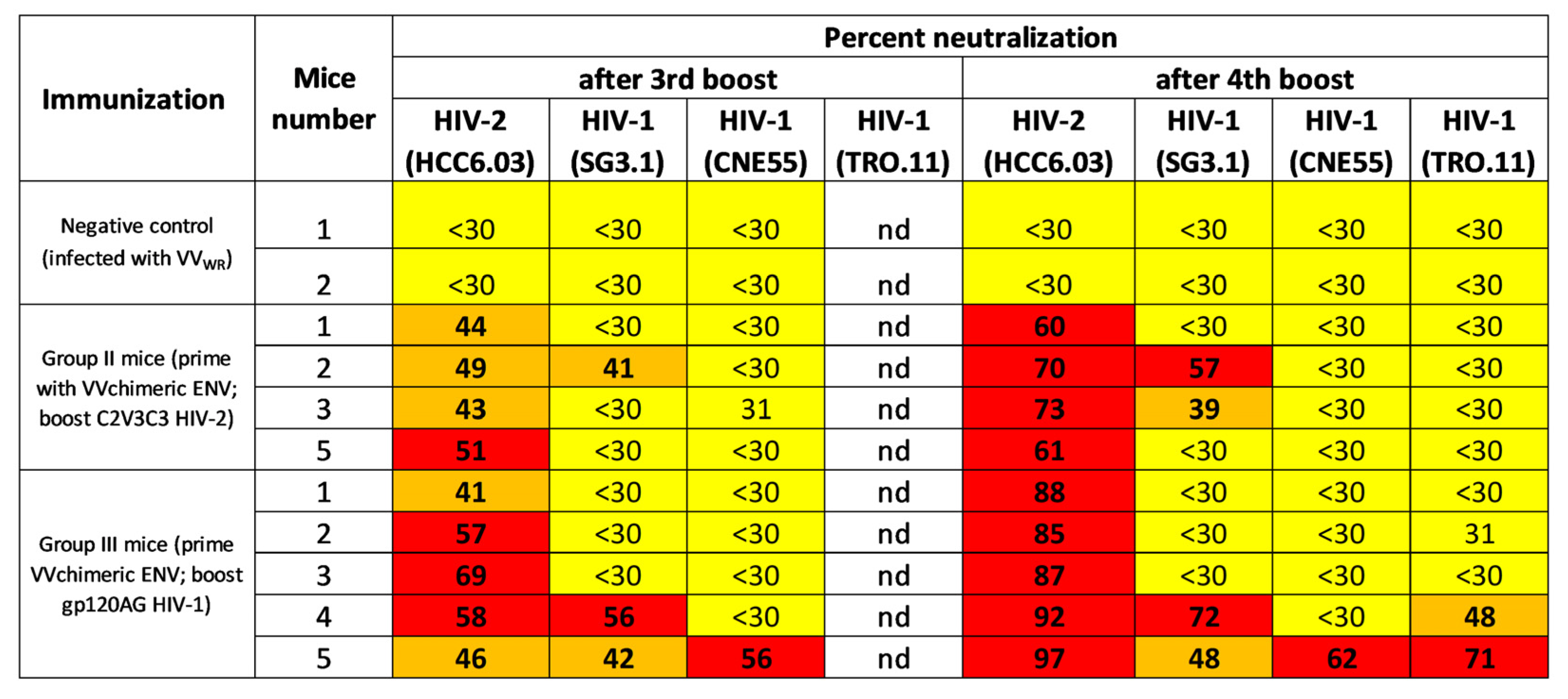
2.2. Chimeric Env Induces HIV-1 and HIV2 Binding and Neutralising Antibodies in Mice

Balb/c mice were inoculated with recombinant vaccinia virus expressing the chimeric Env and boosted four times with soluble gp120_AG or HIV-1 ALI C2V3C3 polypeptide (Figure 2). The mice produced increasing levels of antibodies that bound to the autologous antigens as assessed by ELISA (Figure 3). Mice boosted with the HIV-2 C2V3C3 polypeptide produced high-binding antibody responses to the cognate polypeptide (Figure 3A) and did not produce antibodies to gp120_AG (Figure 3C). In contrast, mice boosted with gp120_AG produced 2.2-fold higher binding antibody responses to the HIV-2 C2V3C3 polypeptide than to the cognate antigen after boost 4 (Figure 3, compare panels B and D). After the final booster immunisation, all mice produced antibodies that neutralised ≥60% (serum dilution 1:40) of the primary HIV-2 isolate HCC6.03, a CCR5-using virus (Figure 4; Figure S1) [44]. Two mice boosted with C2V3C3 polypeptide (group II) or gp120_AG (group III) also produced antibodies that neutralized the HIV-1 strain SG3.1. Finally, two mice boosted with gp120_AG (group III) produced antibodies that neutralized at least one tier 2 HIV-1 isolate. Of these, mouse 5 was able to produce antibodies that neutralized 97% of the HIV-2 isolate and ≥48% of the three HIV-1 isolates tested.

2.3. Epitope Mapping Analyses

HIV-1 neutralizing epitope specificities were assessed using a panel of HIV-1 TRO.11 mutant pseudoviruses whose neutralizing epitopes in V2, CD4 binding site or V3 high mannose patch were disrupted by the alanine substitution of selected amino acids defining the epitopes, namely, N160A in V2, N278A in the CD4 binding site and N332A in the V3 high mannose patch. In this assay, a decrease in the neutralizing potency of the mutants relative to wild-type TRO.11 indicates that the neutralizing antibodies target the epitopes disrupted by the alanine mutation [48]. Group III mouse 5 was unable to neutralize the TRO.11 mutants, indicating that its neutralizing antibodies were directed against the three epitopes (Figure 5). In contrast, group III mouse 4 still retained some neutralizing activity (>30%) against all mutants, suggesting that its neutralizing response was directed against other neutralising epitopes in gp120.

3. Discussion

The development of an Env-based immunogen capable of eliciting broadly neutralizing antibodies is a priority in the roadmap to produce an effective HIV vaccine and end the AIDS epidemic by 2030 [6]. This study investigated the antigenicity and immunogenicity of a novel chimeric envelope gp120 comprising the C2, V3 and C3 regions of HIV-2 and the remaining parts of the HIV-1 envelope. This construct was designed to allow the exposure of broadly reactive neutralizing epitopes in the envelope gp120 of HIV-1 (V2 apex and the CD4bs) and HIV-2 (V3 and the CD4bs) and to promote the elicitation of neutralizing antibodies against both types of virus. Chimeric or mosaic constructs containing T- and/or B-cell epitopes of Gag, Pol, Vif, Nef and Env designed to elicit HIV-specific antibodies and CD4- and CD8- T-cell responses have been used in several HIV-1 vaccine clinical trials (reviewed in [6]). These immunogens elicited Env-specific and V1V2-directed binding antibodies and antigen-specific T-cell responses, but no tier 2 neutralising antibodies were reported. HIV-1/HIV-2 chimeric envelope glycoproteins have not previously been used in vaccine trials. In Western blot, the chimeric gp120 bound to polyclonal serum antibodies from HIV-1- and HIV-2-infected individuals, confirming the presence of antigenic epitopes from both viruses. Mice primed with a vaccinia virus expressing the chimeric envelope and boosted with envelope gp120 from an HIV-1 CRF01_AG isolate [22] or a C2V3C3-polypeptide from HIV-2ALI [47] produced Env-specific binding and neutralizing antibodies against heterologous HIV-2 and tier 1 HIV-1 isolates. Two animals boosted with gp120AG also produced neutralizing antibodies against heterologous tier 2 HIV-1 isolates. Boosting with gp120AG produced broader and more potent neutralizing antibodies than boosting with the C2V3C3 polypeptide. The production of potent HIV-2 neutralizing antibodies by all mice irrespective of boosting modality confirms that the gp120 chimera adequately exposes the C2V3C3-based neutralizing epitopes of HIV-2 [47,49,50,51].
To investigate the exposure of the major neutralizing epitopes of HIV-1, we tested the binding reactivity of the chimeric gp120 to a panel of broadly neutralizing monoclonal antibodies targeting different conformational and N-linked epitopes. Previous studies have shown that the affinity of most bNAbs for monomeric envelope gp120/gp140 or core constructs of gp120 is variable and may not be related to their neutralizing efficacy [52,53]. In fact, with the exception of B12 and 447-52B, most monoclonal antibodies did not bind to chimeric gp120 in ELISA assays. The amino acids defining the B12 epitope are scattered in C1 (K97, L125), V2 (D167, D185, T202), C2 (N276), C3 (S365, P369), C4 (V430, T455, G458) and C5 (G473) [53,54,55]. Except for the substitutions E for P at position 369 in C3 and T for V at position 430 in C4, all amino acids are conserved in our chimeric construct, even those derived from HIV-2 in C2 (N276) and C3 (S365 and P369) (Figure S2). This high level of amino acid conservation in the context of a similar HIV-1 and HIV-2 gp120 structure [56] likely contributes to the binding of B12 to our chimeric gp120. While binding to B12 confirms that the chimeric gp120 exposes a CD4bs epitope, the lack of binding to the other antibodies that mimic the CD4 binding site suggests an altered CD4 binding site and impaired binding to the CD4 receptor in the cell surface. This is an advantageous feature for an Env immunogen as binding to cellular CD4 can lead to sequestration by CD4+ T cells and the occlusion of the CD4bs neutralizing epitopes [17]. The binding of 447-52B to the chimeric gp120 was unexpected because this antibody recognizes the conserved tip of the HIV-1 V3 loop in a β-turn conformation and preferentially binds and neutralizes HIV-1 isolates with the GPGR motif in the V3 crown [57,58,59]. Our chimera contains the HIV-2ALI V3 loop, the sequence (Figure S2) and structure of which, as determined by homology modelling [47], differs significantly from that of the 447-52B epitope. In addition, previous studies have shown that 447-52 is unable to neutralize two different strains of HIV-2, UC1 and 7312A [60]. Further studies are needed to determine whether a 447-52B-like neutralizing epitope is formed in our chimeric gp120.
Epitope mapping experiments with the subtype B strain TRO.11 showed that the neutralizing antibodies elicited in mouse 5 target broadly reactive epitopes in V2, the CD4 binding site and the high mannose patch centred on N332 at the base of the V3 loop. The presence of CD4bs antibodies was not surprising given the conservation of the CD4 binding site amino acids in the chimeric gp120 (see above). The elicitation of neutralizing antibodies against the conserved N160/V2 epitope suggests that our gp120 chimera exposes this broadly reactive epitope in a way that is not captured by the PG16 monoclonal antibody but allows the expansion of B lymphocytes expressing germline precursors of this class of bNAbs [61]. The production of neutralizing antibodies targeting the N332 glycan-dependent supersite in V3 was surprising given that the asparagine residues supporting the major N332 glycan and other N-glycans that can contribute to antibody interaction, such as N295 in C2, N301 in V3 and N339 in C3, are not present in our construct and that the homologous region in the HIV-2 envelope lacks N-linked glycosylation sites [51,56,62]. However, other N-glycans around V3 and underlying peptides that come together in the tertiary structure of HIV-1 gp120 and may interact with antibodies of this class, namely, N133 in C1, N137 and N156 in V1, N160 in V2 and N385 and N392 in V4, are conserved in our construct [36,63,64]. This suggests that some neutralizing antibodies elicited in our animals interact with N-glycans and underlying peptides outside C2, V3 and C3, whose exposure in TRO.11 is significantly affected by the alanine substitution of N332. An example of this type of V3-glycan-directed bNAbs is PGT121, which also targets glycans at positions 137 and 156 in V1 and 160 in V2 [65].
There are several limitations to this study. First, the number of animals in each group was small. Second, the neutralizing antibody response in mice was tested against a limited number of HIV isolates. Third, the epitope mapping was focused only on three broadly neutralizing epitopes in HIV-1 gp120. The small amounts of serum obtained from these animals prevented the testing of neutralizing activity against a broader panel of isolates and more detailed epitope mapping studies. Further studies in larger animal models are required to better characterize the immunogenic potential of our gp120 chimera. Monoclonal antibody isolation is required to confirm the neutralizing epitope specificities found in the best-responding animal.

4. Materials and Methods

4.1. Cells, Plasmids, Viruses, and Antibodies

Rat2 (TK) cells were obtained from the American Type Culture Collection (ATCC) (Rockville, MD, USA). HeLa cells (ATCC®CCL-2™) were obtained from the American Type Culture Collection. TZM-bl cells were provided by the AIDS Research and Reference Reagent Program (ARRRP), National Institutes of Health. HeLa, Rat2 and TZM-bl cells were cultured in complete growth medium consisting of Dulbecco’s minimal essential medium (DMEM) supplemented with 10% v/v fetal bovine serum (FBS), 100 U/mL penicillin-streptomycin (Gibco/Invitrogen, Waltham, MA, USA), 1 mM sodium pyruvate (Gibco/Invitrogen, Waltham, MA, USA), 1 mM L-glutamine (Gibco/Invitrogen, Waltham, MA, USA) and 1 mM non-essential amino acids (Gibco/Invitrogen, Waltham, MA, USA). All cell cultures were maintained at 37 °C in 5% CO2.
The following were also obtained from the ARRRP: Western Reserve strain of vaccinia virus (VVWR), recombinant vaccinia virus vPE16 expressing the envelope gp120 of HTLV-IIIB clone BH8, a panel of global tier 2 HIV-1 Env clones (cat#12670) designed to assess neutralisation responses, HuMAbs PG9 (anti-V1V2), PG16 (anti-V2), 447-52D (anti-V3), 2G12 (anti-N-linked glycans in C2, C3, V4, C4), VRC01 (anti-CD4bs), VRC03 (anti-CD4bs), 3BNC117 (anti-CD4bs), HJ16 (anti-CD4bs), b12 (anti-CD4bs) and 2F5 (anti-MPER), and recombinant proteins M. CON.SD11 gp120 and SF162 gp140 trimer.

4.2. Production and Expression of Recombinant Vaccinia Viruses

A codon-optimized chimeric envelope gene containing gp120 region of HIV-192TH023 (CRF01_AE, GenBank accession number: KU562843.1) with the C2V3C3 region of HIV-2ALI (group A, GenBank accession number: L25445.1) [66] and gp41 of HIV-1Ri112W2R074B10 (subtype B, GenBank accession number: JN205576) was produced by gene synthesis at NZYTech, Lisbon, Portugal (Figure 6; Figure S2).
The chimeric env gene was subcloned into the BamHI and HindIII sites of the vaccinia virus insertion vector pMJ602. HeLa cells were transfected with recombinant pMJ602 plasmid and infected with 0.5 PFU of vaccinia virus strain VVWR, and recombinant vaccinia virus resistant to 5-bromodeoxyuridine and expressing β-galactosidase was selected in Rat2 cells and then propagated in HeLa cells [22,47,67]. The recombinant virus was titrated in Rat2 cells according to the method of Reed and Muench [68].

4.3. Characterization of Chimeric Envelope Glycoproteins

HeLa cells were infected with 5 PFU of recombinant vaccinia virus per cell and incubated for 1 h. After 24 h of infection, the cells were washed with cold phosphate-buffered saline (PBS) and lysed with RIPA+DOC buffer (0.15 M NaCl, 0.05 M Tris-HCl, 1% Triton X-100, 1% DOC, 0.1% SDS). The lysates were centrifuged at 35,000 rpm for 60 min at 4 °C and the supernatant containing the proteins was collected. The expression and antigenic reactivity of envelope glycoproteins were analysed by Western blot (WB) and ELISA. For the WB analysis, proteins separated by 7.5% SDS-PAGE were transferred to 0.45 µM nitrocellulose membranes (Bio-Rad) treated with blocking buffer (1X TBST with 4% w/v nonfat dry milk) and incubated with serum from individuals with HIV-1 and HIV-2 diluted (1:200) in primary antibody buffer (1× TBST with 4% w/v nonfat dry milk and 5% goat serum). The membrane was then washed with TBST 0.25% and incubated with goat anti-human IgG alkaline phosphatase antibody (Sigma, St. Louis, MO, USA) and/or goat anti-human IgG peroxidase antibody (Sigma). The colourimetric detection of proteins was performed using the AP Conjugate Substrate Kit (Bio-Rad, Hercules, CA, USA), and chemiluminescent detection was performed using Pierce™ ECL Western Blotting Substrate (ThermoFisher Scientific, Waltham, MA, USA). Envelope gp120 from HTLV-IIIB clone BH8 expressed by recombinant vaccinia virus VVPE16 was used as an HIV-1 positive control and recombinant vaccinia viruses expressing the full-length HIV-2ALI envelope (VVALI), and a truncated form of gp125 (M2) were used as HIV-2 positive controls [47].
The method of Rose et al. [69] was used to obtain the soluble chimeric gp120. HeLa cells were infected with 5 PFU of recombinant vaccinia virus per cell and incubated for 3 h. The medium containing the infecting virus was replaced with DMEM supplemented with 2.5% FBS 3 h after infection. After 24 h of infection, medium containing released gp120 was collected, clarified by centrifugation at 3000× g for 10 min and filtered through a 0.2 µM pore size filter to remove the vaccinia virus.
The binding specificity of the chimeric gp120 was analysed in ELISA assays against ten broadly neutralizing monoclonal antibodies at a final concentration of 10 µg/mL: 2G12, b12, 447-52D, HJ16, PG9, PG16, 3BNC117, VRC01, VRC03 and 2F5. Immuno MaxiSorp 96-well microplates (Nunc) were coated with the chimeric env (~2.3 to 4 µg per well) and reference SF162 gp140 (2 µg/mL) and M.CON.S D11 gp120 (2 µg/mL) glycoproteins in 0.05M bicarbonate buffer. HIV-1 serum sample at a final dilution of 1:200 was used as a positive control and 2F5 antibody (anti-gp41) as a negative control. The cut-off value of the assay with the chimeric and M.CON.S D11 gp120s was calculated as the mean OD value of the 2F5 reactivity plus 3 times the standard deviation (SD). The cut-off value of the assay with HIV-positive serum and SF162 gp140 (which reacts with 2F5) was calculated as the mean OD value of HIV seronegative samples plus 3 times the SD.

4.4. Balb/c Mice Immunizations

Three groups (I, II and III) of 6-week-old female Balb/c mice were immunised intraperitoneally with 107 PFU of wild-type vaccinia virus WR (VVWR) or vaccinia virus expressing the chimeric Env (VVChimeric ENV) in 100 µL phosphate-buffered saline (PBS) (priming) and on days 15, 30, 45 and 60 with 10 µg of purified recombinant HIV-2ALI C2V3C3 polypeptide or soluble gp120 from a clade CRF01_AG isolate (gp120AG) emulsified in incomplete Freud adjuvant (Figure 2). The production and purification of these booster proteins were previously described by Calado et al. [22]. Group I (two mice) was the control group and received VVWR as priming and a supernatant of VVWR-infected cells as a boost; group II (five mice) received VVChimeric ENV as priming and C2V3C3 polypeptide as a boost; and group III (five mice) received VVChimeric ENV as priming and gp120AG as a boost. Mice were bled before immunization and 14 days after each immunization to determine binding and neutralizing antibodies.

4.5. Envelope-Specific Antibody Binding in Mice Sera

To analyse serum antibody responses in sera from immunized animals, Immuno MaxiSorp 96-well microplates (Nunc) were coated with soluble gp120AG (2 µg/mL), M.CON.S D11 gp120 (2 µg/mL), C2V3C3 polypeptide (2 µg/mL) or supernatant from VVWR-infected cells. After overnight incubation at 4 °C, the microplates were blocked with 2% gelatin (Bio-Rad). Mouse sera from days 0, 15, 30, 45 and 60 were heat-inactivated at 56 °C for 1 h, added to the microplates (1:200 final dilution in a total volume of 100 µL) and incubated for 2 h at room temperature. Goat anti-mouse IgG alkaline phosphatase antibody (Sigma) at 1:2000 dilution was added to the microplates as a secondary antibody. The colourimetric reaction was developed as described above. Negative controls were pre-immune serum and serum from mice immunised with VVWR. Positive controls were sera from individuals with HIV-1 and HIV-2. In this case, the secondary antibody was a goat anti-human IgG alkaline phosphatase antibody (Sigma). Sera with an optical density (OD) greater than that of the preimmune controls were considered positive.

4.6. Neutralization Assays

The neutralizing activity of mouse post-immunization sera was analysed in a one-round viral infectivity assay using TZM-bl against laboratory-adapted HIV-1SG3.1 (a Tier 1 subtype B isolate), two Tier 2 Env-pseudotyped HIV-1 isolates (TRO.11, subtype B, and CNE55, CRF01_AE) and a primary isolate of HIV-2 (03PTHCC6, group A). Env-pseudotyped viruses were generated by the transfection of Env-expressing plasmids into 293T cells using pSG3.1Δenv as a backbone and titrated in TZM-bl cells.
The neutralizing activity of mouse sera was tested in a one-round viral infectivity assay using a luciferase reporter gene assay in TZM-bl cells, as previously described [22]. Briefly, 10,000 cells in 100 µL DMEM supplemented with 10% heat-inactivated foetal bovine serum were added to each well of 96-well flat-bottom culture plates (Nunc) and allowed to attach overnight. Subsequently, 100 µL of each virus (200 TCID50/well) was incubated with heat-inactivated (56 °C, 1 h) mouse sera for 1 h at 37 °C in a total volume of 200 µL of growth medium (DMEM supplemented with 10% heat-inactivated foetal bovine serum) containing DEAE-dextran (19.7 µg/mL) and added to the cells. After 48 h, the medium was removed from each well and the plates were analysed for luciferase activity on a luminometer (TECAN) using the Pierce™ Firefly Luciferase Glow Assay Kit (ThermoFisher scientific). Wells with medium were used as background controls, and virus-only controls were included as infection controls. The effect of preimmune serum on infection was used as the baseline neutralizing activity. Percent neutralization was determined by calculating the difference in mean RLU (relative light units) between test wells containing post-immune sera and test wells containing pre-immune sera, after the normalization of the results using the mean RLU of the cell controls. The results were considered valid if the mean RLU of the virus control wells was ≥10 times the mean RLU of the cell control wells. To monitor the specificity of neutralization, each serum sample was also tested against pseudoviruses carrying the vesicular stomatitis virus (VSV) envelope. Neutralization assays were performed with sera collected after the third and fourth boosts. All mouse sera were tested for neutralizing antibodies once in triplicate at a dilution of 1:40. Neutralization assays were repeated for animals that neutralized at least one tier 2 isolate >30% in the first assay.

4.7. Epitope Mapping

Epitope specificities were assessed using a panel of HIV-1 TRO.11 pseudoviruses in which the major neutralizing epitopes in V2, the CD4 binding site and V3 were disrupted by mutagenesis at specific amino acids (N160A in V2, N278A in C3 and N332A in the V3 base) [48,70,71]. Mutant Env-pseudotyped viruses were produced by the transfection of Env-expressing plasmids into 293T cells using pSG3.1Δenv as a backbone and titrated in TZM-bl cells as described [72]. Target epitopes were identified by the reduction in neutralization scores compared to the wild-type pseudovirus.

5. Conclusions

In conclusion, a chimeric gp120 containing the C2V3C3 region of HIV-2 ALI and the remaining parts of an HIV-1 clade AE isolate elicited neutralising antibodies targeting some of the most broadly reactive epitopes in HIV-1 and HIV-2. Our results provide proof of concept for chimeric HIV-1/HIV-2 envelope glycoproteins as HIV vaccine immunogens.

Supplementary Materials

The following supporting information can be downloaded at: https://www.mdpi.com/article/10.3390/ijms24109077/s1.

Author Contributions

Conceptualization, N.T. and H.B.; Data curation, F.C.; Formal analysis, N.T., F.C. and H.B.; Funding acquisition, N.T. and H.B.; Investigation, N.T., I.F., R.C., F.M., I.B., J.M.M., P.B. and F.C.; Methodology, N.T., F.C. and H.B.; Resources, R.C., F.M. and F.C.; Supervision, H.B.; Validation, N.T. and H.B.; Writing—original draft, N.T., I.F. and H.B.; Writing—review and editing, N.T., I.F., R.C., F.M., I.B., J.M.M., P.B., F.C. and H.B. All authors have read and agreed to the published version of the manuscript.

Funding

This work was funded by national funds through the FCT-Foundation for Science and Technology, I.P., under the project UIDB/04585/2020, by FCT and Aga Khan Development Network (AKDN)—Portugal Collaborative Research Network in Portuguese speaking countries in Africa, project 332821690 and by Ministry of Health, Portugal, project HIV/SAU/0008.11.

Institutional Review Board Statement

Balb/cByJ (H-2d) mice were maintained under specific pathogen-free conditions at the Instituto de Higiene e Medicina Tropical, New University of Lisbon, where all animal procedures were performed in accordance with Directive 2010/63/EU. The animal study was reviewed and approved by the Ethics and Animal Welfare Committee of the Instituto de Higiene e Medicina Tropical, New University of Lisbon.

Data Availability Statement

Data collected for this study will be shared with publication upon reasonable request, with a signed data access agreement.

Acknowledgments

The following reagent was obtained through the NIH HIV Reagent Program, Division of AIDS, NIAID, NIH: vaccinia virus, Recombinant Expressing Human Immunodeficiency Virus Type 1 BH8 Env Protein, ARP-2734, contributed by Christopher Broder and Bernard Moss. Envelope genes with mutations disrupting neutralizing epitopes in the HIV-1 TRO.11 envelope were a generous gift of Penny Moore.

Conflicts of Interest

The authors declare that the research was conducted in the absence of any commercial or financial relationships that could be construed as a potential conflict of interest.

References

  1. Faria, N.R.; Esbjörnsson, J.; Lemey, P. Phylogeographic Insights into the Origins and Epidemic History of the Human Immunodeficiency Virus Type 2. In Encyclopedia of AIDS; Springer Science: New York, NY, USA, 2013. [Google Scholar]
  2. van Tienen, C.; van der Loeff, M.S. Epidemiology of HIV-2 Infection in West Africa. In Encyclopedia of AIDS; Hope, T.J., Stevenson, M., Richman, D., Eds.; Springer: New York, NY, USA, 2016; pp. 1–11. [Google Scholar]
  3. UNAIDS Global HIV & AIDS Statistics—Fact Sheet. Available online: https://www.unaids.org/en/resources/fact-sheet (accessed on 12 May 2023).
  4. Collaborators, G.H. Global, regional, and national sex-specific burden and control of the HIV epidemic, 1990–2019, for 204 countries and territories: The Global Burden of Diseases Study 2019. Lancet HIV 2021, 8, e633–e651. [Google Scholar]
  5. De Lay, P.R.; Benzaken, A.; Karim, Q.A.; Aliyu, S.; Amole, C.; Ayala, G.; Chalkidou, K.; Chang, J.; Clayton, M.; Couto, A.; et al. Ending AIDS as a public health threat by 2030: Time to reset targets for 2025. PLoS Med. 2021, 18, e1003649. [Google Scholar] [CrossRef] [PubMed]
  6. Lee, J.H.; Crotty, S. HIV vaccinology: 2021 update. Semin. Immunol. 2021, 51, 101470. [Google Scholar] [CrossRef] [PubMed]
  7. Landais, E.; Huang, X.; Havenar-Daughton, C.; Murrell, B.; Price, M.A.; Wickramasinghe, L.; Ramos, A.; Bian, C.B.; Simek, M.; Allen, S.; et al. Broadly Neutralizing Antibody Responses in a Large Longitudinal Sub-Saharan HIV Primary Infection Cohort. PLoS Pathog. 2016, 12, e1005369. [Google Scholar] [CrossRef] [PubMed]
  8. Hu, X.; Hu, Y.; Zhao, C.; Gao, H.; Greene, K.M.; Ren, L.; Ma, L.; Ruan, Y.; Sarzotti-Kelsoe, M.; Montefiori, D.C.; et al. Profiling the neutralizing antibody response in chronically HIV-1 CRF07_BC-infected intravenous drug users naive to antiretroviral therapy. Sci. Rep. 2017, 7, 46308. [Google Scholar] [CrossRef] [PubMed]
  9. Rusert, P.; Kouyos, R.D.; Kadelka, C.; Ebner, H.; Schanz, M.; Huber, M.; Braun, D.L.; Hoze, N.; Scherrer, A.; Magnus, C.; et al. Determinants of HIV-1 broadly neutralizing antibody induction. Nat. Med. 2016, 22, 1260–1267. [Google Scholar] [CrossRef]
  10. Hraber, P.; Korber, B.T.; Lapedes, A.S.; Bailer, R.T.; Seaman, M.S.; Gao, H.; Greene, K.M.; McCutchan, F.; Williamson, C.; Kim, J.H.; et al. Impact of clade, geography, and age of the epidemic on HIV-1 neutralization by antibodies. J. Virol. 2014, 88, 12623–12643. [Google Scholar] [CrossRef]
  11. Tomaras, G.D.; Binley, J.M.; Gray, E.S.; Crooks, E.T.; Osawa, K.; Moore, P.L.; Tumba, N.; Tong, T.; Shen, X.; Yates, N.L.; et al. Polyclonal B cell responses to conserved neutralization epitopes in a subset of HIV-1-infected individuals. J. Virol. 2011, 85, 11502–11519. [Google Scholar] [CrossRef]
  12. Simek, M.D.; Rida, W.; Priddy, F.H.; Pung, P.; Carrow, E.; Laufer, D.S.; Lehrman, J.K.; Boaz, M.; Tarragona-Fiol, T.; Miiro, G.; et al. Human immunodeficiency virus type 1 elite neutralizers: Individuals with broad and potent neutralizing activity identified by using a high-throughput neutralization assay together with an analytical selection algorithm. J. Virol. 2009, 83, 7337–7348. [Google Scholar] [CrossRef]
  13. Saunders, K.O.; Nicely, N.I.; Wiehe, K.; Bonsignori, M.; Meyerhoff, R.R.; Parks, R.; Walkowicz, W.E.; Aussedat, B.; Wu, N.R.; Cai, F.; et al. Vaccine Elicitation of High Mannose-Dependent Neutralizing Antibodies against the V3-Glycan Broadly Neutralizing Epitope in Nonhuman Primates. Cell Rep. 2017, 18, 2175–2188. [Google Scholar] [CrossRef]
  14. Saunders, K.O.; Verkoczy, L.K.; Jiang, C.; Zhang, J.; Parks, R.; Chen, H.; Housman, M.; Bouton-Verville, H.; Shen, X.; Trama, A.M.; et al. Vaccine Induction of Heterologous Tier 2 HIV-1 Neutralizing Antibodies in Animal Models. Cell Rep. 2017, 21, 3681–3690. [Google Scholar] [CrossRef] [PubMed]
  15. Barouch, D.H.; Tomaka, F.L.; Wegmann, F.; Stieh, D.J.; Alter, G.; Robb, M.L.; Michael, N.L.; Peter, L.; Nkolola, J.P.; Borducchi, E.N.; et al. Evaluation of a mosaic HIV-1 vaccine in a multicentre, randomised, double-blind, placebo-controlled, phase 1/2a clinical trial (APPROACH) and in rhesus monkeys (NHP 13-19). Lancet 2018, 392, 232–243. [Google Scholar] [CrossRef] [PubMed]
  16. Xu, K.; Acharya, P.; Kong, R.; Cheng, C.; Chuang, G.Y.; Liu, K.; Louder, M.K.; O’Dell, S.; Rawi, R.; Sastry, M.; et al. Epitope-based vaccine design yields fusion peptide-directed antibodies that neutralize diverse strains of HIV-1. Nat. Med. 2018, 24, 857–867. [Google Scholar] [CrossRef] [PubMed]
  17. Zhang, P.; Gorman, J.; Geng, H.; Liu, Q.; Lin, Y.; Tsybovsky, Y.; Go, E.P.; Dey, B.; Andine, T.; Kwon, A.; et al. Interdomain Stabilization Impairs CD4 Binding and Improves Immunogenicity of the HIV-1 Envelope Trimer. Cell Host Microbe 2018, 23, 832–844.e6. [Google Scholar] [CrossRef]
  18. Asbach, B.; Kibler, K.V.; Kostler, J.; Perdiguero, B.; Yates, N.L.; Stanfield-Oakley, S.; Tomaras, G.D.; Kao, S.F.; Foulds, K.E.; Roederer, M.; et al. Priming with a Potent HIV-1 DNA Vaccine Frames the Quality of Immune Responses prior to a Poxvirus and Protein Boost. J. Virol. 2019, 93, e01529-18. [Google Scholar] [CrossRef]
  19. Dubrovskaya, V.; Tran, K.; Ozorowski, G.; Guenaga, J.; Wilson, R.; Bale, S.; Cottrell, C.A.; Turner, H.L.; Seabright, G.; O’Dell, S.; et al. Vaccination with Glycan-Modified HIV NFL Envelope Trimer-Liposomes Elicits Broadly Neutralizing Antibodies to Multiple Sites of Vulnerability. Immunity 2019, 51, 915–929.e7. [Google Scholar] [CrossRef]
  20. Escolano, A.; Gristick, H.B.; Abernathy, M.E.; Merkenschlager, J.; Gautam, R.; Oliveira, T.Y.; Pai, J.; West, A.P., Jr.; Barnes, C.O.; Cohen, A.A.; et al. Immunization expands B cells specific to HIV-1 V3 glycan in mice and macaques. Nature 2019, 570, 468–473. [Google Scholar] [CrossRef]
  21. Francica, J.R.; Laga, R.; Lynn, G.M.; Muzikova, G.; Androvic, L.; Aussedat, B.; Walkowicz, W.E.; Padhan, K.; Ramirez-Valdez, R.A.; Parks, R.; et al. Star nanoparticles delivering HIV-1 peptide minimal immunogens elicit near-native envelope antibody responses in nonhuman primates. PLoS Biol. 2019, 17, e3000328. [Google Scholar] [CrossRef]
  22. Calado, R.; Duarte, J.; Borrego, P.; Marcelino, J.M.; Bartolo, I.; Martin, F.; Figueiredo, I.; Almeida, S.; Graca, L.; Vitor, J.; et al. A Prime-Boost Immunization Strategy with Vaccinia Virus Expressing Novel gp120 Envelope Glycoprotein from a CRF02_AG Isolate Elicits Cross-Clade Tier 2 HIV-1 Neutralizing Antibodies. Vaccines 2020, 8, 171. [Google Scholar] [CrossRef]
  23. Haynes, B.F.; Wiehe, K.; Borrow, P.; Saunders, K.O.; Korber, B.; Wagh, K.; McMichael, A.J.; Kelsoe, G.; Hahn, B.H.; Alt, F.; et al. Strategies for HIV-1 vaccines that induce broadly neutralizing antibodies. Nat. Rev. Immunol. 2023, 23, 142–158. [Google Scholar] [CrossRef]
  24. Burton, D.R.; Mascola, J.R. Antibody responses to envelope glycoproteins in HIV-1 infection. Nat. Immunol. 2015, 16, 571–576. [Google Scholar] [CrossRef] [PubMed]
  25. Stephenson, K.E.; Wagh, K.; Korber, B.; Barouch, D.H. Vaccines and Broadly Neutralizing Antibodies for HIV-1 Prevention. Annu. Rev. Immunol. 2020, 38, 673–703. [Google Scholar] [CrossRef] [PubMed]
  26. Gray, G.E.; Bekker, L.G.; Laher, F.; Malahleha, M.; Allen, M.; Moodie, Z.; Grunenberg, N.; Huang, Y.; Grove, D.; Prigmore, B.; et al. Vaccine Efficacy of ALVAC-HIV and Bivalent Subtype C gp120-MF59 in Adults. N. Engl. J. Med. 2021, 384, 1089–1100. [Google Scholar] [CrossRef] [PubMed]
  27. Sanders, R.W.; Derking, R.; Cupo, A.; Julien, J.P.; Yasmeen, A.; de Val, N.; Kim, H.J.; Blattner, C.; de la Pena, A.T.; Korzun, J.; et al. A next-generation cleaved, soluble HIV-1 Env trimer, BG505 SOSIP.664 gp140, expresses multiple epitopes for broadly neutralizing but not non-neutralizing antibodies. PLoS Pathog. 2013, 9, e1003618. [Google Scholar] [CrossRef] [PubMed]
  28. Julien, J.P.; Lee, J.H.; Ozorowski, G.; Hua, Y.; Torrents de la Pena, A.; de Taeye, S.W.; Nieusma, T.; Cupo, A.; Yasmeen, A.; Golabek, M.; et al. Design and structure of two HIV-1 clade C SOSIP.664 trimers that increase the arsenal of native-like Env immunogens. Proc. Natl. Acad. Sci. USA 2015, 112, 11947–11952. [Google Scholar] [CrossRef]
  29. Sanders, R.W.; van Gils, M.J.; Derking, R.; Sok, D.; Ketas, T.J.; Burger, J.A.; Ozorowski, G.; Cupo, A.; Simonich, C.; Goo, L.; et al. HIV-1 VACCINES. HIV-1 neutralizing antibodies induced by native-like envelope trimers. Science 2015, 349, aac4223. [Google Scholar] [CrossRef]
  30. Pugach, P.; Ozorowski, G.; Cupo, A.; Ringe, R.; Yasmeen, A.; de Val, N.; Derking, R.; Kim, H.J.; Korzun, J.; Golabek, M.; et al. A native-like SOSIP.664 trimer based on an HIV-1 subtype B env gene. J. Virol. 2015, 89, 3380–3395. [Google Scholar] [CrossRef]
  31. Zhang, P.; Narayanan, E.; Liu, Q.; Tsybovsky, Y.; Boswell, K.; Ding, S.; Hu, Z.; Follmann, D.; Lin, Y.; Miao, H.; et al. A multiclade env-gag VLP mRNA vaccine elicits tier-2 HIV-1-neutralizing antibodies and reduces the risk of heterologous SHIV infection in macaques. Nat. Med. 2021, 27, 2234–2245. [Google Scholar] [CrossRef]
  32. Escolano, A.; Gristick, H.B.; Gautam, R.; DeLaitsch, A.T.; Abernathy, M.E.; Yang, Z.; Wang, H.; Hoffmann, M.A.G.; Nishimura, Y.; Wang, Z.; et al. Sequential immunization of macaques elicits heterologous neutralizing antibodies targeting the V3-glycan patch of HIV-1 Env. Sci. Transl. Med. 2021, 13, eabk1533. [Google Scholar] [CrossRef]
  33. Julg, B.; Barouch, D.H. Neutralizing antibodies for HIV-1 prevention. Curr. Opin. HIV AIDS 2019, 14, 318–324. [Google Scholar] [CrossRef]
  34. Cohen, Y.Z.; Caskey, M. Broadly neutralizing antibodies for treatment and prevention of HIV-1 infection. Curr. Opin. HIV AIDS 2018, 13, 366–373. [Google Scholar] [CrossRef] [PubMed]
  35. Spencer, D.A.; Shapiro, M.B.; Haigwood, N.L.; Hessell, A.J. Advancing HIV Broadly Neutralizing Antibodies: From Discovery to the Clinic. Front. Public Health 2021, 9, 690017. [Google Scholar] [CrossRef] [PubMed]
  36. Kumar, S.; Singh, S.; Luthra, K. An Overview of Human Anti-HIV-1 Neutralizing Antibodies against Diverse Epitopes of HIV-1. ACS Omega 2023, 8, 7252–7261. [Google Scholar] [CrossRef] [PubMed]
  37. Kwong, P.D.; Mascola, J.R. HIV-1 Vaccines Based on Antibody Identification, B Cell Ontogeny, and Epitope Structure. Immunity 2018, 48, 855–871. [Google Scholar] [CrossRef]
  38. Cizmeci, D.; Lofano, G.; Rossignol, E.; Dugast, A.S.; Kim, D.; Cavet, G.; Nguyen, N.; Tan, Y.C.; Seaman, M.S.; Alter, G.; et al. Distinct clonal evolution of B-cells in HIV controllers with neutralizing antibody breadth. Elife 2021, 10, e62648. [Google Scholar] [CrossRef]
  39. Visseaux, B.; Damond, F.; Matheron, S.; Descamps, D.; Charpentier, C. Hiv-2 molecular epidemiology. Infect. Genet. Evol. 2016, 46, 233–240. [Google Scholar] [CrossRef]
  40. Azevedo-Pereira, J.M.; Santos-Costa, Q. HIV Interaction With Human Host: HIV-2 As a Model of a Less Virulent Infection. AIDS Rev. 2016, 18, 44–53. [Google Scholar]
  41. Ceccarelli, G.; Giovanetti, M.; Sagnelli, C.; Ciccozzi, A.; d’Ettorre, G.; Angeletti, S.; Borsetti, A.; Ciccozzi, M. Human Immunodeficiency Virus Type 2: The Neglected Threat. Pathogens 2021, 10, 1377. [Google Scholar] [CrossRef]
  42. Matheron, S.; Pueyo, S.; Damond, F.; Simon, F.; Lepretre, A.; Campa, P.; Salamon, R.; Chene, G.; Brun-Vezinet, F. Factors associated with clinical progression in HIV-2 infected-patients: The French ANRS cohort. AIDS 2003, 17, 2593–2601. [Google Scholar] [CrossRef]
  43. Esbjornsson, J.; Mansson, F.; Kvist, A.; da Silva, Z.J.; Andersson, S.; Fenyo, E.M.; Isberg, P.E.; Biague, A.J.; Lindman, J.; Palm, A.A.; et al. Long-term follow-up of HIV-2-related AIDS and mortality in Guinea-Bissau: A prospective open cohort study. Lancet HIV 2018, 6, e25–e31. [Google Scholar] [CrossRef]
  44. Corey, L.; Gilbert, P.B.; Tomaras, G.D.; Haynes, B.F.; Pantaleo, G.; Fauci, A.S. Immune correlates of vaccine protection against HIV-1 acquisition. Sci. Transl. Med. 2015, 7, 310rv7. [Google Scholar] [CrossRef]
  45. Sadanand, S.; Suscovich, T.J.; Alter, G. Broadly Neutralizing Antibodies Against HIV: New Insights to Inform Vaccine Design. Annu. Rev. Med. 2016, 67, 185–200. [Google Scholar] [CrossRef] [PubMed]
  46. Montefiori, D.C.; Roederer, M.; Morris, L.; Seaman, M.S. Neutralization tiers of HIV-1. Curr. Opin. HIV AIDS 2018, 13, 128–136. [Google Scholar] [CrossRef] [PubMed]
  47. Marcelino, J.M.; Borrego, P.; Rocha, C.; Barroso, H.; Quintas, A.; Novo, C.; Taveira, N. Potent and broadly reactive HIV-2 neutralizing antibodies elicited by a vaccinia virus vector prime-C2V3C3 polypeptide boost immunization strategy. J. Virol. 2010, 84, 12429–12436. [Google Scholar] [CrossRef] [PubMed]
  48. Mikell, I.; Sather, D.N.; Kalams, S.A.; Altfeld, M.; Alter, G.; Stamatatos, L. Characteristics of the earliest cross-neutralizing antibody response to HIV-1. PLoS Pathog. 2011, 7, e1001251. [Google Scholar] [CrossRef]
  49. Marcelino, J.M.; Borrego, P.; Nilsson, C.; Familia, C.; Barroso, H.; Maltez, F.; Doroana, M.; Antunes, F.; Quintas, A.; Taveira, N. Resistance to antibody neutralization in HIV-2 infection occurs in late stage disease and is associated with X4 tropism. AIDS 2012, 26, 2275–2284. [Google Scholar] [CrossRef]
  50. Rocha, C.; Duarte, J.; Borrego, P.; Calado, R.; Marcelino, J.M.; Tendeiro, R.; Valadas, E.; Sousa, A.E.; Taveira, N. Potency of HIV-2-specific antibodies increase in direct association with loss of memory B cells. AIDS 2017, 31, 2431–2433. [Google Scholar] [CrossRef]
  51. Rocha, C.; Calado, R.; Borrego, P.; Marcelino, J.M.; Bartolo, I.; Rosado, L.; Cavaco-Silva, P.; Gomes, P.; Familia, C.; Quintas, A.; et al. Evolution of the human immunodeficiency virus type 2 envelope in the first years of infection is associated with the dynamics of the neutralizing antibody response. Retrovirology 2013, 10, 110. [Google Scholar] [CrossRef]
  52. Scheid, J.F.; Mouquet, H.; Ueberheide, B.; Diskin, R.; Klein, F.; Oliveira, T.Y.; Pietzsch, J.; Fenyo, D.; Abadir, A.; Velinzon, K.; et al. Sequence and structural convergence of broad and potent HIV antibodies that mimic CD4 binding. Science 2011, 333, 1633–1637. [Google Scholar] [CrossRef]
  53. Pantophlet, R.; Ollmann Saphire, E.; Poignard, P.; Parren, P.W.; Wilson, I.A.; Burton, D.R. Fine mapping of the interaction of neutralizing and nonneutralizing monoclonal antibodies with the CD4 binding site of human immunodeficiency virus type 1 gp120. J. Virol. 2003, 77, 642–658. [Google Scholar] [CrossRef]
  54. McInerney, T.L.; McLain, L.; Armstrong, S.J.; Dimmock, N.J. A human IgG1 (b12) specific for the CD4 binding site of HIV-1 neutralizes by inhibiting the virus fusion entry process, but b12 Fab neutralizes by inhibiting a postfusion event. Virology 1997, 233, 313–326. [Google Scholar] [CrossRef]
  55. McKeating, J.A.; Thali, M.; Furman, C.; Karwowska, S.; Gorny, M.K.; Cordell, J.; Zolla-Pazner, S.; Sodroski, J.; Weiss, R.A. Amino acid residues of the human immunodeficiency virus type I gp120 critical for the binding of rat and human neutralizing antibodies that block the gp120-sCD4 interaction. Virology 1992, 190, 134–142. [Google Scholar] [CrossRef] [PubMed]
  56. Davenport, Y.W.; West, A.P., Jr.; Bjorkman, P.J. Structure of an HIV-2 gp120 in Complex with CD4. J. Virol. 2016, 90, 2112–2118. [Google Scholar] [CrossRef] [PubMed]
  57. Yusim, K.; Korber, B.T.; Brander, C.; Barouch, D.; de Boer, R.; Haynes, B.F.; Koup, R.; Moore, J.P.; Walker, B.D.; Watkins, D. HIV Molecular Immunology 2015; Los Alamos National Lab. (LANL): Los Alamos, NM, USA, 2016.
  58. Zolla-Pazner, S.; Zhong, P.; Revesz, K.; Volsky, B.; Williams, C.; Nyambi, P.; Gorny, M.K. The cross-clade neutralizing activity of a human monoclonal antibody is determined by the GPGR V3 motif of HIV type 1. AIDS Res. Hum. Retrovir. 2004, 20, 1254–1258. [Google Scholar] [CrossRef]
  59. Gorny, M.K.; Conley, A.J.; Karwowska, S.; Buchbinder, A.; Xu, J.Y.; Emini, E.A.; Koenig, S.; Zolla-Pazner, S. Neutralization of diverse human immunodeficiency virus type 1 variants by an anti-V3 human monoclonal antibody. J. Virol. 1992, 66, 7538–7542. [Google Scholar] [CrossRef] [PubMed]
  60. Decker, J.M.; Bibollet-Ruche, F.; Wei, X.; Wang, S.; Levy, D.N.; Wang, W.; Delaporte, E.; Peeters, M.; Derdeyn, C.A.; Allen, S.; et al. Antigenic conservation and immunogenicity of the HIV coreceptor binding site. J. Exp. Med. 2005, 201, 1407–1419. [Google Scholar] [CrossRef] [PubMed]
  61. Escolano, A.; Dosenovic, P.; Nussenzweig, M.C. Progress toward active or passive HIV-1 vaccination. J. Exp. Med. 2017, 214, 3–16. [Google Scholar] [CrossRef] [PubMed]
  62. Borrego, P.; Marcelino, J.M.; Rocha, C.; Doroana, M.; Antunes, F.; Maltez, F.; Gomes, P.; Novo, C.; Barroso, H.; Taveira, N. The role of the humoral immune response in the molecular evolution of the envelope C2, V3 and C3 regions in chronically HIV-2 infected patients. Retrovirology 2008, 5, 78. [Google Scholar] [CrossRef] [PubMed]
  63. Kong, L.; Lee, J.H.; Doores, K.J.; Murin, C.D.; Julien, J.P.; McBride, R.; Liu, Y.; Marozsan, A.; Cupo, A.; Klasse, P.J.; et al. Supersite of immune vulnerability on the glycosylated face of HIV-1 envelope glycoprotein gp120. Nat. Struct. Mol. Biol. 2013, 20, 796–803. [Google Scholar] [CrossRef]
  64. Daniels, C.N.; Saunders, K.O. Antibody responses to the HIV-1 envelope high mannose patch. Adv. Immunol. 2019, 143, 11–73. [Google Scholar]
  65. Sok, D.; Doores, K.J.; Briney, B.; Le, K.M.; Saye-Francisco, K.L.; Ramos, A.; Kulp, D.W.; Julien, J.P.; Menis, S.; Wickramasinghe, L.; et al. Promiscuous glycan site recognition by antibodies to the high-mannose patch of gp120 broadens neutralization of HIV. Sci. Transl. Med. 2014, 6, 236ra63. [Google Scholar] [CrossRef] [PubMed]
  66. Taveira, N.C.; Bex, F.; Burny, A.; Robertson, D.; Ferreira, M.O.; Moniz-Pereira, J. Molecular characterization of the env gene from a non-syncytium-inducing HIV-2 isolate (HIV-2ALI). AIDS Res. Hum. Retrovir. 1994, 10, 223–224. [Google Scholar] [CrossRef] [PubMed]
  67. Davison, A.J.; Moss, B. New vaccinia virus recombination plasmids incorporating a synthetic late promoter for high level expression of foreign proteins. Nucleic Acids Res. 1990, 18, 4285–4286. [Google Scholar] [CrossRef] [PubMed]
  68. Reed, L.J.; Muench, H. A simple method of estimating fifty percent endpoints. Am. J. Hyg. 1938, 27, 493–497. [Google Scholar]
  69. Rose, N.F.; Roberts, A.; Buonocore, L.; Rose, J.K. Glycoprotein exchange vectors based on vesicular stomatitis virus allow effective boosting and generation of neutralizing antibodies to a primary isolate of human immunodeficiency virus type 1. J. Virol. 2000, 74, 10903–10910. [Google Scholar] [CrossRef]
  70. Nandi, A.; Lavine, C.L.; Wang, P.; Lipchina, I.; Goepfert, P.A.; Shaw, G.M.; Tomaras, G.D.; Montefiori, D.C.; Haynes, B.F.; Easterbrook, P.; et al. Epitopes for broad and potent neutralizing antibody responses during chronic infection with human immunodeficiency virus type 1. Virology 2010, 396, 339–348. [Google Scholar] [CrossRef]
  71. Mkhize, N.N.; Durgiah, R.; Ashley, V.; Archary, D.; Garrett, N.J.; Karim, Q.A.; Karim, S.S.; Moore, P.L.; Yates, N.; Passmore, J.A.; et al. Broadly neutralizing antibody specificities detected in the genital tract of HIV-1 infected women. AIDS 2016, 30, 1005–1014. [Google Scholar] [CrossRef]
  72. deCamp, A.; Hraber, P.; Bailer, R.T.; Seaman, M.S.; Ochsenbauer, C.; Kappes, J.; Gottardo, R.; Edlefsen, P.; Self, S.; Tang, H.; et al. Global panel of HIV-1 Env reference strains for standardized assessments of vaccine-elicited neutralizing antibodies. J. Virol. 2014, 88, 2489–2507. [Google Scholar] [CrossRef]
Figure 1. Reactivity of the HIV1/HIV2 chimeric gp120 expressed by recombinant vaccinia virus in Western blot against sera from individuals with HIV-1 (A) and HIV-2 (B). VVWR is the wildtype vaccinia virus; VVPE16 is a recombinant vaccinia virus expressing the HIV-1 envelope and was used as HIV-1 positive control; VVALI (+) is a recombinant vaccinia virus expressing the HIV-2ALI envelope and was used as an HIV-2 positive control. VVChimeric ENV is the recombinant vaccinia virus expressing the chimeric gp120. M2 is a recombinant vaccinia virus expressing a soluble truncated envelope gp125 glycoprotein from HIV-2ALI and was previously described in Marcelino et al. [47]. Mocks are uninfected Hela cells. The molecular weights of the molecular weight marker bands (Precision Plus Protein Standards, Bio-Rad, Hercules, CA, USA) are given.
Ijms 24 09077 g001
Figure 2. Schedule and regimens for immunization of mice and analysis of immune response. VVWR is the wildtype vaccinia virus strain WR. VVChimeric ENV is the recombinant vaccinia virus expressing the chimeric envelope. C2V3C3 HIV-2ALI is the polypeptide comprising the C2, V3 and C3 regions of HIV-2ALI. gp120AG HIV-1 is the purified surface glycoprotein of a CRF01_AG isolate. The production and purification of these proteins were previously described by Calado et al. [22]. IFA, incomplete Freud adjuvant.
Ijms 24 09077 g002
Figure 3. Kinetics of IgG binding response in mice primed with vaccinia virus expressing the chimeric Env and boosted with purified C2V3C3-polypeptide (A,C) or boosted with gp120AG (B,D). The binding antibody response was tested by ELISA assay against the C2V3C3 polypeptide (A,B) and against a soluble gp120 from a CRF01_AG isolate (gp120AG) (C,D). Control animals (C1 and C2) were primed with wild-type vaccinia virus (strain WR, VVWR) and boosted with supernatant of VVWR infected cells.
Ijms 24 09077 g003
Figure 4. Neutralisation potency of mouse sera against tier 1 and tier 2 HIV isolates as assessed by the percentage of neutralisation at a 1:40 serum dilution. Animals were primed with vaccinia virus expressing the chimeric Env glycoprotein and boosted with either a C2V3C3 polypeptide from HIV-2 (group II) or a CRF01_AG gp120 (group III). Neutralisation results are shown after the third and fourth booster vaccinations. Yellow highlighting indicate less than 30% of neutralization; brown highlighting indicate ≥31–49% neutralization; red highlighting indicate equal or more than 50% of neutralization.
Ijms 24 09077 g004
Figure 5. Neutralization potency of mice sera (% neutralization at 1:40 serum dilution) against HIV-1 TRO.11 mutant pseudoviruses. Animals were primed with vaccinia virus expressing the chimeric Env glycoprotein and boosted with a CRF01_AG gp120 (Group III). Neutralization results are shown after the fourth booster vaccination. Yellow highlighting indicate less than 30% of neutralization; brown highlighting indicate ≥31–49% neutralization; red highlighting indicate equal or more than 50% of neutralization.
Ijms 24 09077 g005
Figure 6. Schematic representation of the chimeric envelope glycoprotein expressed in vaccinia virus. Numbers indicate the position of the HIV-1 and HIV-2 regions in gp120. FP—fusion peptide; HR1—helical region 1; HR2—helical region 2; TMD—transmembrane domain.
Ijms 24 09077 g006
Table 1. Binding of human monoclonal bNAbs to the chimeric Env gp120 and reference gp120 and gp140 glycoproteins in an ELISA assay.
HuMAbsEpitopesChimeric Env gp120M Con. S D-11 gp120SF162
gp140
PG9V1V20.5821.370.23
PG16V20.911.660.28
447-52DV31.5223.0516.05
2G12N-linked glycans0.5823.0516.05
B12CD4bs2.0023.0516.05
VRCO1CD4bs0.3623.0514.63
VRCO3CD4bs0.381.0315.59
HJ16CD4bs0.9023.050.71
3BNC117CD4bs0.4623.0512.54
2F5MPER (gp41)0.910.4216.05
HIV +Multiple2.6334.4316.05
Bold numbers indicate positive results as defined by OD/cut-off ≥ 1.
Disclaimer/Publisher’s Note: The statements, opinions and data contained in all publications are solely those of the individual author(s) and contributor(s) and not of MDPI and/or the editor(s). MDPI and/or the editor(s) disclaim responsibility for any injury to people or property resulting from any ideas, methods, instructions or products referred to in the content.

