Article

Chemical Composition, Antioxidant Activity, and Milk-Clotting Activity of Aqueous Extracts from Leaves, Stems, and Flowers of Three Tunisian Ecotypes of Spontaneous and Cultivated Onopordum nervosum ssp. platylepis Murb.: A Potential Novel Vegetable Rennet Option

1
Higher Institute of Biotechnology of Monastir (ISBM), University of Monastir, Monastir 5000, Tunisia
2
Laboratory of Agrobiodiversity and Ecotoxicology LR21AGR02, High Agronomic Institute of Chott Mariem, University of Sousse, Sousse 4042, Tunisia
3
Department of Biology, College of Sciences, Imam Mohammad Ibn Saud Islamic University (IMSIU), Riyadh 11564, Saudi Arabia
*
Author to whom correspondence should be addressed.
Agronomy 2024, 14(5), 987; https://doi.org/10.3390/agronomy14050987
Submission received: 1 April 2024 / Revised: 26 April 2024 / Accepted: 3 May 2024 / Published: 8 May 2024
(This article belongs to the Section Plant-Crop Biology and Biochemistry)

Abstract

:
This study aimed to compare the chemical composition of aqueous extracts from different aerial plant parts (leaves, stems, and flowers) of Onopordum nervosum ssp. platylepis growing in different regions (Sousse, Kairouan, and Nabeul) in Tunisia, as well as their antioxidant and milk-clotting properties for both spontaneously grown and cultivated plants. Results showed that phenolic composition varies significantly among ecotypes and plant organs (p < 0.05), with flowers containing the highest amounts of total phenols and flavonoids in both plant types. The flowers from Nabeul (NA) region showed the highest amounts of total phenols and higher phenolic contents compared to leaves and stems, with 44.75 mg GAE/g and 39.79 mg GAE/g in spontaneous and cultivated plants, respectively. However, flowers of spontaneously plants grown in Sousse (SO) showed the highest total flavonoid contents (11.42 mg QE/g). Additionally, the findings indicated that flowers contained higher concentrations of mono- and disaccharides than leaves and stems. The antioxidant activity showed that the radical scavenging activity of O. platylepis aqueous extracts is significantly affected by the organ and genotype (p < 0.05). NA genotype revealed the highest potency in inhibiting free radicals, with flowers having the lowest IC50s values in 2,2-diphenyl-1-picrylhydrazyl (DPPH) and 2,2′-azino-bis(3-ethylbenzothiazoline-6-sulfonic acid) (ABTS) tests, registering 0.13 ± 0.02 and 0.14 ± 0.01 mg/mL, respectively. Milk-clotting activity was limited to flowers of this thistle. Moreover, SO and NA genotypes were identified as the most potent populations to coagulate milk in spontaneous and cultivated plants. The principal component analysis confirms the organ and genotype variability in this thistle, with the first two axes explaining 64.15% of the variance and highlighting a distinct flower group. The obtained results suggest that the domestication of this thistle could be useful for the conservation of biodiversity and the promotion of genotypes with potential technological properties.

1. Introduction

Medicinal plants contain bioactive metabolites that play an important role in natural therapies, providing a diverse range of beneficial properties such as anticancer, antiviral, antimicrobial, and antioxidant activities [1,2]. Recently, wild plants have gained attention for their therapeutic properties, polyphenol contents, and growing use in both food and technology sectors, from innovative biomaterials to the use of plant proteases in cheese production. Plants benefit from polyphenols both in protecting themselves against external stresses and in providing the color that defines their appearance [3]. The chemical compositions of plants are influenced by factors such as plant parts, environmental conditions, and genetic factors [4,5]. The destruction of natural ecosystems has led to a significant reduction in the genetic variability of wild species. To preserve the wild genetic background, effective selection and breeding programs are needed [6].
The Asteraceae family, a notable constituent of angiosperms, is recognized as the largest family globally, exhibiting diverse species and genera organized into three subfamilies [7,8]. Within the Asteraceae family, the genus Onopordum comprises approximately 50 species, mostly located in Mediterranean region and western Asia, with four of them widely distributed in Tunisia [9,10].
Thistle plants are rich in bioactive substances such as mono- and dicaffeoylquinic acids, anthocyanins, and flavonoids like apigenin and luteolin [11,12]. Numerous previous studies have reported the presence of prebiotics, including ketose, nystose, and inulin, in the flower heads of Cynara, a species relatively close to Onopordum [13,14,15].
Milk coagulation occurs through acidification or enzymatic action on phenylalanine and methionine within milk’s κ-casein. This destabilizes the milk, causing separation into water and a mixture of fat and proteins, forming a new solid mass [16]. Since ancient times, thistles have been characterized by their milk-clotting properties and have been used for making traditional cheeses in western and central Mediterranean. The most studied thistle species, and the most exploited in cheese making, is Cynara cardunculus L. [17]. Over the years, there has been an increasing demand for cheeses made with plant-based substances, which is a good option for vegetarians without using animal-derived ingredients [16]. The rising prices of animal rennet, with the growing popularity of vegetarianism and adherence to religious customs, have all contributed to the interest in researching vegetable and microbial rennet [18]. Several studies have explored the use of aqueous crude extracts of thistles as clotting agents for milk [19]. The flowers of Cynara cardunculus present a compelling alternative to both calf and microbial rennet [20]. Pistils of flowers of these species contain aspartic proteinases called cardosins, which contribute to cheese’s properties, particularly its texture, during milk coagulation [21]. Different genotypes of Cynara cardunculus from Sicilian areas were evaluated to select the most effective genotypes for cheese production [22]. This cardoon flower extract is commonly used as a vegetable coagulant in the production of PDO-designated Mediterranean cheeses, distinguished by their flavor and creamy texture [23]. Enzymatic aqueous flower extracts of two varieties of Cynara cardunculus used as coagulants in cheese production were evaluated phytochemically and biologically, revealing significant amounts of phenolic compounds, proteins, and sugars [24]. However, other thistle species have not yet been fully described concerning chemical, nutritional, and technological properties. In particular, the coagulant and antioxidant properties as well as phytochemical composition in aerial parts of Onopordum nervosum ssp. platylepis have been rarely studied, including the influence of genotype in these characteristics. The latter species is a perennial thistle endemic to North Africa (Tunisia, Libya, and Algeria), belonging to the same family as artichokes [10,25]. This thistle is used in North Tunisia in rural areas to produce traditional raw cheese. The production of traditional raw cheese contributes to the preservation of traditional knowledge and sustainable food systems in these rural areas. Ancient extraction methods for these extracts are typically passed down through generations, and involve manual harvesting of fresh thistle leaves or flowers from wild-grown plants. These cheeses are produced occasionally since thistle vegetables are only available for a few months and are subject to seasonal variations affecting plant biomass [26]. While Onopordum species are generally well adapted to the Mediterranean climate, with low rainfall and irregular weather patterns, Onopordum nervosum ssp. platylepis faces threats in Tunisia where its distribution is limited due to some factors such as habitat loss and climate change. However, its perennial natural growth suggests potential resilience, making it a candidate for agricultural cultivation in dry and marginal Mediterranean lands [27]. The sustainable cultivation of this thistle suggests that thistle rennet could be utilized on a semi-industrial or industrial scale [28].
Thistles have been utilized for both culinary and medicinal purposes since ancient times in Mediterranean regions [29,30]. Notably, Onopordum illyricum L. (Asteraceae) is consumed raw in salads by Sardinians as a regular part of their daily diet, in the form of capitula and scapes [31]. In folk medicine, the powder and decoction of the aerial part of O. platylepis are known to be diuretics, while seeds are used to treat kidney and biliary disease. Additionally, as a rich source of bioactive compounds, Onopordum biomass has been shown to possess antioxidative, anticarcinogenic, and antibacterial properties [9,32,33]. Moreover, it has contributed to the production of bioactive molecules for the biological control of agricultural and food bacteria [34].
Commercial production of the biomass of Cynara cardunculus species makes a significant contribution to the agricultural economy due to its important role in the diet, human health, and industrial processing. However, there are currently no available literature data regarding the phytochemical composition of O. platylepis biomass, and information regarding its antioxidant and coagulant properties is limited [28]. Therefore, the objective of the current study was to evaluate the chemical composition of different plant aerial parts (leaves, stems, and flowers) as well as the influence of genotype on the chemical composition in both wild and cultivated O. platylepis, and to select the ecotypes with most promising sources of antioxidants and milk-clotting properties, for potential applications in the food industry. This study can be recognized as the first report comparing the chemical composition of different organs and the biological activity of this thistle.

2. Materials and Methods
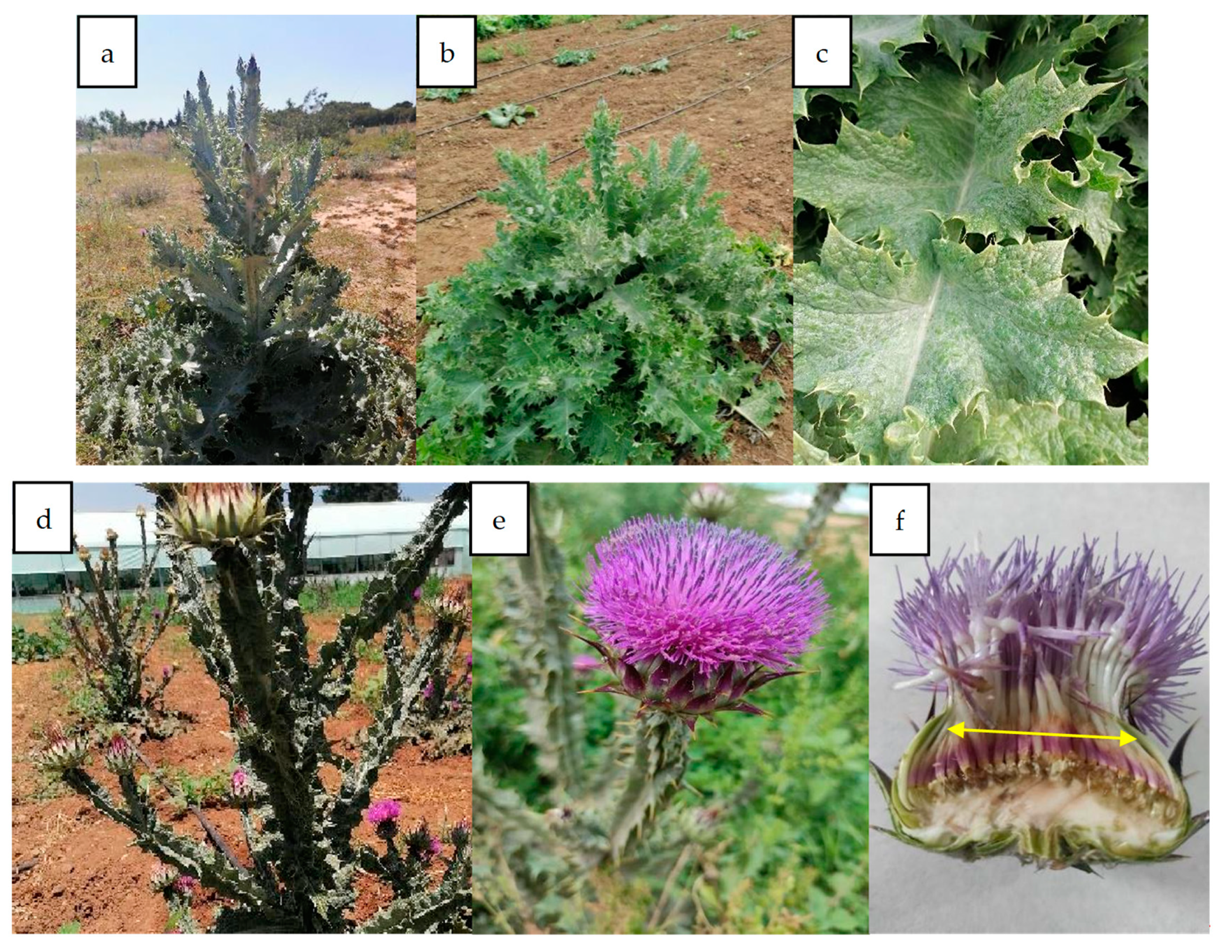
2.1. Plant Material

The aerial parts (stems, leaves, and flower petals) of mature wild Onopordum platylepis were collected from three regions in Tunisia (Sousse, Kairouan, and Nabeul) during the summer season of the year 2021 (Table 1). All studied areas are characterized by a semi-arid climate and sandy clayey soil. The cultivated culture (Figure 1b) was performed to observe the behavior of the plant outside its natural environment. Mature seeds were collected from wild plants in August 2021. Subsequently, seeds were cleaned and disinfected in a 5% sodium hypochlorite solution for 5 min. They were then sown in peat substrate within an uncontrolled greenhouse at the High Agronomic Institute of Chott Mariem (ISA-CM), Sousse, Tunisia, with an average temperature of 26 °C and under natural daylight conditions in September 2021. After germination, seedlings were transplanted into pots. The humidity was maintained through regular watering. Finally, at the end of October 2021, thistle plants were transferred to an experimental field at ISA-CM, where they were planted in spaced lines (1.2 m) with 1.5 m between plants. No fertilizer was used during this process. The irrigation was carried out according to the needs of the plant and precipitation. The plant material was harvested at the end of the flowering season (June 2022), and flowers were harvested when fully opened, before becoming inedible and acquiring a hard texture. Then, petals were hand-selected from the receptacle and cut into small pieces of 2 cm2 (Figure 1f). All collected plant material was immediately stored in a freezer at −20 °C.

2.2. Preparation of Extracts

The plant material (leaves, stems, and flowers) was lyophilized in a freeze drier at a temperature of −40 °C under a pressure of 10−1 Torr for a total duration of 72 h. The plant material was then ground in an electric mill to obtain a thin powder and stored in spittoons marked with the date of preparation and the organ name for further use. Water extracts were obtained by magnetic stirring for 2 h of 1 g of dry sample with 100 mL of H2O at room temperature (23–25 °C) after filtering with a Whatman No. 4 filter. Afterwards, each batch was centrifuged twice for 20 min at 3500× g rpm at room temperature. The collected supernatant was then stored at 4 °C and used in all investigations.

2.3. Chemical Composition of O. platylepis Aerial Parts

2.3.1. Dry Matter Content

The dry matter content of plant samples was obtained through lyophilization of various plant organs. Initially, fresh samples were weighed to determine their initial weight. Then, they were subjected to lyophilization until reaching a constant weight, indicating complete removal of water [35]. Finally, the samples were weighed again to obtain their final dry weight, and the dry matter content of each sample was calculated using the following Formula (1):
D r y   m a t t e r   c o n t e n t % = D r y   s a m p l e   W e i g h t g F r e s h   s a m p l e   W e i g h t g × 100

2.3.2. Determination of Total Phenols

Polyphenols were determined using the Folin–Ciocalteu reagent following the protocol proposed by Al-Farsi et al. [36]. In this assay, the total concentration of hydroxyl groups in the extract is quantified. Folin–Ciocalteu reagent was diluted tenfold to determine the total phenolic content (TPC). A volume of 200 μL of each extract was added to 1500 μL of Folin–Ciocalteu reagent and left to stand for six minutes after stirring at room temperature. In the next step, 1.5 mL of a 6% sodium carbonate solution was added to the mixture, which was then stored at 40 °C for 30 min. At 760 nm, a UV-Vis spectrophotometer (Macy model, UV-1300, Shanghai Aesthetic analysis Instrument Co., Ltd., Shanghai, China) was employed to measure the absorbance of the mixture against the blank. Results are reported in mg equivalent gallic acid (mg GAE/g dry matter).

2.3.3. Total Flavonoid Content

Total flavonoids (TF) were determined using the colorimetric method proposed by Zhishen et al. [37]. Flavonoids were determined by mixing 250 μL of plant extracts with 1.25 mL of distilled water. To the mixture, 5 μL of 5% NaNO2 was then added and left to stand for 6 min, followed by 150 μL of 10% AlCl3. Afterward, 0.5 mL of NaOH (1M) and 250 μL of distilled water were added to the solution six minutes later. As a final step, the mixture was vigorously stirred and the absorbance at 510 nm was measured using a UV-Vis spectrophotometer (Macy model, UV-1300, China). Total flavonoid content (TF) was determined by calibration against quercetin, and the results are reported as mg quercetin equivalent per gram of dry matter (mg QE/g dry matter).

2.3.4. Total Tannin Content

Condensed tannins (CTs) depolymerize in the presence of concentrated hydrochloric acid and are converted into anthocyanidins, which are spectrophotometrically measurable at 500 nm when they react with vanillin [38].
A volume of 50 μL of each extract was added to 1500 μL of the 4% vanillin/methanol solution and vigorously mixed. Following this, 750 μL of concentrated hydrochloric acid (HCl) was added. The resulting mixture was left then at room temperature for 20 min and the absorbance was measured at 500 nm against a blank. A calibration curve of catechin was used to determine the condensed tannin (CT) content, and results are expressed in mg catechin equivalents per gram of dry matter (mg CE/g dry matter).

2.3.5. Determination of Sugar Profile

The chromatographic conditions for sugar analyses were established on an Agilent Technologies HPLC series 1100 system. A refractive index detector (RID) was employed for compound detection. The Aminex 87C column was chosen for sugar separation with a flow rate set at 0.6 mL/min to ensure optimal peak resolution [39]. Sample injection volumes of 20 μL were pre-filtered using a 0.22-micron syringe filter. The column temperature was maintained at 80 °C to enhance component separation. The total analysis time was set at 30 min to cover a comprehensive range of target compounds. These chromatographic parameters were carefully selected to ensure effective separation and accurate quantification of sugars in the analyzed samples. A calibration curve-based method was employed to determine the carbohydrate concentrations in this study. This was achieved by comparing the retention times of unknown analytes with those of analytical grade standards (glucose, fructose, galactose, xylose, arabinose, sucrose, and mannose) obtained from Sigma-Aldrich (St. Louis, MO, USA), and results are expressed in milligrams per gram of dry matter (mg/g dry matter).

2.3.6. Protein Content

The Bradford method [40] was employed to determine protein concentration in extracts by interacting Coomassie blue with proteins. To initiate the process, 50 µL of extract was mixed with 50 µL of distilled water and 2 mL of Bradford reagent. The absorbance of the solution at 595 nm was measured using a UV-Vis spectrophotometer (Macy model, UV-1300, China) with distilled water serving as blank. Subsequently, the protein concentration was estimated based on a calibration curve for bovine serum albumin (BSA), and the results are expressed in milligrams of bovine serum albumin equivalent per gram of dry matter (mg BSAE/g dry matter).

2.3.7. Antioxidant Activity

DPPH Test

The extracts were evaluated for their electron-donating ability and their capacity to trap 2.2- diphenyl-1-picrylhydrazyl (DPPH) radicals using the method of Lee et al. [41]. Specifically, 1 mL of freshly prepared DPPH (0.2 mM) in methanol was combined with 1 mL of O. platylepis extract at various concentrations (0.05, 0.1, 0.2, 0.5, and 1 mg/mL). The absorbance was then measured at 517 nm using a spectrometer after 30 min of incubation in the dark at room temperature. A logarithmic regression was used to calculate the inhibitory concentration (IC50) needed to reduce DPPH by 50%.
DPPH radical neutralization was calculated using the following Formula (2):
P e r c e n t a g e   o f   i n h i b i t i o n   o f   D P P H   r a d i c a l s % = 1 A b s   E x t r a c t A b s   C o n t r o l   × 100
where Abs Control: absorbance of the control; Abs Extract: absorbance of the extract.

ABTS Test

ABTS tests were conducted according to the method outlined by Re et al. [42], which is based on the ability of antioxidants to stabilize ABTS+ cationic radicals, forming a colorless ABTS radical. Two solutions were prepared: ABTS (7 mM) and potassium persulfate (2.45 mM).
The cationic radical ABTS+ was formed by mixing equal amounts of these two solutions. The resulting mixture was then stored at 4 °C and protected from light for 12 to 16 h.
ABTS solution was diluted with ethanol until an absorbance of 0.7 (±0.02) was obtained at 734 nm. Subsequently, the diluted ABTS solution was added to aqueous extracts at various concentrations (0.05 mg/mL, 0.1 mg/mL, 0.2 mg/mL, 0.5 mg/mL, 1 mg/mL). After a 10 min incubation at room temperature in the dark, the reduction reaction of the ABTS solution was measured at 734 nm. The antioxidant capacity of the tested extracts is expressed relative to their concentration, providing valuable insights into their antioxidant potential. The anti-radical power of the extract was quantified as the percentage inhibition of the ABTS+ radical. A logarithmic regression was used to calculate the inhibitory concentration (IC50) needed to reduce ABTS by 50%.
P e r c e n t a g e   o f   i n h i b i t i o n   o f   A B T S   r a d i c a l s % = 1 A b s   E x t r a c t A b s   C o n t r o l   × 100
where Abs Control: absorbance of the control; Abs Extract: absorbance of the extract.

2.4. Milk-Clotting Activity

Milk-clotting activity was evaluated according to the method of Libouga [43], which is based on the examination of the appearance of the first clotting flakes. The assay was conducted by adding 1 mL of the crude extract to a glass test tube containing 10 mL of milk powder solution, which was prepared by dissolving 12 g of milk powder in 100 mL of distilled water, followed by the addition of 0.01 M CaCl2 solution. Finally, the mixture was transferred to a 35 °C water bath, where the clotting time (t) was measured using a chronometer.
The milk-clotting activity is expressed in rennet units (UP) and was calculated using Equation (4):
M C A U P = 10 × V C t × V
where UP: rennet unit; V: volume of milk; V′: volume of coagulant extract (1 mL); Ct: milk-clotting time in seconds

2.5. Statistical Analyses

All statistical analyses were performed using SPSS version 20. Three replicates were employed to calculate all data, which are expressed as means ± standard errors. The chemical composition as well as IC50 for ABTS and DPPH scavenging activities were all analyzed using a general linear model, and the Duncan test was used to compare means of these parameters when significant differences were detected. An independent samples t-test was further used to compare the means of spontaneous and cultivated plants. Principal component analysis (PCA) and dendrograms were performed using the XLSTAT 2023 software package.

3. Results and Discussion

3.1. Chemical Composition of O. platylepis Aerial Parts

3.1.1. Dry Matter Content

The dry matter content in various aerial parts of O. platylepis varied between 20.79% and 38.86% in wild plants and between 23.12% and 39.87% in cultivated plants (Table 2). Flowers showed the highest dry matter content, with no significant differences observed between both plant types (spontaneous and cultivated). However, lower contents were observed in leaves.

3.1.2. Total Phenols, Flavonoids, and Condensed Tannins

Phytochemical levels of phenols, flavonoids, and condensed tannins differ according to ecotypes and organs (Table 2). Significant differences were observed in phenolic compounds between aerial parts of O. platylepis plants (p = 0.000). In all ecotypes, results indicated that flowers contain higher phenolic contents, compared to leaves and stems, in both spontaneous and cultivated plants. Furthermore, significant differences were obtained between studied populations in terms of total phenols (p = 0.031). Flowers of the Nabeul population (NA) showed the highest concentration of total phenols for both plant types (spontaneous and cultivated), recording 44.75 and 39.79 mg EAG/g, respectively. The total phenol content in leaves varied from 18.67 to 28.74 mg EAG/g in spontaneous plants and between 13.66 and 17.64 mg EAG/g in cultivated plants. In contrast, stems showed the lowest phenolic contents, with values ranging from 4.52 to 17.51 EAG/g in naturally grown and cultivated thistles. No significant differences were obtained between the two plant types in O. platylepis aerial parts (p > 0.05).
Results further indicated variations and significant differences (p = 0.000) in flavonoid content among different plant aerial parts and populations in both spontaneous and cultivated plants (Table 2). The best flavonoid content was observed in spontaneous SO population flowers, registering 11.42 ± 0.23 mg QE/g. In comparison with leaves and stems, flowers showed the highest amounts of flavonoids, except in the KA population, where leaves showed higher contents of flavonoids compared to flowers. However, lower contents were observed in stems. Finally, condensed tannins were detected in every studied plant organ, with contents varying between 1.50 mg CE/g, and 3 mg CE/g in spontaneous plants, and between 1.56 mg CE/g and 2.81 mg CE/g in cultivated plants.
In general, when observing variation among organs, flowers emerge with the highest total phenol and flavonoid contents compared to the other organs. Our findings are in agreement with those reported by Ewais et al. [44], who observed an increase in the percentages of total flavonoids and total phenolic acids in flowers and a decrease in those of stems. Moreover, the findings reported by Mandim et al. [45], who assessed the phenolic composition and bioactive properties of various inflorescence tissues (stigma, corolla, bracts, pappus, and receptacle) across seven genotypes of Cynara cardunculus L., revealed a greater variation in phenolic compounds within the corolla, and this is consistent with our results. Furthermore, the findings of this study align with those of Fratianni et al. [46], who reported that cardoon organs play a crucial role in determining the level of phenolic compounds. The elevated concentration of phenolics in flowers, as opposed to leaves and stems, could be attributed to specific plant adaptations, including the attraction of pollinators, reproductive investment, and responses to environmental stress. These adaptations collectively contribute to the biological significance of increased phenolic content in flowers. The attraction of pollinators is essential for successful reproduction, and the investment in reproductive structures, such as flowers, is reflected in the elevated phenolic levels [47]. Additionally, higher phenolic contents can serve as a response to environmental stressors, providing the plant with a defense mechanism against potential threats [48].
Furthermore, in comparison with stems, leaves could be considered as a potential source of phenolic compounds. These findings are corroborated by the research realized by Velez et al. [49], which demonstrated that leaf extracts of Cynara cardunculus L. manifested higher levels of total phenols than those of stem extracts. Beyond organ-specific effects, the results of phenolic compound contents of this study indicate genotype effects, which may be explained by differences in genetic origin. Moreover, the composition of plant phenolics can be influenced by soil conditions; specifically, elevated nitrogen levels and reduced soil moisture may result in lower synthesis and, consequently, decreased levels of certain phenolics [50].
Our results reveal significant differences in total phenols and flavonoids between different studied populations (p < 0.05). This variability could be attributed to genetic and environmental factors. Our results are in agreement with those obtained by Raudone et al. [51], who observed a high diversity of chemical profiles of Achillea spp., which suggests a high potential for discovering new sources of multifunctional phenolic compounds. Furthermore, for cultivated plants, it is possible to discern a genetic basis for these variations among plants grown under similar conditions. Each region preserves its cultivars, potentially differing in chemical composition and consequently in characteristics [52].
Regardless of ecotypes or organs, the values observed in this study surpass those reported by Khaldi et al. [53] and Velez et al. [49] in Cynara cardunculus, suggesting that O. platylepis serves as a notable source of antioxidants due to its higher content of phenolic compounds.
Phenolic compounds, characterized by abundant hydroyl functional groups, are considered promising antioxidants due to their ability to react with free radicals. Specifically, within phenolics, catechols play a crucial role in redox activity [54]. In addition to their safety compared to synthetic food additives, these compounds may also offer a safer option for humans, considering their potential to reduce carcinogenic effects [55]. Statistical analysis showed no significant differences between spontaneous and cultivated plants in terms of total phenols (p > 0.05). Therefore, the O. platylepis species holds promising potential in both industrial and food sectors.

3.1.3. Evaluation of Sugar Profile

Carbohydrate composition findings from extracts of flowers, leaves, and stems of O. platylepis are summarized in Table 3. Results indicated significant differences in mono- and disaccharide content between organs (p = 0.000). Comparison across different plant parts revealed that mono- and disaccharide components were more abundant in flower extracts, notably with higher contents of fructose, xylose, sucrose, and glucose. The abundance of carbohydrates in flowers could be explained by their crucial role in the reproduction and development of thistles, serving as essential energy sources for their growth [56]. Flowering and fruitlet development compete with vegetative growth and accumulation of reserves for most of the necessary metabolites [57].
Moreover, as shown in Table 3, xylose and fructose were undetected in leaves, while stems showed fructose and glucose as major compounds, except in those of the cultivated NA population. Soluble carbohydrates, such as xylose, arabinose, mannose, and galactose, are known to occur in low amounts in shoots. Additionally, our results contradict those reported by Ewais et al. [44], who investigated soluble carbohydrates in leaves, stems, and flowers of Pulicaria incisa subsp. Incisa, a species belonging to the Asteraceae family. Their findings indicated that fructose was detected in lower concentrations among the separated free sugars in the aerial parts of P. incisa.
Moreover, plant flowers from the Kairouan population (KA) revealed the highest glucose contents compared to Sousse (SO) and Nabeul (NA) populations. Moreover, the KA population showed important xylose contents in both plant types, which could be an important source of prebiotics. Sugar contents in our plant flowers are higher than those obtained by Ben Amira et al. [58], who evaluated the chemical composition and monosaccharides composition of wild, lyophilized, cardoon flowers collected in Tunisia, where xylose content was 2.61 ± 0.07% DM. These results revealed the potential of flowers of O. platylepis as a prebiotic source. Statistical analysis showed that carbohydrate composition is significantly affected by genotype (p = 0.000). The observed variation between these different populations could be explained by genetic influence, as well as the geographical location of this thistle. Similar results were obtained by Raccuia and Melilli [59] in their analysis of genetic variation among different genotypes of Cynara cardunculus L., which influences sugar production levels in wild cardoon populations across Sicily (Italy). Both genetic variability and environmental factors contribute to determining the total sugar content within this thistle species.
The investigation into the chemical composition and biological functions of plant extracts is crucial for identifying innovative materials with potential applications in food industry. Plant foods, which are the primary sources of dietary fiber, also serve as the major contributors of carbohydrates in fruits and vegetables, highlighting their importance in human nutrition [60]. O. platylepis emerges as a notable source of carbohydrates, especially flowers, which are suitable for integration into food products. This current research paper could be recognized as the first comparative survey of the carbohydrate composition of O. platylepis.

3.1.4. Protein Content

Results in Table 2 reveal variability in protein content among the different aerial plant parts and genotypes (p < 0.05). The observed values range from 0.28 to 2.31 mg BSA/g and from 0.16 to 2.43 mg BSA/g in spontaneous and cultivated plants, respectively. The highest protein levels were found in flowers, followed by leaves and stems, in both plant types. Additionally, NA and SO populations showed the highest protein contents in the different aerial plant parts. Results further indicate comparable protein amounts between naturally grown and cultivated plants (p > 0.05). Our results align with the findings of Essaidi et al. [28], who compared protein amounts in spontaneously grown and cultivated O. platylepis flowers with various drying methodologies and revealed no significant differences between plant types.

3.1.5. Antioxidant Activity

Evaluation of Radical Scavenging Activity Using DPPH Test

Results showed that free radical inhibition percentages of all extracts increased with extract concentration and varied with extract type (Figure S1). The inhibitory concentration IC50, representing the antioxidant concentration required to inhibit 50% of free radicals, and inversely correlated with the antioxidant activity, recorded values ranging from 0.13 to 1.82 mg/mL in spontaneous plants and from 0.09 to 1.01 mg/mL in cultivated plants (Table 4).
The radical scavenging activity of O. platylepis is significantly affected by the organ (p = 0.000). For both spontaneously grown and cultivated plants, flower extracts showed the highest percentage of DPPH inhibition; except in the Kairouan (KA) population, leaves showed a more pronounced efficacy against DPPH with IC50 values of 0.23 mg/mL and 0.09 mg/mL, respectively. Results further indicate significant differences (p < 0.05) between different genotypes in both plant types (cultivated and spontaneous), with the NA genotype being, in general, the most potent in inhibiting free radicals. In spontaneous plants, the most pronounced radical scavenging activity was noted in flowers from Nabeul (NA) and Sousse (SO) ecotypes, showcasing IC50s values of 0.13 and 0.2 mg/mL. However, stems showed lower potency in inhibiting free radicals compared to other plant parts in both spontaneous and cultivated plants.
In particular, flowers derived from spontaneous plants NA and SO populations showed the highest concentration of total phenols. This finding aligns with their elevated antioxidant potential determined through the DPPH test, surpassing that of the KA population. This variation could be related to the harsher climatic conditions in NA and the SO provenance of O. platylepis.
Some earlier studies of cardoon plants have explored the fact that corolla scavenges DPPH better than other plant tissues [61]. This observation aligns with the results obtained in this study, corroborating that thistle’s corolla possesses significant antioxidant potential. Antioxidant activities of various parts of cardoon plants may be impacted by growing conditions and cultivation practices, as they significantly contribute to the composition of phenolic compounds, recognized for their robust antioxidant effects [62]. Notable differences in antioxidant properties were observed among hydromethanolic extracts of studied plant parts of Cynara cardunculus. Specifically, seeds had the highest scavenging activity in both DPPH and reducing power assays, with IC50 values of 143 and 87 μg mL−1, respectively [63].
Valizadeh et al. [33] previously assessed the antioxidant potential of Onopordum leptolepis, employing the ferric reducing antioxidant potential (FRAP) and (DPPH) free radical methods, and found that flowers manifested lower antioxidant activity than vegetative parts. In comparison, the antioxidant activity of vegetative parts in our results was lower than that of O. platylepis flowers. This difference could be due to the distinct geographical origins of the two species, indicating species-specific variations in the distribution of antioxidant activity between floral and vegetative components. In another study, Petropoulos et al. [64] previously documented variations in the antioxidant properties of morphological organs in wild cardoon. Their findings indicated that among the studied organs, heads showed the highest potency, surpassing leaf blades, midribs, and petioles.

Evaluation of Radical Scavenging Activity Using ABTS Test

The results obtained from the ABTS+ test (Figure S2) show comparable antioxidant activity in flowers compared to leaves and stems, as indicated by the DPPH test. However, in the cultivated KA population, both leaves and stems presented higher antioxidant activities than flowers, with IC50c values of 0.12 mg/mL and 0.15 mg/mL, respectively (Table 4). Additionally, in all studied populations, stems showed a notable capacity to scavenge free radicals, both in spontaneous and cultivated plants, in ABTS radicals. However, flowers manifested more pronounced efficacy against DPPH radicals than ABTS. These results could be explained by the fact that the DPPH radical dissolves only in organic solvents and does not allow the determination of hydrophilic antioxidants. Thus, these raw materials show a diversity of antioxidants and stems may contain a slightly higher proportion of hydrophilic antioxidants. Moreover, significant differences were obtained between the different genotypes in inhibiting free radicals (p < 0.05), with better-reducing power in NA flowers (IC50s = 0.14 mg/mL) > SO flowers (IC50s = 0.23 mg/mL) > KA flowers (IC50s = 0.4 mg/mL).
The findings presented for Cynara cardunculus, a species relatively close to Onopordum nervosum ssp. platylepis, showed that cardoon flowers are a potential source of antioxidant compounds [65]. According to Ksouri et al. [66], the antioxidant activity of plants is dependent on their organs, which aligns with our results. The statistical analysis indicated significant differences between the IC50 values of extracts from cultivated and spontaneous plants (p < 0.05) in both tests, with lower values, in general, in cultivated plants. Consequently, the domestication of Onopordum nervosum ssp. platylepis could be useful for therapeutic properties and industrial applications. Furthermore, additional studies are needed to explore the physiological relevance and potential health benefits associated with these antioxidant properties. The antioxidant properties of natural compounds such as phenolics and flavonoids may influence oxidative stress-related conditions, including cardiovascular diseases, neurodegenerative disorders, metabolic syndromes, and inflammatory conditions [67].

3.2. Milk-Clotting Trial

Results in Table 4 showed that a large variability in the capacity to coagulate milk was obtained between different regions and aerial parts of O. platylepis plants (p = 0.000), with clotting time ranging from 65 to 108 s in spontaneous plants and from 58 to 100 s in cultivated plants.
Furthermore, results indicated that only flowers possessed milk-clotting properties, with SO and NA genotypes revealing the best milk-clotting activity in both plant types. However, the KA genotype showed higher clotting time and, consequently, lower milk-clotting potential (p < 0.05).
The exclusive milk-clotting activity detected in flowers could be explained by the fact that during pollination, protease enzymes are active in mature flowers and pistils, serving to protect them from viruses or microorganisms [16]. In previous studies, cardoon flower ecotypes used for coagulation were found to vary significantly, which is in accordance with our findings [68]. Additionally, our values (Table 4) are higher than those obtained by Ordiales et al. [19], who evaluated the milk-clotting activity of aqueous crude extracts from Cynara cardunculus flowers. Their results showed 0.131 RU/mL after one hour of flower maceration. This could be attributed to O. platylepis’ capacity to produce more protein proteases than other species. Furthermore, according to previous studies, the obtained milk-clotting activities are greater than those of the crude extracts of some plants, such as Silybum marianum (0.08 RU/mL) and Onopordum acanthium (0.54 RU/mL) [69,70]. The coagulant extract of Onopordum platylepis flowers is therefore expected to be more specific for κ-casein. According to these findings, the crude extracts from this thistle hold promise as a vegetable rennet.
Statistical analyses revealed no significant differences between spontaneously grown and cultivated plants (p > 0.05). Thus, our finding suggests that O. platylepis flowers are a good source of milk-clotting agents and, therefore, encourage the technological use of selected genotypes, as an alternative to animal rennet in cheese production.
Cultivation techniques should be continually monitored and improved in order to improve long-term conservation success of this species. Although this thistle grows in Mediterranean climates with low rainfall and irregular weather patterns, sandy clay soils are most suitable for its cultivation. For optimal plant growth, appropriate irrigation would be necessary according to plant needs. Current cultivation methods will need to be continuously evaluated and adjusted as necessary to avoid the loss of desirable traits such as the potential of milk-clotting activity. In this way, it ensures that cultivated plants thrive in their environment, contributing to the protection of biodiversity and the restoration of ecosystems. In addition to domestication, conservation efforts should be conducted to protect genetic diversity, including establishing protected areas, seed banks, and ex situ conservation measures [6]. Furthermore, in order to facilitate effective breeding efforts, it is important to understand the genetic basis for desirable traits. Genetic markers associated with specific traits can be identified using techniques such as genome sequencing and association mapping [71].

3.3. Correlation Analysis between Chemical Composition and Biological Activities

Table 5 presents the results of correlation analysis, aiming to evaluate the relationships between the chemical composition (total phenols, total flavonoids, condensed tannins, protein content, and carbohydrate composition) and the activities of both antioxidant and milk clotting.
Results indicated that the phytochemical composition (total phenols, total flavonoids, condensed tannins, and sugar composition) of aerial parts of O. platylepis was correlated with their antioxidant properties (IC50 of DPPH and ABTS•+). A negative correlation was observed between total phenols, total flavonoids, and IC50 values for inhibiting DPPH and ABTS radicals, with coefficients ranging from −0.605 to −0.394 demonstrating a high level of significance. Additionally, the results revealed a negative correlation between sucrose, glucose, and IC50 values. A significant correlation was found between sucrose, glucose, and IC50 of DPPH, as well as between sucrose and IC50 of ABTS•+. Statistical analysis showed a negative and significant (p < 0.05) correlation between glucose, sucrose, and antioxidant activity, which indicates their potential contribution to the production of various phenolic compounds. In plants, phenolic compounds are primarily synthesized through two fundamental pathways: the shikimic acid pathway and the acetate–malonate (polyketide) pathway.
Glucose plays a vital role as a precursor in the shikimate metabolic pathway, which is fundamental for producing phenolic compounds, including flavonoids, lignins, and lignans [72]. As a carbon transporter, sucrose provides energy and a metabolic precursor to phenolic compound synthesis in plants. Through enzymatic cleavage, it produces glucose and fructose; these serve as substrates in the shikimate pathway, which is crucial for phenolic compound biosynthesis [73]. Phenolic compounds play a crucial role in many biological processes, including antioxidant activity. The antioxidant properties of phenolic compounds are primarily associated with their oxidation properties, which enable them to reduce free radicals by donating electrons or hydrogen atoms [74]. Among phenolic compounds, flavonoids are known for their antioxidant properties, which are related to their ability to scavenge free radicals and chelate metal ions [75]. Results further indicate that protein content was negatively correlated with the DPPH and ABTS IC50 values in our studied thistle aerial parts, suggesting some proteins could be related to antioxidant activity. This finding is in agreement with a previous study, which suggested that the antioxidant activity was highlighted in correlation with amino acid composition [76]. Additionally, a high-significance positive correlation between protein content and milk-clotting activity was observed, indicating the potential effect of plant proteases within the flower extracts. Plant proteases, typically belonging to the class of serine or aspartic proteases, initiate milk coagulation by cleaving specific bonds within milk protein κ-casein. This process leads to the formation of a gel network that traps the milk fat and other proteins, consequently resulting in curd formation [77]. In this context, O. platylepis flower extract could offer a natural alternative to traditional rennet-derived enzymes, aligning with consumer requests for plant-based and environmentally friendly ingredients.

3.4. Principal Component and Dendrogram Analysis

According to principal component analysis (PCA), the first two components account for approximately 64.15% of total variability. The first axis explains 44.94% of the variability and is associated with the following variables: total phenolic compounds (TPCs), xylose (Xyl), sucrose (Suc), glucose (Glu), milk-clotting activity (MCA), and protein content (PC). The second axis, which accounts for 19.2% of the variance, includes total flavonoid content (TFC), condensed tannins (CTs), IC50 ABTS and DPPH, mannose (Man), galactose (Gal), fructose (Fru), and arabinose (Ara). These results indicate that raw materials obtained from O. platylepis plants show distinct proximate compositions depending on organs and genotypes, as revealed by PCA analysis. Moreover, a strong negative correlation between total phenolic contents, total flavonoids, and IC50 of ABTS•+ and DPPH can be clearly seen (Figure 2). Furthermore, the dendrogram confirms the results of PCA analysis and shows that there is a discernible tendency to isolate two major groups of individuals. However, spontaneous and cultivated flowers of the studied populations form a distinct group characterized by significant phenolic compound contents, low IC50 values, and either high protein or carbohydrate contents (sucrose, glucose, xylose, fructose) among the evaluated raw materials. The second group is divided into two subgroups including the leaves and stems of this thistle. All these results confirm the chemical diversity present in O. platylepis extracts across different organs and genotypes.

4. Conclusions

This study is the first to explore Tunisian wild and cultivated germplasm of Onopordum nervosum ssp. platylepis involving the chemical analysis of populations in the center of Tunisia. The findings from this study demonstrate the impact of both plant tissues and genetic background on the biochemical potential of Onopordum. Consequently, Onopordum populations found in a semi-arid climate represent a promising reservoir of compounds with notable antioxidant and milk-clotting properties, making them a compelling genetic pool for inclusion in breeding programs. The identified carbohydrate contents in flowers showed the potential of this plant as a source of prebiotics for human nutrition. Furthermore, the significant amounts of bioactive molecules in various plant parts contribute to medicinal properties and diverse health benefits. The current study suggests that aerial parts of O. platylepis have potential applications in the pharmaceutical and food industries, with flowers potentially serving as reservoirs of bioactive compounds and milk-clotting agents. Additionally, this study indicates that the domestication of this endangered Tunisian plant can contribute to its conservation efforts by maintaining genetic differentiation, and highlights the importance of selecting the most promising populations with valuable sources of antioxidants, carbohydrates, and milk-clotting agents for breeding programs.

Supplementary Materials

The following supporting information can be downloaded at: https://www.mdpi.com/article/10.3390/agronomy14050987/s1, Figure S1. Comparative analysis of antioxidant activity (DPPH) in aqueous extracts of O. nervosum ssp. platylepis (A: flowers, B: leaves, C: stems), from both spontaneous (a) and cultivated (b) thistle. Figure S2. Comparative analysis of antioxidant activity (ABTS•+) in aqueous extracts of O. nervosum ssp. platylepis (A: flowers, B: leaves, C: stems) from both spontaneous (a) and cultivated thistle (b).

Author Contributions

R.K.: Conceptualization, Investigation, Methodology, Writing—original draft. I.E.: Investigation, Methodology, Writing—review and editing. K.A.: Investigation, Methodology, Writing—review and editing. N.D.: Methodology, Writing—review and editing. F.H.: Investigation, Methodology, Writing—review and editing. A.M.A.: Writing—review and editing. H.A.A.: Writing—review and editing. S.A.B. Methodology, Writing—review and editing. O.A.: Methodology, Writing—review and editing. B.A.M.D.: Supervision, Conceptualization, Methodology, Investigation, Writing—review and editing. All authors have read and agreed to the published version of the manuscript.

Funding

This work was supported and funded by the Deanship of Scientific Research at Imam Mohammad Ibn Saud Islamic University (IMSIU) (grant number IMSIU-RG23096).

Data Availability Statement

The original contributions presented in the study are included in the article and Supplementary Material.

Conflicts of Interest

The authors declare no conflicts of interest.

References

  1. Kaur, N.; Ahmed, T. Bioactive secondary metabolites of medicinal and aromatic plants and their disease-fighting properties. In Medicinal and Aromatic Plants; Springer Nature: Basingstoke, UK, 2021; pp. 113–142. ISBN 978-3-030-58974-5. [Google Scholar]
  2. Saboon; Chaudhari, S.K.; Arshad, S.; Amjad, M.S.; Akhtar, M.S. Natural compounds extracted from medicinal plants and their applications. Nat. Bio-Act. Compd. 2019, 1, 193–207. [Google Scholar] [CrossRef] [PubMed]
  3. Singh, S.; Kaur, I.; Kariyat, R. The Multifunctional Roles of Polyphenols in Plant-Herbivore Interactions. Int. J. Mol. Sci. 2021, 22, 1442. [Google Scholar] [CrossRef] [PubMed]
  4. Qaderi, M.M.; Martel, A.B.; Strugnell, C.A. Environmental Factors Regulate Plant Secondary Metabolites. Plants 2023, 12, 447. [Google Scholar] [CrossRef] [PubMed]
  5. Bigdeloo, M.; Hadian, J.; Nazeri, V. Composition of essential oil compounds from different populations of Thymus caramanicus Jalas. J. Appl. Res. Med. Aromat. Plants. 2017, 7, 95–98. [Google Scholar] [CrossRef]
  6. Salgotra, R.K.; Chauhan, B.S. Genetic Diversity, Conservation, and Utilization of Plant Genetic Resources. Genes 2023, 14, 174. [Google Scholar] [CrossRef] [PubMed]
  7. Mandel, J.R.; Dikow, R.B.; Siniscalchi, C.M.; Thapa, R.; Watson, L.E.; Funk, V.A. A fully resolved backbone phylogeny reveals numerous dispersals and explosive diversifications throughout the history of Asteraceae. Proc. Natl. Acad. Sci. USA 2019, 116, 14083–14088. [Google Scholar] [CrossRef] [PubMed]
  8. Sobrinho, A.C.N.; de Souza, E.B.; Rocha, M.F.G.; Albuquerque, M.R.J.R.; Bandeira, P.N.; dos Santos, H.S.; de Paula Cavalcante, C.S.; Oliveira, S.S.; Aragão, P.R.; de Morais, S.M.; et al. Chemical composition, antioxidant, antifungal and hemolytic activities of essential oil from Baccharis trinervis (Lam.) Pers. (Asteraceae). Ind. Crops Prod. 2016, 84, 108–115. [Google Scholar] [CrossRef]
  9. Formisano, C.; Sanna, C.; Ballero, M.; Chianese, G.; Sirignano, C.; Rigano, D.; Millán, E.; Muñoz, E.; Taglialatela-Scafati, O. Anti-inflammatory sesquiterpene lactones from Onopordum illyricum L. (Asteraceae), an Italian medicinal plant. Fitoterapia 2017, 116, 61–65. [Google Scholar] [CrossRef]
  10. Pottier-Alapetite, G.A. Flore de la Tunisie: Angiospermes-Dicotylédones-Gamopétales; Ministère de l’Enseignement Supérieur et de la Recherche Scientifique et le Ministère de l’Agriculture: Tunis, Tunisia, 1981. [Google Scholar]
  11. Pandino, G.; Lombardo, S.; Williamson, G.; Mauromicale, G. Polyphenol profile and content in wild and cultivated Cynara cardunculus L. Ital. J. Agron. 2012, 7, e35. [Google Scholar] [CrossRef]
  12. Sharma, P.; Verma, P.K.; Pankaj, N.K.; Agarwal, S. The Phytochemical Ingredients and Therapeutic Potential of Cynara scolymus L. Pharm. Biomed. Res. 2021, 7, 141–160. [Google Scholar] [CrossRef]
  13. Petkova, N.; Hambarlyiska, I.; Angelova, E.; Ivanov, I. Fructans, Polyphenols and antioxidant activity in edible roots and thistles from seven medicinal plants. Chiang Mai Univ. J. Nat. Sci. 2021, 20, 2021082. [Google Scholar]
  14. Pari, L.; Alfano, V.; Stefanoni, W.; Latterini, F.; Liuzzi, F.; De Bari, I.; Valerio, V.; Ciancolini, A. Inulin content in chipped and whole roots of cardoon after six months storage under natural conditions. Sustainability 2021, 13, 3902. [Google Scholar] [CrossRef]
  15. Barracosa, P.; Barracosa, M.; Pires, E. Cardoon as a sustainable crop for biomass and bioactive compounds production. Chem. Biodivers. 2019, 16, e1900498. [Google Scholar] [CrossRef] [PubMed]
  16. Bravo Bolívar, M.S.; Pasini, F.; Marzocchi, S.; Ravagli, C.; Tedeschi, P. Future Perspective and Technological Innovation in Cheese Making Using Artichoke (Cynara scolymus) as Vegetable Rennet: A Review. Foods 2023, 12, 3032. [Google Scholar] [CrossRef] [PubMed]
  17. Tejada, L.; Vioque, M.; Gómez, R.; Fernández-Salguero, J. Effect of lyophilisation, refrigerated storage and frozen storage on the coagulant activity and microbiological quality of Cynara cardunculus L. extracts. J. Sci. Food Agric. 2008, 88, 1301–1306. [Google Scholar] [CrossRef]
  18. Silva, G.M.d.S.; da Costa, J.S.; Freire, J.O.; Santos, L.S.; Bonomo, R.C.F. Artichoke Leaf Extracts: Proteolytic Activity, Coagulant and Hplc Analysis. Cienc. E Agrotecnol. 2021, 45, e001721. [Google Scholar] [CrossRef]
  19. Ordiales, E.; Martín, A.; Benito, M.J.; Hernández, A.; Ruiz-Moyano, S.; Córdoba, M.D.G. Technological Characterisation by Free Zone Capillary Electrophoresis (FCZE) of the Vegetable Rennet (Cynara cardunculus) Used in “Torta Del Casar” Cheese-Making. Food Chem. 2012, 133, 227–235. [Google Scholar] [CrossRef]
  20. Folgado, A.; Abranches, R. Plant aspartic proteases for industrial applications: Thistle get better. Plants 2020, 9, 147. [Google Scholar] [CrossRef]
  21. Pimentel, C.; Van Der Straeten, D.; Pires, E.; Faro, C.; Rodrigues-Pousada, C. Characterization and expression analysis of the aspartic protease gene family of Cynara cardunculus L. FEBS J. 2007, 274, 2523–2539. [Google Scholar] [CrossRef] [PubMed]
  22. Barbagallo, R.N.; Chisari, M.; Spagna, G.; Mauromicale, G. Natural ingredients for cheese-making: Vegetable rennets extracts from Cynara cardunculus L. Ingredienti Aliment. 2009, 8, 6–12. [Google Scholar]
  23. Barracosa, P.; Simões, I.; Martins, A.P.; Barros, M.; Pires, E. Biochemical diversity of cardoon flowers (Cynara cardunculus L.): Predicting PDO Mediterranean cheese textures. Food Biosci. 2021, 39, 100805. [Google Scholar] [CrossRef]
  24. Saci, F.; Zikiou, A.; Fiala, S.; Bensouici, C. Bioactive compounds and pharmacological activities of enzymatic extracts of cardoon flowers (Cynara cardunculus L.) used as coagulants in cheese production. Biotechnol. Res. Cent. 2023. manuscript in preparation; to be submitted; Available at Research Square. [Google Scholar] [CrossRef]
  25. El Oualidi, J.; Khamar, H.; Fennane, M.; Ibn Tattou, M.; Chauvet, S.; Taleb, M.S. Checklist des endémiques et spécimens types de la flore vasculaire de l’Afrique du Nord. Doc. L’Institut. Sci. 2012, 25, 1–189. [Google Scholar]
  26. Cardinali, F.; Taccari, M.; Milanović, V.; Osimani, A.; Polverigiani, S.; Garofalo, C.; Foligni, R.; Mozzon, M.; Zitti, S.; Raffaelli, N.; et al. Yeast and mould dynamics in Caciofiore della Sibilla cheese coagulated with an aqueous extract of Carlina acanthifolia All. Yeast 2016, 33, 403–414. [Google Scholar] [CrossRef] [PubMed]
  27. Fernando, A.L.; Costa, J.; Barbosa, B.; Monti, A.; Rettenmaier, N. Environmental impact assessment of perennial crops cultivation on marginal soils in the Mediterranean Region. Biomass Bioenergy 2018, 111, 174–186. [Google Scholar] [CrossRef]
  28. Essaidi, I.; Dhen, N.; Lassoued, G.; Kouki, R.; Haouala, F.; Alhudhaibi, A.M.; Dridi Al Mohandes, B. Onopordum nervosum ssp. platylepis Flowers as a Promising Source of Antioxidant and Clotting Milk Agents: Behavior of Spontaneous and Cultivated Plants under Different Drying Methodologies. Processes 2023, 11, 2962. [Google Scholar] [CrossRef]
  29. Mattalia, G.; Sõukand, R.; Corvo, P.; Pieroni, A. Wild food thistle gathering and pastoralism: An inextricable link in the biocultural landscape of Barbagia, central Sardinia (Italy). Sustainability 2020, 12, 5105. [Google Scholar] [CrossRef]
  30. Camini, F.C.; Costa, D.C. Silymarin: Not just another antioxidant. J. Basic Clin. Physiol. Pharmacol. 2020, 31, 20190206. [Google Scholar] [CrossRef] [PubMed]
  31. Verotta, L.; Belvisi, L.; Bertacche, V.; Loi, M.C. Complete Characterization of Extracts of Onopordum illyricum L. (Asteraceae) by HPLC/PDA/ESIMS and NMR. Nat. Prod. Commun. 2008, 3, 1934578X0800301219. [Google Scholar] [CrossRef]
  32. Al-Snafi, A.E. Constituents and pharmacology of Onopordum acanthium. IOSR J. Pharm. 2020, 10, 7–14. [Google Scholar]
  33. Valizadeh, E.; Zonouz, N.F.; Zand, A.; Shahbazi, S.; Malekian, A. Evaluation of antioxidant potentials of extracts of cotton thistle (‘Onopordum leptolepis’ DC.) obtained by various solvents. Aust. J. Crop Sci. 2011, 5, 1163–1166. [Google Scholar]
  34. Caddeo, C.; Tuberoso, C.I.G.; Floris, S.; Masala, V.; Sanna, C.; Pintus, F. A Nanotechnological Approach to Exploit and Enhance the Bioactivity of an Extract from Onopordum illyricum L. Leaves. Plants 2023, 12, 1453. [Google Scholar] [CrossRef]
  35. Brahmakshatriya, R.D.; Donker, J.D. Five methods for determination of silage dry matter. J. Dairy Sci. 1971, 54, 1470–1474. [Google Scholar] [CrossRef]
  36. Al-Farsi, M.; Alasalvar, C.; Morris, A.; Baron, M.; Shahidi, F. Comparison of antioxidant activity, anthocyanins, carotenoids. and phenolics of three native fresh and sun-dried date (Phoenix dactylifera L.) varieties grown in Oman. J. Agric. Food Chem. 2005, 53, 7592–7599. [Google Scholar] [CrossRef] [PubMed]
  37. Zhishen, J.; Mengcheng, T.; Jianming, W. The determination of flavonoid contents in mulberry and their scavenging effects on superoxide radicals. Food Chem. 1999, 64, 555–559. [Google Scholar] [CrossRef]
  38. Julkunen-Titto, R. Phenolic constituents in the leaves of northern willows: Methods for the analysis of certain phenolics. J. Agric. Food Chem. 1985, 33, 213–217. [Google Scholar] [CrossRef]
  39. Lorenzoni, A.S.; Aydos, L.F.; Klein, M.P.; Rodrigues, R.C.; Hertz, P.F. Fructooligosaccharides synthesis by highly stable immobilized β-fructofuranosidase from Aspergillus aculeatus. Carbohydr. Polym. 2014, 103, 193–197. [Google Scholar] [CrossRef] [PubMed]
  40. Kruger, N.J. The Bradford Method for protein quantitation. In The Protein Protocols Handbook; Walker, J.M., Ed.; Springer Protocols Handbooks; Humana Press: Totowa, NJ, USA, 2019. [Google Scholar] [CrossRef]
  41. Lee, J.Y.; Hwang, W.I.; Lim, S.T. Antioxidant and anticancer activities of organic extracts from Platycodon grandiforum A. De Candolle roots. J. Ethnopharmacol. 2004, 93, 409–415. [Google Scholar] [CrossRef] [PubMed]
  42. Re, R.; Pellegrini, N.; Proteggente, A.; Pannala, A.; Yang, M.; Rice-Evans, C. Antioxidant activity applying an improved ABTS radical cation decolorization assay. Free. Radic. Biol. Med. 1999, 26, 1231–1237. [Google Scholar] [CrossRef] [PubMed]
  43. Libouga, D.G.; Ngah, E.; Nono, Y.J.; Bitjoka, L. Clotting of cow (Bos taurus) and goat milk (Capra hircus) using calve and kid rennets. Afr. J. Biotechnol. 2008, 7, 3490–3496. [Google Scholar]
  44. Ewais, E.A.; El-Maboud, M.M.A.; Haggag, M.I. Studies on chemical constituents and biological activity of Pulicaria incisa subsp. Incisa (Asteraceae). Rep. Opin. 2014, 6, 27–33. [Google Scholar]
  45. Mandim, F.; Pinela, J.; Marcelino, S.; Dias, M.I.; Barracosa, P.; Ivanov, M.; Soković, M.; Santos-Buelga, C.; Barros, L. Insights into the phenolic composition and in vitro bioactivity of cardoon capitulum: A nutraceutical-oriented valorization study. Food Chem. 2024, 435, 137480. [Google Scholar] [CrossRef]
  46. Fratianni, F.; Tucci, M.; De Palma, M.; Pepe, R.; Nazzaro, F. Polyphenolic composition in different parts of some cultivars of globe artichoke (Cynara cardunculus L. var. scolymus (L.) Fiori). Food Chem. 2007, 104, 1282–1286. [Google Scholar] [CrossRef]
  47. Bohman, B.; Phillips, R.D.; Flematti, G.R.; Peakall, R. (Methylthio) phenol semio chemicals are exploited by deceptive orchids as sexual attractants for Campylothynnus than nine wasps. Fitoterapia 2018, 126, 78–82. [Google Scholar] [CrossRef]
  48. Lattanzio, V. Phenolic compounds: Introduction. In Natural Products: Phytochemistry, Botany and Metabolism of Akaloids, Phenolics and Terpenes; Ramawat, K.G., Mérillon, J.M., Eds.; Springer: Berlin/Heidelberg, Germany, 2013; pp. 1543–1580. [Google Scholar]
  49. Velez, Z.; Campinho, M.A.; Guerra, A.R.; García, L.; Ramos, P.; Guerreiro, O.; Felício, L.; Schmitt, F.; Duarte, M. Biological Characterization of Cynara cardunculus L. Methanolic extracts: Antioxidant, anti-proliferative, anti-migratory and anti-angiogenic activities. Agriculture 2012, 2, 472492. [Google Scholar] [CrossRef]
  50. Parzhanova, A.B.; Petkova, N.T.; Ivanov, I.G.; Ivanova, S.D. Evaluation of biologically active substance and antioxidant potential of medicinal plants extracts for food and cosmetic purposes. J. Pharm. Sci. Res. 2018, 10, 1804–1809. [Google Scholar]
  51. Raudone, L.; Radušiene, J.; Seyis, F.; Yayla, F.; Vilkickyte, G.; Marksa, M.; Ivanauskas, L.; Cırak, C. Distribution of phenolic compounds and antioxidant activity in plant parts and populations of seven under-utilized wild Achillea species. Plants 2022, 11, 447. [Google Scholar] [CrossRef] [PubMed]
  52. Alamanni, M.C.; Cossu, M. Antioxidant activity of the extracts of the edible part of artichoke (Cynara scolymus L.) var spinososardo. Ital. J. Food Sci. 2003, 15, 187–195. [Google Scholar]
  53. Khaldi, S.; Chaouachi, F.; Ksouri, R.; El Gazeh, M. Polyphenolic Composition in Different Organs of Tunisia Populations of Cynara Cardunculus L. and Their Antioxidant Activity. J. Food Nutr. Res. 2013, 1, 1–6. [Google Scholar] [CrossRef]
  54. Sedó, J.; Saiz-Poseu, J.; Busqué, F.; Ruiz-Molina, D. Catechol-based biomimetic functional materials. Adv. Mater. 2013, 25, 653–701. [Google Scholar] [CrossRef] [PubMed]
  55. Saricaoglu, B.; Subaşı, B.G.; Karbancioglu-Guler, F.; Lorenzo, J.M.; Capanoglu, E. Phenolic compounds as natural microbial toxin detoxifying agents. Toxicon 2023, 222, 106989. [Google Scholar] [CrossRef]
  56. Tworkoski, T. Developmental and environmental effects on assimilate partitioning in Canada thistle (Cirsium arvense). Weed Sci. 1992, 40, 79–85. [Google Scholar] [CrossRef]
  57. Tiley, G.E. Biological flora of the British Isles: Cirsium arvense (L.) scop. J. Ecol. 2010, 98, 938–983. [Google Scholar] [CrossRef]
  58. Ben Amira, A.; Blecker, C.; Richel, A.; Arias, A.A.; Fickers, P.; Francis, F.; Besbes, S.; Attia, H. Influence of the ripening stage and the lyophilization of wild cardoon flowers on their chemical composition, enzymatic activities of extracts and technological properties of cheese curds. Food Chem. 2018, 245, 919–925. [Google Scholar] [CrossRef] [PubMed]
  59. Raccuia, S.A.; Melilli, M.G. Cynara cardunculus L.; a potential source of inulin in the Mediterranean environment: Screening of genetic variability. Aust. J. Agric. Res. 2004, 55, 693–698.66. [Google Scholar] [CrossRef]
  60. Mena, P.; Angelino, D. Plant food nutrition. and human health. Nutrients 2020, 12, 2157. [Google Scholar] [CrossRef] [PubMed]
  61. Dias, M.I.; Barros, L.; Barreira, J.C.M.; Alves, M.J.; Barracosa, P.; Ferreira, I.C.F.R. Phenolic profile and bioactivity of cardoon (Cynara cardunculus L.) inflorescence parts:Selecting the best genotype for food applications. Food Chem. 2018, 268, 196–202. [Google Scholar] [CrossRef] [PubMed]
  62. Colla, G.; Rouphael, Y.; Cardarelli, M.; Svecova, E.; Rea, E.; and Lucini, L. Effects of saline stress on mineral composition. phenolic acids and flavonoids in leaves of artichoke and cardoon genotypes grown in floating system. J. Sci. Food Agric. 2013, 93, 1119–1127. [Google Scholar] [CrossRef] [PubMed]
  63. Petropoulos, S.A.; Pereira, C.; Tzortzakis, N.; Barros, L.; Ferreira, I.C. Nutritional value and bioactive compounds characterization of plant parts from Cynara cardunculus L. (Asteraceae) cultivated in central Greece. Front. Plant Sci. 2018, 9, 459. [Google Scholar] [CrossRef] [PubMed]
  64. Petropoulos, S.A.; Pereira, C.; Barros, L.; Ferreira, I.C. Leaf parts from Greek artichoke genotypes as a good source of bioactive compounds and antioxidants. Food Funct. 2017, 8, 2022–2029. [Google Scholar] [CrossRef] [PubMed]
  65. Barbosa, C.H.; Andrade, M.A.; Vilarinho, F.; Castanheira, I.; Fernando, A.L.; Loizzo, M.R.; Sanches Silva, A. A New Insight on Cardoon: Exploring New Uses besides Cheese Making with a View to Zero Waste. Foods 2020, 9, 564. [Google Scholar] [CrossRef]
  66. Ksouri, R.; Megdiche, W.; Falleh, H.; Trabelsi, N.; Boulaaba, M.; Smaoui, A.; Abdelly, C. Influence of biological. environmental and technical factors on phenolic content and antioxidant activities of Tunisian halophytes. Comptes Rendus. Biol. 2008, 331, 865–873. [Google Scholar] [CrossRef]
  67. Devi, S.; Kumar, V.; Singh, S.K.; Dubey, A.K.; Kim, J.J. Flavonoids: Potential candidates for the treatment of neurodegenerative disorders. Biomedicines 2021, 9, 99. [Google Scholar] [CrossRef]
  68. Guiné, R.P.; Tenreiro, M.I.; Correia, A.C.; Correia, P.M.; Barracosa, P. Analysis of factors influencing the physical, chemical and sensorial properties of Serra da Estrela cheeses. J. Food Meas. Charact. 2016, 10, 643–657. [Google Scholar] [CrossRef]
  69. Vairo-Cavalli, S.; Claver, S.; Priolo, N.; Natalucci, C. Extraction and partial characterization of a coagulant preparation from Silybum marianum flowers. J. Dairy Res. 2005, 72, 271–275. [Google Scholar] [CrossRef]
  70. Brutti, C.B.; Pardo, M.F.; Caffini, N.O.; Natalucci, C.L. Onopordum acanthium L. (Asteraceae) flowers as coagulating agent for cheesemaking. LWT-Food Sci. Technol. 2012, 45, 172–179. [Google Scholar] [CrossRef]
  71. Han, B.; Huang, S.; Huang, G.; Wu, X.; Jin, H.; Liu, Y.; Xiao, Y.; Zhou, R. Genetic relatedness and association mapping of horticulturally valuable traits for the ceiba plants using ddRAD sequencing. Hortic. Plant J. 2023, 9, 826–836. [Google Scholar] [CrossRef]
  72. Khatri, D.; Chhetri, S.B.B. Reducing sugar, total phenolic content, and antioxidant potential of nepalese plants. Biomed. Res. Int. 2020, 2020, 7296859. [Google Scholar] [CrossRef]
  73. Bolouri-Moghaddam, M.R.; Le Roy, K.; Xiang, L.; Rolland, F.; Van den Ende, W. Sugar signaling and antioxidant network connections in plant cells. FEBS J. 2010, 277, 2022–2037. [Google Scholar] [CrossRef]
  74. Sun, W.; Shahrajabian, M.H. Therapeutic potential of phenolic compounds in medicinal plants—Natural health products for human health. Molecules 2023, 28, 1845. [Google Scholar] [CrossRef]
  75. Zargoosh, Z.; Ghavam, M.; Bacchetta, G.; Tavili, A. Effects of ecological factors on the antioxidant potential and total phenol content of Scrophularia striata Boiss. Sci. Rep. 2019, 9, 16021. [Google Scholar] [CrossRef]
  76. Onsaard, W.; Kate-Ngam, S.; Onsaard, E. Physicochemical and antioxidant properties of rice bran protein hydrolysates obtained from different proteases. J. Food Meas. Charact. 2023, 17, 2374–2385. [Google Scholar] [CrossRef]
  77. Nicosia, F.D.; Puglisi, I.; Pino, A.; Caggia, C.; Randazzo, C.L. Plant Milk-Clotting Enzymes for Cheesemaking. Foods 2022, 11, 871. [Google Scholar] [CrossRef] [PubMed]
Figure 1. Spontaneous and cultivated Onopordum nervosum ssp. platylepis plants and its different aerial parts. (a): spontaneous plant. (b): cultivated plant. (c): leaf. (d): stem. (e): flower. (f): hand-selected petals.
Agronomy 14 00987 g001
Figure 2. PCA and classification analysis of O. nervosum ssp. platylepis flowers leaves and stems from three different populations. SOfl: Sousse flowers; NAfl: Nabeul flowers; KAfl: Kairouan flowers; SOlv: Sousse leaves; NAlv: Nabeul leaves KAlv: Kairouan leaves; SOst: Sousse stems; NAst: Na stems; KAst: Kairouan stems; S: spontaneous; C: cultivated; TPC: total phenol content; TFC: total flavonoid content; CT: condensed tannins; Glu: glucose; Fru: fructose; Gal: galactose; Xyl: xylose; Ara: arabinose; Suc: sucrose; Man: mannose; PC: protein content; IC50: inhibitory concentration 50%; MCA: milk-clotting activity.
Agronomy 14 00987 g002
Table 1. Origin of the studied wild Onopordum populations.
PopulationProvinceLocalityCoordinate Points
NANabeulNabeul36°28′12.3″ N, 10°40′08.7″ E
SOSousseChott Mariem35°55′01.2″ N, 10°33′43.5″ E
KAKairouanSbikha35°53′44.6″ N, 9°54′43.8″ E
Table 2. Chemical composition of O. platylepis aerial parts across different populations.
SOKANA
LeavesStemsFlowersLeavesStemsFlowersLeavesStemsFlowers
Dry Matter
(%)
SP20.79 ±
2.29% b(ns)
24.41 ±
3.3% b(ns)
30.04 ±
2.15% a(ns)
21.69 ±
1.28% b(ns)
25.55 ±
1.57% b(ns)
35.44 ±
3.96% a(ns)
21.8 ±
2.45% b(ns)
26.2 ±
2.19% b(ns)
38.86 ±
2.8% a(ns)
CL26.73 ±
1.2% e
31.33 ±
1.62% cd
33.65 ±
2.55% bc
23.12 ±
1.52%f
27.9 ±
1.93% de
36.1 ±
2.34% b
23.3 ±
2.08% f
29.2 ±
2.23% de
39.87 ±
1.24% a
Total Phenols
(mg EAG/g)
SP23.57 ± 0.76 d**17.51 ± 1.98 e**41.52 ± 2.06 a**28.74 ± 0.95 c**11.93 ± 0.57 f**32.39 ± 4.14 b (ns)18.67 ± 1.86 e (ns)10.86 ± 1.79 f (ns)44.75 ± 0.16 a**
CL15.18 ± 0.21 c4.52 ± 0.19 d27.87 ± 2.31 b13.66 ± 2.74 c7.10 ± 0.71 d32.71 ± 3.09 b 17.64 ± 1.27 c 6.26 ± 2.74 d 39.79 ± 2.59 a
Flavonoids
(mg QE/g)
SP9.79 ± 0.72 a**4.21 ± 0.50 d**11.42 ± 0.23 a**10.67 ± 1.82 a**3.27 ± 0.13 d(ns)6.77 ± 0.97 b(ns)6.23 ± 0.89 bc**4.75 ± 1.16 cd(ns)7.63 ± 1.02 b**
CL4.28 ± 0.47 b1.79 ± 0.47 d6.72 ± 0.92 a7.29 ± 1.23 a2.72 ± 0.60 cd6.7 ± 0.54 a3.99 ± 0.93 bc4.77 ± 0.58 b5.19 ± 0.60 b
Condensed Tannins
(mg CE/g)
SP1.95 ± 0.09 bc(ns)2.81 ± 0.24 ab(ns)1.92 ± 0.02 bc(ns)2.84 ± 0.97 ab(ns)3 ± 0.475 a
(ns)
2.20 ± 0.11 bc(ns)1.50 ± 0.18 c(ns)2.17 ± 0.22 bc(ns)2.24 ± 0.02 bc(ns)
CL1.64 ± 0.13 b2.81 ± 0.65 a1.90 ±
0.65 ab
2 ± 0.65 ab2.51 ± 0.47 ab1.90 ± 0.33 ab1.56 ± 0.22 b2.41 ± 0.116 ab2.30 ± 0.76 ab
Protein content
(mg BSA/g)
SP0.55 ± 0.03 d(ns)0.32 ± 0.08 e(ns)2.31 ± 0.09 a(ns)0.33 ± 0.01 e**0.1 ± 0.01 f(ns)1.9 ± 0.06 c(ns)0.46 ± 0.09 de(ns)0.28 ± 0.04 e**2.11 ± 0.23 b(ns)
CL0.49 ± 0.06 d0.28 ± 0.01 e2.43 ± 0.1 a0.21 ± 0.03 ef0.16 ± 0.02 f1.96 ± 0.13 c0.48 ± 0.01 d0.2 ± 0.02 e2.2 ± 0.15 b
SO: Sousse population; KA: Kairouan population; NA: Nabeul population; SP: spontaneous plants; CL: cultivated plants; a,b,c,d,e,f: letters in different columns indicate significant differences (p < 0.05); **: The means expressed in milligrams per gram of dry matter are significant between spontaneous and cultivated plants (p < 0.05), ns: not significant.
Table 3. Carbohydrate content in aerial parts of O. platylepis across studied populations in both plant types.
PTSOKANA
LeavesStemsFlowersLeavesStemsFlowersLeavesStemsFlowers
Xyl (mg/g)SPNDND95.6 ± 1.64 a**NDND40.7 ± 1.52 b(ns)NDND36.7 ± 1.41 c**
CLND3.75 ± 0.37 **d60.7 ± 2.66 aNDND43.3 ± 1.73 b(ns)NDND26.7 ±1.37 c
Suc (mg/g)SPNDND75.4 ±1.1 c**NDND96 ±.0.95 aNDND87.4 b**± 0.73
CLNDNDNDND55.3 ±1.02 **c156.8 ± 3.2 a**53.1 ** ± 2.33 d22.53 ** ± 1.39 e64.9 ± 1.88 b
Man (mg/g)SPND20.5 ± 2.03 a**ND3.4 ± 0.98 b**NDNDNDNDND
CLND7.4 ± 0.5NDNDNDNDNDNDND
Glu (mg/g)SP67.1 ± 0.79 **f32.89 ± 1.35 h136.1 ± 4.9 b68.18 ± 2.86 **e37.34 ± 1.97 g214.8 ± 2.08 **a75.76 ± 2.74 **d17.3 ± 1.3 **i129.48 ± 3.13 c (ns)
CLND46.45 ± 0.88 e**205.2 ± 4.59 **a26.5 ± 1.02 f83.36 ± 2.34 **d133.8 ± 4.65 bNDND122.2 ± 3.42 c (ns)
Gal (mg/g)SP42.72 ± 1.51 b**NDND6.35 ± 0.68 e (ns)8.49 ± 0.52 dND28.08 ± 2.11 c55.24 ± 0.65 a**ND
CL4.36 ±
0.47 d
NDND6 ± 1.21 c(ns)13.52 ± 1.89 b**ND51.0 ±
2.64 a**
NDND
Fru (mg/g)SPND158 ± 0.9 d**219.8 ± 1.8 b**ND95 ± 1.57 e191.5 ± 2.87 cND303.3 ± 7.88 a**ND
CLND153.3 ± 0.26 d173 ± 4.81 cND117.84 ± 1.45 e**252.9 ± 3.12 a**NDND233.1 ± 1.76 b**
Ara (mg/g)SP51.2 ± 0.86 b**NDNDNDNDND45.95 ± 1.79 c59.13 ± 7.66 a**ND
CLNDNDND24.5 ± 0.94 b**NDND63.4 ±
1.53 a**
NDND
PT: plant type; SO: Sousse population; KA: Kairouan population; NA: Nabeul population; SP: spontaneous plants; CL: cultivated plants; Xyl: xylose (detection limit: 0.021 mg/g); Suc: sucrose (detection limit: 0.03 mg/g); Man: mannose (detection limit: 0.007 mg/g); Glu: glucose (detection limit: 0.005 mg/g); Gal: galactose (detection limit: 0.07 mg/g); Fru: fructose (detection limit: 0.012 mg/g); Ara: arabinose (detection limit: 0.023 mg/g); ND: not detected; a,b,c,d,e,f,g,h,i: letters in different columns indicate significant differences (p < 0.05); **: The means expressed in milligrams per gram of dry matter are significant between spontaneous and cultivated plants (p < 0.05), ns: not significant.
Table 4. Antioxidant and milk-clotting activities in different O. platylepis aerial parts and genotypes in spontaneously grown (SP) and cultivated (CL) plants.
SOKANA
LeavesStemsFlowersLeavesStemsFlowersLeavesStemsFlowers
IC50 (mg/mL)DPPHSP0.24 ± 0.01 de1.173 ± 0.11 b**0.2 ± 0.05 de0.23 ± 0.01 de**1.82 ± 0.244 a**0.34 ± 0.01 cd**0.46 ± 0.01 c**0.49 ± 0.05 c0.13 ± 0.02 e (ns)
CL0.54 ± 0.02 c**1.01 ± 0.12 a**0.366 ± 0.03 d**0.09 ± 0.01 f0.74 ± 0.04 b0.26 ± 0.02 e0.13 ± 0.03 f0.9 ± 0.01 a**0.12 ± 0.03 f(ns)
ABTSSP0.28 ± 0.07 d0.74 ± 0.01 b0.23 ± 0.02 d0.1 ± 0.01 e(ns)0.98 ± 0.04 a**0.4 ± 0.02 c**0.48 ± 0.02 c**0.38 ± 0.03 cd(ns)0.14 ± 0.01 e
CL0.58 ± 0.04 b**0.88 ± 0.11 a**0.53 ± 0.02 b**0.12 ± 0.01 d(ns)0.15 ± 0.01 d0.3 ± 0.07 c0.3 ± 0.02 c0.35 ± 0.04 c(ns)0.27 ± 0.03 c**
Ct (s)SPNDND65 cNDND108 aNDND91 b
CLNDND58 cNDND100 aNDND83 b
MCA (UP)SPNDND1.54 ± 0.11 a(ns)NDND0.92 ± 0.06 cNDND1.1 ± 0.13 b(ns)
CLNDND1.74 ± 0.29 a(ns)NDND1 ± 0.05 c**NDND1.2 ± 0.08 b(ns)
SO: Sousse population; KA: Kairouan population; NA: Nabeul population; ND: not determined; Ct: coagulation time in seconds; MCA: milk-clotting activity; IC50: inhibitory concentration. a,b,c,d,e,f: letters in different columns indicate significant differences (p < 0.05). **: The means are significant between spontaneous and cultivated plants (p < 0.05), ns: not significant.
Table 5. Correlation analysis between chemical composition and biological activities in both plant types.
TPCTFCTXylSacManGluGalFruAraPC
IC50 (ABTS)Pearson Correlation−0.394 **−0.605 **0.030−0.224−0.348 **0.424 **−0.258−0.0880.151−0.134−0.255
Sig. (2-tailed)0.0030.0000.8320.1030.0100.0010.0600.5260.2760.3350.062
IC50 (DPPH)Pearson Correlation−0.562 **−0.577 **0.263−0.348 **−0.372 **0.434 **−0.349 **−0.1880.041−0.304 *−0.470 **
Sig. (2-tailed)0.0000.0000.0540.0100.0060.0010.0100.1740.7660.0250.000
MCA (UP)Pearson Correlation0.728 **0.391 **−0.347 *0.925 **0.556 **−0.2400.844 **−0.443 **0.481 **−0.398 **0.962 **
Sig. (2-tailed)0.0000.0030.0100.0000.0000.0810.0000.0010.0000.0030.000
TPC: Total phenolic compounds; TF: total flavonoids; CT: condensed tannins, Xyl: xylose; Sac: saccharose; Man: mannose, Glu: glucose; Gal: galactose; Fru: fructose; Ara: arabinose; PC: protein content; IC50: inhibitory concentration 50%; MCA: milk-clotting activity. **: Correlation is significant at the 0.01 level (2-tailed). *: Correlation is significant at the 0.05 level (2-tailed).
Disclaimer/Publisher’s Note: The statements, opinions and data contained in all publications are solely those of the individual author(s) and contributor(s) and not of MDPI and/or the editor(s). MDPI and/or the editor(s) disclaim responsibility for any injury to people or property resulting from any ideas, methods, instructions or products referred to in the content.

