Article

miR-329b-5p Affects Sheep Intestinal Epithelial Cells against Escherichia coli F17 Infection

1
College of Animal Science and Technology, Yangzhou University, Yangzhou 225009, China
2
Institute of Animal Husbandry, Xinjiang Academy of Animal Sciences, Urumqi 830013, China
3
Dongshan Animal Epidemic Prevention Station, Wuzhong District, Suzhou 215100, China
4
Suzhou Taihu Dongshang Sheep Industry Development Co., Ltd., Suzhou 215000, China
5
Suzhou Stud Farm Co., Ltd., Suzhou 215200, China
6
International Centre for Agricultural Research in the Dry Areas, Addis Ababa 999047, Ethiopia
7
Joint International Research Laboratory of Agriculture and Agri-Product Safety of Ministry of Education, Yangzhou University, Yangzhou 225009, China
8
International Joint Research Laboratory in Universities of Jiangsu Province of China for Domestic Animal Germplasm Resources and Genetic Improvement, Yangzhou 225009, China
*
Author to whom correspondence should be addressed.
Vet. Sci. 2024, 11(5), 206; https://doi.org/10.3390/vetsci11050206
Submission received: 10 April 2024 / Revised: 3 May 2024 / Accepted: 6 May 2024 / Published: 8 May 2024

Abstract

:

Simple Summary

Sheep diarrhea is one of the most common illnesses in sheep farming and diarrhea in sheep caused by E. coli occupies a significant proportion of them. E. coli F17 is one of the main members of E. coli causing diarrhea in sheep. This study is devoted to improving the resistance of sheep itself to E. coli F17. This study provides theoretical support for the solution of E. coli F17-caused diarrhea in sheep.

Abstract

Diarrhea is the most common issue in sheep farms, typically due to pathogenic Escherichia coli (E. coli) infections, such as E. coli F17. microRNA, a primary type of non-coding RNA, has been shown to be involved in diarrhea caused by pathogenic E. coli. To elucidate the profound mechanisms of miRNA in E. coli F17 infections, methods such as E. coli F17 adhesion assay, colony counting assay, relative quantification of bacterial E. coli fimbriae gene expression, indirect immune fluorescence (IF), Cell Counting Kit-8 (CCK-8), 5-ethynyl-2′-deoxyuridine (EdU), Western blotting (WB), and scratch assay were conducted to investigate the effect of miR-329b-5p overexpression/knock-down on E. coli F17 susceptibility of sheep intestinal epithelial cells (IECs). The findings indicated that miR-329b-5p enhances the E. coli F17 resistance of sheep IECs to E.coli F17 by promoting adhesion between E. coli F17 and IEC, as well as IEC proliferation and migration. In summary, miR-329b-5p plays a crucial role in the defense of sheep IECs against E. coli F17 infection, providing valuable insights into its mechanism of action.

1. Introduction

Diarrhea is a common problem in sheep farms, which causes huge economic loss [1]. Enterotoxigenic Escherichia coli (ETEC), as one of the major classes of pathogenic E. coli, is considered to be the most common pathogenic bacteria of E. coli-associated diarrhea [2]. Mechanismly, ETEC can produce lipopolysaccharide (LPS) and enterotoxin, which interact to cause watery diarrhea in livestock. Escherichia coli F17 (E. coli F17), a major subtype of ETEC, has been detected in the feces of diarrheal lambs and calves in many areas and proven to be an important cause of diarrheal deaths in calves and lambs, and is the major cause of E. coli causing diarrhea in newborn alpacas [3,4,5,6]. This shows the widespread of E. coli F17 across multiple regions and species, and its study is becoming increasingly urgent.
The intestinal epithelium, consisting of IECS and cells of the immune system located within the intestinal mucosa, separates the internal environment of the intestine from the external environment and is one of the keys to the intestinal barrier [7]. IECs can maintain gut barrier health by modulating the immune response. They can regulate intestinal mucosal immunity through the production of cytokines and respond to cytokines secreted by immune cells through cell surface receptors [8]. IECs have been studied among many species. LGR5 and BMI1 increase the proliferation of porcine intestinal epithelial cells by stimulating Wnt/β-catenin signaling, which in turn promotes intestinal renewal [9]. STIM1 accelerates porcine epithelial cell recovery through the TRPC1 signaling pathway [10]. STC-1 overexpression increases the antioxidant capacity of bovine IECs [11]. However, there are few studies on sheep IECs.
Small RNAs of about 20nt in length, known as microRNAs, are vital to numerous organisms. microRNAs have been proven to affect diverse cellular progress, including cell proliferation, apoptosis, migration, etc. [12,13,14]. miR-329, located at 14q32.31, has also been proved to affect diverse cellular progress, including cell proliferation, apoptosis, and migration [15,16,17,18]. Upregulation of miR-329-3p inhibited the proliferation of osteosarcoma cells and hepatocellular carcinoma cells [19,20]. Overexpression of miR-329-5P decreases fibroblast activation protein (FAP) expression, whereas circNOX4 adsorbs miR-329-5p to upregulate FAP and induce fibroblast activation [21]. miR-329b-5p, a member of the miR-329 family, is an miRNA that is differentially expressed in the sensitive and antagonistic groups based on pre-sequencing and has rarely been studied. miRNAs can be used as markers for inflammatory bowel disease (IBD); e.g., miR-16, 31, and 223 are all significantly different in IBD-related tests [22]. miRNAs also have the ability to promote epithelial regeneration after injury; for example, miR-31 promotes epithelial regeneration after injury by reducing inflammatory signaling [23]. A stable internal intestinal environment is essential for intestinal health. Down-regulation of MiR-30 inhibits intestinal epithelial cells and promotes intestinal epithelial cell differentiation [24]. miR-200b affects cell cycle progression by regulating CCND1, thereby promoting IECS proliferation [25]. miR-29b can affect IECs proliferation and influence intestinal homeostasis [26]. In addition to its vital role in cell growth and development and maintenance of intestinal health, microRNA has an important regulatory role in viral and bacterial diseases [27,28,29]. Although E. coli can colonize the brain and cause neuroinflammation, the inflammatory response induced by E. coli can be consistent through miR-155 and miR-146a [30]. Knocking down of miR-192 enhanced the expression of target genes and improved the adhesion of E. coli strains F18ab, F18ac, and K88ac, according to Sun et al. [31]. MiR-215 has been found by Dai et al. to target the EREG, NIPAL1, and PTPRU genes to modulate resistance to E. coli F18 in weaned piglets [32]. Ge et al. found that sheep β-Defensin 2 regulates E. coli F17 resistance in sheep intestinal epithelial cells through NF-κB and MAPK signaling pathways [33]. In contrast, little research has been carried out on microRNA regulation of E. coli F17 resistance in sheep.
To investigate the impact of miR-329b-5p on the resistance of sheep IECs to E. coli F17 infection, we transfected miR-329b-5p mimics and inhibitor and the corresponding controls into sheep IECs, respectively, and then carried out the E. coli F17 adhesion assay, colony counting assay, relative quantification of bacterial E. coli fimbriae gene expression, indirect IF, CCK8, EDU, WB and scratch assay to investigate the impact of miR-329b-5p on IECs. This article studies the resistance of sheep to E. coli F17 from the sheep themselves and provides a theoretical basis for solving the diarrhea caused by E. coli F17 in sheep.

2. Materials and Methods

2.1. Cell culture and Transfection

Sheep IECs used in this study were derived from previously established cell lines in our laboratory [33]. DMEM/F12 (HyClone, Logan, UT, USA) complemented with 15% fetal bovine serum (FBS) (Gibco, Grand Island, NY, USA) were used to culture IECs at 37 °C in 5% CO2, and 1% of penicillin–streptomycin was used to prevent the pollution. miR-329b-5p mimics, miR-329b-5p inhibitor, and mimics/inhibitor negative control (NC) and were designed and produced by GenePharma (Suzhou, China), mimics/inhibitor/NC were transfected into IECs at the cell density of 60~70% for 48 h before the subsequent experiment.

2.2. E. coli F17 Adherence Assay

E. coli F17 strain (DN1401) was obtained from Prof. Dongfang Shi’ lab at the Northeast Agricultural University. E. coli F17 were inoculated to LB solid culture medium, inverted, and incubated overnight, then single colonies were picked and shaken at 37 °C for 4 h. Transfected cells were added to E. coli F17 and DMEM/F12 in a 1:1 configuration and incubated at 37 °C for 3 h. Then, the adhesion capacity of E. coli F17 to sheep IECs via colony counting assay, relative quantification of bacterial E. coli fimbriae gene expression, and indirect immune fluorescence were carried out.

2.3. Cell Proliferation

After cell transfection, the OD values of the cells at 0 h, 24 h, 48 h, and 72 h were detected using the Cell Counting Kit-8 (CCK-8, Vazyme, Nanjing, China). Cell proliferation was detected after 48 h using an EdU kit (RiboBio, Guangzhou, China) according to the instructions.

2.4. RT-qPCR

The transfection efficiency of miR-329b-5p mimics and miR-329b-5p inhibitor was tested using the stem-loop method and ChamQ SYBR qPCR Master Mix kit (Vazyme, Nanjing, China). IECs were infected with E. coli F17 48 h after transfection. DNA was extracted using TIANamp Bacteria DNA kit (TIANGEN, Beijing, China). Extracted DNA was used as a template and GAPDH as an internal reference to detect the expression of the fimbriae gene F17b-A and F17b-G using the E. coli F17 fimbriae gene primers (Table 1). To verify the effect of miR-329b-5p on tight junctions between IECs, the expression of mRNA levels of the vimentin, which are associated with tight junctions, was examined. The role of miR-329b-5p on IEC’s proliferation was further investigated using RT-qPCR detection of proliferation markers, including proliferating cell nuclear antigen (PCNA) and cyclin D1 (CCND1). GAPDH was used as a reference gene to estimate the relative expression level of the target gene.

2.5. Western Blot

Using RIPA lysate (Beyotime, Shanghai, China), cells were lysed, and proteins were collected. We used a BCA kit to detect the protein concentration and performed the protein denaturation according to protein concentration. After SDS-PAGE electrophoresis, proteins were transferred to PVDF membranes. PVDF membranes were incubated with primary antibodies with PCNA (1:2000), vimentin (1:10,000), GAPDH (1:5000), and then with rabbit secondary antibodies and mouse secondary antibodies. Detection was performed using the ECL Western Blot kit (BioSharp, Hefei, China).

2.6. Scratch Assay

Sheep IECs were inoculated in 12-well plates and transfected when the cells reached 60%. Forty-eight hours later, a line was traced across the 12-well plate, and 2% FBS medium was applied. Using a microscope, images were collected at 0 and 12 h. The migratory capacity of the cells was analyzed based on the healed area of the scratch.

2.7. Statistical Analysis

All experiments were performed using the −2ΔΔCT method. Software for statistical analysis, SPSS 26.0, was utilized. An independent sample t-test was used to perform variance analysis and significance test. All experimental data were expressed as mean ± SEM * p < 0.05 and ** p < 0.01. A Benjamini–Hochberg correction was performed.

3. Results

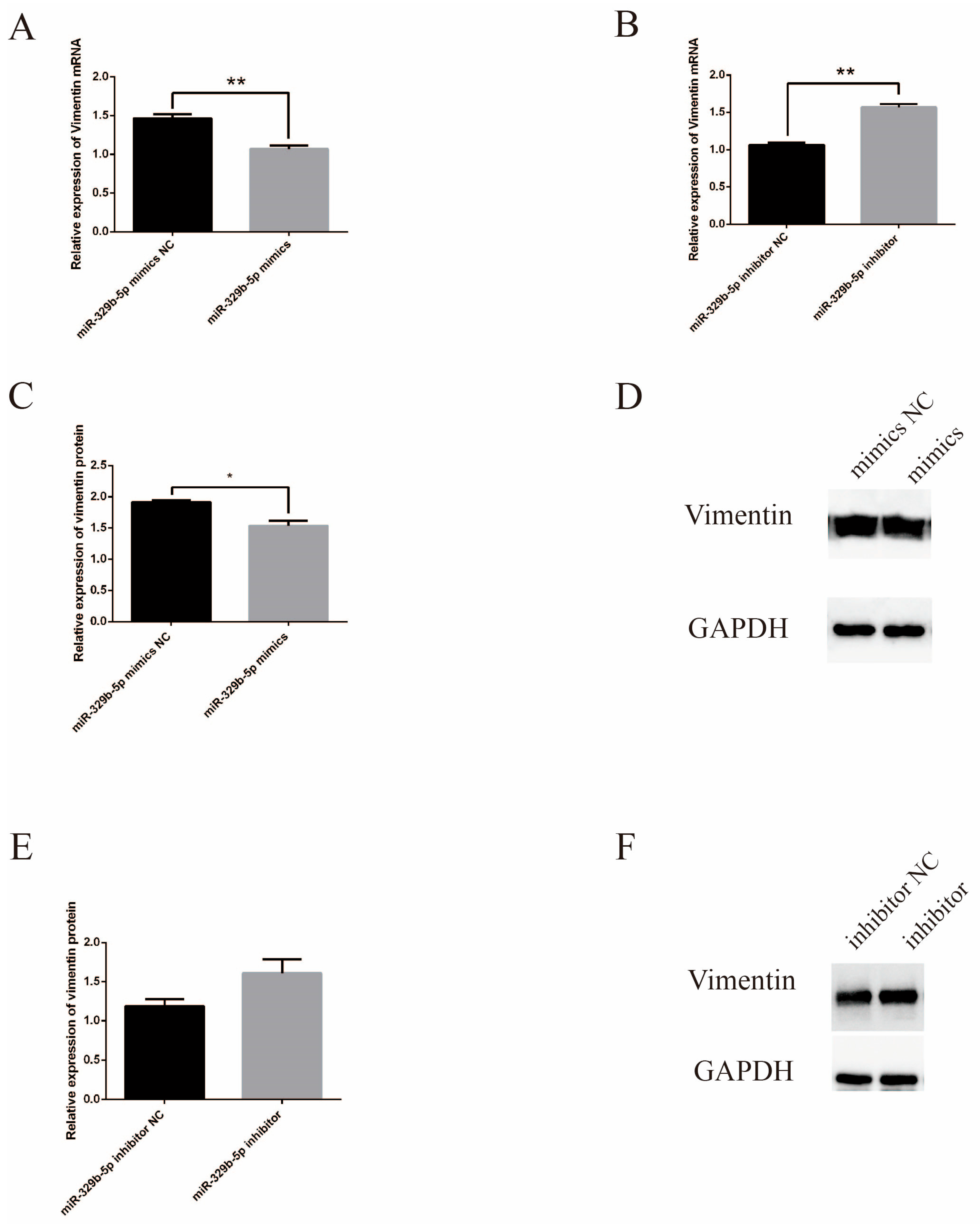
3.1. Effects of miR-329b-5p Mimics and miR-329b-5p Inhibitor

The mRNA expression level of miR-329b-5p in IECs was detected by RT-qPCR. The expression of miR-329b-5p mimics was significantly increased at the mRNA level after transfection with miR-329b-5p mimics (p < 0.05) (Figure 1A), whereas the opposite was true after transfection with miR-329b-5p inhibitor (p < 0.05) (Figure 1B). The above results indicate that the miR-329b-5p mimics and miR-329b-5p inhibitors are efficiently transfected and can be used for subsequent assays.

3.2. miR-329b-5p Influences the E. coli F17 Susceptibility of Sheep IEC s

E. coli F17 infection assay was performed in sheep IECs transfected with miR-329b-5p mimics, miR-329b-5p NC, and miR-329b-5p inhibitor, miR-329b-5p inhibitor NC, and the effects of miR-329b-5p on E. coli F17 infection were verified by E. coli F17 colony counting and fimbriae gene RT-qPCR respectively and immunofluorescence assay to verify the effect of miR-329b-5p on sheep IECs against E. coli F17 infection.
The results of colony counting showed (Figure 2A,B) that the E. coli F17 adhering to the IECs in the miR-329b-5p mimics transfection group was significantly higher than that in the miR-329b-5p mimics NC (p < 0.01), and correspondingly, the E. coli F17 adhering to the IECs in the miR-329b-5p inhibitor transfection group was significantly lower than that of miR-329b-5p inhibitor NC (p < 0.05).
The RT-qPCR of the fimbriae gene was performed targeting F17b-A and F17b-G, respectively, and the results showed (Figure 2C) that both F17b-A and F17b-G fimbriae genes were significantly higher in the miR-329b-5p mimics transfection group than in the miR-329b-5p mimics NC (p < 0.05). In contrast (Figure 2D), the miR-329b-5p inhibitor transfected group F17b-A fimbriae gene was lower than the miR-329b-5p inhibitor NC, whereas F17b-G fimbriae gene was significantly lower than the miR-329b-5p inhibitor NC (p < 0.05).
Immunofluorescence results showed (Figure 2E) that more E. coli F17 than miR-329b-5p mimics NC adhered to the miR-329b-5p mimics transfection group, whereas less E. coli F17 than miR-329b-5p inhibitor NC adhered to the miR-329b-5p inhibitor transfection group. In summary, miR-329b-5p has an important effect on the ability of IECs to resist E. coli F17. In addition, we detected the expression of vimentin in IECs at the mRNA level after transfection with miR-329b-5p mimics and NC as well as miR-329b-5p inhibitor and inhibitor NC by RT-qPCR assay, and we detected the expression of the vimentin gene at the protein level by WB.
Figure 3A showed that after overexpression of miR-329b-5p, vimentin was extremely significantly lower than the NC. After inhibition of miR-329b-5p, vimentin was significantly higher than the inhibitor NC at the mRNA level (Figure 3B). Vimentin was significantly lower at the protein level after transfection with miR-329b-5p mimics than NC, while after transfection with miR-329b-5p inhibitor, the protein levels of vimentin were higher than inhibitor NC (Figure 3C–F) (A detailed picture of the WB was shown in Figure S1). In. In summary, up-regulation or down-regulation of miR-329b-5p affects vimentin expression at both the mRNA and protein levels.

3.3. miR-329b-5p Suppress the Proliferation of IECs

In order to investigate the effect of miR-329b-5p on the proliferation of sheep IECs, we performed CCK-8 and EDU assays. The results of CCK-8 assay showed (Figure 4A,B) that the OD values of cell growth after transfection with miR-329b-5p mimics were extremely significantly lower than that of the corresponding control group at 24/48/72 h (p < 0.01), while OD values of cell growth at 24/72 h after transfection with miR-329b-5p inhibitor were highly significantly higher than the corresponding control group (p < 0.01), and significantly lower at 48 h (p < 0.05). The results of the EDU assay showed (Figure 4C–F) that the rate of EDU positive cells detected after transfection by miR-329b-5p mimics was significantly lower than the corresponding control group (p < 0.05), and the rate of EDU-positive cells detected after transfection using miR-329b-5p inhibitor was significantly higher than the corresponding control group (p < 0.01).
In addition, the expression of PCNA and CCND1 at the mRNA level was reduced after miR-329p-5p mimic transfection compared to the miR-329b-5p mimic NC group (Figure 5A), while the opposite was true for miR-329b-5p inhibitor (Figure 5B). Western blot results showed (Figure 5C–F) that the expression of PCNA protein, a marker associated with cell proliferation, was significantly decreased (p < 0.05) after miR-329b-5p mimics transfection, while PCNA expression was significantly increased (p < 0.05) after miR-329b-5p inhibitor transfection (A detailed picture of the WB was shown in Figure S2). In summary, up-regulation of miR-329b-5p inhibits sheep IEC’s proliferation, while down-regulation of miR-329 promotes sheep IEC’s proliferation.

3.4. miR-329b-5p Suppress the Migration of IECs

In this study, the effect of miR-329b-5p on the migration of sheep IECs was examined by cell scratch assay. Figure 6 showed that the wound healing speed of cells in the transfected miR-329b-5p mimics group was significantly slower than that of the miR-329b-5p mimics NC (p < 0.05), whereas the wound healing speed of cells in the transfected miR-329b-5p inhibitor group was highly significantly faster than that of the miR-329b-5p inhibitor NC (p < 0.05), thus indicating that miR-329b-5p can inhibit the migration of sheep IECs.

4. Discussion

One of the most prevalent illnesses on sheep farms is Escherichia coli-caused sheep diarrhea. ETEC is one of the major pathogenic E. coli. ETEC mainly colonizes the proximal small intestine, and ETEC colonization of the small intestine is predominantly dependent on the expression of several different protein surface structures, which are commonly referred to as colonization factors (CFs) [34]. One of the primary members of the ETEC family is E. coli F17. E. coli F17 is widespread in Southern Peru, Russia, Iran, and other places around the world [3,6,35]. E. coli F17 fimbriae are mainly composed of the pilin F17A and F17G adhesin. F17G adhesin is one of the most important adhesion factors that cause diarrhea in ruminants by mediating on the intestinal microvilli [36]. The study by Ana Umpiérrez et al. tested calves for Escherichia coli adhesin-related genes with a high abundance of F17G [37]. E. coli causes host damage by colonizing mucosal sites, then evading host defenses and beginning to multiply. Although the intestinal flora itself competes for nutrients, E. coli has specific fimbrial antigens that enhance its intestinal colonization and adherence to the small intestinal mucosa, causing host damage [38]. Therefore, the present study focused on enhancing the resistance of sheep IECs to E. coli F17 adhesion through a host receptor cell perspective. This study shows that IECs were found to have enhanced resistance to F17 adhesion after inhibition of miR-329b-5p expression, both in the detection of the E. coli F17 fimbriae gene, the E. coli F17 colony counting assay, and indirect immunofluorescence.
miRNAs have been the subject of numerous studies in gut homeostasis, inflammation, and damage repair. Increasing evidence has shown that miRNAs play a vital role in intestinal innate immunity, e.g., mice with miR-223 deficient in macrophages and dendritic cells exhibit a strong pro-inflammatory phenotype [39]. Liang et al. showed that mice with knockout of miR-146b exhibited enhanced M1 macrophage polarization [40]. miRNAs can also be involved in adaptive immunity. miR-221 and miR-222, specifically lost in T cells, promote colitis infection in mice [41]. In addition, it has been suggested that miRNAs may affect gut microbiota composition and function, thereby influencing gut homeostasis and host health [42]. We therefore speculate that miR-329b-5p may influence the ability of sheep to resist E. coli F17 infection by affecting intestinal innate immunity, adaptive immunity, or gut microbes. Lipid metabolism and immune function in the bovine intestine may be altered by miRNAs, which may affect the interactions between the host and E. coli O157 most, leading to excessive shedding of E. coli O157 in cattle, affecting the farm environment and contaminating farm crops [43]. A. Jaeger et al. found that many miRNAs were significantly up-regulated in porcine mammary epithelial cells attacked for 3 and 24 h [44]. Chen et al. conducted RNA sequencing on the ileal tissues of sheep that were either E. coli F17-sensitive or antagonistic, identifying differentially expressed miRNAs and circRNAs, and constructing the corresponding ceRNA networks [45]. Therefore, in this study, we selected the screened differentially expressed miR-329b-5p to investigate the role of miRNAs against E. coli infection in sheep. MiR-329 has been found to inhibit the proliferation, invasion, and migration of melanoma cells, which is similar to its function in small intestinal epithelial cells. This suggests that similar mechanisms may also exist in small intestinal cells, such as genes such as HMGB2. According to reports, the HMGB2 gene is associated with proliferation, apoptosis, and tumors. In melanoma cells, silencing of the HMGB2 gene can weaken cell viability, and there is a targeted relationship between miR-329 and the HMGB2 gene, which can negatively regulate the HMGB2 gene and inhibit β- catenin pathway, thereby regulating cellular life activities [46].
Intestinal barriers include mechanical, immune, biological, and chemical barriers [47]. The mechanical barrier is one of the crucial components of the intestinal barrier and is maintained mainly by IECs and intercellular junctions [48]. Massive epithelial cell death leads to increased intestinal permeability and microflora dysbiosis, thus providing an opportunity for pathogens to breach the intestinal barrier [49]. In addition, small intestinal mucus contains high concentrations of antimicrobial peptides and proteins secreted by panniculus cells and enterocytes, which kill or trap bacteria, thus preventing the epithelial cells from coming into contact with the bacteria [50]. miRNAs regulate intestinal epithelial homeostasis by altering IEC’s proliferation, migration, and cell–cell interactions [51]. miR-138-5p overexpression inhibits pyroptosis, promotes tight junctions, and ameliorates intestinal barrier breakdown [52]. Inhibition of miR-379-5p promotes IEC’s proliferation, restores barrier function, and improves survival after intestinal injury [53]. This is consistent with the current study’s results, which found that inhibition of miR-329b-5p promotes the proliferation of IECs, which are important for the stability of the intestinal barrier, based on CCK-8, EDU. PCNA is a gene that is extremely important in organisms and is involved in DNA replication, damage repair, and transcription in organisms. In addition, PCNA is able to participate in various cellular activities and is involved in energy metabolism [54]. PCNA is a cofactor for DNA polymerase δ, which is involved in cell proliferation [55]. PCNA expression is elevated in the G1/S phase of cells and is low in quiescent and senescent cells [56]. Wang et al. found that PCNA may interact with KCTD10 and have an effect on cell proliferation [57]. It has been suggested that growth factors or damaged DNA may induce an increase in PCNA [56]. CCND1 specifically regulates the cell cycle and functions primarily in the nucleus. Both CCND1 and PCNA are commonly used as markers to detect cell proliferation. CCND1, located on chromosome 11q13, is a member of the CCND family and is commonly used as a marker for cell proliferation and cancer. Its primary function is to facilitate the transition of cells from the G1 phase to the S phase, resulting in the acceleration of cell proliferation and playing a crucial role in cell migration [58,59]. It has been confirmed that CCND1 can be regulated by miR-193a-3p to inhibit the rapid growth and metastasis of pancreatic cancer [60]. CircCCND1 can interact with HuR and miR-646, synergistically enhancing the stability of CCND1 mRNA, thereby promoting LSCC cell proliferation [61]. These results also demonstrate the regulatory ability of CCND1 on cell proliferation and its reliability as a biomarker. CCND1 can activate the MAPK/PI3K-AKT signaling pathway [62]. In this study, we examined the expression of proliferation markers CCND1 and PCNA at the mRNA level and protein level and found that after down-regulation of miR-329b-5p, the expression of PCNA and CCND1 increased, which was consistent with the results of CCK-8 and EDU assays. Thus, inhibition of miR-329b-5p promotes the proliferation of intestinal epithelial cells, which leads to a more stable intestinal barrier and increases the ability of intestinal epithelial cells to resist E. coli F17 infection.
Vimentin is an intermediate filament protein involved in a variety of functions in living organisms, including maintenance of cytoskeletal integrity, cytokinesis, intracellular signal transduction, cell adhesion, and cell migration [63,64,65,66,67]. Vimentin is also a typical marker of epithelial–mesenchymal transition (EMT) [68]. During EMT, epithelial cells differentiate functionally and behaviorally into the mesenchymal cell type, which is essential for tissue regeneration and wound healing [69]. Knocking mice out of vimentin results in mice with defects associated with the ability to repair wound damage [70]. It has been demonstrated that vimentin deficiency prevents normal wound healing, causing slow and incomplete tissue recovery [71]. Yang et al. showed that overexpression of CLDN6 in human triple-negative breast cancer cells inhibited cell proliferation and resulted in downregulation of the mesenchymal marker vimentin [72]. Overexpression of UNC13C in the human tongue squamous carcinoma cell downregulated vimentin expression [73]. In addition, miR-17-5p overexpression reduced vimentin expression at the mRNA level and protein level [74]. This is consistent with the current study’s results, which revealed that down-regulation of miR-329b-5p expression in IECs up-regulates vimentin expression at both the mRNA and protein level.
One of the foundations for establishing and maintaining the normal organization of an organism is cell migration [75]. Cell migration plays an emphatic role in tissue homeostasis, immune response, and wound repair. When damage occurs in the body, cells migrate quickly to repair the damage [76]. The results of Xu et al. found that miR-301a knockout mice had reduced macrophage migration capacity, which affected subsequent damage repair in damaged tissues [77]. Liu et al. discovered that miR-874-3p upregulation diminishes the migration capability of osteosarcoma cells [78]. This resembled the results of the present study, where the results of the scratch assay indicated that down-regulation of miR-329b-5p improves the migration of IEC, thereby enhancing the resistance of IEC to E. coli F17 in the infection.
Although, in this study, we found that up-regulation of miR-329b-5p decreased the ability of IECs to resist E. coli F17 by E. coli F17 fimbriae gene assay, colony counting, and immunofluorescence and inhibited the proliferation and migration of IECs. However, this study still has shortcomings, as it did not include any cellular attack during the testing of cell proliferation and migration ability. As the cells after tapping, with higher mortality rate, could not support proliferation and migration-related experiments, the experimental protocols could be subsequently optimized for further attempts to assay cell proliferation and migration ability after attacking. miRNAs often act on cells by targeting genes, while miRNAs can often be adsorbed by circRNAs or bind to lncRNAs to play corresponding roles. In contrast, the present study lacks relevant research, which can be followed up with the target genes of miR-329b-5p to further investigate the molecular mechanisms affecting the resistance of sheep to E. coli F17 infection. This study is currently conducted only at the cellular level and lacks animal in vivo tests, which can be followed up with in vivo knockout tests for validation.

5. Conclusions

The up-regulation of miR-329b-5p can improve the susceptibility of sheep IECs to E. coli F17, while the down-regulation of miR-329b-5p inhibits the proliferation and migration of sheep IECs. In conclusion, miR-329b-5p plays a crucial role in the resistance of sheep IECs against E. coli F17 infection. This study offers a scientific foundation for elucidating the mechanism of miR-329b-5p on the E. coli F17 susceptibility of sheep IECs.

Supplementary Materials

The following supporting information can be downloaded at: https://www.mdpi.com/article/10.3390/vetsci11050206/s1, Figure S1: Vimentin WB images after up-/down-regulation of miR-329b-5p. Figure S2: PCNA WB images after up-/down-regulation of miR-329b-5p.

Author Contributions

Conceptualization, Y.X., W.C., H.Y., Z.S., Y.W., R.S., J.M.M., X.L. and W.S.; methodology, W.C.; validation, H.Y. and Z.S.; formal analysis, Y.X. and Y.W.; data curation, Y.X.; writing—original draft preparation, Y.X.; writing—review and editing, W.C. and X.L.; visualization, R.S. and J.M.M.; funding acquisition, W.S. All authors have read and agreed to the published version of the manuscript.

Funding

This research was funded by the National Natural Science Foundation of China-CGIAR (32061143036), the National Natural Science Foundation of China (32172689), the Natural Science Foundation of the Jiangsu Higher Education Institutions of China (22KJA230001, 22KJD230003), the Major New Varieties of Agricultural Projects in Jiangsu Province (PZCZ201739), the Jiangsu Agricultural Science and Technology Innovation Fund (CX(23)1036), the Project of Jiangsu Innovation Support Programme for International Science and Technology Cooperation—“Belt and Road” Innovation Cooperation (BZ2023009), the High-end Foreign Expert Introduction Project (G2022014148L), the Jiangsu 333 Distinguished Talents Project Foundation ((2022)2-323), the Yangzhou Meat Sheep Industry Public Service Platform (YZ2023222), the Distinguished Talents Project Foundation of Yangzhou University, and the Open Project Program of Joint International Research Laboratory of Agriculture and Agri-Product Safety, of the Ministry Education of China, Yangzhou University (JILAR-KF202205).

Institutional Review Board Statement

The animal study protocol was approved by the Experimental Animal Welfare and Ethical of Institute of Animal Science, Yangzhou University (No: NFNC2020-NFY-6) and was performed in accordance with the Regulations for the Administration of Affairs Concerning Experimental Animals approved by the State Council of the People’s Republic of China.

Informed Consent Statement

Not applicable.

Data Availability Statement

All data are contained within the article.

Conflicts of Interest

Yeqing Wang and Rui Su are employed in Suzhou Stud Farm Co., Ltd. The remaining authors have no conflicts of interest to declare.

References

  1. Kaper, J.B.; Nataro, J.P.; Mobley, H.L. Pathogenic Escherichia coli. Nat. Rev. Microbiol. 2004, 2, 123–140. [Google Scholar] [CrossRef] [PubMed]
  2. Dubreuil, J.D.; Isaacson, R.E.; Schifferli, D.M. Animal Enterotoxigenic Escherichia coli. EcoSal Plus 2016, 7, 10–1128. [Google Scholar] [CrossRef] [PubMed]
  3. Bihannic, M.; Ghanbarpour, R.; Auvray, F.; Cavalié, L.; Châtre, P.; Boury, M.; Brugère, H.; Madec, J.Y.; Oswald, E. Identification and detection of three new F17 fimbrial variants in Escherichia coli strains isolated from cattle. Vet. Res. 2014, 45, 76. [Google Scholar] [CrossRef] [PubMed]
  4. Kolenda, R.; Burdukiewicz, M.; Schierack, P. A systematic review and meta-analysis of the epidemiology of pathogenic Escherichia coli of calves and the role of calves as reservoirs for human pathogenic E. coli. Front. Cell. Infect. Microbiol. 2015, 5, 23. [Google Scholar] [CrossRef] [PubMed]
  5. Coşkun, M.R.; Şahin, M. Prevalence of neonatal calf diarrhea caused by Escherichia coli and investigation of virulence factors, serotypes, and antibiotic susceptibility. Pol. J. Vet. Sci. 2023, 26, 335–341. [Google Scholar] [CrossRef] [PubMed]
  6. Siuce, J.; Maturrano, L.; Wheeler, J.C.; Rosadio, R. Diarrheagenic Escherichia coli isolates from neonatal alpacas mainly display F17 fimbriae adhesion gene. Trop. Anim. Health Prod. 2020, 52, 3917–3921. [Google Scholar] [CrossRef] [PubMed]
  7. Zouiten-Mekki, L.; Serghini, M.; Fekih, M.; Kallel, L.; Matri, S.; Ben Mustapha, N.; Boubaker, J.; Filali, A. Epithelial cell in intestinal homeostasis and inflammatory bowel diseases. Med. Sci. M/S 2013, 29, 1145–1150. [Google Scholar]
  8. Xiao, X.; Mao, X.; Chen, D.; Yu, B.; He, J.; Yan, H.; Wang, J. miRNAs Can Affect Intestinal Epithelial Barrier in Inflammatory Bowel Disease. Front. Immunol. 2022, 13, 868229. [Google Scholar] [CrossRef]
  9. Li, X.G.; Wang, Z.; Chen, R.Q.; Fu, H.L.; Gao, C.Q.; Yan, H.C.; Xing, G.X.; Wang, X.Q. LGR5 and BMI1 Increase Pig Intestinal Epithelial Cell Proliferation by Stimulating WNT/β-Catenin Signaling. Int. J. Mol. Sci. 2018, 19, 1036. [Google Scholar] [CrossRef]
  10. Mo, W.; Liu, G.; Wu, C.; Jia, G.; Zhao, H.; Chen, X.; Wang, J. STIM1 promotes IPEC-J2 porcine epithelial cell restitution by TRPC1 signaling. Anim. Biotechnol. 2022, 33, 1492–1503. [Google Scholar] [CrossRef]
  11. Wu, L.M.; Guo, R.; Hui, L.; Ye, Y.G.; Xiang, J.M.; Wan, C.Y.; Zou, M.; Ma, R.; Sun, X.Z.; Yang, S.J.; et al. Stanniocalcin-1 protects bovine intestinal epithelial cells from oxidative stress-induced damage. J. Vet. Sci. 2014, 15, 475–483. [Google Scholar] [CrossRef] [PubMed]
  12. Liang, J.F.; Li, P.H.; Zhu, Y.; Zheng, S.S.; Liu, J.W.; Song, S.Q. MicroRNA-186 suppresses cell proliferation and metastasis in bladder cancer. Afr. Health Sci. 2022, 22, 56–63. [Google Scholar] [CrossRef] [PubMed]
  13. Lynam-Lennon, N.; Maher, S.G.; Reynolds, J.V. The roles of microRNA in cancer and apoptosis. Biol. Rev. Camb. Philos. Soc. 2009, 84, 55–71. [Google Scholar] [CrossRef] [PubMed]
  14. Sohn, E.J.; Park, H.T. MicroRNA Mediated Regulation of Schwann Cell Migration and Proliferation in Peripheral Nerve Injury. BioMed Res. Int. 2018, 2018, 8198365. [Google Scholar] [CrossRef] [PubMed]
  15. Jiang, W.; Liu, J.; Xu, T.; Yu, X. MiR-329 suppresses osteosarcoma development by downregulating Rab10. FEBS Lett. 2016, 590, 2973–2981. [Google Scholar] [CrossRef] [PubMed]
  16. Wang, X.; Lu, X.; Zhang, T.; Wen, C.; Shi, M.; Tang, X.; Chen, H.; Peng, C.; Li, H.; Fang, Y.; et al. mir-329 restricts tumor growth by targeting grb2 in pancreatic cancer. Oncotarget 2016, 7, 21441–21453. [Google Scholar] [CrossRef] [PubMed]
  17. Lin, D.; Shi, Y.; Hu, Y.; Du, X.; Tu, G. miR-329-3p regulates neural stem cell proliferation by targeting E2F1. Mol. Med. Rep. 2019, 19, 4137–4146. [Google Scholar] [CrossRef] [PubMed]
  18. Wu, L.; Pei, F.; Men, X.; Wang, K.; Ma, D. miR-329 inhibits papillary thyroid cancer progression via direct targeting WNT1. Oncol. Lett. 2018, 16, 3561–3568. [Google Scholar] [CrossRef] [PubMed]
  19. Li, G.; Li, Y.; Wang, D.Y. Overexpression of miR-329-3p sensitizes osteosarcoma cells to cisplatin through suppression of glucose metabolism by targeting LDHA. Cell Biol. Int. 2021, 45, 766–774. [Google Scholar] [CrossRef]
  20. Xin, R.Q.; Li, W.B.; Hu, Z.W.; Wu, Z.X.; Sun, W. MiR-329-3p inhibits hepatocellular carcinoma cell proliferation and migration through USP22-Wnt/β-Catenin pathway. Eur. Rev. Med. Pharmacol. Sci. 2020, 24, 9932–9939. [Google Scholar]
  21. Zhao, Y.; Jia, Y.; Wang, J.; Chen, X.; Han, J.; Zhen, S.; Yin, S.; Lv, W.; Yu, F.; Wang, J.; et al. circNOX4 activates an inflammatory fibroblast niche to promote tumor growth and metastasis in NSCLC via FAP/IL-6 axis. Mol. Cancer 2024, 23, 47. [Google Scholar] [CrossRef]
  22. Archanioti, P.; Gazouli, M.; Theodoropoulos, G.; Vaiopoulou, A.; Nikiteas, N. Micro-RNAs as regulators and possible diagnostic bio-markers in inflammatory bowel disease. J. Crohn’s Colitis 2011, 5, 520–524. [Google Scholar] [CrossRef]
  23. Tian, Y.; Xu, J.; Li, Y.; Zhao, R.; Du, S.; Lv, C.; Wu, W.; Liu, R.; Sheng, X.; Song, Y.; et al. MicroRNA-31 Reduces Inflammatory Signaling and Promotes Regeneration in Colon Epithelium, and Delivery of Mimics in Microspheres Reduces Colitis in Mice. Gastroenterology 2019, 156, 2281–2296.e6. [Google Scholar] [CrossRef] [PubMed]
  24. Peck, B.C.; Sincavage, J.; Feinstein, S.; Mah, A.T.; Simmons, J.G.; Lund, P.K.; Sethupathy, P. miR-30 Family Controls Proliferation and Differentiation of Intestinal Epithelial Cell Models by Directing a Broad Gene Expression Program That Includes SOX9 and the Ubiquitin Ligase Pathway. J. Biol. Chem. 2016, 291, 15975–15984. [Google Scholar] [CrossRef]
  25. Chen, Y.; Xiao, Y.; Ge, W.; Zhou, K.; Wen, J.; Yan, W.; Wang, Y.; Wang, B.; Qu, C.; Wu, J.; et al. miR-200b inhibits TGF-β1-induced epithelial-mesenchymal transition and promotes growth of intestinal epithelial cells. Cell Death Dis. 2013, 4, e541. [Google Scholar] [CrossRef]
  26. Xiao, L.; Ma, X.X.; Luo, J.; Chung, H.K.; Kwon, M.S.; Yu, T.X.; Rao, J.N.; Kozar, R.; Gorospe, M.; Wang, J.Y. Circular RNA CircHIPK3 Promotes Homeostasis of the Intestinal Epithelium by Reducing MicroRNA 29b Function. Gastroenterology 2021, 161, 1303–1317.e3. [Google Scholar] [CrossRef] [PubMed]
  27. He, G.; Ding, J.; Zhang, Y.; Cai, M.; Yang, J.; Cho, W.C.; Zheng, Y. microRNA-21: A key modulator in oncogenic viral infections. RNA Biol. 2021, 18, 809–817. [Google Scholar] [CrossRef]
  28. Poltronieri, P.; Sun, B.; Huang, K.Y.; Chang, T.H.; Lee, T.Y. State-of-the-Art on Viral microRNAs in HPV Infection and Cancer Development. MicroRNA 2018, 7, 85–91. [Google Scholar] [CrossRef] [PubMed]
  29. Staedel, C.; Darfeuille, F. MicroRNAs and bacterial infection. Cell. Microbiol. 2013, 15, 1496–1507. [Google Scholar] [CrossRef]
  30. Yang, B.; Yang, R.; Xu, B.; Fu, J.; Qu, X.; Li, L.; Dai, M.; Tan, C.; Chen, H.; Wang, X. miR-155 and miR-146a collectively regulate meningitic Escherichia coli infection-mediated neuroinflammatory responses. J. Neuroinflammation 2021, 18, 114. [Google Scholar] [CrossRef]
  31. Sun, L.; Wu, S.; Dai, C.H.; Sun, S.Y.; Zhu, G.Q.; Wu, S.L.; Bao, W.B. Insight into the molecular mechanism of miR-192 regulating Escherichia coli resistance in piglets. Biosci. Rep. 2018, 38, BSR20171160. [Google Scholar] [CrossRef] [PubMed]
  32. Dai, C.H.; Wang, F.; Wang, S.Q.; Wu, Z.C.; Wu, S.L.; Bao, W.B. miR-215 Targeting Novel Genes EREG, NIPAL1 and PTPRU Regulates the Resistance to E.coli F18 in Piglets. Genes 2020, 11, 1053. [Google Scholar] [CrossRef] [PubMed]
  33. Ge, L.; Zou, S.; Yuan, Z.; Chen, W.; Wang, S.; Cao, X.; Lv, X.; Getachew, T.; Mwacharo, J.M.; Haile, A.; et al. Sheep β-Defensin 2 Regulates Escherichia coli F17 Resistance via NF-κB and MAPK Signaling Pathways in Ovine Intestinal Epithelial Cells. Biology 2021, 10, 1356. [Google Scholar] [CrossRef] [PubMed]
  34. Torres, A.G.; Zhou, X.; Kaper, J.B. Adherence of diarrheagenic Escherichia coli strains to epithelial cells. Infect. Immun. 2005, 73, 18–29. [Google Scholar] [CrossRef] [PubMed]
  35. Knirel, Y.A.; Ivanov, P.A.; Senchenkova, S.N.; Naumenko, O.I.; Ovchinnikova, O.O.; Shashkov, A.S.; Golomidova, A.K.; Babenko, V.V.; Kulikov, E.E.; Letarov, A.V. Structure and gene cluster of the O antigen of Escherichia coli F17, a candidate for a new O-serogroup. Int. J. Biol. Macromol. 2019, 124, 389–395. [Google Scholar] [CrossRef] [PubMed]
  36. Buts, L.; Bouckaert, J.; De Genst, E.; Loris, R.; Oscarson, S.; Lahmann, M.; Messens, J.; Brosens, E.; Wyns, L.; De Greve, H. The fimbrial adhesin F17-G of enterotoxigenic Escherichia coli has an immunoglobulin-like lectin domain that binds N-acetylglucosamine. Mol. Microbiol. 2003, 49, 705–715. [Google Scholar] [CrossRef] [PubMed]
  37. Umpiérrez, A.; Ernst, D.; Fernández, M.; Oliver, M.; Casaux, M.L.; Caffarena, R.D.; Schild, C.; Giannitti, F.; Fraga, M.; Zunino, P. Virulence genes of Escherichia coli in diarrheic and healthy calves. Rev. Argent. Microbiol. 2021, 53, 34–38. [Google Scholar] [CrossRef] [PubMed]
  38. Nataro, J.P.; Kaper, J.B. Diarrheagenic Escherichia coli. Clin. Microbiol. Rev. 1998, 11, 142–201. [Google Scholar] [CrossRef] [PubMed]
  39. Zhou, H.; Xiao, J.; Wu, N.; Liu, C.; Xu, J.; Liu, F.; Wu, L. MicroRNA-223 Regulates the Differentiation and Function of Intestinal Dendritic Cells and Macrophages by Targeting C/EBPβ. Cell Rep. 2015, 13, 1149–1160. [Google Scholar] [CrossRef]
  40. Peng, L.; Zhang, H.; Hao, Y.; Xu, F.; Yang, J.; Zhang, R.; Lu, G.; Zheng, Z.; Cui, M.; Qi, C.F.; et al. Reprogramming macrophage orientation by microRNA 146b targeting transcription factor IRF5. EBioMedicine 2016, 14, 83–96. [Google Scholar] [CrossRef]
  41. Mikami, Y.; Philips, R.L.; Sciumè, G.; Petermann, F.; Meylan, F.; Nagashima, H.; Yao, C.; Davis, F.P.; Brooks, S.R.; Sun, H.W.; et al. MicroRNA-221 and -222 modulate intestinal inflammatory Th17 cell response as negative feedback regulators downstream of interleukin-23. Immunity 2021, 54, 514–525.e6. [Google Scholar] [CrossRef] [PubMed]
  42. Liu, S.; da Cunha, A.P.; Rezende, R.M.; Cialic, R.; Wei, Z.; Bry, L.; Comstock, L.E.; Gandhi, R.; Weiner, H.L. The Host Shapes the Gut Microbiota via Fecal MicroRNA. Cell Host Microbe 2016, 19, 32–43. [Google Scholar] [CrossRef] [PubMed]
  43. Wang, O.; Zhou, M.; Chen, Y.; McAllister, T.A.; Plastow, G.; Stanford, K.; Selinger, B.; Guan, L.L. MicroRNAomes of Cattle Intestinal Tissues Revealed Possible miRNA Regulated Mechanisms Involved in Escherichia coli O157 Fecal Shedding. Front. Cell. Infect. Microbiol. 2021, 11, 634505. [Google Scholar] [CrossRef] [PubMed]
  44. Jaeger, A.; Hadlich, F.; Kemper, N.; Lübke-Becker, A.; Muráni, E.; Wimmers, K.; Ponsuksili, S. MicroRNA expression profiling of porcine mammary epithelial cells after challenge with Escherichia coli in vitro. BMC Genom. 2017, 18, 660. [Google Scholar] [CrossRef] [PubMed]
  45. Chen, W.; Lv, X.; Zhang, W.; Hu, T.; Cao, X.; Ren, Z.; Getachew, T.; Mwacharo, J.M.; Haile, A.; Sun, W. Non-Coding Transcriptome Provides Novel Insights into the Escherichia coli F17 Susceptibility of Sheep Lamb. Biology 2022, 11, 348. [Google Scholar] [CrossRef] [PubMed]
  46. Mo, Y.; Fang, R.H.; Wu, J.; Si, Y.; Jia, S.Q.; Li, Q.; Bai, J.Z.; She, X.N.; Wang, J.Q. MicroRNA-329 upregulation impairs the HMGB2/β-catenin pathway and regulates cell biological behaviors in melanoma. J. Cell Physiol. 2019, 234, 23518–23527. [Google Scholar] [CrossRef] [PubMed]
  47. Liu, Y.; Liu, G.; Fang, J. Progress on the mechanisms of Lactobacillus plantarum to improve intestinal barrier function in ulcerative colitis. J. Nutr. Biochem. 2024, 124, 109505. [Google Scholar] [CrossRef] [PubMed]
  48. He, W.; Wang, Y.; Wang, P.; Wang, F. Intestinal barrier dysfunction in severe burn injury. Burn. Trauma 2019, 7, 24. [Google Scholar] [CrossRef] [PubMed]
  49. Song, Y.; Li, Y.; Hu, W.; Li, F.; Sheng, H.; Huang, C.; Gou, X.; Hou, J.; Zheng, J.; Xiao, Y. Luminol-conjugated cyclodextrin biological nanoparticles for the treatment of severe burn-induced intestinal barrier disruption. Burn. Trauma 2024, 12, tkad054. [Google Scholar] [CrossRef]
  50. Johansson, M.E.; Sjövall, H.; Hansson, G.C. The gastrointestinal mucus system in health and disease. Nat. Rev. Gastroenterol. Hepatol. 2013, 10, 352–361. [Google Scholar] [CrossRef]
  51. Cairns, C.A.; Xiao, L.; Wang, J.Y. Posttranscriptional Regulation of Intestinal Mucosal Growth and Adaptation by Noncoding RNAs in Critical Surgical Disorders. J. Investig. Surg. Off. J. Acad. Surg. Res. 2024, 37, 2308809. [Google Scholar] [CrossRef] [PubMed]
  52. Li, Y.; Ling, P.; Li, Y.; Wang, Y.; Li, G.; Qiu, C.; Wang, J.; Gong, K. miR-138-5p ameliorates intestinal barrier disruption caused by acute superior mesenteric vein thrombosis injury by inhibiting the NLRP3/HMGB1 axis. PeerJ 2024, 12, e16692. [Google Scholar] [CrossRef] [PubMed]
  53. Jia, Z.; Wang, Y.; Gao, J.; Zheng, M.; Wang, P.; Zu, G. miR-379-5P inhibition enhances intestinal epithelial proliferation and barrier function recovery after ischemia/reperfusion by targeting EIF4G2. Shock 2023, 60, 594–602. [Google Scholar] [CrossRef] [PubMed]
  54. Cardano, M.; Tribioli, C.; Prosperi, E. Targeting Proliferating Cell Nuclear Antigen (PCNA) as an Effective Strategy to Inhibit Tumor Cell Proliferation. Curr. Cancer Drug Targets 2020, 20, 240–252. [Google Scholar] [CrossRef] [PubMed]
  55. Paunesku, T.; Mittal, S.; Protić, M.; Oryhon, J.; Korolev, S.V.; Joachimiak, A.; Woloschak, G.E. Proliferating cell nuclear antigen (PCNA): Ringmaster of the genome. Int. J. Radiat. Biol. 2001, 77, 1007–1021. [Google Scholar] [CrossRef] [PubMed]
  56. Bologna-Molina, R.; Mosqueda-Taylor, A.; Molina-Frechero, N.; Mori-Estevez, A.D.; Sánchez-Acuña, G. Comparison of the value of PCNA and Ki-67 as markers of cell proliferation in ameloblastic tumors. Med. Oral Patol. Oral Cir. Bucal 2013, 18, e174–e179. [Google Scholar] [CrossRef] [PubMed]
  57. Wang, Y.; Zheng, Y.; Luo, F.; Fan, X.; Chen, J.; Zhang, C.; Hui, R. KCTD10 interacts with proliferating cell nuclear antigen and its down-regulation could inhibit cell proliferation. J. Cell. Biochem. 2009, 106, 409–413. [Google Scholar] [CrossRef] [PubMed]
  58. Chen, D.G.; Zhu, B.; Lv, S.Q.; Zhu, H.; Tang, J.; Huang, C.; Li, Q.; Zhou, P.; Wang, D.L.; Li, G.H. Inhibition of EGR1 inhibits glioma proliferation by targeting CCND1 promoter. J. Exp. Clin. Cancer Res. CR 2017, 36, 186. [Google Scholar] [CrossRef]
  59. Nardone, V.; Barbarino, M.; Angrisani, A.; Correale, P.; Pastina, P.; Cappabianca, S.; Reginelli, A.; Mutti, L.; Miracco, C.; Giannicola, R.; et al. CDK4, CDK6/cyclin-D1 Complex Inhibition and Radiotherapy for Cancer Control: A Role for Autophagy. Int. J. Mol. Sci. 2021, 22, 8391. [Google Scholar] [CrossRef]
  60. Chen, Z.M.; Yu, Q.; Chen, G.; Tang, R.X.; Luo, D.Z.; Dang, Y.W.; Wei, D.M. MiR-193a-3p inhibits pancreatic ductal adenocarcinoma cell proliferation by targeting CCND1. Cancer Manag. Res. 2019, 11, 4825–4837. [Google Scholar] [CrossRef]
  61. Zang, Y.; Li, J.; Wan, B.; Tai, Y. circRNA circ-CCND1 promotes the proliferation of laryngeal squamous cell carcinoma through elevating CCND1 expression via interacting with HuR and miR-646. J. Cell. Mol. Med. 2020, 24, 2423–2433. [Google Scholar] [CrossRef] [PubMed]
  62. Yang, Y.; Lu, T.; Li, Z.; Lu, S. FGFR1 regulates proliferation and metastasis by targeting CCND1 in FGFR1 amplified lung cancer. Cell Adhes. Migr. 2020, 14, 82–95. [Google Scholar] [CrossRef] [PubMed]
  63. Lowery, J.; Kuczmarski, E.R.; Herrmann, H.; Goldman, R.D. Intermediate Filaments Play a Pivotal Role in Regulating Cell Architecture and Function. J. Biol. Chem. 2015, 290, 17145–17153. [Google Scholar] [CrossRef] [PubMed]
  64. Duarte, S.; Viedma-Poyatos, Á.; Navarro-Carrasco, E.; Martínez, A.E.; Pajares, M.A.; Pérez-Sala, D. Vimentin filaments interact with the actin cortex in mitosis allowing normal cell division. Nat. Commun. 2019, 10, 4200. [Google Scholar] [CrossRef] [PubMed]
  65. Karoii, D.H.; Azizi, H.; Amirian, M. Signaling Pathways and Protein-Protein Interaction of Vimentin in Invasive and Migration Cells: A Review. Cell. Reprogramming 2022, 24, 165–174. [Google Scholar] [CrossRef] [PubMed]
  66. Dave, J.M.; Bayless, K.J. Vimentin as an integral regulator of cell adhesion and endothelial sprouting. Microcirculation 2014, 21, 333–344. [Google Scholar] [CrossRef] [PubMed]
  67. Battaglia, R.A.; Delic, S.; Herrmann, H.; Snider, N.T. Vimentin on the move: New developments in cell migration. F1000Research 2018, 7, 1796. [Google Scholar] [CrossRef] [PubMed]
  68. Thiery, J.P. Epithelial-mesenchymal transitions in tumour progression. Nat. Rev. Cancer 2002, 2, 442–454. [Google Scholar] [CrossRef]
  69. Ostrowska-Podhorodecka, Z.; McCulloch, C.A. Vimentin regulates the assembly and function of matrix adhesions. Wound Repair Regen. 2021, 29, 602–612. [Google Scholar] [CrossRef]
  70. Bornheim, R.; Müller, M.; Reuter, U.; Herrmann, H.; Büssow, H.; Magin, T.M. A dominant vimentin mutant upregulates Hsp70 and the activity of the ubiquitin-proteasome system, and causes posterior cataracts in transgenic mice. J. Cell Sci. 2008, 121 Pt 22, 3737–3746. [Google Scholar] [CrossRef]
  71. Cheng, F.; Shen, Y.; Mohanasundaram, P.; Lindström, M.; Ivaska, J.; Ny, T.; Eriksson, J.E. Vimentin coordinates fibroblast proliferation and keratinocyte differentiation in wound healing via TGF-β-Slug signaling. Proc. Natl. Acad. Sci. USA 2016, 113, E4320–E4327. [Google Scholar] [CrossRef] [PubMed]
  72. Yang, M.; Li, Y.; Ruan, Y.; Lu, Y.; Lin, D.; Xie, Y.; Dong, B.; Dang, Q.; Quan, C. CLDN6 enhances chemoresistance to ADM via AF-6/ERKs pathway in TNBC cell line MDAMB231. Mol. Cell Biochem. 2018, 443, 169–180. [Google Scholar] [CrossRef] [PubMed]
  73. Velmurugan, B.K.; Yeh, K.T.; Hsieh, M.J.; Yeh, C.M.; Lin, C.C.; Kao, C.Y.; Huang, L.R.; Lin, S.H. UNC13C Suppress Tumor Progression via Inhibiting EMT Pathway and Improves Survival in Oral Squamous Cell Carcinoma. Front. Oncol. 2019, 9, 728. [Google Scholar] [CrossRef] [PubMed]
  74. Kim, T.W.; Lee, Y.S.; Yun, N.H.; Shin, C.H.; Hong, H.K.; Kim, H.H.; Cho, Y.B. MicroRNA-17-5p regulates EMT by targeting vimentin in colorectal cancer. Br. J. Cancer 2020, 123, 1123–1130. [Google Scholar] [CrossRef] [PubMed]
  75. Trepat, X.; Chen, Z.; Jacobson, K. Cell migration. Compr. Physiol. 2012, 2, 2369–2392. [Google Scholar] [PubMed]
  76. Shaw, T.J.; Martin, P. Wound repair at a glance. J. Cell Sci. 2009, 122 Pt 18, 3209–3213. [Google Scholar] [CrossRef] [PubMed]
  77. Xu, J.; Fu, L.; Deng, J.; Zhang, J.; Zou, Y.; Liao, L.; Ma, X.; Li, Z.; Xu, Y.; Xu, Y.; et al. miR-301a Deficiency Attenuates the Macrophage Migration and Phagocytosis through YY1/CXCR4 Pathway. Cells 2022, 11, 3952. [Google Scholar] [CrossRef]
  78. Liu, W.G.; Zhuo, L.; Lu, Y.; Wang, L.; Ji, Y.X.; Guo, Q. miR-874-3p inhibits cell migration through targeting RGS4 in osteosarcoma. J. Gene Med. 2020, 22, e3213. [Google Scholar] [CrossRef]
Figure 1. Efficiency verification of miR-329b-5p mimics and miR-329p-5p inhibitors. (A) Transfection of miR-329b-5p mimics and NC into sheep IECs. Expression of miR-329b-5p was detected by RT-qPCR 48 h after transfection. (B) Transfection of miR-329b-5p inhibitor and inhibitor NC into sheep IECs. Expression of miR-329b-5p was detected by RT-qPCR 48 h after transfection. * p < 0.05.
Vetsci 11 00206 g001
Figure 2. miR-329b-5p affects susceptibility of sheep IEC to E. coli F17. (A) Colony count of E. coli F17 after up-regulation of miR-329b-5p. (B) Colony count of E. coli F17 after down-regulation of miR-329b-5p. (C) RT-qPCR of E. coli F17 fimbriae gene after up-regulation of miR-329b-5p. (D) RT-qPCR of E. coli F17 fimbriae gene after down-regulation of miR-329b-5p. (E) In the immunofluorescence assay, cells were observed under the fluorescence microscope (100×). * p < 0.05 and ** p < 0.01.
Vetsci 11 00206 g002
Figure 3. Effect of miR-329b-5p on vimentin expression. (A) RT-qPCR assay of vimentin expression after up-regulation of miR-329b-5p. (B) RT-qPCR assay of vimentin expression after down-regulation of miR-329b-5p. (C,D) WB of vimentin expression after up-regulation of miR-329b-5p. (E,F) WB of vimentin expression after down-regulation of miR-329b-5p. * p < 0.05 and ** p < 0.01.
Vetsci 11 00206 g003
Figure 4. Effect of miR-329b-5p on proliferation of IECs. (A) CCK-8 assay after up-regulation of miR-329b-5p. (B) CCK-8 assay after down-regulation of miR-329b-5p. (C,D) EDU assay after up-regulation of miR-329b-5p (100×). (E,F) EDU assay after -regulation of miR-329b-5p (100×). * p < 0.05 and ** p < 0.01.
Vetsci 11 00206 g004
Figure 5. Effect of miR-329b-5p on proliferation of IECs detected by proliferation markers. (A) mRNA relative expression of PCNA and CCND1 after up-regulation of miR-329b-5p. (B) mRNA relative expression of PCNA and CCND1 after down-regulation of miR-329b-5p. (C,D) Protein expression of PCNA after up-regulation of miR-329b-5p. (E,F) Protein expression of PCNA after down-regulation of miR-329b-5p. * p < 0.05 and ** p < 0.01.
Vetsci 11 00206 g005
Figure 6. Effect of miR-329b-5p on migration of IECs. (A,B) Scratch assay after up-regulation of miR-329b-5p. (C,D) Scratch assay after down-regulation of miR-329b-5p. ** p < 0.01.
Vetsci 11 00206 g006
Table 1. Sequence information of genes used in RT-qPCR.
Gene NameSequences (5′→3′)Product Length/bpAccession No.
F17b-AF: CAACTAACGGGATGTACAGTTTC
R: CTGATAAGCGATGGTGTAATTAAC
323L14318.1
F17b-GF: CGTGGGAAATTATCTATCAACG
R: TGTTGATATTCCGTTAACCGTAC
615L14319.1
VimentinF: CTGCTAACCGCAACAACGAC
R: TAGTCCCTTTGAGCGCATCC
108XM_004014247.6
PCNAF: TCTGCAAGTGGAGAACTTGGAA
R: AGGAGACAGTGGAGTGGCTT
162XM_004014340.5
CCND1F: CCGAGGAGAACAAGCAGATC
R: GAGGGTGGGTTGGAAATG
91XM_027959928.2
GAPDHF: TCTCAAGGGCATTCTAGGCTAC
R: GCCGAATTCATTGTCGTACCAG
151XM_060411593.1
Disclaimer/Publisher’s Note: The statements, opinions and data contained in all publications are solely those of the individual author(s) and contributor(s) and not of MDPI and/or the editor(s). MDPI and/or the editor(s) disclaim responsibility for any injury to people or property resulting from any ideas, methods, instructions or products referred to in the content.

