Open AccessArticle
Single-Cell Analyses Offer Insights into the Different Remodeling Programs of Arteries and Veins
by
Miguel G. Rojas, Simone Pereira-Simon, Zachary M. Zigmond, Javier Varona Santos, Mikael Perla, Nieves Santos Falcon, Filipe F. Stoyell-Conti, Alghidak Salama, Xiaofeng Yang, Xiaochun Long, Juan C. Duque, Loay H. Salman, Marwan Tabbara, Laisel Martinez and Roberto I. Vazquez-Padron
Abstract
Arteries and veins develop different types of occlusive diseases and respond differently to injury. The biological reasons for this discrepancy are not well understood, which is a limiting factor for the development of vein-targeted therapies. This study contrasts human peripheral arteries and veins
[...] Read more.
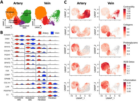
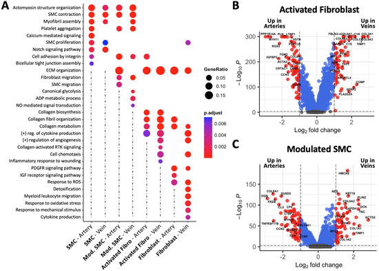
Arteries and veins develop different types of occlusive diseases and respond differently to injury. The biological reasons for this discrepancy are not well understood, which is a limiting factor for the development of vein-targeted therapies. This study contrasts human peripheral arteries and veins at the single-cell level, with a focus on cell populations with remodeling potential. Upper arm arteries (brachial) and veins (basilic/cephalic) from 30 organ donors were compared using a combination of bulk and single-cell RNA sequencing, proteomics, flow cytometry, and histology. The cellular atlases of six arteries and veins demonstrated a 7.8× higher proportion of contractile smooth muscle cells (SMCs) in arteries and a trend toward more modulated SMCs. In contrast, veins showed a higher abundance of endothelial cells, pericytes, and macrophages, as well as an increasing trend in fibroblasts. Activated fibroblasts had similar proportions in both types of vessels but with significant differences in gene expression. Modulated SMCs and activated fibroblasts were characterized by the upregulation of
MYH10,
FN1,
COL8A1, and
ITGA10. Activated fibroblasts also expressed
F2R,
POSTN, and
COMP and were confirmed by F2R/CD90 flow cytometry. Activated fibroblasts from veins were the top producers of collagens among all fibroblast populations from both types of vessels. Venous fibroblasts were also highly angiogenic, proinflammatory, and hyper-responders to reactive oxygen species. Differences in wall structure further explain the significant contribution of fibroblast populations to remodeling in veins. Fibroblasts are almost exclusively located outside the external elastic lamina in arteries, while widely distributed throughout the venous wall. In line with the above, ECM-targeted proteomics confirmed a higher abundance of fibrillar collagens in veins vs. more basement ECM components in arteries. The distinct cellular compositions and transcriptional programs of reparative populations in arteries and veins may explain differences in acute and chronic wall remodeling between vessels. This information may be relevant for the development of antistenotic therapies.
Full article
►▼
Show Figures


