Article

Genome-Wide Identification and Characterization of the PHT1 Gene Family and Its Response to Mycorrhizal Symbiosis in Salvia miltiorrhiza under Phosphate Stress

1
College of Pharmacy, Shandong University of Traditional Chinese Medicine, Jinan 250355, China
2
Experimental Center, Shandong University of Traditional Chinese Medicine, Jinan 250355, China
3
Innovative Institute of Chinese Medicine and Pharmacy, Shandong University of Traditional Chinese Medicine, Jinan 250355, China
*
Author to whom correspondence should be addressed.
These authors contributed equally to this work.
Genes 2024, 15(5), 589; https://doi.org/10.3390/genes15050589
Submission received: 26 March 2024 / Revised: 30 April 2024 / Accepted: 1 May 2024 / Published: 6 May 2024
(This article belongs to the Section Plant Genetics and Genomics)

Abstract

:
Phosphorus (P) is a vital nutrient element that is essential for plant growth and development, and arbuscular mycorrhizal fungi (AMF) can significantly enhance P absorption. The phosphate transporter protein 1 (PHT1) family mediates the uptake of P in plants. However, the PHT1 gene has not yet been characterized in Salvia miltiorrhiza. In this study, to gain insight into the functional divergence of PHT1 genes, nine SmPHT1 genes were identified in the S. miltiorrhiza genome database via bioinformatics tools. Phylogenetic analysis revealed that the PHT1 proteins of S. miltiorrhiza, Arabidopsis thaliana, and Oryza sativa could be divided into three groups. PHT1 in the same clade has a similar gene structure and motif, suggesting that the features of each clade are relatively conserved. Further tissue expression analysis revealed that SmPHT1 was expressed mainly in the roots and stems. In addition, phenotypic changes, P content, and PHT1 gene expression were analyzed in S. miltiorrhiza plants inoculated with AMF under different P conditions (0 mM, 0.1 mM, and 10 mM). P stress and AMF significantly affected the growth and P accumulation of S. miltiorrhiza. SmPHT1;6 was strongly expressed in the roots colonized by AMF, implying that SmPHT1;6 was a specific AMF-inducible PHT1. Taken together, these results provide new insights into the functional divergence and genetic redundancy of the PHT1 genes in response to P stress and AMF symbiosis in S. miltiorrhiza.

1. Introduction

Phosphorus (P) is one of the essential mineral nutrient elements required for plant growth and development and plays an important role in processes such as energy storage and transfer, signal transduction, enzyme regulation, and gene expression [1]. Phosphorus primarily exists in the soil in complex, insoluble organic forms that are inaccessible to plants. The content of inorganic phosphorus is relatively low; when the soil pH exceeds 7.0, inorganic phosphorus is predominantly mineralized and immobilized as calcium phosphates [2,3]. The diffusion rate of soil phosphates is much lower than the absorption rate of phosphates by plant roots, thereby limiting the P supply available to plants. Low P utilization efficiency has long been recognized as a key limiting factor for crop growth worldwide [4]. Thus, to enhance P utilization efficiency and increase crop yield, it is imperative to elucidate the strategies and associated mechanisms for improving P acquisition and utilization.
To address the issue of P deficiency in soil, plants have evolved a variety of strategies, such as increasing the root–soil contact area and establishing symbiotic relationships with arbuscular mycorrhizal fungi (AMF). AMF are widely distributed, as they are capable of forming symbiotic associations with more than 80% of terrestrial higher plants [5,6]. This symbiotic relationship aids in the absorption of mineral nutrients from the soil, particularly P [3]. AMF extract P from the soil containing the rhizosphere by extending their extraradical mycelia away from the root surface [5]. Subsequently, P is transported along fungal hyphae via the mycorrhizal pathway to the specialized symbiotic interface within the root cortex in exchange for plant carbon resources [7]. Furthermore, when plants form a symbiotic relationship with AMF, they can obtain up to 90% of their P requirements from the AMF [8].
The absorption and transport of P in plants are typically mediated by phosphate transporters (PHTs). The PHT gene family has been identified in plant species such as Sorghum [9], Solanum tuberosum [10], Malus domestica [11], and Liriodendron tulipifera [12] through whole-genome analysis. Based on the sequence characteristics and subcellular localization, members of the PHT family can be classified into five types: PHT1, located in the plasma membrane; PHT2, located in chloroplasts; PHT3, located in mitochondria; PHT4, located in chloroplasts, Golgi apparatus, and non-photosynthetic plastids; and PHT5, located in vacuoles [13,14,15]. Among these proteins, PHT1, which belongs to the major facilitator superfamily, plays an important role in the acquisition of inorganic phosphate from soil and its transport within plants [16]. The PHT1 family has been widely studied in plants. In the model plants Arabidopsis thaliana and Oryza sativa, a total of 9 and 13 PHT1 genes, respectively, have been identified [17,18]. Furthermore, multiple P-transport proteins from the PHT1 family induced by AMF have been identified and investigated. For example, NaPT5 expression in roots is triggered by AMF in Nicotiana attenuate [19]. In Lotus japonicus, LjPT4 was expressed in arbusculated plant cells and in the root tips of non-mycorrhizal plants, and impairment of LjPT4 reduced the response of roots to P [20]. The PHT1 gene family exhibited similar expression patterns in Capsicum frutescens, Solanum melongena, and Nicotiana tabacum. The expression of PHT1;1 and PHT1;2 was upregulated in response to P deficiency, but their expression levels decreased after inoculation with AMF. PHT1;3 expression increases with mycorrhizal colonization, while PHT1;4 and PHT1;5 are specifically expressed in response to mycorrhizal symbiosis under low-P conditions [21]. Although some PHT1 members have been functionally studied in plants, a comprehensive analysis of PHT1 proteins in Salvia miltiorrhiza is lacking.
S. miltiorrhiza is well-known for its use in traditional Chinese medicine and is considered a model organism for medicinal plant research [22,23,24]. It is mainly used for the treatment of cardiovascular and cerebrovascular diseases. Phenolic acids, as effective components of S. miltiorrhiza, exhibit various biological properties, such as antioxidative, anti-inflammatory, antibacterial, antitumor, and cardioprotective effects [25]. Previous studies have demonstrated that AMF symbiosis can increase the accumulation of P and promote the synthesis of phenolic acids. However, the regulatory mechanism underlying this phenomenon still requires further investigation [26].
In this study, a total of nine SmPHT1 genes were identified from the genome of S. miltiorrhiza. The characteristics of these genes were analyzed using bioinformatics and experimental methods. The gene expression patterns in response to different P levels (0 mM, 0.1 mM, and 10 mM) during AMF colonization were investigated via RT-qPCR. This study provides a foundation for further research on the functional roles of SmPHT1 genes.

2. Materials and Methods

2.1. AMF Inoculum Preparation, Plant Materials, and Stress Treatments

The original inocula of AMF Rhizophagus irregularis was cultivated through symbiosis with scallion plants. The river sand used in the study after rinsing with clean water 20 times was mixed with perlite at a 1:1 ratio and autoclaved (121 °C, 1 h). Scallion seeds were sown in plastic pots filled with sterilized sand and perlite (1:1 ratio) and moderate AMF original inocula. After about two months of cultivation, the mycorrhizal colonization rate was detected, and inoculum was collected from pot cultures of scallion plants. The inoculum was a mixture of hyphae, spores, root fragments, sand, and perlite that contained approximately 800 spores per 100 g.
The S. miltiorrhiza cultivar ‘Huadan 2’ used in this study was planted in the Shandong University of Traditional Chinese Medicine Medicinal Botanical Garden [27]. One-month-old uniform S. miltiorrhiza seedlings were transplanted into pots (1 L volume) filled with sterilized sand and perlite at a 1:1 ratio. For the mycorrhizal treatments, 50 g of Rhizophagus irregularis inoculum was added to each pot. An equal amount of autoclaved (121 °C, 1 h) inoculum was added to the non-AMF culture pot.
In the experiments, three P concentrations (0 mM, 0.1 mM, and 10 mM) were used, and each treatment was replicated in ten pots. The experiments were repeated in triplicate. The S. miltiorrhiza plants were watered with modified Hoagland solution with different P levels once every two weeks. The plants were grown in a greenhouse under a 16 h light/8 h dark cycle at 23 °C and a relative humidity of 70%. After three months of cultivation, root samples were harvested and one part was used to determine the fresh weight, P concentration, and AMF colonization rate, and an additional part was preserved via rapid freezing in liquid nitrogen at −80 °C for RNA extraction. Total P content was determined using the molybdenum antimony anticolorimetric method [28]. To analyze tissue-specific gene expression, roots, stems, leaves, and flowers were collected from two-year-old S. miltiorrhiza plants at the Shandong University of Traditional Chinese Medicine Medicinal Botanical Garden [27].

2.2. Identification and Phylogenetic Analysis of SmPHT Genes in S. miltiorrhiza

The amino acid sequences of PHT1 proteins in A. thaliana were used as query probes to search the S. miltiorrhiza genome via BLASTP. Furthermore, the conserved domains of the SmPHT1 proteins were analyzed using the Conserved Domain Database (CDD; https://www.ncbi.nlm.nih.gov/Structure/cdd/wrpsb.cgi, accessed on 1 March 2023). The ExPASy server [29] (https://web.expasy.org/protparam/, accessed on 7 March 2023) was subsequently used to predict the physicochemical properties (e.g., number of amino acids, protein molecular weight (MW), theoretical isoelectric point (pI), instability index, aliphatic index, and grand average of hydropathicity (GRAVY)) of the SmPHT1 proteins. The subcellular localization of the proteins was predicted using the Plant-PLoc server [30] (www.csbio.sjtu.edu.cn/bioinf/plant/, accessed on 8 March 2023). Finally, the amino acid sequences of the PHT1 proteins in A. thaliana and O. sativa were downloaded from the Arabidopsis Information Resource (TAIR) database (http://www.arabidopsis.org, accessed on 3 March 2023) and the rice genome database (http://rice.plantbiology.msu.edu/, accessed on 6 March 2023), respectively. A phylogenetic tree of PHT1 proteins was constructed in MEGA-X using the maximum likelihood (ML) method with 1000 bootstrap replicates [31].

2.3. Gene Structure, Conserved Motif, and Cis-Acting Element Analysis

The exon/intron distribution of each SmPHT1 gene was analyzed using the Gene Structure Display Server [32] (GSDS, http://gsds.cbi.pku.edu.cn/, accessed on 14 March 2023). The conserved motifs were analyzed using Multiple EM for Motif Elicitation [33] (MEME, https://meme-suite.org/meme/, accessed on 15 March 2023), with a maximum of 15 motifs per sequence. After cis-acting regulatory element analysis, the 2000 bp upstream sequence of the transcription initiation site was analyzed using the Plant Care website [34] (https://bioinformatics.psb.ugent.be/webtools/plantcare/html/, accessed on 18 March 2023).

2.4. RNA Extraction and SmPHT1 Gene Expression

Total RNA was extracted from the samples using the FastPure Plant Total RNA Isolation Kit (Vazyme, Nanjing, China) and subsequently reverse-transcribed into complementary DNA (cDNA) (TaKaRa, Dalian, China). RT-qPCR analysis was performed using a CFX96 Real-Time System (Bio-Rad, Hercules, CA, USA) with TB Green Premix Ex Taq II (TaKaRa, Dalian, China). Primers for RT-qPCR were designed using Primer3 based on the CDS of the genes, and the NCBI database was used to determine primer specificity. The β-actin gene was chosen as the reference gene. The sequences of the primers used in this study are listed in Table S1. Relative expression was calculated using the 2−ΔΔCt method [35].

2.5. Analysis of Mycorrhizal Colonization

The root of S. miltiorrhiza were cut into segments approximately 1 cm in length. Then, the root segments were placed into test tubes, and Trypan blue staining was performed according to the methods of Phillips and Hayman [36]. First, the root segments were immersed in a 10% KOH solution and heated in a water bath at 90 °C for 40 min. Next, the segments were immersed in a 2% HCl solution for 5 min, 0.05% phenol cotton blue staining solution was added, and the test tubes were heated in a water bath at 90 °C for 30 min. Finally, after rinsing with distilled water, an appropriate amount of decolorizing solution (water–glycerol = 1:1) was added for 24 h of decolorization treatment, after which the results were observed. The mycorrhizal colonization rate was calculated via light microscopy using the gridline intersection method [37].

2.6. Data Statistics and Analysis

The statistical data were analyzed using SPSS software and Student’s t test. All experiments were repeated in triplicate. Differences were considered significant at p < 0.01 and p < 0.05.

3. Results

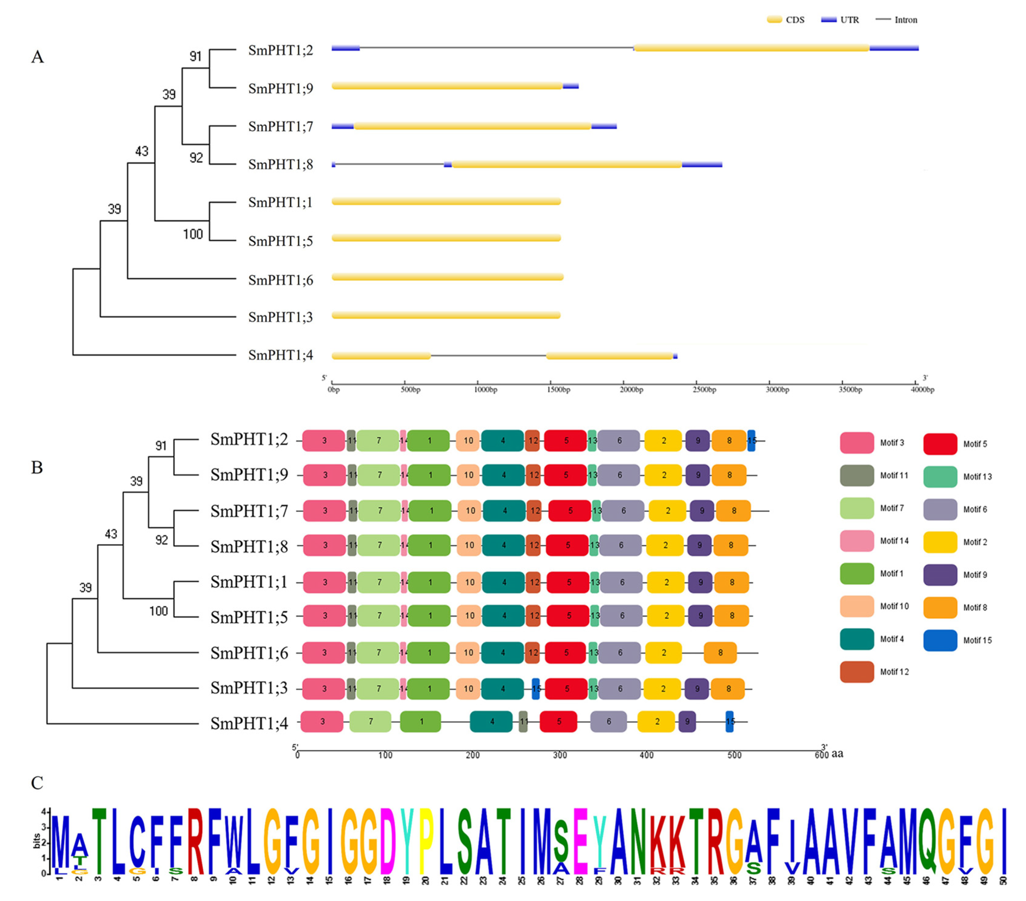
3.1. Identification and Phylogenetic Relationships of SmPHT1

We identified nine SmPHT1 genes from the S. miltiorrhiza genome (Table 1). The amino acid sequence length of SmPHT1 ranged from 516 aa (SmPHT1;4) to 542 aa (SmPHT1;7), with an average length of 527 aa. The molecular weight ranged from 55,704.84 to 59,451.05 Da, and the theoretical pI ranged from 8.32 (SmPHT1;1) to 9.15 (SmPHT1;8). Except for SmPHT1;4, the instability indices of the remaining proteins were less than 40, indicating that all the proteins were stable (S). The hydrophobicity of the nine PHT1 proteins was positive, indicating that they are hydrophobic proteins. Subcellular localization prediction revealed that all the SmPHT1 proteins were located on the plasma membrane.
To investigate the phylogenetic relationships and functional associations, phylogenetic trees were constructed from S. miltiorrhiza (Sm), A. thaliana (At), and O. sativa (Os). The PHT1 proteins were clustered into three subgroups: Group I, Group II, and Group III (Figure 1). There were six SmPHT1 (SmPHT1;1, SmPHT1;2, SmPHT1;5, SmPHT1;7, SmPHT1;8, SmPHT1;9) proteins in Group I, two SmPHT1 (SmPHT1;3, SmPHT1;6) in Group II, and one SmPHT1 (SmPHT1;4) in Group III.

3.2. Gene Structures and Conserved Motifs

To understand the structural diversity of SmPHT1, exons and introns were further analyzed (Figure 2A). According to the results of the gene structure analysis, the number of exons varied from 1 to 2. The SmPHT1;1, SmPHT1;3, SmPHT1;5, SmPHT1;6, SmPHT1;7, and SmPHT1;9 genes contained no introns, while the SmPHT1;2, SmPHT1;4, and SmPHT1;8 genes contained 1 intron.
To further investigate the structural features of SmPHT1, a total of 15 putative motifs were identified (Figure 2B). The number of conserved domains in each protein ranged from 10 to 15. Motif 1 was present in all the SmPHT1 proteins detected in S. miltiorrhiza (Figure 2C). Motifs 3 and 8 were located in the N- and C-terminal domains of SmPHT1, respectively. Motifs 1–7 were present in all the SmPHT1 proteins. Motif 15 was detected only in SmPHT1;2, SmPHT1;3, and SmPHT1;4. In addition, SmPHT1;3 lacked motif 12; SmPHT1;4 lacked motif 8, motif 10, motif 13, and motif 14; and SmPHT1;6 lacked motif 9 and motif 15. These differences in motif components might lead to different gene functions.

3.3. Cis-Acting Element Analysis

To understand the transcriptional regulation and potential functional roles of SmPHT1, the region 2000 bp upstream of the initiation codon was analyzed. Among the PHT1 promoters, seven hormone-related, six stress-related, six development-related, and twenty-two light-related elements were identified (Table S4). Among the hormone-related elements, ABREs (ABA responsiveness), CGTCA motifs (MeJA responsiveness), TGACG motifs (MeJA responsiveness), and TCA elements (SA responsiveness) were found in all SmPHT1 promoters. The gibberellin-responsive element was found only in SmPHT1;3 (TATC-box) and SmPHT1;9 (P-box). Sp1 (drought inducibility), Box4 (light responsiveness), G-Box (light responsiveness), and G-box (light responsiveness) were also observed in the promoter regions of all the SmPHT1s. However, SmPHT1;9 lacked any elements associated with development-related genes. These results implied that the SmPHT1 genes could be involved in hormone signal responsiveness and stress adaptation.

3.4. Mycorrhizal Colonization

To investigate the symbiotic relationship between AMF and the roots of S. miltiorrhiza under different P concentrations, mycorrhizal colonization was analyzed. In the absence of AMF, the mycorrhizal colonization structure of S. miltiorrhiza was not established (Figure 3A). After inoculation with AMF, the colonization rate was greater at 0.1 mM P than 0 mM and 10 mM P (Figure 3B). These results indicated that a P concentration of 0.1 mM significantly increased the colonization rate of the AMF.

3.5. Effect of AMF Colonization on the Growth and P uptake of S. miltiorrhiza

To study the effects of AMF colonization on the growth and nutrient absorption of S. miltiorrhiza, the biomass of S. miltiorrhiza plants and the total P content in the roots were measured. Compared with those in the non-AMF inoculation treatment, the S. miltiorrhiza plants that were inoculated with AMF had significantly greater root biomass at 0 mM and 0.1 mM P (Figure 4A,B). Moreover, the total P content in the roots of AMF-inoculated plants was significantly greater than that in the roots of non-AMF-inoculated plants under the 0.1 mM P concentration (Figure 4C). However, no significant differences in either the biomass or P content were observed between the AMF-inoculated and non-AMF plants under high P supply conditions (Figure 4B,C). The results indicated that AMF inoculation significantly promoted S. miltiorrhiza plant growth and P absorption.

3.6. Tissue-Specific Expression Patterns of SmPHT1

To investigate the potential biological functions of SmPHT1 genes, the expression patterns of the PHT1 genes in the roots, stems, leaves, and flowers were analyzed. As shown in Figure 5, SmPHT1;7 and SmPHT1;8 were highly expressed in the roots; SmPHT1;1, SmPHT1;3, SmPHT1;4, and SmPHT1;6 were highly expressed in the stems; SmPHT1;2 was highly expressed in the leaves; and SmPHT1;5 and SmPHT1;9 were highly expressed in the flowers. Based on the results of the organizational expression pattern analysis, the PHT1 genes in S. miltiorrhiza were shown to be expressed mainly in roots and stems and less expressed in leaves and flowers.

3.7. Expression Patterns of SmPHT1 Genes in Response to P and AMF

To further investigate the functional role of SmPHT1 genes in S. miltiorrhiza, their expression levels were examined in S. miltiorrhiza roots inoculated with AMF under different P concentrations (Figure 6). First, with increasing P concentration, there was a significant decrease in the expression of most SmPHT1 genes between the colonized and the noncolonized plants. At a P concentration of 10 mM with or without AMF colonization, SmPHT1;5 and SmPHT1;8 expression was almost absent. AMF inoculation under P-free conditions could induce SmPHT1;5 and SmPHT1;9 expression, but they were suppressed when 0.1 mM P was added. Moreover, there were no significant differences in the expression levels of SmPHT1;2, SmPHT1;3, SmPHT1;4, SmPHT1;7, or SmPHT1;8 between the colonized and the noncolonized plants. Notably, the expression level of SmPHT1;6 was significantly greater in the AMF-colonized plants than in the non-AMF-colonized plants. The present study provides a valuable description of SmPHT1 gene expression regulation during P stress and AMF colonization.

4. Discussion

The PHT1 family plays a crucial role in the uptake and transport of P from the soil to ensure the optimal growth and development of plants. Investigating the specificity and biochemical characteristics of PHT1 will contribute to a better understanding of P homeostasis and utilization efficiency in plants [38]. In the present study, we conducted a comprehensive analysis of SmPHT1 genes in S. miltiorrhiza. A total of nine SmPHT1 genes were identified in the S. miltiorrhiza genome. Nine PHT1 genes were found in A. thaliana [39], thirteen PHT1 genes were found in O. sativa [18], six PHT1 genes were found in Astragalus sinicus [40], twenty-one PHT1 genes were found in Triticum aestivum [41], and forty-nine PHT1 genes were found in Brassica napus [42]. The variation in the number of PHT1 genes among different species may be attributed to the occurrence of gene duplications and gene loss during evolution [43,44]. Gene duplication followed by functional differentiation has played a crucial role in driving evolutionary novelty, enabling plants to increase their adaptability to new environments [45]. The results of this study provide information for the functionality of the PHT1 gene in S. miltiorrhiza under P stress.
Phylogenetic analysis serves as an important tool in comparative research on any biological process. Phylogenetic analysis of SmPHT1 proteins in S. miltiorrhiza, together with those of A. thaliana and O. sativa, revealed that PHT1s can be classified into three main groups. Similar results have also been found in Gossypium hirsutum [46]. These results also demonstrated that PHT1 was unevenly distributed among the groups and the PHT1 gene family members in the S. miltiorrhiza, A. thaliana, and O. sativa subfamilies. Consistent with the current information on plant evolution, the phylogenetic tree indicated that SmPHT1 was more closely related to its counterparts in A. thaliana (eudicot) than to its counterparts in O. sativa (monocot) (Figure 1). Phylogenetic analysis of PHT1s revealed similar evolutionary divergences that reflected differences in biochemical and functional properties [7]. The distribution patterns of PHT1 proteins suggested that the SmPHT1 protein could perform similar biological processes as its counterparts in A. thaliana or O. sativa.
PHT1 proteins have a conserved structure containing 12 transmembrane (TM) domains with a large hydrophilic loop between TM6 and TM7, and the C- and N-termini are expected to be oriented inside the cell, with the protein inserted in the plasma membrane [47,48]. PHT1 has the conserved feature sequence GGDYPLSATIxSE [49]. PHT1 in plants is responsible for the expression of H+/Pi symporters, which exhibit similarities in sequence, structure, and size across various plant species [50]. Our present findings align with the existing knowledge on this subject. Motif analysis revealed that the domain was located in motif 1 of SmPHT1 and was present in all the PHT1s in S. miltiorrhiza. Furthermore, genes within the same branch exhibited a similar distribution of basic sequences (Figure 2C). Similar results were found in tea plants (Camellia sinensis) [51] and tomatoes (Solanum lycopersicum) [7]. These conserved motifs possibly have functional and/or structural roles in active proteins [52]. Analysis of the gene structures indicated that the numbers and distributions of exons and introns within each branch were relatively similar. Of the 20 coding sequences of SmPHT1 genes, 6 contained no introns, and the rest were destroyed by introns [10]. The variations in motifs and structures between different branches demonstrated the diversity in the functionality of the SmPHT1 gene family in S. miltiorrhiza. These findings indicate that the SmPHT1 gene family is relatively conserved throughout the process of evolution, guiding subsequent functional research.
Most members of the PHT1 family exhibit strong expression in the roots, regardless of whether they are found in dicotyledonous or monocotyledonous plants [12,40,53,54]. However, the expression of the PHT1 gene has also been detected in other organs, such as leaves, flowers, seeds, fruits, and aleurones [11,55,56]. To investigate the functions of these SmPHT1 genes in more detail, we examined the transcriptional patterns of these genes in the roots, stems, leaves, and flowers of S. miltiorrhiza, which could provide important clues about their functions. In the present study, the expression of SmPHT1 varied to different degrees across the roots, stems, leaves, and flowers of S. miltiorrhiza (Figure 5), where it may play a role in P uptake and translocation. SmPHT1;1, SmPHT1;3, SmPHT1;4, SmPHT1;7, and SmPHT1;8 were highly expressed in the stem and root, implying that they may play roles in P absorption and transport [43,57,58]. Similarly, in Gossypium hirsutum, the expression of GhPHT1;3-At, GhPHT1;4-At, GhPHT1;5-At, GhPHT1;3-Dt, GhPHT1;4-Dt, and GhPHT1;5-Dt is increased in the roots and stems [46]. SmPHT1;2, SmPHT1;5, and SmPHT1;9 were highly expressed in leaves and flowers. The transcript levels of LbPT1, LbPT2, and LbPT7 in Lycium barbarum were highest in senescent leaves [59], and SgPT3 in Stylosanthes guianensis was highly expressed in flowers [60], indicating the potential role of these genes in reproductive growth [46]. These variable expression patterns indicate that the PHT1s involved in development and other physiological processes are functionally diverse.
AMF are among the most widely distributed symbiotic organisms in plant roots. The existence of AMF symbiosis in many early diverging land plants, including liverworts, hornworts, lycophytes, and ferns, reveals that AMF symbiosis predates the development of true root systems. AMF symbiosis provides a pathway for the acquisition of P and other mineral nutrients [60]. Studies have shown that the development of AMF is influenced by the availability of P in the environment and by the P transport proteins within plants [10,52]. Moreover, the extent of symbiotic associations between different types of AMF and their hosts substantially influences P absorption by mycorrhizae. For Sorghum bicolor, mycorrhizal colonization by Rhizophagus irregularis and Funneliformis mosseae was only visible in the treatments in which plants were inoculated with AMF, ranging from 52% to 93% for arbuscular colonization [61]. Similar results were found in Petunia hybrida; with increasing P concentration, AMF colonization was inhibited and was already very low at a concentration of 10 mM P [62]. In the present study, AMF colonization was inhibited by 10 mM P (Figure 3B). Our findings indicate that AMF play an important role in plant P uptake.
The inoculation of AMF can promote the adaptability of host plants, especially by providing additional P and improving plant growth [63]. S. miltiorrhiza, a mycotrophic plant, readily forms symbiotic associations with AMF [26]. Comparisons of the growth status of S. miltiorrhiza between the AMF-inoculated and non-AMF-inoculated plants under different P supply conditions showed that the symbiotic relationship of AMF significantly promoted the growth of S. miltiorrhiza (Figure 4A). In addition, under low-P conditions (0.1 mM), inoculation with AMF significantly increased the fresh weight and P content of the roots (Figure 4B,C), which is consistent with the findings of previous studies of S. lycopersicum [43] and G. hirsutum [63].
The symbiotic relationship between AMF and plants can induce the expression of PHT1 under P stress, as observed in Hordeum vulgare [53], Glycine max [64], and S. bicolor and Linum usitatissimum [65]. In the present study, the transcript levels of SmPHT1 in S. miltiorrhiza roots were examined after AMF inoculation under different P concentrations (0 mM, 0.1 mM, and 10 mM). These findings indicated that the SmPHT1 gene was differentially expressed in response to symbiotic AMF and P stress. The expression of SmPHT1 was significantly repressed under high-P conditions. PHT1 reportedly responds to P stress, as observed in S. lycopersicum [43] and Zea mays [66]. In addition, PHT1s, such as EgPT8 in R. irregularis [67], AsPT1 in A. sinicus [40], and ZEAma: Pht1;6 in Z. mays [68], are specifically induced during mycorrhization and have been found to be crucial for mycorrhizal P uptake. Notably, in the present study, the expression of SmPHT1;6 was markedly greater in AMF-inoculated roots than in non-inoculated roots. Furthermore, SmPHT1;6 exhibited orthologous relationships with OsPHT1;11 (Figure 1). OsPHT1;11 plays a role in the mycorrhizal symbiosis process, and its expression is induced by mycorrhizal infection; it is responsible for the uptake of inorganic P from mycorrhizal fungi [69]. Similarly, ZmPHT1;6 clustered with OsPHT1;11 and was found to be a specific AMF-inducible PHT1 [66]. Therefore, SmPHT1;6 may play an important role in mycorrhizal symbiosis and P absorption.

5. Conclusions

In the present study, a total of nine SmPHT1 genes were identified. The phylogenetic relationships, gene structures, conserved motifs, and cis-acting elements of the genes were analyzed, and the results provided insights into their biological functions. Furthermore, tissue expression analysis revealed that most of the SmPHT1 genes were expressed in the roots and stems. SmPHT1 genes were differentially expressed upon inoculation with AMF under P stress. Notably, SmPHT1;6 expression was strongly induced by AMF symbiosis. These results provide a useful foundation for future work and could lead to a better understanding of the evolutionary regulatory mechanisms of PHT1 genes in response to P stress and AMF symbiosis in S. miltiorrhiza.

Supplementary Materials

The following supporting information can be downloaded at https://www.mdpi.com/article/10.3390/genes15050589/s1: Table S1: RT-qPCR primer sequence; Table S2: Protein sequences of PHT1 in Salvia miltiorrhiza, Arabidopsis thaliana, and Oryza sativa; Table S3: The motif sequence information of SmPHT1; Table S4: Analysis of cis-elements in SmPHT1 genes.

Author Contributions

Conceptualization, X.C. and Y.B.; writing-original draft preparation, X.C. and Y.B.; methodology, X.C., Y.B. and Y.L.; data curation, X.C., Y.B., Y.L., H.L. and F.H.; software, Y.L. and H.L.; investigation, F.H.; resources, H.C. and M.L.; validation, H.C. and M.L.; supervision, Q.L.; writing-review & editing, Q.L. All authors have read and agreed to the published version of the manuscript.

Funding

This research was funded by the Natural Science Foundation of Shandong Province, grant numbers ZR2021QH202 and ZR2023MH338; the Scientific and Technological Innovation Breakthrough Project of Heze Yellow River Basin Ecological Protection and High-quality Development, grant number KJTP202308; the National Modern Agricultural Industry Technology System, grant number CARS-21; and the Traditional Chinese Medicine Science and Technology Project of Shandong Province, grant number 2021Q074.

Institutional Review Board Statement

Not applicable.

Informed Consent Statement

Not applicable.

Data Availability Statement

All data analyzed during this study are included in this article and its additional files.

Conflicts of Interest

The authors declare no conflicts of interest.

References

  1. Wang, D.; Lv, S.; Jiang, P.; Li, Y. Roles, Regulation, and Agricultural Application of Plant Phosphate Transporters. Front. Plant Sci. 2017, 8, 817. [Google Scholar] [CrossRef]
  2. Zhang, Y.; Hu, L.; Yu, D.; Xu, K.; Zhang, J.; Li, X.; Wang, P.; Chen, G.; Liu, Z.; Peng, C.; et al. Integrative Analysis of the Wheat PHT1 Gene Family Reveals A Novel Member Involved in Arbuscular Mycorrhizal Phosphate Transport and Immunity. Cells 2019, 8, 490. [Google Scholar] [CrossRef] [PubMed]
  3. Etesami, H.; Jeong, B.R.; Glick, B.R. Contribution of Arbuscular Mycorrhizal Fungi, Phosphate–Solubilizing Bacteria, and Silicon to P Uptake by Plant. Front. Plant Sci. 2021, 12, 699618. [Google Scholar] [CrossRef] [PubMed]
  4. Karandashov, V.; Bucher, M. Symbiotic Phosphate Transport in Arbuscular Mycorrhizas. Trends Plant Sci. 2005, 10, 22–29. [Google Scholar] [CrossRef] [PubMed]
  5. Smith, S.E.; Smith, F.A. Roles of Arbuscular Mycorrhizas in Plant Nutrition and Growth: New Paradigms from Cellular to Ecosystem Scales. Annu. Rev. Plant Biol. 2011, 62, 227–250. [Google Scholar] [CrossRef] [PubMed]
  6. Gu, M.; Chen, A.; Sun, S.; Xu, G. Complex Regulation of Plant Phosphate Transporters and the Gap between Molecular Mechanisms and Practical Application: What Is Missing? Mol. Plant 2016, 9, 396–416. [Google Scholar] [CrossRef]
  7. Rui, W.; Ma, J.; Wei, N.; Zhu, X.; Li, Z. Genome-Wide Analysis of the PHT Gene Family and Its Response to Mycorrhizal Symbiosis in Tomatoes under Phosphate Starvation Conditions. Int. J. Mol. Sci. 2023, 24, 10246. [Google Scholar] [CrossRef]
  8. Van Der Heijden, M.G.A.; Bardgett, R.D.; Van Straalen, N.M. The Unseen Majority: Soil Microbes as Drivers of Plant Diversity and Productivity in Terrestrial Ecosystems. Ecol. Lett. 2008, 11, 296–310. [Google Scholar] [CrossRef]
  9. Wang, J.; Yang, Y.; Liao, L.; Xu, J.; Liang, X.; Liu, W. Genome-Wide Identification and Functional Characterization of the Phosphate Transporter Gene Family in Sorghum. Biomolecules 2019, 9, 670. [Google Scholar] [CrossRef]
  10. Liu, B.; Zhao, S.; Wu, X.; Wang, X.; Nan, Y.; Wang, D.; Chen, Q. Identification and Characterization of Phosphate Transporter Genes in Potato. J. Biotechnol. 2017, 264, 17–28. [Google Scholar] [CrossRef]
  11. Sun, T.; Li, M.; Shao, Y.; Yu, L.; Ma, F. Comprehensive Genomic Identification and Expression Analysis of the Phosphate Transporter (PHT) Gene Family in Apple. Front. Plant Sci. 2017, 8, 426. [Google Scholar] [CrossRef] [PubMed]
  12. Zhang, C.; Meng, S.; Li, M.; Zhao, Z. Genomic Identification and Expression Analysis of the Phosphate Transporter Gene Family in Poplar. Front. Plant Sci. 2016, 7, 218945. [Google Scholar] [CrossRef] [PubMed]
  13. Wang, F.; Deng, M.; Xu, J.; Zhu, X.; Mao, C. Molecular Mechanisms of Phosphate Transport and Signaling in Higher Plants. Semin. Cell Dev. Biol. 2018, 74, 114–122. [Google Scholar] [CrossRef] [PubMed]
  14. Srivastava, S.; Upadhyay, M.; Srivastava, A.; Abdelrahman, M.; Suprasanna, P.; Tran, L.-S. Cellular and Subcellular Phosphate Transport Machinery in Plants. Int. J. Mol. Sci. 2018, 19, 1914. [Google Scholar] [CrossRef] [PubMed]
  15. Wang, Y.; Wang, F.; Lu, H.; Liu, Y.; Mao, C. Phosphate Uptake and Transport in Plants: An Elaborate Regulatory System. Plant Cell Physiol. 2021, 62, 564–572. [Google Scholar] [CrossRef] [PubMed]
  16. Ceasar, S.A.; Baker, A.; Muench, S.P.; Ignacimuthu, S.; Baldwin, S.A. The Conservation of Phosphate-Binding Residues among PHT1 Transporters Suggests That Distinct Transport Affinities Are Unlikely to Result from Differences in the Phosphate-Binding Site. Biochem. Soc. Trans. 2016, 44, 1541–1548. [Google Scholar] [CrossRef] [PubMed]
  17. Ayadi, A.; David, P.; Arrighi, J.-F.; Chiarenza, S.; Thibaud, M.-C.; Nussaume, L.; Marin, E. Reducing the Genetic Redundancy of Arabidopsis PHOSPHATE TRANSPORTER1 Transporters to Study Phosphate Uptake and Signaling. Plant Physiol. 2015, 167, 1511–1526. [Google Scholar] [CrossRef] [PubMed]
  18. Paszkowski, U.; Kroken, S.; Roux, C.; Briggs, S.P. Rice Phosphate Transporters Include an Evolutionarily Divergent Gene Specifically Activated in Arbuscular Mycorrhizal Symbiosis. Proc. Natl. Acad. Sci. USA 2002, 99, 13324–13329. [Google Scholar] [CrossRef]
  19. Wang, M.; Wilde, J.; Baldwin, I.T.; Groten, K. Nicotiana Attenuata ’s Capacity to Interact with Arbuscular Mycorrhiza Alters Its Competitive Ability and Elicits Major Changes in the Leaf Transcriptome. J. Integr. Plant Biol. 2018, 60, 242–261. [Google Scholar] [CrossRef]
  20. Volpe, V.; Giovannetti, M.; Sun, X.; Fiorilli, V.; Bonfante, P. The Phosphate Transporters LjPT4 and MtPT4 Mediate Early Root Responses to Phosphate Status in Non Mycorrhizal Roots. Plant Cell Environ. 2016, 39, 660–671. [Google Scholar] [CrossRef]
  21. Chen, A.; Hu, J.; Sun, S.; Xu, G. Conservation and Divergence of Both Phosphate- and Mycorrhiza-regulated Physiological Responses and Expression Patterns of Phosphate Transporters in Solanaceous Species. New Phytol. 2007, 173, 817–831. [Google Scholar] [CrossRef] [PubMed]
  22. Xu, Z.; Peters, R.J.; Weirather, J.; Luo, H.; Liao, B.; Zhang, X.; Zhu, Y.; Ji, A.; Zhang, B.; Hu, S.; et al. Full-length Transcriptome Sequences and Splice Variants Obtained by a Combination of Sequencing Platforms Applied to Different Root Tissues of Salvia miltiorrhiza and Tanshinone Biosynthesis. Plant J. 2015, 82, 951–961. [Google Scholar] [CrossRef] [PubMed]
  23. Ma, Y.; Yuan, L.; Wu, B.; Li, X.; Chen, S.; Lu, S. Genome-Wide Identification and Characterization of Novel Genes Involved in Terpenoid Biosynthesis in Salvia Miltiorrhiza. J. Exp. Bot. 2012, 63, 2809–2823. [Google Scholar] [CrossRef] [PubMed]
  24. Li, J.; Li, C.; Lu, S. Identification and Characterization of the Cytosine-5 DNA Methyltransferase Gene Family in Salvia miltiorrhiza. PeerJ 2018, 6, e4461. [Google Scholar] [CrossRef] [PubMed]
  25. Shi, M.; Huang, F.; Deng, C.; Wang, Y.; Kai, G. Bioactivities, Biosynthesis and Biotechnological Production of Phenolic Acids in Salvia miltiorrhiza. Crit. Rev. Food Sci. Nutr. 2019, 59, 953–964. [Google Scholar] [CrossRef] [PubMed]
  26. Yang, Y.; Ou, X.; Yang, G.; Xia, Y.; Chen, M.; Guo, L.; Liu, D. Arbuscular Mycorrhizal Fungi Regulate the Growth and Phyto-Active Compound of Salvia Miltiorrhiza Seedlings. Appl. Sci. 2017, 7, 68. [Google Scholar] [CrossRef]
  27. Bai, Y.; Zhou, Y.; Lei, Q.; Wang, Y.; Pu, G.; Liu, Z.; Chen, X.; Liu, Q. Analysis of the HD-Zip I Transcription Factor Family in Salvia Miltiorrhiza and Functional Research of SmHD-Zip12 in Tanshinone Synthesis. PeerJ 2023, 11, e15510. [Google Scholar] [CrossRef] [PubMed]
  28. Veneklaas, E.J.; Stevens, J.; Cawthray, G.R.; Turner, S.; Lambers, H. Chickpea and White Lupin Rhizosphere Carboxylates Vary with Soil Properties and Enhance Phosphorus Uptake. Plant Soil 2003, 248, 187–197. [Google Scholar] [CrossRef]
  29. Gasteiger, E.; Hoogland, C.; Gattiker, A.; Duvaud, S.; Wilkins, M.R.; Appel, R.D.; Bairoch, A. Protein Identification and Analysis Tools on the ExPASy Server. In The Proteomics Protocols Handbook; Walker, J.M., Ed.; Humana Press: Totowa, NJ, USA, 2005; pp. 571–607. ISBN 978-1-58829-343-5. [Google Scholar]
  30. Chou, K.-C.; Shen, H.-B. Cell-PLoc: A Package of Web Servers for Predicting Subcellular Localization of Proteins in Various Organisms. Nat. Protoc. 2008, 3, 153–162. [Google Scholar] [CrossRef]
  31. Kumar, S.; Stecher, G.; Li, M.; Knyaz, C.; Tamura, K. MEGA X: Molecular Evolutionary Genetics Analysis across Computing Platforms. Mol. Biol. Evol. 2018, 35, 1547–1549. [Google Scholar] [CrossRef]
  32. Hu, B.; Jin, J.; Guo, A.-Y.; Zhang, H.; Luo, J.; Gao, G. GSDS 2.0: An Upgraded Gene Feature Visualization Server. Bioinformatics 2015, 31, 1296–1297. [Google Scholar] [CrossRef] [PubMed]
  33. Song, A.; Li, P.; Xin, J.; Chen, S.; Zhao, K.; Wu, D.; Fan, Q.; Gao, T.; Chen, F.; Guan, Z. Transcriptome-Wide Survey and Expression Profile Analysis of Putative Chrysanthemum HD-Zip I and II Genes. Genes 2016, 7, 19. [Google Scholar] [CrossRef] [PubMed]
  34. Lescot, M. PlantCARE, a Database of Plant Cis-Acting Regulatory Elements and a Portal to Tools for in Silico Analysis of Promoter Sequences. Nucleic Acids Res. 2002, 30, 325–327. [Google Scholar] [CrossRef]
  35. Schmittgen, T.D.; Livak, K.J. Analyzing Real-Time PCR Data by the Comparative CT Method. Nat. Protoc. 2008, 3, 1101–1108. [Google Scholar] [CrossRef]
  36. Phillips, J.M.; Hayman, D.S. Improved Procedures for Clearing Roots and Staining Parasitic and Vesicular-Arbuscular Mycorrhizal Fungi for Rapid Assessment of Infection. Trans. Br. Mycol. Soc. 1970, 55, 158-IN18. [Google Scholar] [CrossRef]
  37. McGONIGLE, T.P.; Miller, M.H.; Evans, D.G.; Fairchild, G.L.; Swan, J.A. A New Method Which Gives an Objective Measure of Colonization of Roots by Vesicular—Arbuscular Mycorrhizal Fungi. New Phytol. 1990, 115, 495–501. [Google Scholar] [CrossRef]
  38. Fontenot, E.B.; Ditusa, S.F.; Kato, N.; Olivier, D.M.; Dale, R.; Lin, W.; Chiou, T.; Macnaughtan, M.A.; Smith, A.P. Increased Phosphate Transport of Arabidopsis thaliana PHt1;1 by Site-directed Mutagenesis of Tyrosine 312 May Be Attributed to the Disruption of Homomeric Interactions. Plant Cell Environ. 2015, 38, 2012–2022. [Google Scholar] [CrossRef]
  39. Mudge, S.R.; Rae, A.L.; Diatloff, E.; Smith, F.W. Expression Analysis Suggests Novel Roles for Members of the Pht1 Family of Phosphate Transporters in Arabidopsis. Plant J. 2002, 31, 341–353. [Google Scholar] [CrossRef] [PubMed]
  40. Xie, X.; Huang, W.; Liu, F.; Tang, N.; Liu, Y.; Lin, H.; Zhao, B. Functional Analysis of the Novel Mycorrhiza-specific Phosphate Transporter AsPT 1 and PHT 1 Family from Astragalus sinicus during the Arbuscular Mycorrhizal Symbiosis. New Phytol. 2013, 198, 836–852. [Google Scholar] [CrossRef]
  41. Teng, W.; Zhao, Y.-Y.; Zhao, X.-Q.; He, X.; Ma, W.-Y.; Deng, Y.; Chen, X.-P.; Tong, Y.-P. Genome-Wide Identification, Characterization, and Expression Analysis of PHT1 Phosphate Transporters in Wheat. Front. Plant Sci. 2017, 8, 543. [Google Scholar] [CrossRef]
  42. Li, Y.; Wang, X.; Zhang, H.; Wang, S.; Ye, X.; Shi, L.; Xu, F.; Ding, G. Molecular Identification of the Phosphate Transporter Family 1 (PHT1) Genes and Their Expression Profiles in Response to Phosphorus Deprivation and Other Abiotic Stresses in Brassica napus. PLoS ONE 2019, 14, e0220374. [Google Scholar] [CrossRef] [PubMed]
  43. Chen, A.; Chen, X.; Wang, H.; Liao, D.; Gu, M.; Qu, H.; Sun, S.; Xu, G. Genome-Wide Investigation and Expression Analysis Suggest Diverse Roles and Genetic Redundancy of Pht1 Family Genes in Response to Pi Deficiency in Tomato. BMC Plant Biol 2014, 14, 61. [Google Scholar] [CrossRef] [PubMed]
  44. Grün, A.; Buchner, P.; Broadley, M.R.; Hawkesford, M.J. Identification and Expression Profiling of Pht1 Phosphate Transporters in Wheat in Controlled Environments and in the Field. Plant Biol. J. 2018, 20, 374–389. [Google Scholar] [CrossRef] [PubMed]
  45. Chen, A.; He, S.; Li, F.; Li, Z.; Ding, M.; Liu, Q.; Rong, J. Analyses of the Sucrose Synthase Gene Family in Cotton: Structure, Phylogeny and Expression Patterns. BMC Plant Biol. 2012, 12, 85. [Google Scholar] [CrossRef] [PubMed]
  46. Cai, S.; Liu, F.; Zhou, B. Genome-Wide Identification and Expression Profile Analysis of the PHT1 Gene Family in Gossypium Hirsutum and Its Two Close Relatives of Subgenome Donor Species. Int. J. Mol. Sci. 2020, 21, 4905. [Google Scholar] [CrossRef] [PubMed]
  47. Wang, L.; Xiao, L.; Yang, H.; Chen, G.; Zeng, H.; Zhao, H.; Zhu, Y. Genome-Wide Identification, Expression Profiling, and Evolution of Phosphate Transporter Gene Family in Green Algae. Front. Genet. 2020, 11, 590947. [Google Scholar] [CrossRef] [PubMed]
  48. Wang, J.; Li, T.; Wu, X.; Zhao, Z. Molecular Cloning and Functional Analysis of a H+-Dependent Phosphate Transporter Gene from the Ectomycorrhizal Fungus Boletus Edulis in Southwest China. Fungal Biol. 2014, 118, 453–461. [Google Scholar] [CrossRef]
  49. Liu, P.; Chen, S.; Song, A.; Zhao, S.; Fang, W.; Guan, Z.; Liao, Y.; Jiang, J.; Chen, F. A Putative High Affinity Phosphate Transporter, CmPT1, Enhances Tolerance to Pi Deficiency of Chrysanthemum. BMC Plant Biol. 2014, 14, 18. [Google Scholar] [CrossRef]
  50. Liu, F.; Chang, X.-J.; Ye, Y.; Xie, W.-B.; Wu, P.; Lian, X.-M. Comprehensive Sequence and Whole-Life-Cycle Expression Profile Analysis of the Phosphate Transporter Gene Family in Rice. Mol. Plant 2011, 4, 1105–1122. [Google Scholar] [CrossRef]
  51. Cao, D.; Liu, Y.; Ma, L.; Liu, Z.; Li, J.; Wen, B.; Zhang, X.; Yin, P.; Jin, X.; Huang, J. Genome-Wide Identification and Characterization of Phosphate Transporter Gene Family Members in Tea Plants (Camellia sinensis L. O. Kuntze) under Different Selenite Levels. Plant Physiol. Biochem. 2021, 166, 668–676. [Google Scholar] [CrossRef]
  52. Yamasaki, K.; Kigawa, T.; Seki, M.; Shinozaki, K.; Yokoyama, S. DNA-Binding Domains of Plant-Specific Transcription Factors: Structure, Function, and Evolution. Trends Plant Sci. 2013, 18, 267–276. [Google Scholar] [CrossRef] [PubMed]
  53. Srivastava, R.; Sirohi, P.; Chauhan, H.; Kumar, R. The Enhanced Phosphorus Use Efficiency in Phosphate-Deficient and Mycorrhiza-Inoculated Barley Seedlings Involves Activation of Different Sets of PHT1 Transporters in Roots. Planta 2021, 254, 38. [Google Scholar] [CrossRef] [PubMed]
  54. Nussaume, L. Phosphate Import in Plants: Focus on the PHT1 Transporters. Front. Plant Sci. 2011, 2, 83. [Google Scholar] [CrossRef] [PubMed]
  55. Shukla, V.; Kaur, M.; Aggarwal, S.; Bhati, K.K.; Kaur, J.; Mantri, S.; Pandey, A.K. Tissue Specific Transcript Profiling of Wheat Phosphate Transporter Genes and Its Association with Phosphate Allocation in Grains. Sci. Rep. 2016, 6, 39293. [Google Scholar] [CrossRef] [PubMed]
  56. An, N.; Huang, J.; Xue, Y.; Liu, P.; Liu, G.; Zhu, S.; Chen, Z. Characterization of Phosphate Transporter Genes and the Function of SgPT1 Involved in Phosphate Uptake in Stylosanthes Guianensis. Plant Physiol. Biochem. 2023, 194, 731–741. [Google Scholar] [CrossRef] [PubMed]
  57. Liu, J.; Versaw, W.K.; Pumplin, N.; Gomez, S.K.; Blaylock, L.A.; Harrison, M.J. Closely Related Members of the Medicago Truncatula PHT1 Phosphate Transporter Gene Family Encode Phosphate Transporters with Distinct Biochemical Activities. J. Biol. Chem. 2008, 283, 24673–24681. [Google Scholar] [CrossRef] [PubMed]
  58. Qin, L.; Guo, Y.; Chen, L.; Liang, R.; Gu, M.; Xu, G.; Zhao, J.; Walk, T.; Liao, H. Functional Characterization of 14 Pht1 Family Genes in Yeast and Their Expressions in Response to Nutrient Starvation in Soybean. PLoS ONE 2012, 7, e47726. [Google Scholar] [CrossRef] [PubMed]
  59. Hu, W.; Zhang, H.; Zhang, X.; Chen, H.; Tang, M. Characterization of Six PHT1 Members in Lycium Barbarum and Their Response to Arbuscular Mycorrhiza and Water Stress. Tree Physiol. 2017, 37, 351–366. [Google Scholar] [CrossRef] [PubMed]
  60. MacLean, A.M.; Bravo, A.; Harrison, M.J. Plant Signaling and Metabolic Pathways Enabling Arbuscular Mycorrhizal Symbiosis. Plant Cell 2017, 29, 2319–2335. [Google Scholar] [CrossRef]
  61. Raphael, B.; Nicolás, M.; Martina, J.; Daphnée, B.; Daniel, W.; Pierre-Emmanuel, C. The Fine-tuning of Mycorrhizal Pathway in Sorghum Depends on Both Nitrogen−phosphorus Availability and the Identity of the Fungal Partner. Plant Cell Environ. 2022, 45, 3354–3366. [Google Scholar] [CrossRef]
  62. Breuillin, F.; Schramm, J.; Hajirezaei, M.; Ahkami, A.; Favre, P.; Druege, U.; Hause, B.; Bucher, M.; Kretzschmar, T.; Bossolini, E.; et al. Phosphate Systemically Inhibits Development of Arbuscular Mycorrhiza in Petunia Hybrida and Represses Genes Involved in Mycorrhizal Functioning: Phosphate and Petunia Mycorrhiza Development and Functioning. Plant J. 2010, 64, 1002–1017. [Google Scholar] [CrossRef] [PubMed]
  63. Gao, X.; Guo, H.; Zhang, Q.; Guo, H.; Zhang, L.; Zhang, C.; Gou, Z.; Liu, Y.; Wei, J.; Chen, A.; et al. Arbuscular Mycorrhizal Fungi (AMF) Enhanced the Growth, Yield, Fiber Quality and Phosphorus Regulation in Upland Cotton (Gossypium hirsutum L.). Sci. Rep. 2020, 10, 2084. [Google Scholar] [CrossRef] [PubMed]
  64. Bulgarelli, R.G.; De Oliveira, V.H.; De Andrade, S.A.L. Arbuscular Mycorrhizal Symbiosis Alters the Expression of PHT1 Phosphate Transporters in Roots and Nodules of P-Starved Soybean Plants. Theor. Exp. Plant Physiol. 2020, 32, 243–253. [Google Scholar] [CrossRef]
  65. Walder, F.; Brulé, D.; Koegel, S.; Wiemken, A.; Boller, T.; Courty, P. Plant Phosphorus Acquisition in a Common Mycorrhizal Network: Regulation of Phosphate Transporter Genes of the Pht1 Family in Sorghum and Flax. New Phytol. 2015, 205, 1632–1645. [Google Scholar] [CrossRef] [PubMed]
  66. Liu, F.; Xu, Y.; Jiang, H.; Jiang, C.; Du, Y.; Gong, C.; Wang, W.; Zhu, S.; Han, G.; Cheng, B. Systematic Identification, Evolution and Expression Analysis of the Zea Mays PHT1 Gene Family Reveals Several New Members Involved in Root Colonization by Arbuscular Mycorrhizal Fungi. Int. J. Mol. Sci. 2016, 17, 930. [Google Scholar] [CrossRef]
  67. Che, X.; Wang, S.; Ren, Y.; Xie, X.; Hu, W.; Chen, H.; Tang, M. A Eucalyptus Pht1 Family Gene EgPT8 Is Essential for Arbuscule Elongation of Rhizophagus irregularis. Microbiol. Spectr. 2022, 10, e01470-22. [Google Scholar] [CrossRef] [PubMed]
  68. Tian, H.; Drijber, R.A.; Li, X.; Miller, D.N.; Wienhold, B.J. Arbuscular Mycorrhizal Fungi Differ in Their Ability to Regulate the Expression of Phosphate Transporters in Maize (Zea mays L.). Mycorrhiza 2013, 23, 507–514. [Google Scholar] [CrossRef]
  69. Yang, S.-Y.; Grønlund, M.; Jakobsen, I.; Grotemeyer, M.S.; Rentsch, D.; Miyao, A.; Hirochika, H.; Kumar, C.S.; Sundaresan, V.; Salamin, N.; et al. Nonredundant Regulation of Rice Arbuscular Mycorrhizal Symbiosis by Two Members of the PHOSPHATE TRANSPORTER1 Gene Family. Plant Cell 2012, 24, 4236–4251. [Google Scholar] [CrossRef]
Figure 1. Phylogenetic tree of the PHT1 proteins from S. miltiorrhiza, A. thaliana, and O. sativa. Proteins from S. miltiorrhiza, A. thaliana, and O. sativa are denoted by red circles, blue circles, and yellow circles, respectively. Bootstrap values was 27–100. The sequences used in this study are listed in Table S2.
Genes 15 00589 g001
Figure 2. Conserved motifs and gene structures of the SmPHT1 in S. miltiorrhiza. (A) Gene structures. The yellow boxes indicate exons, the blue boxes indicate untranslated 5′ and 3′ regions, and the black lines indicate introns. (B) Motif composition. The motifs are displayed in different colored boxes. The sequence information for each motif is provided in Table S3. (C) Visualization of motif 1.
Genes 15 00589 g002
Figure 3. Impact of external P concentration on AMF symbiosis in S. miltiorrhiza roots: (A) typical structure of AMF in the root system of S. miltiorrhiza; (B) percentage of total mycorrhizal colonization in S. miltiorrhiza plants. * p < 0.05, ** p < 0.01.
Genes 15 00589 g003
Figure 4. The impact of AMF colonization on the growth of S. miltiorrhiza and P absorption was investigated under the influence of different P concentrations: (A) growth status of S. miltiorrhiza under different P concentrations with AMF colonization; (B) fresh root biomass; (C) root total P content. The asterisks indicate significant differences. C stands for non-mycorrhizal. * p < 0.05, ** p < 0.01.
Genes 15 00589 g004
Figure 5. Expression analysis of SmPHT1 genes in S. miltiorrhiza. Heatmap of SmPHT1 expression in the roots, stems, leaves, and flowers. The color scale limits were artificially set to −1.50 to 1.50 according to the normalized values. The gradient from blue to red represents low to high expression.
Genes 15 00589 g005
Figure 6. Expression patterns of SmPHT1 genes under P and AMF in S. miltiorrhiza. Transcript profiling of the root tissues of AMF and non-AMF plants. SmActin was used as an endogenous control. The asterisks indicate significant differences. * p < 0.05, ** p < 0.01.
Genes 15 00589 g006
Table 1. Characteristics of SmPHT1 identified from S. miltiorrhiz.
NameAmino AcidsMolecular Weight (Da)Theoretical pIInstability IndexGrand Average of HydropathicitySubcellular Localization
SmPHT1;152357,218.78.3230.35 S0.376Plasma membrane
SmPHT1;253758,947.838.8632.44 S0.312Plasma membrane
SmPHT1;352256,466.879.0135.33 S0.417Plasma membrane
SmPHT1;451655,704.848.8343.73 US0.443Plasma membrane
SmPHT1;552357,184.768.6430.96 S0.37Plasma membrane
SmPHT1;652958,482.948.6732.11 S0.249Plasma membrane
SmPHT1;754259,451.058.9831.81 S0.281Plasma membrane
SmPHT1;852557,433.079.1530.96 S0.336Plasma membrane
SmPHT1;952757,588.119.0738.79 S0.32Plasma membrane
Disclaimer/Publisher’s Note: The statements, opinions and data contained in all publications are solely those of the individual author(s) and contributor(s) and not of MDPI and/or the editor(s). MDPI and/or the editor(s) disclaim responsibility for any injury to people or property resulting from any ideas, methods, instructions or products referred to in the content.

