
SNP-Associated Substitutions of Amino Acid Residues in the dNTP Selection Subdomain Decrease Polβ Polymerase Activity
 , , and
Abstract
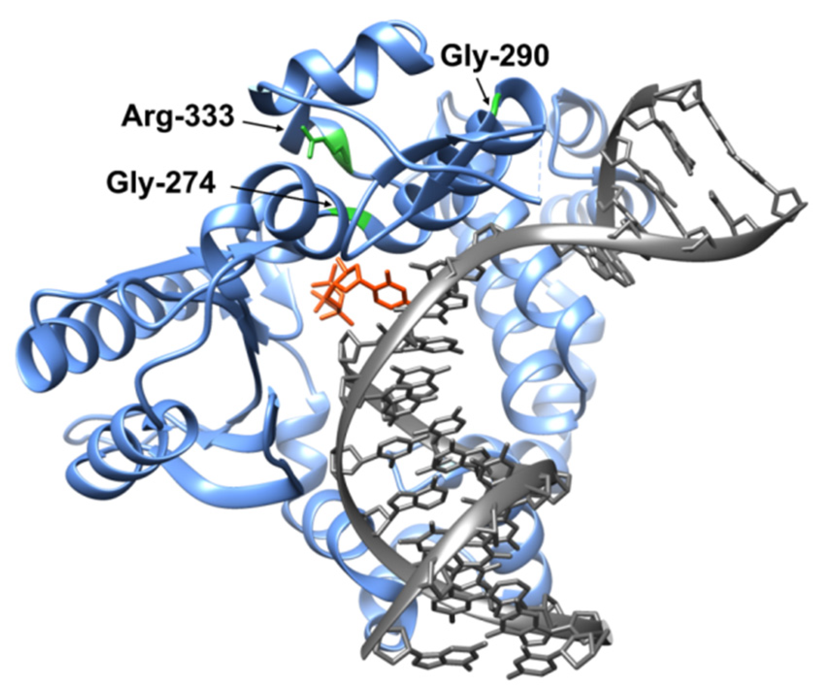
:1. Introduction
2. Materials and Methods
2.1. Protein Purification
2.2. DNA Substrates
2.3. Circular Dichroism Spectroscopy (CD)
2.4. Analysis of the Melting Point of the Enzymes
2.5. DNA-Binding Analysis
2.6. Analysis of Polβ Variants’ Polymerase Activity
2.7. Registration of Conformational Changes in the DNA Substrate by the Stopped-Flow Method and Determination of Polymerization Reaction Rate Constant kpol and Observed Constant Kd,app(dATP) for the G290C Variant
2.8. Determination of Polymerization Reaction Rate Constant kpol and Observed Constant Kd,app(dATP) for Variants G274R and R333W
2.9. Molecular Dynamic Simulations (MD)
3. Results
3.1. Effects of the Substitutions on Protein Structure and Melting Temperature
3.2. Determining the Effect on the Formation of the Complex with DNA
3.3. Determination of the Gap-Filling Efficiency of the Polβ Variants
3.4. dNTP Binding and Incorporation
4. Conclusions
Supplementary Materials
Author Contributions
Funding
Institutional Review Board Statement
Informed Consent Statement
Data Availability Statement
Acknowledgments
Conflicts of Interest
References
- Lindahl, T. Instability and Decay of the Primary Structure of DNA. Nature 1993, 362, 709–715. [Google Scholar] [CrossRef] [PubMed]
- Retèl, J.; Hoebee, B.; Braun, J.E.; Lutgerink, J.T.; van den Akker, E.; Wanamarta, A.H.; Joenje, H.; Lafleur, M.V. Mutational Specificity of Oxidative DNA Damage. Mutat. Res. 1993, 299, 165–182. [Google Scholar] [CrossRef] [PubMed]
- Marnett, L.J. Oxy Radicals, Lipid Peroxidation and DNA Damage. Toxicology 2002, 181–182, 219–222. [Google Scholar] [CrossRef]
- Whitaker, A.M.; Schaich, M.A.; Smith, M.S.; Flynn, T.S.; Freudenthal, B.D. Base Excision Repair of Oxidative DNA Damage: From Mechanism to Disease. Front. Biosci.—Landmark 2017, 22, 1493–1522. [Google Scholar] [CrossRef] [PubMed]
- Lomax, M.E.; Folkes, L.K.; O’Neill, P. Biological Consequences of Radiation-Induced DNA Damage: Relevance to Radiotherapy. Clin. Oncol. 2013, 25, 578–585. [Google Scholar] [CrossRef] [PubMed]
- Hsieh, P. Molecular Mechanisms of DNA Mismatch Repair. Mutat. Res. 2001, 486, 71–87. [Google Scholar] [CrossRef] [PubMed]
- Krokan, H.E.; Bjørås, M. Base Excision Repair. Cold Spring Harb. Perspect. Biol. 2013, 5, a012583. [Google Scholar] [CrossRef]
- Fromme, J.C.; Verdine, G.L. Base Excision Repair. In Advances in Protein Chemistry; Academic Press: Cambridge, MA, USA, 2004; Volume 69, pp. 1–41. [Google Scholar]
- Ide, H.; Kotera, M. Human DNA Glycosylases Involved in the Repair of Oxidatively Damaged DNA. Biol. Pharm. Bull. 2004, 27, 480–485. [Google Scholar] [CrossRef] [PubMed]
- Jacobs, A.L.; Schär, P. DNA Glycosylases: In DNA Repair and Beyond. Chromosoma 2012, 121, 1–20. [Google Scholar] [CrossRef] [PubMed]
- Liu, P.; Burdzy, A.; Sowers, L.C. Substrate Recognition by a Family of Uracil-DNA Glycosylases: UNG, MUG, and TDG. Chem. Res. Toxicol. 2002, 15, 1001–1009. [Google Scholar] [CrossRef]
- Tell, G.; Quadrifoglio, F.; Tiribelli, C.; Kelley, M.R. The Many Functions of APE1/Ref-1: Not Only a DNA Repair Enzyme. Antioxid. Redox Signal 2009, 11, 601–619. [Google Scholar] [CrossRef]
- Fung, H.; Demple, B. A Vital Role for Ape1/Ref1 Protein in Repairing Spontaneous DNA Damage in Human Cells. Mol. Cell 2005, 17, 463–470. [Google Scholar] [CrossRef] [PubMed]
- Sobol, R.W.; Wilson, S.H. Mammalian DNA Beta-Polymerase in Base Excision Repair of Alkylation Damage. Prog. Nucleic Acid Res. Mol. Biol. 2001, 68, 57–74. [Google Scholar] [PubMed]
- Sawaya, M.R.; Prasad, R.; Wilson, S.H.; Kraut, J.; Pelletier, H. Crystal Structures of Human DNA Polymerase Beta Complexed with Gapped and Nicked DNA: Evidence for an Induced Fit Mechanism. Biochemistry 1997, 36, 11205–11215. [Google Scholar] [CrossRef] [PubMed]
- Dianov, G.L.; Prasad, R.; Wilson, S.H.; Bohr, V.A. Role of DNA Polymerase β in the Excision Step of Long Patch Mammalian Base Excision Repair. J. Biol. Chem. 1999, 274, 13741–13743. [Google Scholar] [CrossRef] [PubMed]
- Prasad, R.; Lavrik, O.I.; Kim, S.-J.; Kedar, P.; Yang, X.-P.; Vande Berg, B.J.; Wilson, S.H. DNA Polymerase β-Mediated Long Patch Base Excision Repair. Available online: https://reader.elsevier.com/reader/sd/pii/S0021925820777550?token=AB86DB76EB9167C69BF33CC20A6473F65E81FDCB8BC20B7DF2D8C1DE8FD345E4A99A08EA7CDDAB5B6FD6F55CB7B5A220&originRegion=eu-west-1&originCreation=20210618070314 (accessed on 18 June 2021).
- Kumar, A.; Widen, S.G.; Williams, K.R.; Kedar, P.; Karpel, R.L.; Wilson, S.H. Studies of the Domain Structure of Mammalian DNA Polymerase Beta. Identification of a Discrete Template Binding Domain. J. Biol. Chem. 1990, 265, 2124–2131. [Google Scholar] [CrossRef] [PubMed]
- Menge, K.L.; Hostomsky, Z.; Nodes, B.R.; Hudson, G.O.; Rahmati, S.; Moomaw, E.W.; Almassy, R.J.; Hostomska, Z. Structure-Function Analysis of the Mammalian DNA Polymerase Beta Active Site: Role of Aspartic Acid 256, Arginine 254, and Arginine 258 in Nucleotidyl Transfer. Biochemistry 1995, 34, 15934–15942. [Google Scholar] [CrossRef]
- Brookes, A.J. The Essence of SNPs. Gene 1999, 234, 177–186. [Google Scholar] [CrossRef]
- Hall, J.; Marcel, V.; Bolin, C.; Fernet, M.; Tartier, L.; Vaslin, L.; Hainaut, P. The Associations of Sequence Variants in DNA-Repair and Cell-Cycle Genes with Cancer Risk: Genotype-Phenotype Correlations. Biochem. Soc. Trans. 2009, 37, 527–533. [Google Scholar] [CrossRef]
- Starcevic, D.; Dalal, S.; Sweasy, J.B. Is There a Link between DNA Polymerase β and Cancer? Taylor and Francis Inc.: Abingdon, UK, 2004; Volume 3, pp. 998–1001. [Google Scholar]
- Kladova, O.A.; Fedorova, O.S.; Kuznetsov, N.A. The Role of Natural Polymorphic Variants of DNA Polymerase β in DNA Repair. Int. J. Mol. Sci. 2022, 23, 2390. [Google Scholar] [CrossRef]
- Murphy, D.L.; Donigan, K.A.; Jaeger, J.; Sweasy, J.B. The E288K Colon Tumor Variant of DNA Polymerase β Is a Sequence Specific Mutator. Biochemistry 2012, 51, 5269–5275. [Google Scholar] [CrossRef] [PubMed]
- Alnajjar, K.S.; Garcia-Barboza, B.; Negahbani, A.; Nakhjiri, M.; Kashemirov, B.; McKenna, C.; Goodman, M.F.; Sweasy, J.B. A Change in the Rate-Determining Step of Polymerization by the K289M DNA Polymerase β Cancer-Associated Variant. Biochemistry 2017, 56, 2096. [Google Scholar] [CrossRef]
- Batra, V.K.; Alnajjar, K.S.; Sweasy, J.B.; McKenna, C.E.; Goodman, M.F.; Wilson, S.H. Revealing an Internal Stabilization Deficiency in the DNA Polymerase β K289M Cancer Variant through the Combined Use of Chemical Biology and X-ray Crystallography. Biochemistry 2020, 59, 955–963. [Google Scholar] [CrossRef] [PubMed]
- Lang, T.; Dalal, S.; Chikova, A.; DiMaio, D.; Sweasy, J.B. The E295K DNA Polymerase Beta Gastric Cancer-Associated Variant Interferes with Base Excision Repair and Induces Cellular Transformation. Mol. Cell Biol. 2007, 27, 5587–5596. [Google Scholar] [CrossRef] [PubMed]
- Yang, L.; Beard, W.A.; Wilson, S.H.; Broyde, S.; Schlick, T. Highly Organized but Pliant Active Site of DNA Polymerase b: Compensatory Mechanisms in Mutant Enzymes Revealed by Dynamics Simulations and Energy Analyses. Biophys. J. 2004, 86, 3392–3408. [Google Scholar] [CrossRef] [PubMed]
- Murphy, D.L.; Jaeger, J.; Sweasy, J.B. A Triad Interaction in the Fingers Subdomain of DNA Polymerase Beta Controls Polymerase Activity. J. Am. Chem. Soc. 2011, 133, 6279. [Google Scholar] [CrossRef] [PubMed]
- Skandalis, A.; Loeb, L.A. Enzymatic Properties of Rat DNA Polymerase β Mutants Obtained by Randomized Mutagenesis. Nucleic Acids Res. 2001, 29, 2418–2426. [Google Scholar] [CrossRef] [PubMed]
- Maciejewski, M.W.; Liu, D.; Prasad, R.; Wilson, S.H.; Mullen, G.P. Backbone Dynamics and Refined Solution Structure of the N-Terminal Domain of DNA Polymerase Beta. Correlation with DNA Binding and DRP Lyase Activity. J. Mol. Biol. 2000, 296, 229–253. [Google Scholar] [CrossRef] [PubMed]
- Kladova, O.A.; Tyugashev, T.E.; Mikushina, E.S.; Soloviev, N.O.; Kuznetsov, N.A.; Novopashina, D.S.; Kuznetsova, A.A. Human Polβ Natural Polymorphic Variants G118V and R149I Affects Substate Binding and Catalysis. Int. J. Mol. Sci. 2023, 24, 5892. [Google Scholar] [CrossRef]
- Micsonai, A.; Bulyáki, É.; Kardos, J. BeStSel: From Secondary Structure Analysis to Protein Fold Prediction by Circular Dichroism Spectroscopy. Methods Mol. Biol. 2021, 2199, 175–189. [Google Scholar] [CrossRef]
- Micsonai, A.; Moussong, É.; Wien, F.; Boros, E.; Vadászi, H.; Murvai, N.; Lee, Y.H.; Molnár, T.; Réfrégiers, M.; Goto, Y.; et al. BeStSel: Webserver for Secondary Structure and Fold Prediction for Protein CD Spectroscopy. Nucleic Acids Res. 2022, 50, W90–W98. [Google Scholar] [CrossRef] [PubMed]
- Micsonai, A.; Wien, F.; Bulyáki, É.; Kun, J.; Moussong, É.; Lee, Y.H.; Goto, Y.; Réfrégiers, M.; Kardos, J. BeStSel: A Web Server for Accurate Protein Secondary Structure Prediction and Fold Recognition from the Circular Dichroism Spectra. Nucleic Acids Res. 2018, 46, W315–W322. [Google Scholar] [CrossRef] [PubMed]
- Dunlap, C.A.; Tsai, M.D. Use of 2-Aminopurine and Tryptophan Fluorescence as Probes in Kinetic Analyses of DNA Polymerase β. Biochemistry 2002, 41, 11226–11235. [Google Scholar] [CrossRef]
- Gridley, C.L.; Rangarajan, S.; Firbank, S.; Dalal, S.; Sweasy, J.B.; Jaeger, J. Structural Changes in the Hydrophobic Hinge Region Adversely Affect the Activity and Fidelity of the I260Q Mutator DNA Polymerase β. Biochemistry 2013, 52, 4422–4432. [Google Scholar] [CrossRef]
- Freudenthal, B.D.; Beard, W.A.; Shock, D.D.; Wilson, S.H. Observing a DNA Polymerase Choose Right from Wrong. Cell 2013, 154, 157. [Google Scholar] [CrossRef] [PubMed]
- Bayly, C.I.; Merz, K.M.; Ferguson, D.M.; Cornell, W.D.; Fox, T.; Caldwell, J.W.; Kollman, P.A.; Cieplak, P.; Gould, I.R.; Spellmeyer, D.C. A Second Generation Force Field for the Simulation of Proteins, Nucleic Acids, and Organic Molecules. J. Am. Chem. Soc. 1995, 117, 5179–5197. [Google Scholar] [CrossRef]
- Zgarbová, M.; Šponer, J.; Otyepka, M.; Cheatham, T.E.; Galindo-Murillo, R.; Jurečka, P. Refinement of the Sugar-Phosphate Backbone Torsion Beta for AMBER Force Fields Improves the Description of Z- and B-DNA. J. Chem. Theory Comput. 2015, 11, 5723–5736. [Google Scholar] [CrossRef]
- Maier, J.A.; Martinez, C.; Kasavajhala, K.; Wickstrom, L.; Hauser, K.E.; Simmerling, C. Ff14SB: Improving the Accuracy of Protein Side Chain and Backbone Parameters from Ff99SB. J. Chem. Theory Comput. 2015, 11, 3696–3713. [Google Scholar] [CrossRef]
- Webb, B.; Sali, A. Protein Structure Modeling with MODELLER. In Methods in Molecular Biology; Spring: Berlin/Heidelberg, Germany, 2014; Volume 1137. [Google Scholar] [CrossRef]
- Jurrus, E.; Engel, D.; Star, K.; Monson, K.; Brandi, J.; Felberg, L.E.; Brookes, D.H.; Wilson, L.; Chen, J.; Liles, K.; et al. Improvements to the APBS Biomolecular Solvation Software Suite. Protein Sci. 2018, 27, 112–128. [Google Scholar] [CrossRef]
- Olsson, M.H.M.; SØndergaard, C.R.; Rostkowski, M.; Jensen, J.H. PROPKA3: Consistent Treatment of Internal and Surface Residues in Empirical PKa Predictions. J. Chem. Theory Comput. 2011, 7, 525–537. [Google Scholar] [CrossRef]
- Jorgensen, W.L.; Chandrasekhar, J.; Madura, J.D.; Impey, R.W.; Klein, M.L. Comparison of Simple Potential Functions for Simulating Liquid Water. J. Chem. Phys. 1998, 79, 926. [Google Scholar] [CrossRef]
- Joung, I.S.; Cheatham, T.E. Determination of Alkali and Halide Monovalent Ion Parameters for Use in Explicitly Solvated Biomolecular Simulations. J. Phys. Chem. B 2008, 112, 9020–9041. [Google Scholar] [CrossRef] [PubMed]
- Jiang, Y.; Zhang, H.; Tan, T. Rational Design of Methodology-Independent Metal Parameters Using a Nonbonded Dummy Model. J. Chem. Theory Comput. 2016, 12, 3250–3260. [Google Scholar] [CrossRef]
- Meagher, K.L.; Redman, L.T.; Carlson, H.A. Development of Polyphosphate Parameters for Use with the AMBER Force Field. J. Comput. Chem. 2003, 24, 1016–1025. [Google Scholar] [CrossRef]
- Vanquelef, E.; Simon, S.; Marquant, G.; Garcia, E.; Klimerak, G.; Delepine, J.C.; Cieplak, P.; Dupradeau, F.Y. R.E.D. Server: A Web Service for Deriving RESP and ESP Charges and Building Force Field Libraries for New Molecules and Molecular Fragments. Nucleic Acids Res. 2011, 39, W511–W517. [Google Scholar] [CrossRef] [PubMed]
- Da Silva, A.W.S.; Vranken, W.F. ACPYPE-AnteChamber PYthon Parser InterfacE. BMC Res. Notes 2012, 5, 367. [Google Scholar] [CrossRef]
- Wennberg, C.L.; Murtola, T.; Hess, B.; Lindahl, E. Lennard-Jones Lattice Summation in Bilayer Simulations Has Critical Effects on Surface Tension and Lipid Properties. J. Chem. Theory Comput. 2013, 9, 3527–3537. [Google Scholar] [CrossRef]
- Hess, B.; Bekker, H.; Berendsen, H.J.C.; Fraaije, J.G.E.M. LINCS: A Linear Constraint Solver for Molecular Simulations. J. Comput. Chem. 1997, 18, 14631472. [Google Scholar]
- Bussi, G.; Donadio, D.; Parrinello, M. Canonical Sampling through Velocity Rescaling. J. Chem. Phys. 2007, 126, 014101. [Google Scholar] [CrossRef]
- Bernetti, M.; Bussi, G. Pressure Control Using Stochastic Cell Rescaling. J. Chem. Phys. 2020, 153, 114107. [Google Scholar] [CrossRef]
- Kladova, O.A.; Tyugashev, T.E.; Mikushina, E.S.; Kuznetsov, N.A.; Novopashina, D.S.; Kuznetsova, A.A. The Activity of Natural Polymorphic Variants of Human DNA Polymerase β Having an Amino Acid Substitution in the Transferase Domain. Cells 2023, 12, 1300. [Google Scholar] [CrossRef] [PubMed]
- Prasad, R.; Beard, W.A.; Wilson, S.H. Studies of Gapped DNA Substrate Binding by Mammalian DNA Polymerase Beta. Dependence on 5′-Phosphate Group. J. Biol. Chem. 1994, 269, 18096–18101. [Google Scholar] [CrossRef] [PubMed]
- Guo, Z.; Zheng, L.; Dai, H.; Zhou, M.; Xu, H.; Shen, B. Human DNA Polymerase β Polymorphism, Arg137Gln, Impairs Its Polymerase Activity and Interaction with PCNA and the Cellular Base Excision Repair Capacity. Nucleic Acids Res. 2009, 37, 3431–3441. [Google Scholar] [CrossRef] [PubMed]
- Beard, W.A.; Wilson, S.H. Structure and Mechanism of DNA Polymerase β. Biochemistry 2014, 53, 2768–2780. [Google Scholar] [CrossRef] [PubMed]
- Rachofsky, E.L.; Osman, R.; Ross, J.B.A.A. Probing Structure and Dynamics of DNA with 2-Aminopurine: Effects of Local Environment on Fluorescence †. Biochemistry 2001, 40, 946–956. [Google Scholar] [CrossRef] [PubMed]
- Jean, J.M.; Hall, K.B. 2-Aminopurine Fluorescence Quenching and Lifetimes: Role of Base Stacking. Proc. Natl. Acad. Sci. USA 2001, 98, 37–41. [Google Scholar] [CrossRef]
- Bharill, S.; Sarkar, P.; Ballin, J.D.; Gryczynski, I.; Wilson, G.M.; Gryczynski, Z. Fluorescence Intensity Decays of 2-Aminopurine Solutions: Lifetime Distribution Approach. Anal. Biochem. 2008, 377, 141–149. [Google Scholar] [CrossRef]
- Ghisaidoobe, A.B.T.; Chung, S.J. Intrinsic Tryptophan Fluorescence in the Detection and Analysis of Proteins: A Focus on Förster Resonance Energy Transfer Techniques. Int. J. Mol. Sci. 2014, 15, 22518–22538. [Google Scholar] [CrossRef]


| aa Substitution | HIVE Biochemistry (https://hive.biochemistry.gwu.edu accessed on 21 February 2021) | COSMIC (https://cancer.sanger.ac.uk accessed on 21 February 2021) | cBioportal (https://www.cbioportal.org accessed on 21 February 2021) |
|---|---|---|---|
| L270P | Hepatocellular carcinoma | Hepatocellular carcinoma | |
| G274V | Malignant melanoma | Cutaneous melanoma | |
| I277V | Uterine endometrioid carcinoma | Uterine cancer | |
| K280N | Lung squamous cell carcinoma | Squamous cell lung carcinoma | Lung cancer |
| N281S | Hepatocellular carcinoma | Blastoma | |
| M282I | Cervix squamous cell carcinoma | Squamous cell cervical carcinoma | Cervical cancer |
| R283S | Breast carcinoma | ||
| A284V | Uterine endometrioid carcinoma | Uterine cancer | |
| A286S | Liver cancer | ||
| A286V | Colon adenocarcinoma | ||
| E288K | Breast carcinoma | Invasive ductal carcinoma | Breast cancer |
| G290D | Uterine cancer | ||
| F291L | Upper aerodigestive tract squamous cell carcinoma | ||
| Y296D | Small cell lymphocytic lymphoma | ||
| R299C | Colon adenocarcinoma | ||
| R299S | Upper aerodigestive tract squamous cell carcinoma | Head and neck squamous cell carcinoma | |
| P300L | Malignant melanoma | ||
| G305E | Metaplastic breast carcinoma | Breast cancer | |
| A307T | Malignant melanoma | ||
| E309K | Transitional cell carcinoma | ||
| P310L | Malignant melanoma | ||
| D314Y | Acute myeloid leukemia | Hematological cancer | |
| K317I | Malignant melanoma | ||
| I319V | Glioma | Astrocytoma | Malignant glioma |
| D321N | Colon adenocarcinoma | ||
| Y322C | Colon adenocarcinoma | Melanoma | |
| W325L | Malignant melanoma | Cutaneous melanoma | |
| R328Q | Malignant melanoma | ||
| D332N | Merkel cell carcinoma | ||
| R333W | Prostate adenocarcinoma | Prostate cancer | |
| R333Q | Cecum adenocarcinoma | Colon adenocarcinoma | Colorectal cancer |
| Name | Sequence |
|---|---|
| Pol16 | 5′-TAGTCACCTCAATCCA-3′ |
| Pol19 | 5′-GCCTCGCAGCGGTCCAACC-3′ |
| Pol19_FAM | FAM 5′-GCCTCGCAGCGGTCCAACC-3′ * |
| Pol36_T_Ã | 5′-TGGATTGAGGTGACTÃNGGTTGGACGGCTGCGAGGC-3′ * |
| Pol36_N: | 5′-TGGATTGAGGTGACTANGGTTGGACGGCTGCGAGGC-3′ * |
| WT | G274R | G290C | R333W | |
|---|---|---|---|---|
| α-helices, % | 79 ± 16 * | 48 ± 10 | 33 ± 7 | 34 ± 7 |
| Tm, °C | 44.9 ± 0.2 ** | 42 ± 1 | 42.5 ± 0.9 | 41 ± 1 |
| Gap_A | Gap_T | Gap_G | Gap_C | |
|---|---|---|---|---|
| WT * | 0.38 ± 0.02 | 0.33 ± 0.03 | 0.59 ± 0.07 | 0.38 ± 0.03 |
| G274R | 1.10 ± 0.09 | 1.4 ± 0.1 | 1.2 ± 0.1 | 1.4 ± 0.1 |
| G290C | 1.0 ± 0.1 | 1.8 ± 0.1 | 2.4 ± 0.2 | 1.6 ± 0.1 |
| R333W | 4.4 ± 0.2 | 3.6 ± 0.4 | 4.9 ± 0.6 | 3.6 ± 0.2 |
| Gap_A | Gap_T | Gap_G | Gap_C | |
|---|---|---|---|---|
| WT * | 0.33 ± 0.03 | 0.32 ± 0.04 | 0.25 ± 0.02 | 0.34 ± 0.02 |
| G290C | 0.12 ± 0.02 | 0.081 ± 0.002 | 0.063 ± 0.003 | 0.10 ± 0.02 |
| kpol, s−1 | Kd,app(dATP), µM | |
|---|---|---|
| WT * | 0.93 ± 0.05 | 16 ± 3 |
| G274R | 0.019 ± 0.008 | 306 ± 168 |
| G290C | 1.2 ± 0.1 | 62 ± 21 |
| R333W | 0.025 ± 0.002 | 374 ± 75 |
Disclaimer/Publisher’s Note: The statements, opinions and data contained in all publications are solely those of the individual author(s) and contributor(s) and not of MDPI and/or the editor(s). MDPI and/or the editor(s) disclaim responsibility for any injury to people or property resulting from any ideas, methods, instructions or products referred to in the content. |
© 2024 by the authors. Licensee MDPI, Basel, Switzerland. This article is an open access article distributed under the terms and conditions of the Creative Commons Attribution (CC BY) license (https://creativecommons.org/licenses/by/4.0/).
Share and Cite
Kladova, O.A.; Tyugashev, T.E.; Miroshnikov, A.A.; Novopashina, D.S.; Kuznetsov, N.A.; Kuznetsova, A.A. SNP-Associated Substitutions of Amino Acid Residues in the dNTP Selection Subdomain Decrease Polβ Polymerase Activity. Biomolecules 2024, 14, 547. https://doi.org/10.3390/biom14050547
Kladova OA, Tyugashev TE, Miroshnikov AA, Novopashina DS, Kuznetsov NA, Kuznetsova AA. SNP-Associated Substitutions of Amino Acid Residues in the dNTP Selection Subdomain Decrease Polβ Polymerase Activity. Biomolecules. 2024; 14(5):547. https://doi.org/10.3390/biom14050547
Chicago/Turabian StyleKladova, Olga A., Timofey E. Tyugashev, Aleksandr A. Miroshnikov, Daria S. Novopashina, Nikita A. Kuznetsov, and Aleksandra A. Kuznetsova. 2024. "SNP-Associated Substitutions of Amino Acid Residues in the dNTP Selection Subdomain Decrease Polβ Polymerase Activity" Biomolecules 14, no. 5: 547. https://doi.org/10.3390/biom14050547