


Int. J. Mol. Sci. 2024, 25(10), 5110; https://doi.org/10.3390/ijms25105110 (registering DOI) - 08 May 2024
Abstract
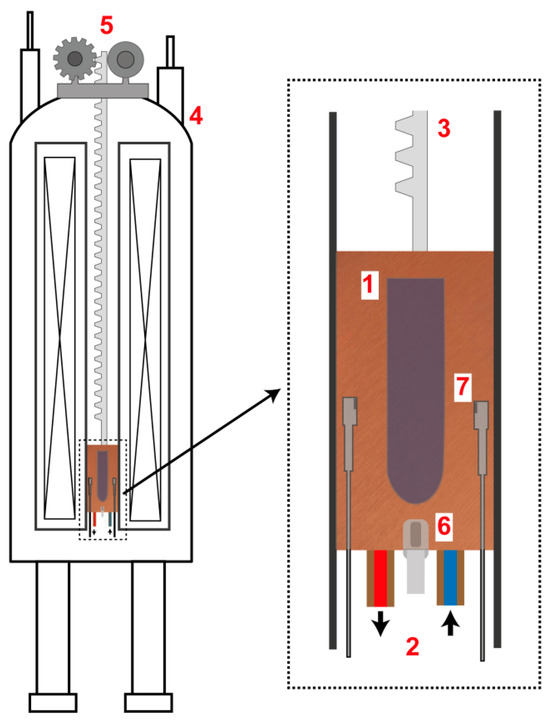
The crystallization of paramagnetic species in a magnetic field gradient under microgravity-like conditions is an area of interest for both fundamental and applied science. In this paper, a setup for the crystallization of paramagnetic species in the magnetic field up to 7 T
[...] Read more.
The crystallization of paramagnetic species in a magnetic field gradient under microgravity-like conditions is an area of interest for both fundamental and applied science. In this paper, a setup for the crystallization of paramagnetic species in the magnetic field up to 7 T generated by a superconducting magnet is described. The research includes calculations of the conditions necessary to compensate for the gravitational force for several types of paramagnetic substances using the magnetic field of superconducting magnets (4.7 T, 7 T, 9.4 T, and 16.4 T). Additionally, for the first time, the crystallization of copper sulfate and cobalt sulfate, as well as a mixture of copper sulfate and cobalt sulfate under gravitational force compensation in a superconducting magnet, was performed. This paper experimentally demonstrates the feasibility of growing paramagnetic crystals within the volume of a test tube on the example of copper and cobalt sulfate crystals. A comparison of crystals grown from the solution of a mixture of copper and cobalt sulfates under the same conditions, with and without the presence of a magnetic field, showed changes in both the number and size of crystals.
Full article
(This article belongs to the Special Issue Original Approaches and Advanced Techniques in Molecular Magnetism Research)
►
Show Figures

Figure 1


