
Surg. Tech. Dev. 2024, 13(2), 205-213; https://doi.org/10.3390/std13020013 - 24 May 2024
Abstract
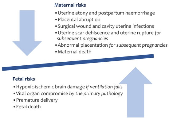
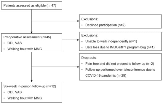
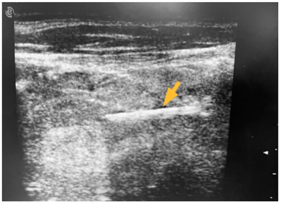
Acute bowel ischemia is a life-threatening abdominal emergency. In many patients, percutaneous endovascular repair of visceral arteries in an antegrade direction across occluding lesionsis challenging and sometimes not possible. We present the case of technically successful percutaneous retrograde recanalization of an occluded superior
[...] Read more.
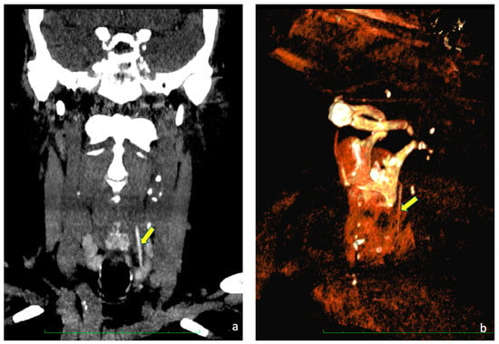
Acute bowel ischemia is a life-threatening abdominal emergency. In many patients, percutaneous endovascular repair of visceral arteries in an antegrade direction across occluding lesionsis challenging and sometimes not possible. We present the case of technically successful percutaneous retrograde recanalization of an occluded superior mesenteric artery in a critically ill 82-year-old patient. The superior mesenteric artery was recanalized via the branches of the celiac trunk; the guidewires were navigated to the target artery through the gastroduodenal and pancreaticoduodenal arteries. Retrograde percutaneous recanalization of the superior mesenteric artery is technically feasible, even in hemodynamically unstable patients.
Full article


