


Viruses 2024, 16(5), 738; https://doi.org/10.3390/v16050738 - 07 May 2024
Abstract
Hepatitis C virus (HCV) infects the human liver, and its chronic infection is one of the major causes of Hepatocellular carcinoma. Translation of HCV RNA is mediated by an Internal Ribosome Entry Site (IRES) element located in the 5’UTR of viral RNA. Several
[...] Read more.
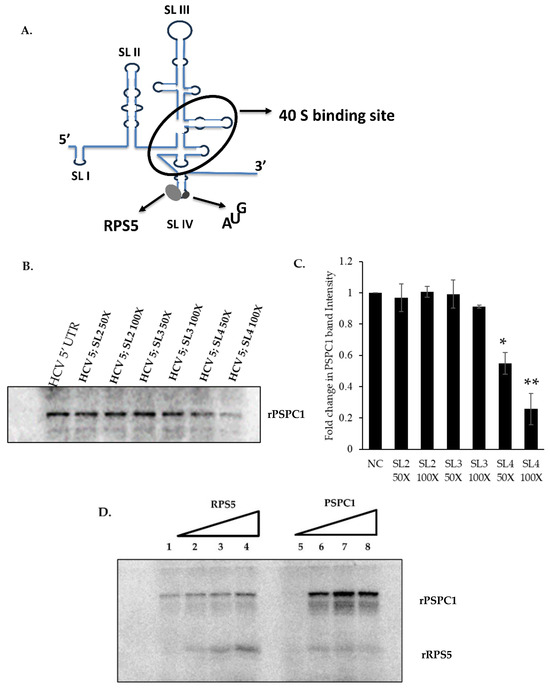
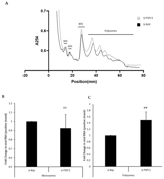
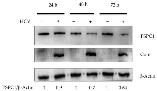
Hepatitis C virus (HCV) infects the human liver, and its chronic infection is one of the major causes of Hepatocellular carcinoma. Translation of HCV RNA is mediated by an Internal Ribosome Entry Site (IRES) element located in the 5’UTR of viral RNA. Several RNA Binding proteins of the host interact with the HCV IRES and modulate its function. Here, we demonstrate that PSPC1 (Paraspeckle Component 1), an essential paraspeckle component, upon HCV infection is relocalized and interacts with HCV IRES to prevent viral RNA translation. Competition UV-crosslinking experiments showed that PSPC1 interacts explicitly with the SLIV region of the HCV IRES, which is known to play a vital role in ribosomal loading to the HCV IRES via interaction with Ribosomal protein S5 (RPS5). Partial silencing of PSPC1 increased viral RNA translation and, consequently, HCV replication, suggesting a negative regulation by PSPC1. Interestingly, the silencing of PSPC1 protein leads to an increased interaction of RPS5 at the SLIV region, leading to an overall increase in the viral RNA in polysomes. Overall, our results showed how the host counters viral infection by relocalizing nuclear protein to the cytoplasm as a survival strategy.
Full article
(This article belongs to the Special Issue Functional and Structural Features of Viral RNA Elements)
►
Show Figures
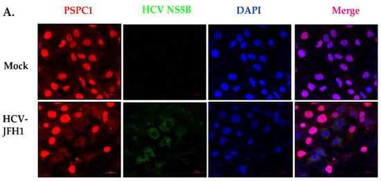
Figure 1


