Possible Further Evidence for the Thixotropic Phenomenon of Water

Abstract
: In this work we review the literature for possible confirmation of a phenomenon that was proposed to develop when water is left to stand for some time undisturbed in closed vessels. The phenomenon has been termed thixotropy of water due to the weak gel-like behaviour which may develop spontaneously over time where ions and contact with hydrophilic surfaces seem to play important roles. Thixotropy is a property of certain gels and liquids that under normal conditions are highly viscous, whereas during mechanical processing their viscosity diminishes. We found experiments indicating water’s self-organizing properties, long-lived inhomogeneities and time-dependent changes in the spectral parameters of aqueous systems. The large-scale inhomogeneities in aqueous solutions seem to occur in a vast number of systems. Long-term spectral changes of aqueous systems were observed even though the source of radiation was switched off or removed. And water was considered to be an active excitable medium in which appropriate conditions for self-organization can be established. In short, the thixotropic phenomenon of water is further indicated by different experimental techniques and may be triggered by large-scale ordering of water in the vicinity of nucleating solutes and hydrophilic surfaces.PACS Codes: 61.20.Qg; 61.80.Ba; 64.70.Ja; 66.10.-x; 66.20.-d;1. Introduction
Liquid water has a complicated molecular-scale structure that is still not perfectly understood. The weak nature of the hydrogen bond allows the water network to rapidly fluctuate and rearrange at rates of tens of femtoseconds to picoseconds [1–4], yet water molecules associate by these weak hydrogen bonds that seem to offer the possibility of forming clusters of molecules with specific shapes and behaviour [5].
In a previous study [6,7] we continued the work of Elia [8] who explored the physico-chemical properties of aqueous solutions of NaHCO3 treated mechanically by iterating dilution and succussion (vigorous shaking). They repeated the processes to extreme dilution, to form extremely dilute solutions (EDS), where the chemical composition of the end solution was identical to that of the solvent. They measured the electrical conductivities, heats of mixing and pH of aged EDS and compared the results with values of one day old untreated analogous solutions [8–17]. They noticed significantly higher electrical conductivities in comparison to the values of analogous fresh untreated solutions. Additionally, the values of excess conductivities were in correlation with excess heats of mixing and increased pH of aged EDS. They found that the addition of electrolyte (0.5 and 1 M NaCl) does not diminish the amount of excess heat [13]. Since the concentration of the unknown impurities was substantially lower in comparison to the amount of added electrolyte (0.5 M NaCl) and the temporal evolution of the electrical conductivity of up to 500 days features a maximum followed by a return to initial values, the presence of the unknown impurities was proven to be insignificant [12]. They attributed their unusual results to water’s self-organizing abilities triggered by the input of kinetic energy during vigorous shaking which induces the ordering of water molecules. Namely, larger water clusters accelerate the Grotthuss mechanism of proton transfer that predominates in aqueous hydrogen carbonate solutions [18]. According to this mechanism proton is transferred through the network of hydrogen-bonded water molecules through the formation or cleavage of hydrogen and covalent bonds [19].
In contrast to the results of Elia [8], we [7] found no differences in conductivities of aged mechanically treated solutions and aged untreated solutions. Yet all aged solutions (treated and untreated) had significantly higher conductivity values than those of freshly prepared chemically analogous aqueous solutions and no excess conductivity was found in frozen samples. The results surprisingly resemble a previously noticed phenomenon in liquid water, which may develop when water is stored in closed vessels. The phenomenon was termed by Vybíral and Voráček [20] as “autothixotropy of water” due to the weak gel-like behaviour which may develop spontaneously over time in liquid distilled water when left to stand undisturbed for some time in closed vessels, where ions seem to play an important role. Thixotropy is a property of certain gels and liquids that under normal conditions are highly viscous, whereas during mechanical processing their viscosity diminishes. We reviewed the possible formation of a structured network of hydrogen bonds between water molecules and ions in aqueous solutions that are left to stand undisturbed for some time where hydrophilic surfaces also may play an important role [21]. We proposed thixotropy to be the cause of the faster proton transfer in aged solutions along the lines of the Grotthuss mechanism.
Similarly, Guckenberger et al. [22] using scanning tunneling microscopy based on the lateral conductivity of ultrathin water films next to biological specimens, and Sasaki [23] by exploring the dielectric properties of slightly hydrated collagen found increased conductivity values in aqueous solutions under constrained conditions where the lifetime of a hydrogen bond is prolonged.
In what follows, we review possible indications of water’s self-organizing properties, long-lived inhomogeneities and time-dependent changes in the spectral parameters of aqueous systems which could further confirm and explain the existence of the thixotropic phenomenon. Additionally, changes in viscosity of aqueous systems upon mechanical treatment are discussed.
2. Discussion
2.1. Water’s Self-organizing Properties
Generally, water is thought of as a homogeneous liquid at ambient temperatures. For instance, small-angle X-ray scattering data on deionized and degassed water at room temperature, as well as molecular dynamics simulations of 32,000 water molecules [24] gave no indications of heterogeneities in the water structure. Furthermore, vibrational spectroscopy in the femtosecond (fs) time domain showed that water is characterized by ultrafast structural fluctuations. The OH stretching and bending excitations of water molecules decay on a subpicosecond time scale, followed by dissipation of excess energy in the hydrogen bond network within a few picoseconds. While the lifetime of hydrogen bonds is of the order of 1 ps, structural dynamics in the form of librational and other motions exist on a much shorter time scale [25].
Nevertheless, a two-state model of water was developed already by Röntgen [26] and also proposed by Robinson et al. [27]. To demonstrate the presence of density fluctuations due to the two structural motifs in water, small-angle X-ray scattering (SAXS) was conducted [28] and the results confirmed a nonhomogeneous distribution of molecules in deionized and degassed water at room temperature. Clark et al. [24] propose that these SAXS data [28] are caused by stochastic number fluctuations present in all liquids which are not qualitatively different for water. However, the dynamic balance between tetrahedral-like and hydrogen-bond distorted structures related to low-(LDL) and high-density liquid (HDL) can be understood from the increasing anticorrelation between fluctuations in local volume and in entropy on cooling the liquid [29]. A pressure-dependent SAXS study of water [30] is also fully consistent with the picture in [28]. Namely, LDL tetrahedral motifs decrease in number with increasing pressure in favor of the HDL disordered motifs undergoing normal stochastic fluctuations. And recently, measurements of optical Kerr effects by Taschin et al. gave strong evidence for two distinct, high-density and low-density water forms [31].
Further, certain investigations confirm that due to the small mass of the hydrogen atom and proton the quantum phenomenon of coherence may give a better description of the properties of water and aqueous solutions than the classical one. Paesani et al. [32] found nuclear quantum effects in the reorientation of water from polarization-resolved fs mid-IR pump-probe spectroscopy and quantum molecular dynamics simulations. The importance of explicitly accounting for nuclear quantum effects when simulating the molecular properties of water was recently also confirmed by Yang and Skinner [33]. Namely, they found coherent vibrational energy transfer in the IR and Raman line shapes of liquid water. Woutersen and Bakker [34] using time-resolved pump-probe laser spectroscopy measurements found that fast resonant intermolecular transfer of OH-stretch excitations occurs over many water molecules before the excitation energy is dissipated. Their findings suggest that liquid water may play an important role in transporting vibrational energy between OH groups located on either different biomolecules or along extended biological structures. This was further confirmed by mid-IR spectroscopy measurements showing that the hydrogen atom becomes delocalized between the oxygen atoms of two neighbouring water molecules [35]. Additionally, broadband dielectric spectroscopy and deep inelastic neutron scattering [36] indicated that water protons reflect delocalization over the surface of a hydrated protein at ambient temperatures.
The research groups we refer to in this subsection have published their papers in good, and some even in the highest ranked journals. However, their viewpoints of water are rather different. The same results obtained from the same experimental technique, like for instance SAXS, were interpreted in different ways. For some water seems to be a homogeneous and for others a heterogeneous liquid. For reviewing possible indications of the thixotropic phenomenon, we shall consider that under some circumstances it is an inhomogeneous liquid with time-dependent properties.
2.2. The Time Domain of Water
It was reported that air-saturated purified water exhibits luminescence in the UV and blue-green regions of the spectrum under ionizing radiation [37], UV light [38,39] and IR radiation [40]. The spectral parameters change even when the source of radiation is removed or switched off [37,38,40,41]. Quickenden and Que Hee [37] observed a nondecaying weak luminescence from air-saturated bidistilled water exposed to ambient ionizing radiation. For several hours after removal from daylight, aqueous samples exhibit slowly decaying luminescence before reaching basal levels after approximately four hours. A substantial portion of the luminescence is non–Cherenkov emission, i.e. not emitted by dissolved charged particles. Lobyshev et al. [38] employing luminescence spectroscopy found that the structural equilibrium in air-saturated bidistilled water is in general not stable and changes after exposure to UV and mild X-ray irradiation and also after dissolution of small amounts of glycyltryptophane. Similar to our own and to Elia’s results, the bands in the emission spectra of aqueous solutions also depend on the duration of storage in a closed vessel. To clarify the possible role of dissolved gasses, the aqueous samples stored for a long time were degassed. The spectra of the degassed samples did not differ from those of the control samples.
Gudkov et al. [40] found that a 5-min exposure of air-saturated bidistilled water to low-intensity laser IR radiation at 1,264 nm after a long latent period of 1.5 to 2 hours induces auto-oscillations of water luminescence that last around 20 hours. Because the energy of the IR radiation was insufficient for ionization of water molecules or generation of free radicals, but sufficient for the electronic transition of oxygen to the singlet state, the authors attributed the phenomenon to dissolved oxygen; singlet oxygen is highly reactive and has a tendency to initiate redox reactions. Further, it was found that water molecules significantly affect electron-transfer and possibly also the triplet–triplet energy transfer properties between redox partners when in the vicinity of chromophores [42].
The time domain of the properties of water may also be monitored by means of a magnetic field. Many experiments show that aqueous systems can be magnetized by a magnetic field and stay magnetized much longer than is theoretically explicable [43–53], even though the magnetization effect is small. However, when water and aqueous solutions are exposed to a magnetic field, the so-called magnetization of water induces changes in its properties, including optical features [54], magnetism [50], thermodynamics [43], increase in melting point [52], surface tension [48], increase in vaporization rate [51,55,56] and electric conductivity [51] compared with those of pure aqueous systems.
Pang and Deng [50] found that the properties of IR and UV absorption, Raman scattering and X-ray diffraction of magnetized water significantly change relative to those of pure water. For the experiment they used doubly distilled and passed through a Millipore water system water. Mostly a static magnetic field of around 0.4 T was applied. The intensities of peaks increase, the frequencies of some peaks shift and new peaks occur. The peak shifts in the X-ray diffraction spectra of magnetized and nonmagnetized water on addition of Fe3O4 hybrid show that the magnetized water possesses magnetism. Similarly to the previous studies of Higashitani et al. [44–46], they found [50] that the magnetization effect correlates with magnetization time, the intensity of the externally applied magnetic field and the temperature of water. Longer magnetization times, higher intensities of the applied magnetic field and lower temperatures of water correlate with enhancement in peak intensities.
Once the externally applied magnetic field is removed from magnetized water, the effect of magnetization does not disappear immediately but persists for a very long time (60 minutes and more) and finally becomes the same as that of pure water [46,50]. The magnetic memory of magnetized water also increases with increasing magnetic field and decreasing water temperature. Since the magnetic effect is usually very small [44–47], such measurements have seen a lot of data that proofed to be wrong, caused by impurities, not the substances under investigation. The effect may have some relation to the unstable structure of water molecules around structure-disordering ions and hydrophobic molecules. The effect can be easily destroyed by external disturbances and appears more effectively with discontinuous magnetic radiation. The magnetic field in some way increases the structuring of the hydration shells of structure-breaking ions [51]. So the magnetic effect may be due to enhanced proton transfer along the lines of the Grotthuss mechanism in more organized water.
Yamashita et al. [53] investigated the effects of a magnetic field on pH and oxidation-reduction potential (ORP) equilibration rates of distilled and deionized water, previously stored for air equilibration. The results showed slow and large fluctuations from ~0.05 to 0.1 in pH units and ~60 mV in ORP during the first few hours. When readings were taken for days, the pH and ORP generally changed slowly toward their equilibrium values. Long-time pH and ORP measurements appear to be essential to characterize purified water’s structural state. O2 and CO2 equilibration of the samples is typically a slow process that is well understood. What is not so well understood is the enhancement effect associated with the presence of a magnetic field. The auto-oscillatory features of the magnetic effect results [53] and other studies [40,41] could allow one to consider water as an active excitable medium. An auto-oscillatory process like the Belousov-Zhabotinski reaction in a particular set-up where lamellar lipid structures are involved stops if the concentration of water in comparison to the concentration of lipids falls below 70 wt. % of lipids, where neither water nor lipids participate in the reaction [57]. This may indicate that water molecules establish the appropriate conditions for self-organization of such systems [58].
2.3. Large-scale Inhomogeneities in Aqueous Systems and/or Exclusion Zones
According to Marchettini et al. [58] the ordering of water molecules next to the hydrophilic surfaces of lamellar lipids could be the trigger for the self-organization of the Belousov-Zhabotinski reaction. Namely, Szent-Györgyi quotes in Bioenergetics [59] the report of Henniker and McBain from 1948 [60] with 175 papers devoted to water hydrating hydrophilic surfaces which showed that interfacial water may have a thickness of from tens to hundreds of molecular layers, and not just a few as usually believed. This result emerged from exploration of the luminescent properties of different aqueous solutions of dyes which depend strongly on the physical state of water, structured by different properties of the surface. Based on these experimental results, Szent-Györgyi proposed that the thick layers of organized water adjacent to hydrophilic surfaces could allow very long lasting electronic excitations that may play a significant role in energy transfer near cell surfaces, where water resides in a state similar to a liquid crystal.
A few decades later Pollack et al. [61,62] rediscovered that water molecules adjacent to hydrophilic gels are ordered in layers a few hundred thousand molecules-thick. He called this region the exclusion zone (EZ), as water molecules in these zones seem to exclude solutes. In some instances the EZs can extend many micrometres from the respective surfaces. Further, they found that EZs also appear next to some solutes [63] and have the capacity to increase effective particle sizes [64].
The properties of EZ water are different from the bulk. They bear a negative charge of as much as 150–200 mV near the nucleating surface, exhibit increased dynamic viscosity and show diminished IR emissivity and smaller T2 relaxation times [62] and seem to be expanded by light [65]. Yoo et al. [66] studied EZ water by NMR spectroscopy to measure chemical shifts, spin-lattice (T1) relaxation and the self-diffusion coefficient of interfacial water. They found two water proton resonances indicating two distinct and magnetically unique water species. According to the authors [66], although the chemical shift is similar to that of the bulk (0.2 ppm), T1 and the self-diffusion coefficient values could be identifiers of the restricted long-range nature of EZ water.
Ovchinnikova and Pollack [67] observed that after standing undisturbed for several hours in a sealed beaker aqueous microsphere suspensions develop large, vertically oriented, microsphere-free cylinders situated near the beaker’s vertical axis. These cylindrical zones were found to be quite stable when not subjected to evaporative forces by sealing the vessels, with their radial positions sensitive to incident light. The authors [67] attributed the low photon energy derived from the laboratory environment to be critical for inducing this surprising formation. We propose these formations to be one of the triggers for ordering of water molecules in aged aqueous solutions.
Investigation of potential clinical significance of the properties of EZ water, according to Davidson et al. [68], is warranted. The features of EZ water, in our opinion, also play an important role in the development of the thixotropic phenomenon that could be further confirmed by large-scale inhomogeneities found in aqueous systems. Literature indicating large scale inhomogeneities in aqueous solutions is accumulating, with emphasis on static and dynamic laser light scattering (LLS) measurements [69–80]. A wide variety of other experimental techniques may confirm that in aqueous under-saturated strong electrolyte solutions, the ions are not homogeneously distributed [63,81–86]. Quantum electrodynamic interactions may cause formation of μm-sized domains composed of solvated ions [87–90].
Possible evidence of inhomogeneities was found by LLS studies of aqueous solutions of 3-methylpyridine and tertiary butyl alcohol [79]. The observed inhomogeneities were attributed to near-spherical Brownian aggregates of sizes from a hundred to a few hundred nm. LLS experiments with mixtures of water and organic molecules like tetrahydrofuran, ethanol, urea and R-cyclodextrin also showed slow relaxation modes [70,71]. Because the phenomenon disappeared on repeated filtration but was regenerated by air injection, the authors attributed their results to the formation of nanobubbles around 100 nm in diameter. Nanobubbles are submicron sized air bubbles and have been the subject of dispute for a long time. However, proofs of their existence are accumulating [91,92]. They may play an important role in the long-range hydrophobic attractions observed in aqueous systems [93].
Bunkin et al. [94] found that any liquid saturated with dissolved gas (for example, atmospheric air) and containing an ionic component is unstable with respect to spontaneous formation of spherical gas cavities on the nanometre scale, so-called “bubstons” which are stable gas nanobubbles. In very dilute aqueous solutions of electrolytes the radii of bubstons are between 10 and 100 nm. Under conditions of mechanical equilibrium the pressure of the gas inside a bubston is precisely equal to the gas pressure over the surface of the liquid.
The stability of bubstons is conditioned by the adsorption of ions of the same sign on the “bubston” surface. Hence, bubstons can be positively or negatively charged. Thus, they are capable of coagulating, resulting in bubston clusters. A bubston cluster includes around 100 separate bubstons and its effective radius is 0.5 μm. The results of laser modulation interference microscopy and Mueller-matrix scatterometry show that macroscopic scatterers of light waves are present in doubly distilled water free from external solid impurities [95]. The authors interpreted the experimental data by bubston clusters composed of polydisperse air bubbles having effective radii of 70–90 nm. Experiments with dynamic light scattering, phase microscopy and polarimetric scatterometry additionally indicated that long-lived gas nanobubbles and clusters composed of such nanobubbles are generated spontaneously in an aqueous solution of salt saturated with dissolved gas [96]. According to Pollack’s [97] embryonic bubble concept in water next to nucleators, like hydrophilic surfaces or some solutes, EZ grow layer by layer and also laterally. Protons build positive charge beyond the EZ. Because of attraction to positive charge EZ deflects and continued deflection creates vesicle. If a vesicle captures enough incident radiant energy, the layers of EZ augment, therefore the concentration of protons inside the vesicle increase, raising the internal pressure, and leading to vesicle expansion. When that happens, the contained water molecules experience lowered pressure leading to their conversion into vapour. Hence, incident radiant energy transforms water vesicles into vapour bubbles.
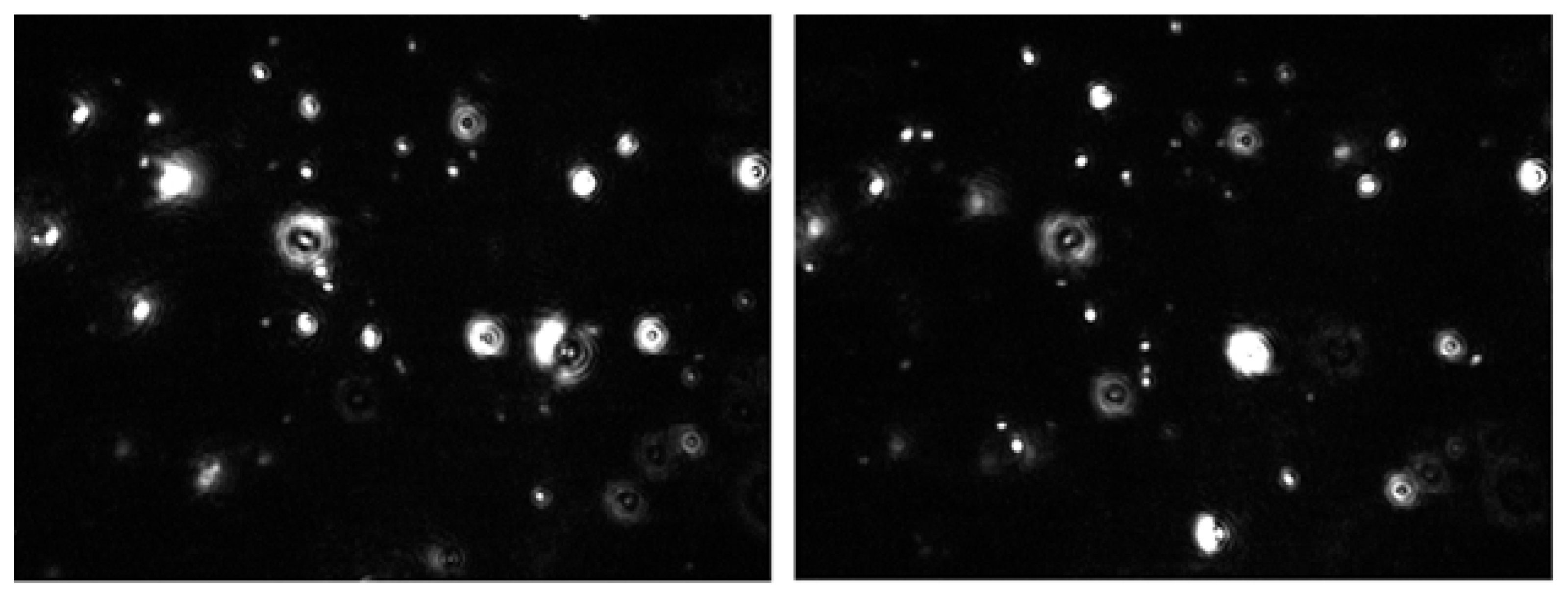
Recently, Sedlák and Rak [77] exploring solutions and mixtures under ordinary conditions (equilibrated with air at 1 atm), with a degassed solvent and solutions and mixtures degassed after formation of large structures, found real discrete objects with macroscopic lifetimes, not fluctuations and not bicontinuous phases with large correlation lengths. Their results, in accordance with the results of Häbich et al. [98], show that these structures are not nanobubbles in all cases. The 100 nm mesoscale inhomogeneities are long-lived, with relatively slow kinetics of formation which was monitored on mixing the components. Visualization of such large-scale structures via nanoparticle tracking analysis (NTA), presented in Figure 1, may additionally confirm their previous LLS work [74–76]. In addition, good agreement between the size distributions obtained from LLS and NTA was obtained.
Sedlák [74–76] investigated systems of D-glucose, NaCl, Na2SO3, MgSO4, Al(NO3)3, acetic acid, dimethyl sulfoxide, ethanol and benzene with double distilled and subsequently deionized (not degassed) water. LLS results show that the solutes were distributed inhomogeneously on large length scales. They found that the number of solute molecules per domain varies in the range of 103–108 and the development of supramolecular structures seems time-related. Furthermore, they found that the time scale on which the supramolecular structures develop varies from minutes to weeks and has a saturation character. It is faster at the beginning and then gradually slows down. After 80 days the domains appear stable with domain sizes from 30 to almost 500 nm. In some systems the domain structure appears stable, whereas in others a very slow process of domain fading out was observed.
Hence, the large-scale inhomogeneities are much larger than the dimensions of individual molecules, typically on the order of 100 nm [74,77,78]. Solutions of ordinary low molar mass compounds and mixtures of freely miscible liquids, according to a vast literature on LLS measurements, may evidently not be as homogeneous as previously thought. These large-scale inhomogeneities could be, as we already mentioned, attributed to the exclusion zone water. Together with their time-dependent spectral parameters they could be further confirmation of the thixotropic phenomenon.
2.4. Thixotropy in Aqueous Systems
Thixotropy is one of the more complex characteristics associated with the behaviour of non-Newtonian liquids. The term originally referred to the reversible changes from a flowable fluid to a solid-like gel where particles can form a loose association which is easily destroyed by shaking but re-establishes itself on standing. The time-scales involved can range from many minutes in the case of the breakdown to many hours or days in rebuilding. The word combines the Greek words thixis (stirring or shaking) and trepo (turning or changing) [99].
A whole variety of systems were found to manifest thixotropic effects, including vanadium pentoxide and aluminium oxide sols, aqueous iron oxide dispersions, paints, starch pastes, gelatin and pectin gels, clays, creams, flour doughs and suspensions, fibre greases, jellies, carbon black and soil suspensions and even human blood [99,100]. For instance, thick albumen demonstrates the ability to transform from a dye excluding gel to a non-dye excluding sol under the influence of pressure or agitation [101]. According to Pollack [97] exclusion zone material such as an egg—a supramolecular assembly of molecules, behaves thixotropically because of the electrostatic nature of its bonds. Those bonds should sustain small deformations more or less elastically. However, “if you tug on the array enough to break those bonds, then it will fracture and the vesicle material will flow” [97].
Thixotropic systems are commonly concentrated solutions of substances of high molecular weight or colloidal suspensions. But all liquids with microstructure can show thixotropy, because thixotropy only reflects the finite time taken to move from any one state of microstructure to another and back again [99]. Vybíral and Voráček observed thixotropy of air-saturated distilled water as a disturbing phenomenon during their gravimetric measurements [20,102]. The thixotropy did not appear in air-saturated deionized water, not even when it was strongly magnetized (magnetic field of 1 T) [102]. The phenomenon was very weak and appeared only when water was left standing in closed vessels for some time (a few days). This resulted in a force of mechanical resistance against an immersed body changing its position. So, they proposed an explanation involving cluster formation by H2O molecules in aged water where ions seem to play an important role. We proposed that hydrophilic surfaces play an important role, too [21].
The true steady-state behaviour of a thixotropic liquid is seen after an infinite shearing time at any shear stress or infinite rest time. Along with the breakdown/formation in structure, other non-rheological features change, such as the dielectric constant, and also conductivity [99]. Therefore, our own and Elia’s increased conductivity results in ~one year and more aged diluted aqueous solutions might, in accordance with Bauer and Collins [103], confirm that aqueous solutions of ions when left to stand undisturbed for some time develop thixotropic properties.
4. Conclusions
It appears that the existence of large-scale, long-lived inhomogeneities is a rather universal phenomenon occurring in a vast number of aqueous systems. The long-lived inhomogeneities may be attributed to the “large” exclusion zones found next to hydrophilic gels and some solutes by Pollack and his group [62,97]. Further, spectral parameters of many aqueous systems exhibit time dependence. In some cases, auto-oscillatory changes in properties were observed.
The common features of the properties of EZ water, large-scale inhomogeneities and changes in spectral parameters of aqueous systems are the following:
Expansion of EZ with radiation correlates with the enhancement of intensities in the spectra of aqueous systems upon radiation;
Increase of effective particle sizes by growth of EZ around some solutes correlates with the observed development of long-lived large-scale inhomogeneities in aqueous systems.
We think that the large-scale inhomogeneities together with time-dependent spectral parameters of aqueous systems could be further confirmation of the thixotropic phenomenon. We propose that the main factor inducing the thixotropic phenomenon is formation of exclusion zones in the vicinity of nucleating solutes and hydrophilic surfaces.
Acknowledgments
The authors gratefully thank Igor Jerman, Rok Krašovec, Matej Plankar, Nigel Dyer, Stephanie Seneff and Robert M. Davidson for fruitful discussions.
Conflicts of Interest
The authors declare no conflict of interest.
- Author ContributionsThe authors contributed equally to the manuscript.
References
- Fecko, C.J.; Eaves, J.D.; Loparo, J.J.; Tokmakoff, A.; Geissler, P.L. Ultrafast hydrogen-bond dynamics in the infrared spectroscopy of water. Science 2003, 301, 1698–1702. [Google Scholar]
- Fecko, C.J.; Loparo, J.J.; Roberts, S.T.; Tokmakoff, A. Local hydrogen bonding dynamics and collective reorganization in water: Ultrafast infrared spectroscopy of HOD/D2O. J. Chem. Phys 2005, 122, 054506–054524. [Google Scholar]
- Ohmine, I. Liquid water dynamics: collective motions, fluctuation and relaxation. J. Phys. Chem 1995, 99, 6767–6776. [Google Scholar]
- Ball, P. Water as an active constituent in cell biology. Chem. Rev 2008, 108, 74–108. [Google Scholar]
- Chaplin, M. Water Structure and Science. Available online: http://www.lsbu.ac.uk/water/ accessed on 4 October 2013.
- Verdel, N.; Jerman, I.; Bukovec, P.; Krašovec, R. Conductivity measurements as a possible means to measure the degree of water ordering. J. Phys. Conf. Ser 2011, 329, 012005. [Google Scholar]
- Verdel, N.; Jerman, I.; Krašovec, R.; Bukovec, P.; Zupančič, M. Possible time-dependent effect of ions and hydrophilic surfaces on the electrical conductivity of aqueous solutions. Int. J. Mol. Sci 2012, 13, 4048–4068. [Google Scholar]
- Elia, V.; Napoli, E.; Niccoli, M.; Marchettini, N.; Tiezzi, E. New physic-chemical properties of extremely diluted solutions. A conductivity study at 25 °C in relation to ageing. J. Solution Chem 2008, 37, 85–96. [Google Scholar]
- Elia, V.; Niccoli, M. Thermodynamics of extremely diluted aqueous solutions. Ann. NY Acad. Sci 1999, 879, 241–248. [Google Scholar]
- Elia, V.; Napoli, E.; Niccoli, M.; Nonatelli, L.; Ramaglia, A.; Ventimiglia, E. New physico-chemical properties of extremely diluted aqueous solutions. A calorimetric and conductivity study at 25 °C. J. Therm. Anal. Calorim 2004, 78, 331–342. [Google Scholar]
- Elia, V.; Marchese, M.; Montanino, M.; Napoli, E.; Niccoli, M.; Nonatelli, L.; Ramaglia, A. Hydrohysteretic phenomena of extremely diluted solutions induced by mechanical treatments: A calorimetric and conductometric study at 25 °C. J. Solut. Chem 2005, 34, 947–960. [Google Scholar]
- Elia, V.; Elia, L.; Montanino, M.; Napoli, E.; Niccoli, M.; Nonatelli, L. Conductometric studies of the serially diluted and agitated solutions on an anomalous effect that depends on the dilution process. J. Mol. Liq 2007, 135, 158–165. [Google Scholar]
- Elia, V.; Napoli, E.; Niccoli, M. On the stability of extremely diluted aqueous solutions at high ionic strength. A calorimetric study at 298 K. J. Therm. Anal. Calorim 2008, 92, 643–648. [Google Scholar]
- Ciavatta, L.; Elia, V.; Napoli, E.; Niccoli, M. New physico-chemical properties of extremely diluted solutions. Electromotive force measurements of Galvanic cells sensible to the activity of NaCl at 25 °C. J. Solut. Chem 2008, 37, 1037–1049. [Google Scholar]
- Elia, V.; Napoli, E.; Niccoli, M. A molecular model of interaction between extremely diluted solutions and NaOH solutions used as titrant. Conductometric and pHmetric titrations. J. Mol. Liq 2009, 148, 45–50. [Google Scholar]
- Elia, V.; Napoli, E.; Niccoli, M. Thermodynamic parameters for the binding process of the OH− ion with the dissipative structures. Calorimetric and conductometric titrations. J. Therm. Anal. Calorim 2010, 102, 1111–1118. [Google Scholar]
- Betti, L.; Elia, V.; Napoli, E.; Trebbi, G.; Zurla, M.; Nani, D.; Peruzzi, M.; Brizzi, M. Biological effects and physico-chemical properties of extremely diluted aqueous solutions as a function of aging-time. HFSP J 2011, 5, 117–126. [Google Scholar]
- Han, J.; Zhou, X.; Liu, H. Ab initio simulation on the mechanism of proton transport in water. J. Power Sources 2006, 161, 1420–1427. [Google Scholar]
- Xantheas, S.S. Computational chemistry: dances with hydrogen cations. Nature 2009, 457, 673–674. [Google Scholar]
- Vybíral, B.; Voráček, P. Long term structural effects in water: autothixotropy of water and its hysteresis. Homeopathy 2007, 96, 183–188. [Google Scholar]
- Verdel, N.; Jerman, I.; Bukovec, P. The “Autothixotropic” phenomenon of water and its role in proton transfer. Int. J. Mol. Sci 2011, 12, 7481–7494. [Google Scholar]
- Guckenberger, R.; Heim, M.; Cevc, G.; Knapp, H.F.; Wiegräbe, W.; Hillebrand, A. Scanning tunneling microscopy of insulators and biological specimens based on lateral conductivity of ultrathin water films. Science 1994, 266, 1538–1540. [Google Scholar]
- Sasaki, N. Dielectric properties of slightly hydrated collagen: time-water content superposition analysis. Biopolymers 1984, 23, 1725–1734. [Google Scholar]
- Clark, G.N.I.; Hura, G.L.; Teixeira, J.; Soper, A.K.; Head-Gordon, T. Small-angle scattering and the structure of ambient liquid water. Proc. Natl. Acad. Sci. USA 2010, 107, 14003–14007. [Google Scholar]
- Elsaesser, T. Ultrafast memory loss and relaxation processes in hydrogen-bonded systems. Biol. Chem 2009, 390, 1125–1132. [Google Scholar]
- Röntgen, W.K. The structure of liquid water. Ann. Phys 1892, 45, 91–97. [Google Scholar]
- Robinson, G.W.; Cho, C.H.; Gellene, G.I. Refractive index mysteries of water. J. Phys. Chem. B 2000, 104, 7179–7182. [Google Scholar]
- Huang, C.; Wikfeldt, K.T.; Tokushima, T.; Nordlund, D.; Harada, Y.; Bergmann, U.; Niebuhr, M.; Weiss, T.M.; Horikawa, Y.; Leetmaa, M.; et al. The inhomogeneous structure of water at ambient conditions. Proc. Natl. Acad. Sci. USA 2009, 106, 15214–15218. [Google Scholar]
- Huang, C.; Wikfeldt, K.T.; Tokushima, T.; Nordlund, D.; Harada, Y.; Bergmann, U.; Niebuhr, M.; Weiss, T.M.; Horikawa, Y.; Leetmaa, M.; et al. Reply to Soper et al.: Fluctuations in water around a bimodal distribution of local hydrogen-bonded structural motifs. Proc. Natl. Acad. Sci. USA 2010, 107, E45. [Google Scholar]
- Cunsolo, A.; Formisano, F.; Ferrero, C.; Bencivenga, F.; Finet, S. Pressure dependence of the large-scale structure of water. J. Chem. Phys 131, 02.
- Taschin, A.; Bartolini, P.; Eramo, R.; Righini, R.; Torre, R. Evidence of two distinct local structures of water from ambient to supercooled conditions. Nat. Commun 2013, 4, 2401. [Google Scholar]
- Paesani, F.; Yoo, S.; Bakker, H.J.; Xantheas, S.S. Nuclear quantum effects in the reorientation of water. J. Phys. Chem. Lett 2010, 1, 2316–2321. [Google Scholar]
- Yang, M.; Skinner, J.L. Signatures of coherent vibrational energy transfer in IR and Raman line shapes for liquid water. Phys. Chem. Chem. Phys 2010, 12, 982–991. [Google Scholar]
- Woutersen, S.; Bakker, H.J. Resonant intermolecular transfer of vibrational energy in liquid water. Nature 1999, 402, 507–509. [Google Scholar]
- Bakker, H.J.; Nienhuys, H.-K. Delocalization of protons in liquid water. Science 2002, 297, 587–590. [Google Scholar]
- Pagnotta, S.E.; Bruni, F.; Senesi, R.; Pietropaolo, A. Quantum behavior of water protons in protein hydration shell. Biophys. J 2009, 96, 1939–1943. [Google Scholar]
- Quickenden, T.I.; Que Hee, S.S. The luminescence of water excited by ambient ionizing radiation. Radiat. Res 1971, 46, 28–35. [Google Scholar]
- Lobyshev, V.I.; Shikhlinskaya, R.E.; Ryzhikov, B.D. Experimental evidence for intrinsic luminescence of water. J. Mol. Liq 1999, 8, 73–81. [Google Scholar]
- Belovolova, L.V.; Glushkov, M.V.; Vinogradov, E.A.; Babintsev, V.A.; Golovanov, V.I. Ultraviolet fluorescence of water and highly diluted aqueous media. Phys. Wave Phenom 2009, 17, 21–31. [Google Scholar]
- Gudkov, S.V.; Bruskov, V.I.; Astashev, M.E.; Chernikov, A.V.; Yaguzhinsky, L.S.; Zakharov, S.D. Oxygen-dependent auto-oscillations of water luminescence triggered by the 1264 nm radiation. J. Phys. Chem. B 2011, 115, 7693–7698. [Google Scholar]
- Belovolova, L.V.; Glushkov, M.V.; Vinogradova, G.I.; Vinogradov, E.A. Nature of long-lived nonequilibrium states of water and glycyltryptophan aqueous solutions. Phys. Wave Phenom 2008, 16, 292–299. [Google Scholar]
- Di Valentin, M.; Tait, C.E.; Salvadori, E.; Orian, L.; Polimeno, A.; Carbonera, D. Evidence for water-mediated triplet-triplet energy transfer in the photoprotective site of the peridinin-chlorophyll a-protein. Biochim. Biophys. Acta 2014, 1837, 85–97. [Google Scholar]
- Ohata, R.; Tomita, N.; Ikada, Y. Effect of a static magnetic field on ion transport in a cellulose membrane. J. Colloid Interface Sci 2004, 270, 413–416. [Google Scholar]
- Higashitani, K.; Okuhara, K.; Hatade, S. Effects of magnetic fields on stability of nonmagnetic ultrafine colloidal particles. J. Colloid Interface Sci 1992, 152, 125–131. [Google Scholar]
- Higashitani, K.; Iseri, H.; Okuhara, K.; Kage, A.; Hatade, S. Magnetic effects on zeta potential and diffusivity of nonmagnetic colloidal particles. J. Colloid Interface Sci 1995, 172, 383–388. [Google Scholar]
- Higashitani, K.; Oshitani, J.; Ohmura, N. Effects of magnetic field on water investigated with fluorescent probes. J. Coloids Surf. A 1996, 109, 167–173. [Google Scholar]
- Higashitani, K.; Oshitani, J. Magnetic effect on the interface between aqueous solution and solid. J. Surf. Sci. Soc. Jpn 1999, 20, 764–769. [Google Scholar]
- Amiri, M.C.; Dadkhah, A.A. On reduction in the surface tension of water due to magnetic treatment. Colloids Surf. A 2006, 278, 252–255. [Google Scholar]
- Chang, K.T.; Weng, C.I. The effect of an external magnetic field on the structure of liquid water using molecular dynamics simulation. J. Appl. Phys 2006, 100, 043917–043922. [Google Scholar]
- Pang, X.F.; Deng, B. Investigation of changes in properties of water under the action of a magnetic field. Sci. China Ser. G 2008, 51, 1621–1632. [Google Scholar]
- Holysz, L.; Szczes, A.; Chibowski, E. Effects of a static magnetic field on water and electrolyte solutions. J. Colloid Interface Sci 2007, 316, 996–1002. [Google Scholar]
- Inaba, H.; Saitou, T.; Tozaki, K.; Hayashi, H. Effect of the magnetic field on the melting transition of H2O and D2O measured by a high resolution and supersensitive differential scanning calorimeter. J. Appl. Phys 2004, 96, 6127–6133. [Google Scholar]
- Yamashita, M.; Duffield, C.; Tiller, W.A. Dirrect current magnetic field and electromagnetic field effects on the pH and oxidation reduction potential equilibration rates of water. 1. Purified water. Langmuir 2003, 19, 6851–6856. [Google Scholar]
- Deng, B.; Pang, X.F. The variations of optical features of water after action of static magnetic-field. Chin. Sci. Bull 2007, 52, 3179–3182. [Google Scholar]
- Nakagawa, J.; Hirota, N.; Kitazawa, K.; Shoda, M. Magnetic field enhancement of water vaporization. J. Appl. Phys 1999, 86, 2923–2925. [Google Scholar]
- Guo, Y.-Z.; Yin, D.-C.; Cao, H.-L.; Shi, J.-Y.; Zhang, C.-Y.; Liu, Y.-M.; Huang, H.-H.; Liu, Y.; Wang, Y.; Guo, W.-H.; Qian, A.-R.; Shang, P. Evaporation rate of water as a function of a magnetic field and field gradient. Int. J. Mol. Sci 2012, 13, 16916–16928. [Google Scholar]
- Magnani, A.; Marchettini, N.; Ristori, S.; Rossi, C.; Rossi, F.; Rustici, M.; Spalla, O.; Tiezzi, E. Chemical waves and pattern formation in the 1,2-dipalmitoyl-sn-glycero-3-phosphocholine/water lamellar system. J. Am. Chem. Soc 2004, 126, 11406–11407. [Google Scholar]
- Marchettini, N.; Del Giudice, E.; Voeikov, V.; Tiezzi, E. Water: A medium where dissipative structures are produced by a coherent dynamics. J. Theor. Biol 2010, 265, 511–516. [Google Scholar]
- Szent-Györgyi, A. Bioenergetics; Academic Press: New York, NY, USA, 1957. [Google Scholar]
- Henniker, J.C.; McBain, J.W. The Depth of a Surface Zone of a Liquid; Technical Report 6 ori 154 T. O. 11; Stanford Research Institute: Stanford, CA, USA, 1948. [Google Scholar]
- Zheng, J.M.; Pollack, G.H. Long-range forces extending from polymer-gel surfaces. Phys. Rev. E 2003, 68, 031408–031414. [Google Scholar]
- Zheng, J.-M.; Chin, W.-C.; Khijniak, E.; Khijniak, E., Jr.; Pollack, G.H. Surfaces and interfacial water: Evidence that hydrophilic surfaces have long-range impact. Adv. Colloid Interface Sci 2006, 127, 19–27. [Google Scholar]
- Chai, B.H.; Zheng, J.M.; Zhao, Q.; Pollack, G.H. Spectroscopic studies of solutes in aqueous solution. J. Phys. Chem. A 2008, 112, 2242–2247. [Google Scholar]
- Bhalerao, A.S.; Pollack, G.H. Light-induced effects on Brownian displacements. J. Biophotonics 2011, 4, 172–177. [Google Scholar]
- Chai, B.; Yoo, H.; Pollack, G.H. Effect of radiant energy on near-surface water. J. Phys. Chem. B 2009, 113, 13953–13958. [Google Scholar]
- Yoo, H.; Paranji, R.; Pollack, G.H. Impact of hydrophilic surfaces on interfacial water dynamics probed with NMR spectroscopy. J. Phys. Chem. Lett 2011, 2, 532–536. [Google Scholar]
- Ovchinnnikova, K.; Pollack, G.H. Cylindrical phase separation in colloidal suspensions. Phys. Rev. E 2009, 79, 036117. [Google Scholar]
- Davidson, R.M.; Lauritzen, A.; Seneff, S. Biological water dynamics and entropy: A biophysical origin of cancer and other diseases. Entropy 2013, 15, 3822–3876. [Google Scholar]
- Georgalis, Y.; Kierzek, A.M.; Saenger, W. Cluster formation in aqueous electrolyte solutions observed by dynamic light scattering. J. Phys. Chem. B 2000, 104, 3405–3406. [Google Scholar]
- Jin, F.; Ye, J.; Hong, L.; Lam, H.; Wu, C. Slow relaxation mode in mixtures of water and organic molecules: Supramolecular structures or nanobubbles? J. Phys. Chem. B 2007, 111, 2255–2261. [Google Scholar]
- Jin, F.; Li, J.; Ye, X.; Wu, C. Effects of pH and ionic strength on the stability of nanobubbles in aqueous solutions of alpha-cyclodextrin. J. Phys. Chem. B 2007, 111, 11745–11749. [Google Scholar]
- Li, L.; Ogawa, T. Clusters and their properties in aqueous solutions of KDP, KCl and sugar. J. Cryst. Gr 2000, 211, 286–289. [Google Scholar]
- Sedlák, M. Real-time monitoring of the origination of multimacroion domains in a polyelectrolyte solution. J. Chem. Phys 2005, 122, 151102. [Google Scholar]
- Sedlák, M. Large-scale supramolecular structure in solutions of low molar mass compounds and mixtures of liquids: I. Light scattering characterization. J. Phys. Chem. B 2006, 110, 4329–4338. [Google Scholar]
- Sedlák, M. Large-scale supramolecular structure in solutions of low molar mass compounds and mixtures of liquids: II. Kinetics of the formation and long-time stability. J. Phys. Chem. B 2006, 110, 4339–4345. [Google Scholar]
- Sedlák, M. Large-scale supramolecular structure in solutions of low molar mass compounds and mixtures of liquids: III. Correlation with molecular properties and interactions. J. Phys. Chem. B 2006, 110, 13976–13984. [Google Scholar]
- Sedlák, M.; Rak, D. Large-scale inhomogeneities in solutions of low molar mass compounds and mixtures of liquids: Supramolecular structures or nanobubbles? J. Phys. Chem. B 2013, 117, 2495–2504. [Google Scholar]
- Subramanian, D.; Anisimov, M.A. Resolving the mystery of aqueous solutions of tertiary butyl alcohol. J. Phys. Chem. B 2011, 115, 9179–9183. [Google Scholar]
- Subramanian, D.; Ivanov, D.A.; Yudin, I.K.; Anisimov, M.A.; Sengers, J.V. Mesoscale inhomogeneities in aqueous solutions of 3-methylpyridine and tertiary butyl alcohol. J. Chem. Eng. Data 2011, 56, 1238–1248. [Google Scholar]
- Liu, T. An unusually slow self-assembly of inorganic ions in dilute aqueous solution. J. Am. Chem. Soc 2003, 125, 312–313. [Google Scholar]
- Bowron, D.T.; Moreno, S.D. Structural correlations of water molecules in a concentrated alcohol solution. J. Phys. Condens. Matter 2003, 15, S121–S127. [Google Scholar]
- Dixit, S.; Crain, J.; Poon, W.C.K.; Finney, J.L.; Soper, A.K. Molecular segregation observed in a concentrated alcohol-water solution. Nature 2002, 416, 829–832. [Google Scholar]
- Max, J.-J.; Daneault, S.; Chapados, C. 1-Propanol hydrate by IR spectroscopy. Can. J. Chem 2002, 80, 113–123. [Google Scholar]
- Max, J.-J.; Gessinger, V.; van Driessche, C.; Larouche, P.; Chapados, C. Infrared spectroscopy of aqueous ionic salt solutions at low concentrations. J. Chem. Phys 2007, 126, 184507–184521. [Google Scholar]
- Misawa, M.; Sato, T.; Onozuka, A.; Maruyama, K.; Mori, K.; Suzuki, S.; Otomo, T. A visualized analysis of small-angle neutron scattering intensity: Concentration fluctuation in alcohol-water mixtures. J. Appl. Cryst 2007, 40, S93–S96. [Google Scholar]
- Yinnon, C.A.; Yinnon, T.A. Domains in aqueous solutions: Theory and experimental evidence. Mod. Phys. Lett. B 2009, 23, 1959–1973. [Google Scholar]
- Del Giudice, E.; Preparata, G.; Fleischmann, M. QED coherence and electrolyte solutions. J. Electroanal. Chem 2000, 482, 110–116. [Google Scholar]
- Del Giudice, E.; Fleischmann, M.; Preparata, G.; Talpo, G. On the “unreasonable” effects of ELF magnetic fields upon a system of ions. Bioelectromagnetics 2002, 23, 522–530. [Google Scholar]
- Yinnon, T.A.; Elia, V. Dynamics in perturbed very dilute aqueous solutions: Theory and experimental evidence. Int. J. Mod. Phys. B 2013, 27, 1350005–1350040. [Google Scholar]
- Yinnon, T.A.; Yinnon, C.A. Domains of solvated ions in aqueous solutions, their characteristics and impact on electric conductivity: Theory and experimental evidence. Mod. Phys. Lett. B 2012, 26, 1150006–1150020. [Google Scholar]
- Attard, P. Nanobubbles and the hydrophobic attraction. Adv. Colloid Interface Sci 2003, 104, 75–91. [Google Scholar]
- Carambasis, A.; Jonker, L.C.; Attard, P.; Rutland, M.W. Forces measured between hydrophobic surfaces due to a submicroscopic bridging bubble. Phys. Rev. Lett 1998, 80, 5357–5360. [Google Scholar]
- Pashley, R.M.; McGuiggan, P.M.; Ninham, B.W.; Evans, D.F. Attractive forces between uncharged hydrophobic surfaces-direct measurements in aqueous solution. Science 1985, 229, 1088–1089. [Google Scholar]
- Bunkin, N.F.; Bunkin, F.V. Screening of strongly charged macroparticles in liquid electrolyte solutions. J. Exp. Theor. Phys 2003, 96, 730–746. [Google Scholar]
- Bunkin, N.F.; Suyazov, N.V.; Shkirin, A.V.; Ignatiev, P.S.; Indukaev, K.V. Nanoscale structure of dissolved air bubbles in water as studied by measuring the elements of the scattering matrix. J. Chem. Phys 2009, 130, 134308. [Google Scholar]
- Bunkin, N.F.; Shkirin, A.V.; Ignatiev, P.S.; Chaikov, L.L.; Burkhanov, I.S.; Starosvetskij, A.V. Nanobubble clusters of dissolved gas in aqueous solutions of electrolyte. I. Experimental proof. J. Chem. Phys 2012, 137, 054706–054716. [Google Scholar]
- Pollack, G.H. The Fourth Phase of Water. Beyond Solid, Liquid and Vapor; Ebner & Sons: Seattle, USA, 2013. [Google Scholar]
- Häbich, A.; Ducker, W.; Dunstan, D.E.; Zhang, X. Do stable nanobubbles exist in mixtures of organic solvents and water? J. Phys. Chem. B 2010, 114, 6962–6967. [Google Scholar]
- Barnes, H.A. Thixotropy—A review. J. Non-Newtonian Fluid Mech 1997, 70, 1–33. [Google Scholar]
- Huang, C.R.; Su, J.A.; Kristol, D.; Cohn, J.D. Temperature Dependency of Thixotropic Behavior of Whole Human Blood. In Rheology; Astarita, G., Marrucci, G., Nicolais, L., Eds.; Springer-Verlag: New York, NY USA, 1980; Volume 3, pp. 581–582. [Google Scholar]
- Cameron, I. Dye exclusion and other physical properties of hen egg white. Water 2010, 2, 83–96. [Google Scholar]
- Vybíral, B. Autothixotropy of Water and its Possible Importance for the Cytoskeletal Structures. J. Phys. Conf. Ser 2011, 329, 012004. [Google Scholar]
- Bauer, W.H.; Collins, E.A. Rheology: Theory and Applications; Eirich, F.R., Ed.; Academic Press: New York, NY, USA, 1967; Volume 4, Chapter 8. [Google Scholar]

© 2014 by the authors; licensee MDPI, Basel, Switzerland This article is an open access article distributed under the terms and conditions of the Creative Commons Attribution license (http://creativecommons.org/licenses/by/3.0/).
Share and Cite
Verdel, N.; Bukovec, P. Possible Further Evidence for the Thixotropic Phenomenon of Water. Entropy 2014, 16, 2146-2160. https://doi.org/10.3390/e16042146
Verdel N, Bukovec P. Possible Further Evidence for the Thixotropic Phenomenon of Water. Entropy. 2014; 16(4):2146-2160. https://doi.org/10.3390/e16042146
Chicago/Turabian StyleVerdel, Nada, and Peter Bukovec. 2014. "Possible Further Evidence for the Thixotropic Phenomenon of Water" Entropy 16, no. 4: 2146-2160. https://doi.org/10.3390/e16042146