Biodegradable Polymers for Microencapsulation of Drugs

Department of Pharmaceutics and Biomedical Engineering, Purdue University, U.S.A
*
Author to whom correspondence should be addressed.
Molecules 2005, 10(1), 146-161; https://doi.org/10.3390/10010146
Submission received: 25 June 2004
/
Accepted: 1 July 2004
/
Published: 31 January 2005
(This article belongs to the Special Issue Macromolecules Applied to Pharmaceutical Chemistry)
Abstract
:Drug delivery has become increasingly important mainly due to the awareness of the difficulties associated with a variety of old and new drugs. Of the many polymeric drug delivery systems, biodegradable polymers have been used widely as drug delivery systems because of their biocompatibility and biodegradability. The majority of biodegradable polymers have been used in the form of microparticles, from which the incorporated drug is released to the environment in a controlled manner. The factors responsible for controlling the drug release rate are physicochemical properties of drugs, degradation rate of polymers, and the morphology and size of microparticles. This review discusses the conventional and recent technologies for microencapsulation of the drugs using biodegradable polymers. In addition, this review presents characteristics and degradation behaviors of biodegradable polymers which are currently used in drug delivery.
Introduction
For the past few decades, biodegradable polymers have been applied as carriers for controlled delivery of low molecular weight drugs as well as bioactive proteins [1,2,3,4,5]. Biodegradable polymers, either synthetic or natural, are capable of being cleaved into biocompatible byproducts through chemical or enzyme-catalyzed hydrolysis. This biodegradable property makes it possible to implant them into the body without the need of subsequent removal by the surgical operation. Drugs formulated with these polymers can be released in a controlled manner, by which the drug concentration in the target site is maintained within the therapeutic window. The release rates of the drugs from biodegradable polymers can be controlled by a number of factors, such as biodegradation kinetics of the polymers [6,7,8], physicochemical properties of the polymers and drugs [9,10], thermodynamic compatibility between the polymers and drugs [11], and the shape of the devices [12,13,14].
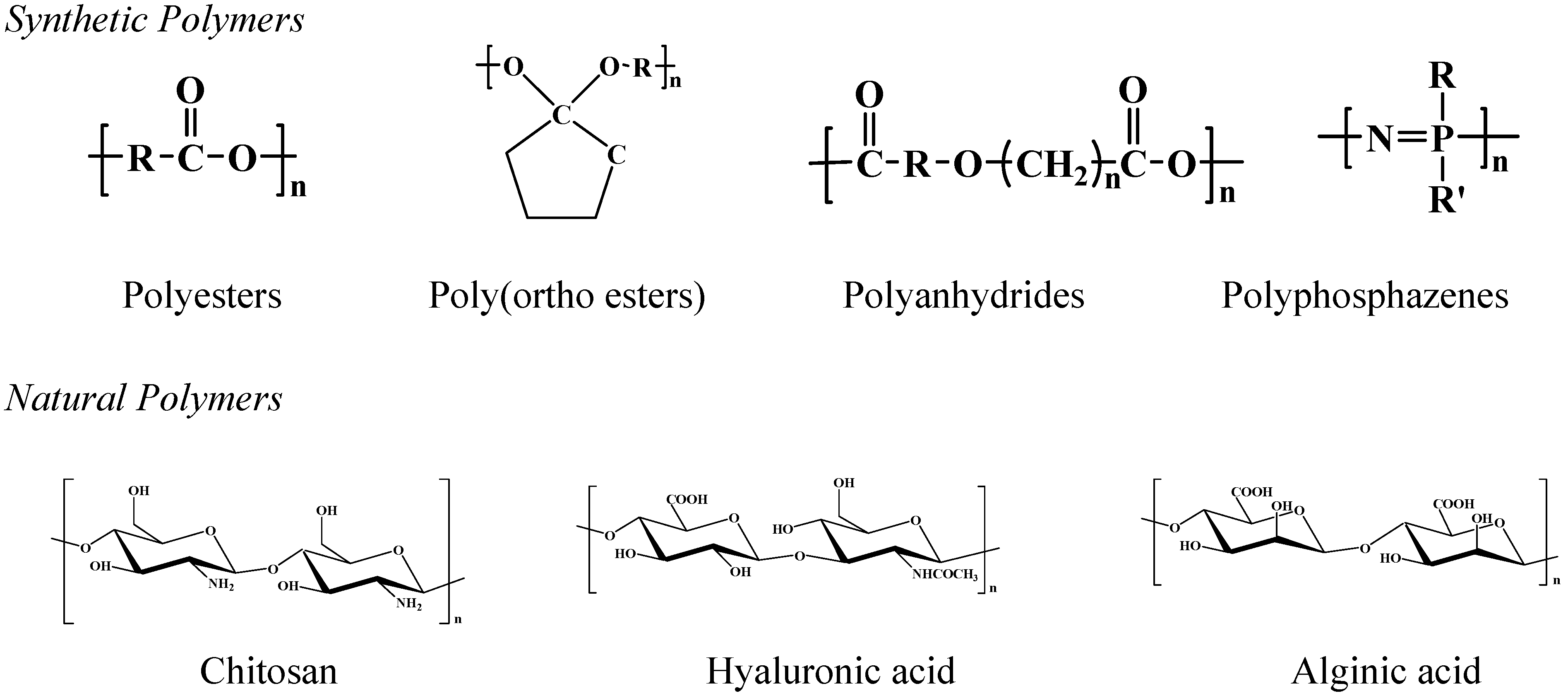
Biodegradable polymer particles (e.g., microspheres, microcapsules, and nanoparticles) are highly useful because they can be administered to a variety of locations in vivo through a syringe needle [15,16]. A variety of drugs, regardless of their molecular weights and water solubility, can be loaded into the biodegradable microparticles using different manufacturing techniques [17,18,19,20,21]. A few examples of biodegradable polymers used in microparticle preparation include polyesters [15,22], polyanhydrides [23,24], poly(ortho esters) [25,26,27], polyphosphazenes [28,29] and polysaccharides [19,30,31]. Figure 1 shows the chemical structures of several biodegradable polymers.
Figure 1.
Chemical structures of several biodegradable polymers.

Techniques of Microparticle Preparation
For preparation of microparticles using biodegradable polymers, it is important to choose an appropriate encapsulation process which meets the following requirements. First, the chemical stability and biological activity of the incorporated drugs should be maintained during the encapsulation process. For example, since most proteins are readily denatured upon contact with hydrophobic organic solvents or acidic/basic aqueous solutions, the process should avoid such harsh environments. Second, the encapsulation efficiency and the yield of the microparticles should be high enough for mass production. Third, the microparticles produced should have the reasonable size range (< 250 μm) that can be administrated using the syringe needle via the parenteral pathway. Fourth, the release profile of the drug should be reproducible without the significant initial burst. Fifth, the process employed should produce free-flowing microparticles, thus making it easy to prepare uniform suspension of the microparticles.
There are a number of techniques available for microencapsulation of drugs such as the emulsion-solvent evaporation/extraction method, spray drying, phase separation-coacervation, interfacial deposition, and in situ polymerization. Each method has its own advantages and disadvantages. The choice of a particular technique depends on the attributes of the polymer and the drug, the site of the drug action, and the duration of the therapy [15,32,33]. This review deals with representative techniques, currently being recognized as effective in microencapsulation of drugs.
Emulsion-solvent evaporation/extraction methods
Single emulsion method
This method has been primarily used to encapsulate hydrophobic drugs through oil-in-water (o/w) emulsification process. The polymer is dissolved in a water-immiscible, volatile organic solvent such as dichloromethane, and the drug is dissolved or suspended into the polymer solution. The resulting mixture is emulsified in a large volume of water in the presence of an emulsifier [15,34,35]. The solvent in the emulsion is removed by either evaporation at elevated temperatures or extraction in a large amount of water, resulting in formation of compact microparticles. The rate of solvent removal is reported to affect the final morphology of microparticles. The solvent removal rate is determined by the temperature of the medium, the solubility characteristics of the polymer, and the solvent used [34,35,36]. This method, however, is only available for the hydrophobic drugs because the hydrophilic drugs may diffuse out or partition from the dispersed oil phase into the aqueous phase, leading to poor encapsulation efficiencies [34,36].
In an attempt to encapsulate hydrophilic drugs (e.g., peptides and proteins), an oil-in-oil (o/o) emulsification method has recently received considerable attention [37,38,39]. In this method, the water-miscible organic solvents are employed to dissolve the drug and polymer, whereas hydrophobic oils are used as a continuous phase of the o/o emulsion. The microparticles are obtained by removing the organic solvents through evaporation or extraction process.
Double emulsion method
Most water-soluble drugs have been encapsulated by water-in-oil-in-water (w/o/w) methods [19,40,41]. The aqueous solution of the water-soluble drug is emulsified with polymer-dissolved organic solution to form the water-in-oil (w/o) emulsion. The emulsification is carried out using either high speed homogenizers or sonicators. This primary emulsion is then transferred into an excess amount of water containing an emulsifier under vigorous stirring, thus forming a w/o/w emulsion. In the subsequent procedure, the solvent is removed by either evaporation or extraction process.
One advantage of this method is encapsulation of hydrophilic drugs in an aqueous phase with the high encapsulation efficiency. For this reason, the w/o/w emulsion system has been used widely for the development of protein delivery systems [19,40,41]. The characteristics of the microspheres prepared by the double emulsion method are dependent on the properties of the polymer (such as composition and molecular weight), the ratio of polymer to drug, the concentration and nature of the emulsifier, temperature, and the stirring/agitation speed during the emulsification process.
Phase separation
This method involves phase separation of a polymer solution by adding an organic nonsolvent [37,42,43]. Drugs are first dispersed or dissolved in a polymer solution. To this mixture solution is added an organic nonsolvent (e.g., silicon oil) under continuous stirring, by which the polymer solvent is gradually extracted and soft coacervate droplets containing the drug are generated. The rate of adding nonsolvent affects the extraction rate of the solvent, the size of microparticles and encapsulation efficiency of the drug. The commonly used nonsolvents include silicone oil, vegetable oil, light liquid paraffin, and low-molecular-weight polybutadiene. The coacervate phase is then hardened by exposing it into an excess amount of another nonsolvent such as hexane, heptane, and diethyl ether. The characteristics of the final microspheres are determined by the molecular weight of the polymer, viscosity of the nonsolvent, and polymer concentration [44,45]. The main disadvantage of this method is a high possibility of forming large aggregates. Extremely sticky coacervate droplets frequently adhere to each other before complete phase separation.
Figure 2.
Formation of mononuclear reservoir-type microcapsules by interfacial phase separation. Two different liquid droplets produced from ink-jet nozzles collide each other in the air. The solvent exchange occurs at the interface between two liquids to form a polymer layer on the aqueous core. The formed micro-capsules are collected in the water bath.
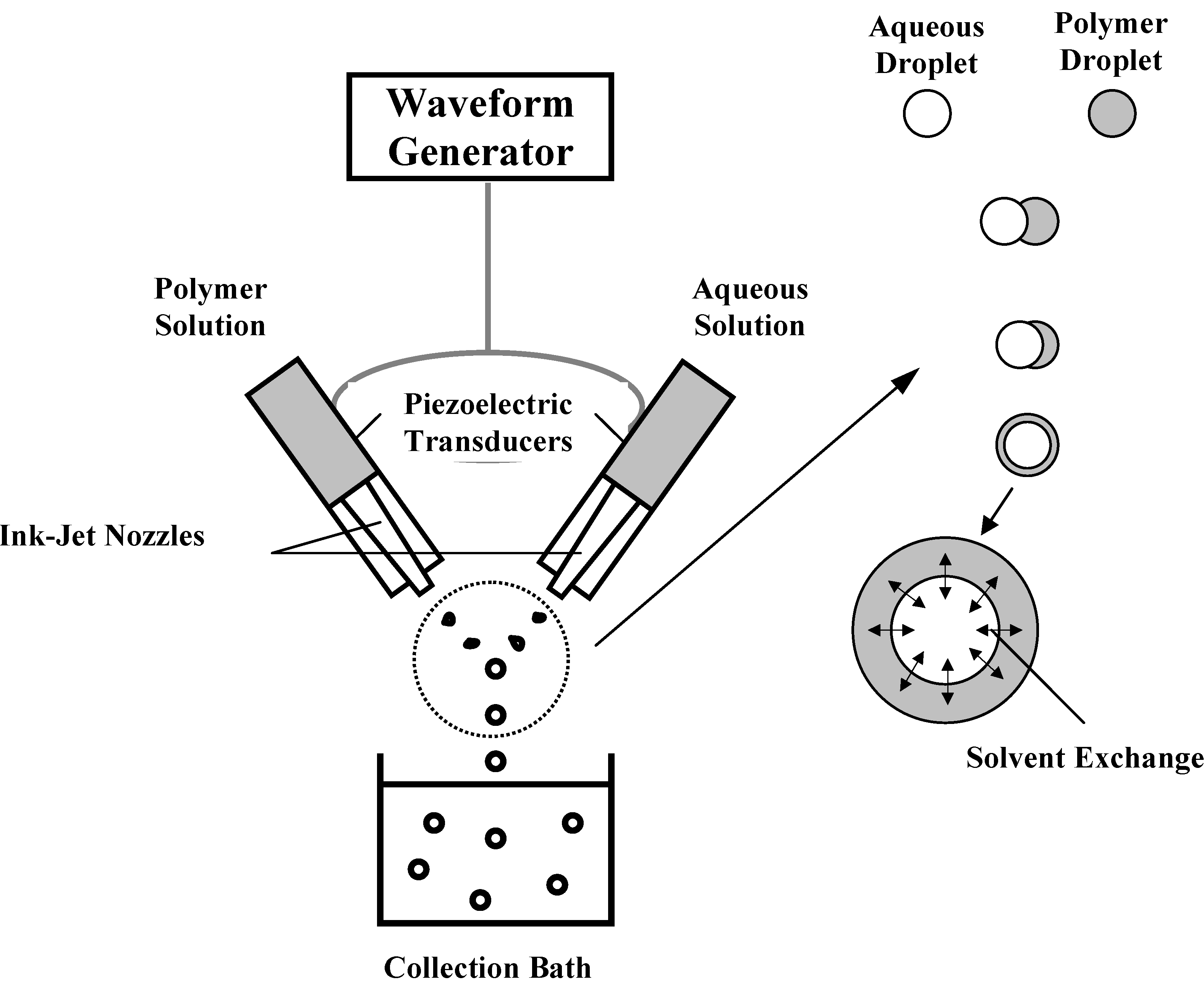
Recently, a novel method of preparing reservoir-type microcapsules, based on interfacial phase separation, was developed [46,47]. Two different types of liquid droplets (i.e., a polymer solution and a drug solution) were separately produced using a dual microdispenser system consisting of two ink-jet nozzles, and the produced droplets were allowed to collide each other in the air (Figure 2). Upon collision, the drug-containing aqueous core remains spherical due to its high surface tension while the polymer-containing droplet spreads over the aqueous core. As a result, a reservoir-type microcapsule is generated due to the interfacial phase separation by the mutual mass transfer of two solvents (i.e., solvent exchange). Successful formation of microcapsules depends on the polymer concentration and the properties of the solvents, such as surface tension, interfacial tension, and the solvent exchange rate.
This technique is promising for preparation of protein-loaded microcapsules. For example, conventional methods of preparing microparticles involve extensive exposure of proteins to the interface between aqueous and organic phases, to hydrophobic polymer matrix, and to acidic/basic microenviroments resulting from degradation of the polymer. These unfavorable interactions are reported to induce conformational changes of proteins [48,49]. On the contrary, the interfacial phase separation technique is shown to minimize these sources of protein inactivation [46,47].
Spray drying
Compared to other conventional methods, spray drying offers several advantages [19,50,51,52]. It shows good reproducibility, involves relatively mild conditions, allows controlling the particle size, and is less dependent on the solubility of the drug and the polymer. The drug is dissolved or dispersed in the polymer solution, in which volatile solvents (e.g., dichloromethane and acetone) are preferred. The resulting solution or suspension is sprayed in a stream of heated air to produce microparticles. The size of the microparticles is determined depending on the atomizing conditions. The main disadvantage of this technique is a loss of a significant amount of product, primarily due to adhesion of the microparticles to the inner wall of the spray-drier. In addition, large aggregates are frequently obtained because the microparticles are very sticky before the complete removal of the solvent.
In an attempt to minimize aggregation of the microparticles, a double-nozzle spray-drying technique was developed [53]. While the polymer/drug solution is sprayed from one nozzle, aqueous mannitol solution is simultaneously sprayed, which enables the surface of the microparticles to be coated with mannitol. The results indicated that the coating of the microsphere with mannitol reduces the extent of aggregation and augments the yield of the product. A cryogenic, non-aqueous process was used to prepare protein-loaded microparticles [54,55]. In this technique, the liquid droplets of the polymer/drug solution are produced through the spraying nozzle, collected in liquid nitrogen containing frozen ethanol, and hardened by placing them at -80 oC where the solvent extraction occurs. This method is known to encapsulate proteins into microparticles without significant loss of their biological activity [55].
Biodegradable Polymers for Microparticles
Polyesters
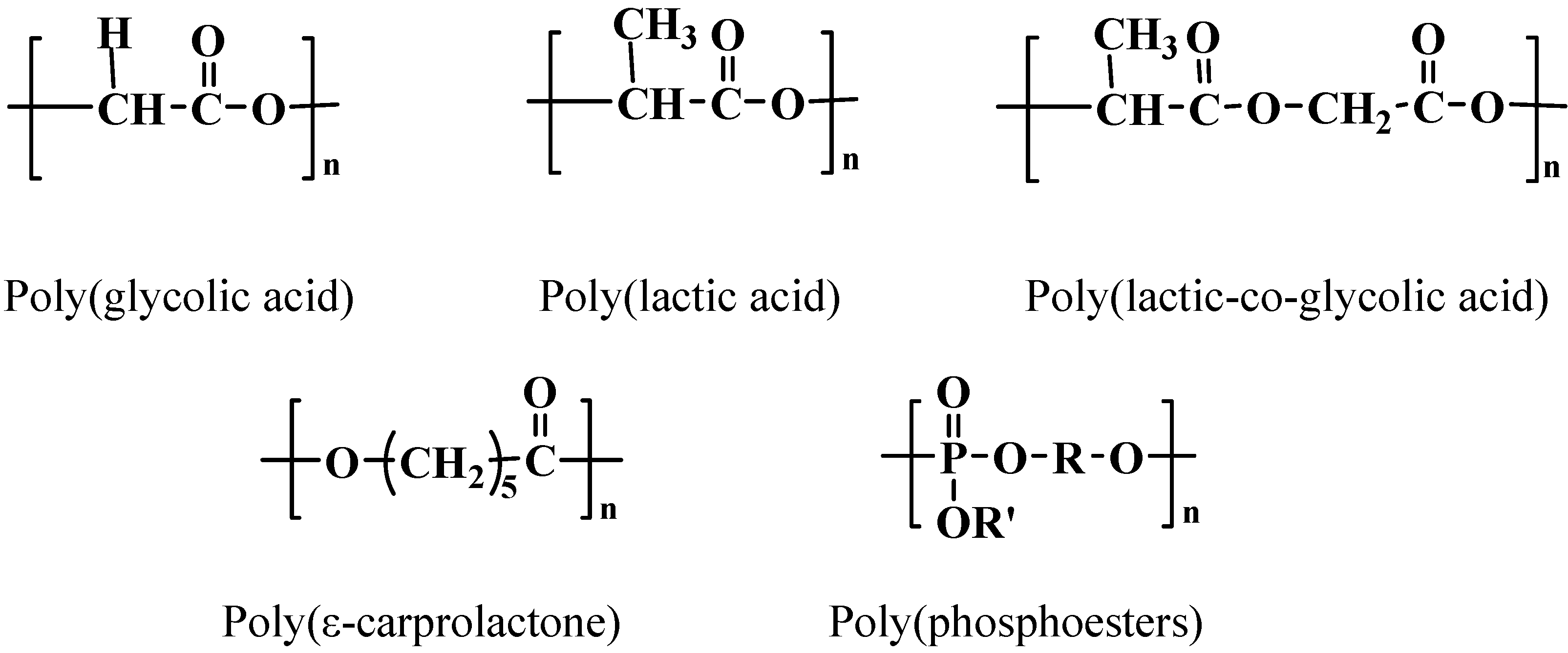
Aliphatic polyesters have attracted significant interest as drug carriers due to their biocompatibility and biodegradability [7,9,15,22,56,57]. This class of polymers degrade via the hydrolytic cleavage of the ester bonds in their backbone, whereas the role of enzymatic involvement in biodegradation is unclear. Chemical structures of the representative polyesters are shown in Figure 3. Poly(lactic-co-glycolic acid) (PLGA) copolymers have been most widely used because their degradation rate and mechanical properties can be precisely controlled by varying the lactic acid/glycolic acid ratio and by altering the molecular weight of the polymers.
Figure 3.
Chemical structures of polyesters.

PLGA polymers are cleaved into monomeric acids (i.e., lactic and glycolic acids) which are consequently eliminated from the body as carbon dioxide and water. The degradation rate of PLGA is critical for determining the release rate of the encapsulated drug and depends on the crystallinity, hydrophobicity, and molecular weight of the polymer [58,59]. In general, glycolic acid-rich PLGA copolymers (up to 70%) are amorphous in nature and degrade more rapidly. As the molecular weight of the polymer decreases, the degradation becomes faster because of the higher content of carboxylic groups at the end of polymer chain which accelerate the acid-catalyzed degradation. The PLGA-based microparticles undergo bulk degradation. The PLGA matrix undergoes random chain scission while preserving the original shape and mass until significant degradation (~ 90%) has occurred. In spite of these promising characteristics of biodegradation, recent studies have demonstrated that PLGA copolymers significantly affect the stability and biological activity of the drugs (e.g., peptide and proteins), primarily due to the hydrophobicity of the polymers and the presence of acidic degradation products
Poly(ε-caprolactone) (PCL) is a biodegradable, semicrystalline polymer having a low glass transition temperature (~60 oC). A number of drugs have been encapsulated using PCL. Due to its crystallinity and hydrophobicity, degradation of PCL is very slow, rendering it suitable for long-term delivery over a period of more than one year [62,63]. It has the ability to form compatible blends with other polymers, which provides opportunities to manipulate the drug release rate from microparticles [63]. The PCL-based devices maintain their shape and weight during the initial phase of biodegradation, where the molecular weight decreases by up to 5000 through bulk hydrolysis of the ester bonds. The second phase of PCL degradation is characterized by the onset of weight loss because the continuous chain cleavage produces a fragment small enough to diffuse out of the polymer matrix. On the other hand, the hydrolysis rate is known to decrease at the second phase, due to the increased crystallinity.
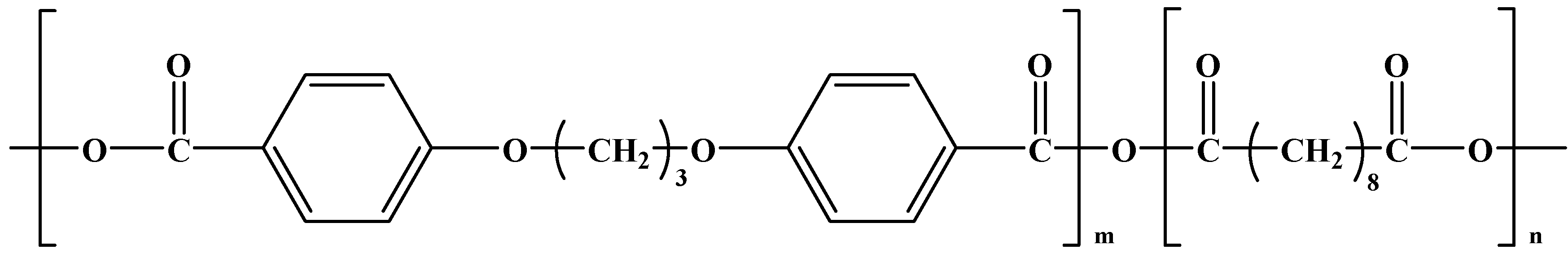
Poly(phosphoesters) (PPEs) have been used recently for delivery of low molecular weight drugs as well as high molecular weight proteins and DNA [5,64,65]. This type of polymer degrades under the physiological conditions via hydrolysis or enzymatic cleavage of the phosphate bonds in the backbone. Since their chemical structure can be tailored by varying R and R’ in Figure 3 during synthesis, it is possible to obtain PPEs with a wide range of physicochemical properties. In particular, by choosing biocompatible building blocks of the polymer, degradation products of PPEs can have minimal toxic effects and good biocompatibility. The degradation rate of PPEs is controllable by the percentage of the phosphate content in the backbone: The degradation rate increases with increasing the phosphate content of the polymer. In contrast to other polyesters, PPEs are known to degrade by a combined mechanism of surface erosion and bulk degradation [66]. Recent studies have demonstrated that PPE-based microparticles are promising for protein delivery because they don’t generate acidic environments [5,65,67].
Poly(ortho esters)
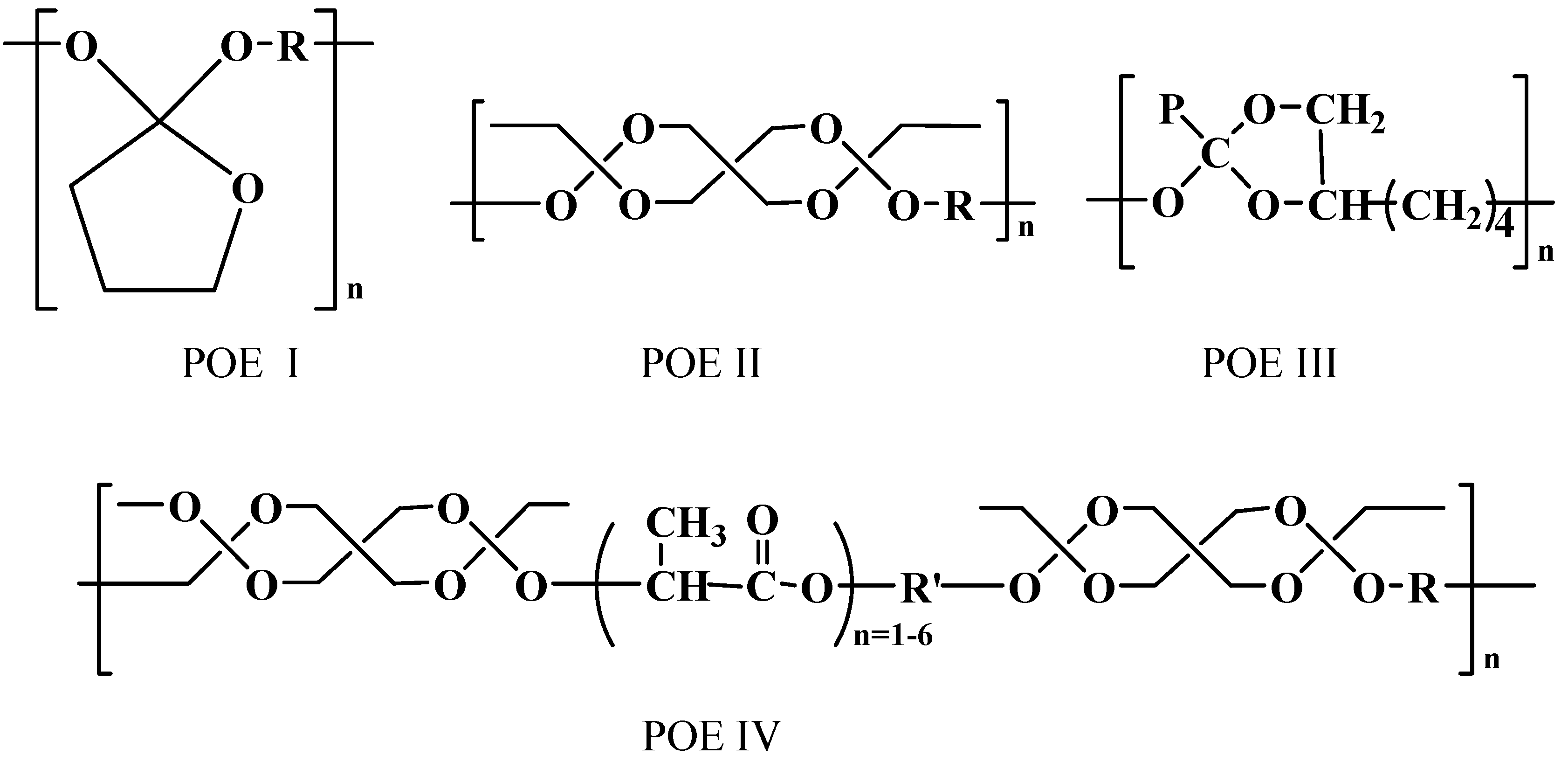
Since the 1970s, poly(ortho esters) (POEs) have evolved through four families as biodegradable polymers [68,69,70]: POE I, POE II, POE III, and POE IV. The general chemical structure of each family is shown in Figure 4. In contrast to polyesters which degrade homogeneously throughout the polymer matrix, POEs undergo surface erosion because of high hydrophobicity and water impermeability. Therefore, depending on the surface erosion rate of the POEs, the drug-loaded device may release the drug at a constant rate without significant burst release [25,60,69].
Figure 4.
Chemical structures of poly(ortho esters).

POE I, developed at Alza Coporation, is hydrolyzed under aqueous environment, thus producing γ‑butyrolactone that is rapidly converted to γ-hydroxybutyric acid. Because the ortho ester linkage of this polymer is highly susceptible to the acids, it should be stabilized with a base such as Na2CO3 to prevent an uncontrolled, autocatalytic hydrolysis reaction. Such disadvantages of POE I have limited applications in biomedical applications.
POE II, developed at the Stanford Research Institute, has several advantages, compared with POE I [70]. Polymer synthesis is simple and highly reproducible. The molecular weight of the polymer can be readily controlled by adjusting the stoichiometry. The initial product of the polymer hydrolysis is neutral, and thus it is not necessarily to use a basic excipient. However, the polymers belonging to this family are extremely hydrophobic, which limits the access of water to the hydrolytically labile ortho ester linkage. Hence, in order to achieve the increase in the surface erosion rate, it is necessary to incorporate acidic excipients into the polymer matrix. This limitation of POE II makes it difficult to design surface eroding devices because the presence of acidic excipients often accelerates the autocatalytic reaction.
POE III, developed at the Stanford Research Institute, is a semisolid material at room temperature. This semisolid material enables to prepare the injectable drug delivery system by a simple mixing with the therapeutic agents without the need of using organic solvents or elevated temperatures. In addition, no autocatalysis occurs during degradation, since the initial hydrolysis generates one or more isomeric monoesters. The ortho ester bonds of the polymer are only sensitive to the acidic products. Despite these advantages of POE III, its biomedical applications have been limited due to difficulties in the synthesis and poor reproducibility of the synthesized polymers [70].
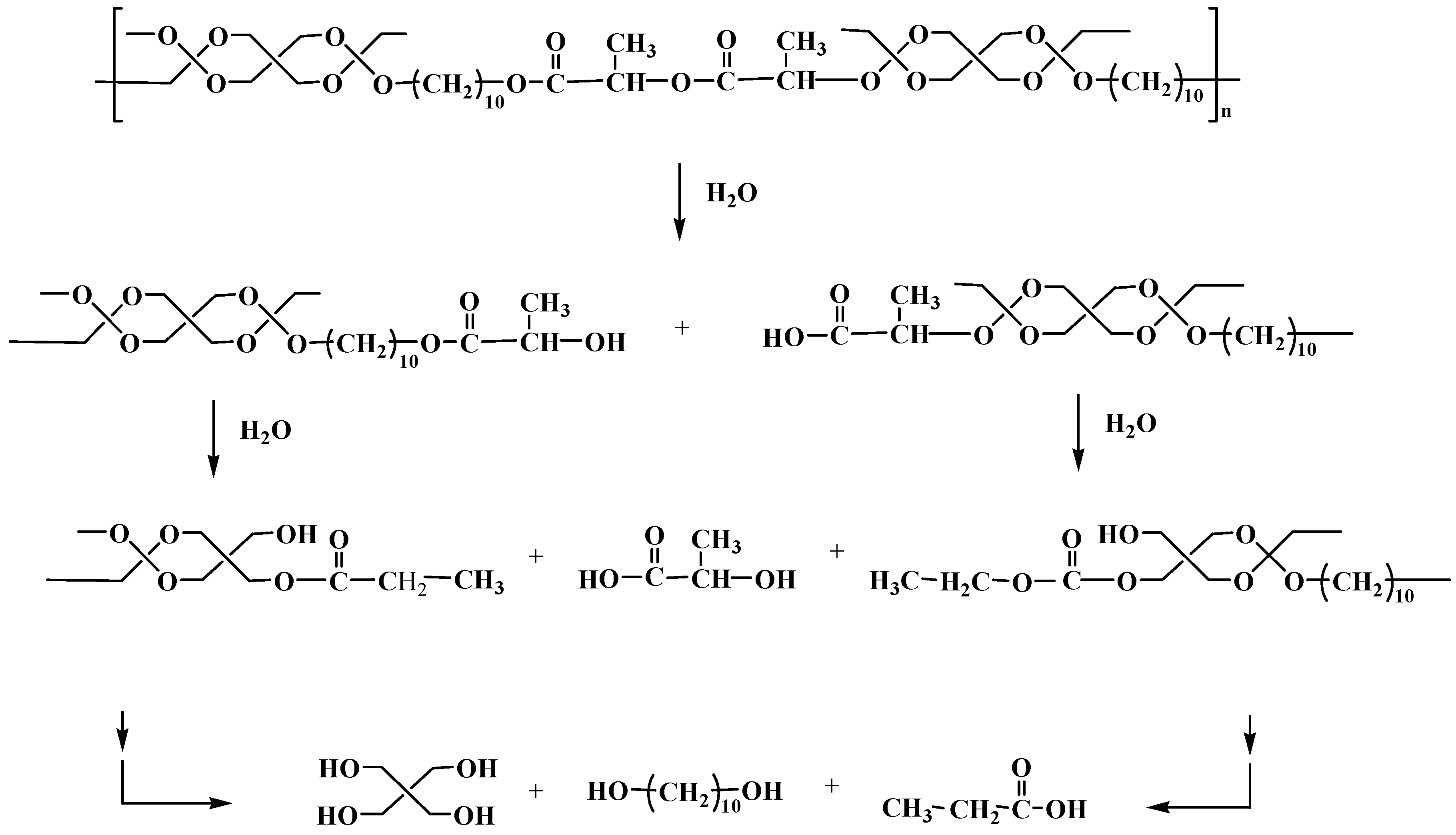
Figure 5.
Hydrolysis pathway of POE IV under aqueous environments.

POE IV, recently developed by Heller et al. [71], is a modified POE II that can be used without acidic excipients. To surmount the disadvantages of POE II, such as extreme hydrophobicity and stability, a short segment based on lactic acid or glycolic acid is incorporated into the backbone of POE IV. Such segments are included as latent acids which catalyze hydrolysis of the ortho ester linkages in the polymer backbone. The erosion rate of the polymer can be precisely controlled by adjusting the content of such segments. When POE IV is exposed to an aqueous environment, hydrolysis proceeds in three consecutive steps, as shown in Figure 5. First, carboxylic acid-terminated polymer fragments are produced by hydrolysis of the lactic acid or glycolic acid segment in the polymer backbone. Second, free α-hydroxy acids are generated and they catalyze hydrolysis of the ortho ester linkages. Third, the ortho esters are cleaved into the diol and pentaerythritol dipropionate, followed by ester hydrolysis to produce pentaerythritol and propionic acid.
The erosion process of POE IV-based devices is primarily confined to the surface layers, and this property provides a number of advantages [68,69,70,71]. For example, it is possible to control the release behavior of the drug by manipulating the surface erosion rate of the polymer. When the drug release is completed, the device is also removed because the drug release and the surface erosion of the polymer take place simultaneously. In addition, the acidic degradation products are readily diffused out of the device, due to principal hydrolysis on the surface layer; this prevents the generation of acidic environments in the bulk of the microparticles.
Polyanhydrides
Over the past two decades, polyanhydrides have been investigated for short-term controlled delivery of the bioactive agents because they exhibit rapid degradation in vivo and have limited mechanical properties [23,24,72]. Polyanhydrides are the hydrophobic polymers with hydrolytically labile anhydride linkages, in which the degradation rate can be manipulated by varying polymer composition; numerous diacids are available for the design of the polymer with desired physicochemical properties. In general, this class of polymers show minimal inflammatory reaction in vivo and degrade into monomeric acids as non-mutagenic and non-cytotoxic products [73,74,75]. Polyanhydrides are known to undergo surface erosion, a desired property to attain near zero-order drug release profile [76]. Degradation of polyanhydrides depends on the rate of the water uptake, determined by hydrophilicity and crystallinity of the polymer. Recently, due to their biocompatibility and biodegradability, the Food and Drug Administraion has approved the use of the polyanhydride derived from sebacic acid and 1,3-bis(p-carboxyphenoxy) propane as the carrier of antitumor agents for the treatment of brain cancer (see Figure 6 for its chemical structure) [77]. One drawback of polyanhydrides is that most of them have to be stored at frozen state under the anhydrous condition because of the hydrolytic instability of the anhydride bond.
Figure 6.
Chemical structure of poly[bis(p-carboxyphenoxy) propane-co-sebacic acid]
Polyphosphazenes
Polyphosphazenes are one of the most versatile and rapidly developing class of biomedical polymers [29,60]. They are typically synthesized as linear polymers, composed of an inorganic backbone with nitrogen and phosphorous atoms. Since the synthetic method of the linear polydichlorophosphazene (PDPP) was found in 1965 [78], a number of biodegradable poly-phosphazenes have been developed by reacting the highly reactive phosphorus-chlorine bonds of PDPP with alkoxide, primary (or secondary) amines, and organometallic reagents [29]. Because there are numerous substituents capable of being introduced into the backbone, a broad spectrum of polyphosphazenes can be synthesized by choosing the type and ratios of appropriate side groups. When exposed to an aqueous solution, these polymers are cleaved into nontoxic, low molecular weight products such as phosphates, ammonia and the corresponding side groups. They can degrade by both surface and bulk erosion, depending on the lability of the bond and hydrophobicity of the polymer [29]. A few examples, synthesized for biomedical applications, are polyposphazene-bearing amino acid ester [28,79], imidazole [28,80], glucosyl amino [81], glycolic acid ester [82], and lactic acid ester side groups [82].
Natural polymers
Although a number of synthetic biodegradable polymers have been developed for biomedical applications, the use of natural biodegradable polymers remains attractive because of their abundance in nature, good biocompatibility, and ability to be readily modified by the simple chemistry. A majority of drug delivery systems using natural polymers have been based on proteins (e.g., collagen, gelatin, and albumin) and polysaccharides (e.g., starch, dextran, hyaluronic acid, and chitosan). Applications of proteins to delivery of protein drugs have been limited due to their poor mechanical properties, low elasticity, possible occurrence of an antigenic response, and the high cost [19]. On the other hand, polysaccharides have attracted increasing interest as the drug carriers because they are commercially available at low cost, are readily modified by simple chemical reactions for specific applications, and they exhibit a broad range of physicochemical properties. For example, chitosan, primarily composed of 2-amino-2-deoxy-β-D-glucopyranose (D-glucosamine), is obtained from chitin, the second most abundant natural polysaccharide. Chitosan and its derivatives have showed excellent biocompatibility, biodegradability, low immunogenicity, and biological activities [83,84]. In particular, their biodegradability can be precisely controlled by modifying the structures with acetic anhydride [84,85,86]. Since they contain primary amino groups in the main backbone that make their surfaces positive in the biological fluid, biodegradable microparticles can be readily prepared by treating them with a variety of biocompatible polyanionic substances such as sulfate, citrate, and tripolyphosphate [87,88]. These unique features of chitosan have stimulated development of delivery systems for a wide range of biological agents [87,88,89,90,91,92,93].
Conclusions
Biodegradable microparticles allow the drug release to be accurately tuned for the treatment of the specific disease through the appropriate choice and formulation of specific drugs and polymers. Based on various microencapsultion techniques, microparticles can be designed for optimum delivery of a selected bioactive agent. The resulting microparticles may offer the ability to improve the stability of therapeutic agents against hydrolytic or enzymatic degradation, to augment the therapeutic effect by releasing the drug into the specific site, and to sustain the therapeutic effect in the target site. Many synthetic and natural biodegradable polymers present exciting opportunities in tailor-making the microparticle formulations for long-term drug release with specific release rates.
Acknowledgments
This study was supported in part by National Institute of Health through GM 67044 and EB003584.
References
- Pekarek, K. J.; Jacob, J. S.; Mathiowitz, E. Double-walled polymer microspheres for controlled drug release. Nature 1994, 367, 258–260. [Google Scholar] [PubMed]
- Jeong, B.; Bae, Y. H.; Lee, D. S.; Kim, S. W. Biodegradable block copolymers as injectable drug-delivery systems. Nature 1997, 388, 860–862. [Google Scholar] [PubMed]
- Ulbrich, K.; Pechar, M.; Strohalm, J.; Subr, V.; Rihova, B. Synthesis of biodegradable polymers for controlled drug release. Ann. N. Y. Acad. Sci. 1997, 831, 47–56. [Google Scholar] [PubMed]
- Hejazi, R.; Amiji, M. Chitosan-based gastrointestinal delivery systems. J. Control. Release 2003, 89, 151–165. [Google Scholar] [PubMed]
- Zhao, Z.; Wang, J.; Mao, H. Q.; Leong, K. W. Polyphosphoesters in drug and gene delivery. Adv. Drug Deliv. Rev. 2003, 55, 483–499. [Google Scholar] [CrossRef]
- Cicek, H.; Tuncel, A.; Tuncel, M.; Piskin, E. Degradation and drug release characteristics of monosize polyethylcyanoacrylate microspheres. J. Biomater. Sci. Polym. Ed. 1995, 6, 845–856. [Google Scholar] [CrossRef] [PubMed]
- Mi, F. L.; Lin, Y. M.; Wu, Y. B.; Shyu, S. S.; Tsai, Y. H. Chitin/PLGA blend microspheres as a biodegradable drug-delivery system: phase-separation, degradation and release behavior. Biomaterials 2002, 23, 3257–3267. [Google Scholar] [CrossRef]
- Zhang, Y.; Chu, C. C. In vitro release behavior of insulin from biodegradable hybrid hydrogel networks of polysaccharide and synthetic biodegradable polyester. J. Biomater. Appl. 2002, 16, 305–325. [Google Scholar] [PubMed]
- Abraham, G. A.; Gallardo, A.; San Roman, J.; Fernandez-Mayoralas, A.; Zurita, M.; Vaquero, J. Polymeric matrices based on graft copolymers of PCL onto acrylic backbones for releasing antitumoral drugs. J. Biomed. Mater. Res. 2003, 64A, 638–647. [Google Scholar] [CrossRef]
- Calandrelli, L.; De Rosa, G.; Errico, M. E.; La Rotonda, M. I.; Laurienzo, P.; Malinconico, M.; Oliva, A.; Quaglia, F. Novel graft PLLA-based copolymers: potential of their application to particle technology. J Biomed Mater Res 2002, 62, 244–253. [Google Scholar] [PubMed]
- Liu, J.; Xiao, Y.; Allen, C. Polymer-drug compatibility: a guide to the development of delivery systems for the anticancer agent, ellipticine. J. Pharm. Sci. 2004, 93, 132–143. [Google Scholar] [PubMed]
- Chen, B. H.; Lee, D. J. Slow release of drug through deformed coating film: effects of morphology and drug diffusivity in the coating film. J. Pharm. Sci. 2001, 90, 1478–1496. [Google Scholar] [PubMed]
- Tunon, A.; Grasjo, J.; Alderborn, G. Effect of intragranular porosity on compression behaviour of and drug release from reservoir pellets. Eur. J. Pharm. Sci. 2003, 19, 333–344. [Google Scholar] [CrossRef] [PubMed]
- Fulzele, S. V.; Satturwar, P. M.; Kasliwal, R. H.; Dorle, A. K. Preparation and evaluation of microcapsules using polymerized rosin as a novel wall forming material. J. Microencapsul. 2004, 21, 83–89. [Google Scholar] [PubMed]
- Jain, R. A. The manufacturing techniques of various drug loaded biodegradable poly(lactide-co-glycolide) (PLGA) devices. Biomaterials 2000, 21, 2475–2490. [Google Scholar] [CrossRef]
- Berkland, C.; King, M.; Cox, A.; Kim, K.; Pack, D. W. Precise control of PLG microsphere size provides enhanced control of drug release rate. J. Control. Release 2002, 82, 137–147. [Google Scholar] [CrossRef] [PubMed]
- Felder Ch, B.; Blanco-Prieto, M. J.; Heizmann, J.; Merkle, H. P.; Gander, B. Ultrasonic atomization and subsequent polymer desolvation for peptide and protein microencapsulation into biodegradable polyesters. J. Microencapsul. 2003, 20, 553–567. [Google Scholar] [CrossRef]
- Kiyoyama, S.; Shiomori, K.; Kawano, Y.; Hatate, Y. Preparation of microcapsules and control of their morphology. J. Microencapsul. 2003, 20, 497–508. [Google Scholar] [PubMed]
- Sinha, V. R.; Trehan, A. Biodegradable microspheres for protein delivery. J. Control. Release 2003, 90, 261–280. [Google Scholar] [PubMed]
- Sinha, V. R.; Goyal, V.; Bhinge, J. R.; Mittal, B. R.; Trehan, A. Diagnostic microspheres: an overview. Crit Rev Ther Drug Carrier Syst 2003, 20, 431–460. [Google Scholar] [PubMed]
- Wang, J.; Chua, K. M.; Wang, C. H. Stabilization and encapsulation of human immunoglobulin G into biodegradable microspheres. J. Colloid Interface Sci. 2004, 271, 92–101. [Google Scholar] [PubMed]
- Kissel, T.; Li, Y.; Unger, F. ABA-triblock copolymers from biodegradable polyester A-blocks and hydrophilic poly(ethylene oxide) B-blocks as a candidate for in situ forming hydrogel delivery systems for proteins. Adv. Drug. Deliv. Rev. 2002, 54, 99–134. [Google Scholar] [PubMed]
- Tabata, Y.; Gutta, S.; Langer, R. Controlled delivery systems for proteins using polyanhydride microspheres. Pharm. Res. 1993, 10, 487–496. [Google Scholar] [CrossRef]
- Kipper, M. J.; Shen, E.; Determan, A.; Narasimhan, B. Design of an injectable system based on bioerodible polyanhydride microspheres for sustained drug delivery. Biomaterials 2002, 23, 4405–4412. [Google Scholar] [PubMed]
- Lin, Y. H.; Vasavada, R. C. Studies on microencapsulation of 5-fluorouracil with poly(ortho ester) polymers. J. Microencapsul. 2000, 17, 1–11. [Google Scholar] [PubMed]
- Deng, J. S.; Li, L.; Tian, Y.; Ginsburg, E.; Widman, M.; Myers, A. In vitro characterization of polyorthoester microparticles containing bupivacaine. Pharm. Dev. Technol. 2003, 8, 31–38. [Google Scholar] [PubMed]
- Wang, C.; Ge, Q.; Ting, D.; Nguyen, D.; Shen, H. R.; Chen, J.; Eisen, H. N.; Heller, J.; Langer, R.; Putnam, D. Molecularly engineered poly(ortho ester) microspheres for enhanced delivery of DNA vaccines. Nat. Mater. 2004, 3, 190–196. [Google Scholar] [PubMed]
- Veronese, F. M.; Marsilio, F.; Lora, S.; Caliceti, P.; Passi, P.; Orsolini, P. Polyphosphazene membranes and microspheres in periodontal diseases and implant surgery. Biomaterials 1999, 20, 91–98. [Google Scholar] [CrossRef]
- Lakshmi, S.; Katti, D. S.; Laurencin, C. T. Biodegradable polyphosphazenes for drug delivery applications. Adv. Drug. Deliv. Rev. 2003, 55, 467–482. [Google Scholar] [PubMed]
- Kas, H. S. Chitosan: properties, preparations and application to microparticulate systems. J. Microencapsul. 1997, 14, 689–711. [Google Scholar] [PubMed]
- Felt, O.; Buri, P.; Gurny, R. Chitosan: a unique polysaccharide for drug delivery. Drug. Dev. Ind. Pharm. 1998, 24, 979–993. [Google Scholar] [CrossRef] [PubMed]
- Fukushima, S.; Kishimoto, S.; Takeuchi, Y.; Fukushima, M. Preparation and evaluation of o/w type emulsions containing antitumor prostaglandin. Adv. Drug. Deliv. Rev. 2000, 45, 65–75. [Google Scholar] [CrossRef]
- Okada, H.; Toguchi, H. Biodegradable microspheres in drug delivery. Crit. Rev. Ther. Drug Carrier Syst. 1995, 12, 1–99. [Google Scholar] [PubMed]
- Hombreiro Perez, M.; Zinutti, C.; Lamprecht, A.; Ubrich, N.; Astier, A.; Hoffman, M.; Bodmeier, R.; Maincent, P. The preparation and evaluation of poly(epsilon-caprolactone) microparticles containing both a lipophilic and a hydrophilic drug. J. Control. Release. 2000, 65, 429–438. [Google Scholar] [PubMed]
- Passerini, N.; Craig, D. Q. Characterization of ciclosporin A loaded poly (D,L lactide-co-glycolide) microspheres using modulated temperature differential scanning calorimetry. J. Pharm. Pharmacol. 2002, 54, 913–919. [Google Scholar] [CrossRef]
- Arshady, R. Preparation of biodegradable microspheres and microcapsules: 2. Polylactides and related polyesters. J. Control. Release 1991, 17, 1–22. [Google Scholar]
- Carrasquillo, K. G.; Stanley, A. M.; Aponte-Carro, J. C.; De Jesus, P.; Costantino, H. R.; Bosques, C. J.; Griebenow, K. Non-aqueous encapsulation of excipient-stabilized spray-freeze dried BSA into poly(lactide-co-glycolide) microspheres results in release of native protein. J. Control. Release 2001, 76, 199–208. [Google Scholar] [PubMed]
- Jiang, W.; Schwendeman, S. P. Stabilization of a model formalinized protein antigen encapsulated in poly(lactide-co-glycolide)-based microspheres. J Pharm Sci 2001, 90, 1558–1569. [Google Scholar] [PubMed]
- Jiang, W.; Schwendeman, S. P. Stabilization and controlled release of bovine serum albumin encapsulated in poly(D, L-lactide) and poly(ethylene glycol) microsphere blends. Pharm Res 2001, 18, 878–885. [Google Scholar] [PubMed]
- Crotts, G.; Park, T. G. Protein delivery from poly(lactic-co-glycolic acid) biodegradable microspheres: release kinetics and stability issues. J. Microencapsul. 1998, 15, 699–713. [Google Scholar] [PubMed]
- Okochi, H.; Nakano, M. Preparation and evaluation of w/o/w type emulsions containing vancomycin. Adv. Drug Deliv. Rev. 2000, 45, 5–26. [Google Scholar] [PubMed]
- Mallarde, D.; Boutignon, F.; Moine, F.; Barre, E.; David, S.; Touchet, H.; Ferruti, P.; Deghenghi, R. PLGA-PEG microspheres of teverelix: influence of polymer type on microsphere characteristics and on teverelix in vitro release. Int. J. Pharm. 2003, 261, 69–80. [Google Scholar] [PubMed]
- Johansen, P.; Moon, L.; Tamber, H.; Merkle, H. P.; Gander, B.; Sesardic, D. Immunogenicity of single-dose diphtheria vaccines based on PLA/PLGA microspheres in guinea pigs. Vaccine 1999, 18, 209–215. [Google Scholar] [PubMed]
- Ruiz, J. P.; Tissier, B.; Benoit, J. P. Microencapsulation of peptide: a study of the phase separation of poly(D,L-lactic acid-co-glycolic acid) copolymers 50/50 by silicone oil. Int. J. Pharm. 1989, 49, 69–77. [Google Scholar]
- Ruiz, J. M.; Busnel, J. P.; Benoit, J. P. Influence of average molecular weights of poly(DL-lactic acid-co-glycolic acid) copolymers 50/50 on phase separation and in vitro drug release from microspheres. Pharm. Res. 1990, 7, 928–934. [Google Scholar] [CrossRef]
- Yeo, Y.; Basaran, O. A.; Park, K. A new process for making reservoir-type microcapsules using ink-jet technology and interfacial phase separation. J. Control. Release 2003, 93, 161–173. [Google Scholar] [CrossRef]
- Yeo, Y.; Chen, A. U.; Basaran, O. A.; Park, K. Solvent exchange method: a novel microencapsulation technique using dual microdispensers. In Pharm. Res.; 2004. (In press) [Google Scholar]
- Sah, H. Protein behavior at the water/methylene chloride interface. J. Pharm. Sci. 1999, 88, 1320–1325. [Google Scholar] [PubMed]
- Kim, H. K.; Park, T. G. Microencapsulation of human growth hormone within biodegradable polyester microspheres: protein aggregation stability and incomplete release mechanism. Biotechnol. Bioeng. 1999, 65, 659–667. [Google Scholar] [PubMed]
- Murillo, M.; Gamazo, C.; Goni, M.; Irache, J.; Blanco-Prieto, M. Development of microparticles prepared by spray-drying as a vaccine delivery system against brucellosis. Int. J. Pharm. 2002, 242, 341–344. [Google Scholar] [PubMed]
- Blanco-Prieto, M. J.; Campanero, M. A.; Besseghir, K.; Heimgatner, F.; Gander, B. Importance of single or blended polymer types for controlled in vitro release and plasma levels of a somatostatin analogue entrapped in PLA/PLGA microspheres. J. Control. Release 2004, 96, 437–448. [Google Scholar] [CrossRef]
- Burke, P. A.; Klumb, L. A.; Herberger, J. D.; Nguyen, X. C.; Harrell, R. A.; Zordich, M. Poly(lactide-co-glycolide) microsphere formulations of darbepoetin alfa: spray drying is an alternative to encapsulation by spray-freeze drying. Pharm. Res. 2004, 21, 500–506. [Google Scholar] [CrossRef] [PubMed]
- Takada, S.; Uda, Y.; Toguchi, H.; Ogawa, Y. Application of a spray drying technique in the production of TRH-containing injectable sustained-release microparticles of biodegradable polymers. PDA J. Pharm. Sci. Technol. 1995, 49, 180–184. [Google Scholar] [PubMed]
- Johnson, O. L.; Jaworowicz, W.; Cleland, J. L.; Bailey, L.; Charnis, M.; Duenas, E.; Wu, C.; Shepard, D.; Magil, S.; Last, T.; Jones, A. J.; Putney, S. D. The stabilization and encapsulation of human growth hormone into biodegradable microspheres. Pharm. Res. 1997, 14, 730–735. [Google Scholar] [PubMed]
- Johnson, O. L.; Cleland, J. L.; Lee, H. J.; Charnis, M.; Duenas, E.; Jaworowicz, W.; Shepard, D.; Shahzamani, A.; Jones, A. J.; Putney, S. D. A month-long effect from a single injection of microencapsulated human growth hormone. Nat. Med. 1996, 2, 795–799. [Google Scholar] [PubMed]
- Bittner, B.; Morlock, M.; Koll, H.; Winter, G.; Kissel, T. Recombinant human erythropoietin (rhEPO) loaded poly(lactide-co-glycolide) microspheres: influence of the encapsulation technique and polymer purity on microsphere characteristics. Eur. J. Pharm. Biopharm. 1998, 45, 295–305. [Google Scholar] [PubMed]
- Morlock, M.; Kissel, T.; Li, Y. X.; Koll, H.; Winter, G. Erythropoietin loaded microspheres prepared from biodegradable LPLG-PEO-LPLG triblock copolymers: protein stabilization and in-vitro release properties. J. Control. Release. 1998, 56, 105–115. [Google Scholar] [PubMed]
- Witschi, C.; Doelker, E. Influence of the microencapsulation method and peptide loading on poly(lactic acid) and poly(lactic-co-glycolic acid) degradation during in vitro testing. J. Control. Release 1998, 51, 327–341. [Google Scholar] [PubMed]
- Jalil, R.; Nixon, J. R. Biodegradable poly(lactic acid) and poly(lactide-co-glycolide) microcapsules: problems associated with preparative techniques and release properties. J. Microencapsul. 1990, 7, 297–325. [Google Scholar] [PubMed]
- Andrianov, A. K.; Payne, L. G. Polymeric carriers for oral uptake of microparticulates. Adv. Drug Deliv. Rev. 1998, 34, 155–170. [Google Scholar] [PubMed]
- Sluzky, V.; Tamada, J. A.; Klibanov, A. M.; Langer, R. Kinetics of insulin aggregation in aqueous solutions upon agitation in the presence of hydrophobic surfaces. Proc. Natl. Acad. Sci. U S A 1991, 88, 9377–9381. [Google Scholar] [PubMed]
- Youan, B. B.; Benoit, M. A.; Baras, B.; Gillard, J. Protein-loaded poly(epsilon-caprolactone) microparticles. I. Optimization of the preparation by (water-in-oil)-in water emulsion solvent evaporation. J. Microencapsul. 1999, 16, 587–599. [Google Scholar] [PubMed]
- Sinha, V. R.; Bansal, K.; Kaushik, R.; Kumria, R.; Trehan, A. Poly-epsilon-caprolactone microspheres and nanospheres: an overview. Int. J. Pharm. 2004, 278, 1–23. [Google Scholar] [PubMed]
- Wang, J.; Mao, H. Q.; Leong, K. W. A novel biodegradable gene carrier based on polyphosphoester. J. Am. Chem. Soc. 2001, 123, 9480–9481. [Google Scholar] [PubMed]
- Xu, X.; Yu, H.; Gao, S.; Ma, H. Q.; Leong, K. W.; Wang, S. Polyphosphoester microspheres for sustained release of biologically active nerve growth factor. Biomaterials 2002, 23, 3765–3772. [Google Scholar] [PubMed]
- Wang, S.; Wan, A. C.; Xu, X.; Gao, S.; Mao, H. Q.; Leong, K. W.; Yu, H. A new nerve guide conduit material composed of a biodegradable poly(phosphoester). Biomaterials 2001, 22, 1157–1169. [Google Scholar] [PubMed]
- Xu, X.; Yee, W. C.; Hwang, P. Y.; Yu, H.; Wan, A. C.; Gao, S.; Boon, K. L.; Mao, H. Q.; Leong, K. W.; Wang, S. Peripheral nerve regeneration with sustained release of poly(phosphoester) microencapsulated nerve growth factor within nerve guide conduits. Biomaterials 2003, 24, 2405–2412. [Google Scholar] [PubMed]
- Heller, J. Poly(ortho esters). Adv. Polym. Sci. 1993, 107, 41–92. [Google Scholar]
- Heller, J.; Barr, J.; Ng, S. Y.; Shen, H. R.; Schwach-Abdellaoui, K.; Einmahl, S.; Rothen-Weinhold, A.; Gurny, R.; Emmahl, S. Poly(ortho esters) - their development and some recent applications. Eur. J. Pharm. Biopharm. 2000, 50, 121–128. [Google Scholar] [PubMed]
- Heller, J.; Barr, J.; Ng, S. Y.; Abdellauoi, K. S.; Gurny, R. Poly(ortho esters): synthesis, characterization, properties and uses. Adv. Drug Deliv. Rev. 2002, 54, 1015–1039. [Google Scholar] [CrossRef]
- Ng, S. Y.; Heller, J. Synthesis and erosion studies of self-catalyzed poly(orthoester)s. Macromolecules 1997, 30, 770–772. [Google Scholar]
- Uhrich, K. E.; Cannizzaro, S. M.; Langer, R.; Shakesheff, K. M. Polymeric systems for controlled drug release. Chem. Rev. 1999, 99, 3181–3198. [Google Scholar] [PubMed]
- Leong, K. W.; Kost, J.; Mathiowitz, E.; Langer, R. Polyanhydrides for controlled release of bioactive agents. Biomaterials 1986, 7, 364–371. [Google Scholar] [PubMed]
- Leong, K. W.; D'Amore, P. D.; Marletta, M.; Langer, R. Bioerodible polyanhydrides as drug-carrier matrices. II. Biocompatibility and chemical reactivity. J Biomed Mater Res 1986, 20, 51–64. [Google Scholar] [CrossRef]
- Kumar, N.; Langer, R. S.; Domb, A. J. Polyanhydrides: an overview. Adv. Drug Deliv. Rev. 2002, 54, 889–910. [Google Scholar] [PubMed]
- Katti, D. S.; Lakshmi, S.; Langer, R.; Laurencin, C. T. Toxicity, biodegradation and elimination of polyanhydrides. Adv. Drug. Deliv. Rev. 2002, 54, 933–961. [Google Scholar] [PubMed]
- Dang, W.; Daviau, T.; Brem, H. Morphological characterization of polyanhydride biodegradable implant gliadel during in vitro and in vivo erosion using scanning electron microscopy. Pharm. Res. 1996, 13, 683–691. [Google Scholar] [PubMed]
- Allcock, H. R.; Kugel, R. L.; Valan, K. J. Synthesis of high polymeric alkoxy and aryloxy phosphonitriles. J. Am. Chem. Soc. 1965, 87, 4216–4217. [Google Scholar] [CrossRef]
- Crommen, J. H.; Schacht, E. H.; Mense, E. H. Biodegradable polymers. I. Synthesis of hydrolysis-sensitive poly[(organo)phosphazenes]. Biomaterials 1992, 13, 511–520. [Google Scholar] [PubMed]
- Laurencin, C. T.; Koh, H. J.; Neenan, T. X.; Allcock, H. R.; Langer, R. Controlled release using a new bioerodible polyphosphazene matrix system. J. Biomed. Mater. Res. 1987, 21, 1231–1246. [Google Scholar] [PubMed]
- Allcock, H. R.; Pucher, A. G. Polyphosphazenes with glucosyl and methyl amino, trifluoroethoxy, phenoxy, or (methoxyethoxy)ethoxy side groups. Macromolecules 1991, 24, 23–34. [Google Scholar]
- Allcock, H. R.; Pucher, A. G.; Scopelianos, A. G. Synthesis of poly(organophosphazenes) with glycolic acid ester and lactic acid ester side groups: prototypes for new bioerodible polymers. Macromolecules 1994, 27, 1–4. [Google Scholar]
- Hirano, S. Chitin and chitosan as novel biotechnological materials. Polym. Int. 1999, 48, 732–734. [Google Scholar]
- Park, J. H.; Cho, Y. W.; Chung, H.; Kwon, I. C.; Jeong, S. Y. Synthesis and characterization of sugar-bearing chitosan derivatives: aqueous solubility and biodegradability. Biomacromolecules 2003, 4, 1087–1091. [Google Scholar] [PubMed]
- Sashiwa, H.; Saimoto, H.; Shigemasa, Y.; Ogawa, R.; Tokura, S. Lysozyme susceptibility of partially deacetylated chitin. Int. J. Biol. Macromol. 1990, 12, 295–296. [Google Scholar] [CrossRef] [PubMed]
- Hirano, S.; Tsuchida, H.; Nagao, N. N-acetylation in chitosan and the rate of its enzymic hydrolysis. Biomaterials 1989, 10, 574–576. [Google Scholar] [CrossRef]
- Kim, S. E.; Park, J. H.; Cho, Y. W.; Chung, H.; Jeong, S. Y.; Lee, E. B.; Kwon, I. C. Porous chitosan scaffold containing microspheres loaded with transforming growth factor-beta1: implications for cartilage tissue engineering. J. Control. Release 2003, 91, 365–374. [Google Scholar] [PubMed]
- Sinha, V. R.; Singla, A. K.; Wadhawan, S.; Kaushik, R.; Kumria, R.; Bansal, K.; Dhawan, S. Chitosan microspheres as a potential carrier for drugs. Int. J. Pharm. 2004, 274, 1–33. [Google Scholar] [PubMed]
- Jameela, S. R.; Kumary, T. V.; Lal, A. V.; Jayakrishnan, A. Progesterone-loaded chitosan microspheres: a long acting biodegradable controlled delivery system. J. Control. Release 1998, 52, 17–24. [Google Scholar] [PubMed]
- Chandy, T.; Rao, G. H.; Wilson, R. F.; Das, G. S. Development of poly(Lactic acid)/chitosan co-matrix microspheres: controlled release of taxol-heparin for preventing restenosis. Drug. Deliv. 2001, 8, 77–86. [Google Scholar] [CrossRef] [PubMed]
- Lee, J. E.; Kim, K. E.; Kwon, I. C.; Ahn, H. J.; Lee, S. H.; Cho, H.; Kim, H. J.; Seong, S. C.; Lee, M. C. Effects of the controlled-released TGF-beta 1 from chitosan microspheres on chondrocytes cultured in a collagen/chitosan/glycosaminoglycan scaffold. Biomaterials 2004, 25, 4163–4173. [Google Scholar] [PubMed]
- Cho, B. C.; Kim, J. Y.; Lee, J. H.; Chung, H. Y.; Park, J. W.; Roh, K. H.; Kim, G. U.; Kwon, I. C.; Jang, K. H.; Lee, D. S.; Park, N. W.; Kim, I. S. The bone regenerative effect of chitosan microsphere-encapsulated growth hormone on bony consolidation in mandibular distraction osteogenesis in a dog model. J. Craniofac. Surg. 2004, 15, 299–311, discussion 312-293. [Google Scholar]
- Park, J. H.; Kwon, S.; Nam, J. O.; Park, R. W.; Chung, H.; Seo, S. B.; Kim, I. S.; Kwon, I. C.; Jeong, S. Y. Self-assembled nanoparticles based on glycol chitosan bearing 5-beta-cholanic acid for RGD peptide delivery. J. Control. Release. 2004, 95, 579–588. [Google Scholar] [CrossRef]
© 2005 by MDPI (http://www.mdpi.org). Reproduction is permitted for noncommercial purposes.
Share and Cite
MDPI and ACS Style
Park, J.; Ye, M.; Park, K. Biodegradable Polymers for Microencapsulation of Drugs. Molecules 2005, 10, 146-161. https://doi.org/10.3390/10010146
AMA Style
Park J, Ye M, Park K. Biodegradable Polymers for Microencapsulation of Drugs. Molecules. 2005; 10(1):146-161. https://doi.org/10.3390/10010146
Chicago/Turabian StylePark, J., M. Ye, and K. Park. 2005. "Biodegradable Polymers for Microencapsulation of Drugs" Molecules 10, no. 1: 146-161. https://doi.org/10.3390/10010146