9-Hydroxyfurodysinin-O-ethyl Lactone: A New Sesquiterpene Isolated from the Tropical Marine Sponge Dysidea arenaria


Department of Chemistry and Biomolecular Sciences, Macquarie University, Australia
*
Author to whom correspondence should be addressed.
Molecules 2005, 10(10), 1292-1297; https://doi.org/10.3390/10101292
Submission received: 28 March 2005
/
Accepted: 2 May 2005
/
Published: 31 October 2005
(This article belongs to the Special Issue 2004 RACI One-day Natural Products Group Symposium)
Abstract
:A new sesquiterpene, 9-hydroxyfurodysinin-O-ethyl lactone, has been isolated from a New Caledonian Dysidea arenaria, along with three known compounds. The possible incorporation of the ethyl ether from the extraction solvent is discussed.
Introduction
Marine sponges belonging to the genus Dysidea generally contain sesquiterpenes [1], polychlorinated amino acids [2,3] or polybrominated diphenyl ethers [4,5], although the latter two classes of compound have been shown to originate from the symbiotic cyanobacterium Oscillatoria spongeliae [6]. Herein, we describe the isolation, purification and structure elucidation of one new and three known compounds from a New Caledonian Dysidea arenaria.
Results and Discussion
A freeze-dried sample of Dysidea arenaria from New Caledonia was extracted exhaustively with aqueous ethanol, and the combined extracts were partitioned against light petroleum and then DCM. The light petroleum partition yielded furodysinin-O-ethyl lactone (1), which has been isolated previously from Dysidea tupha [7], while the DCM partition yielded the known polyhydroxylated sterol (2) [8], the known sesquiterpene, furodysinin lactone (3) [9], and a third compound (4) as a colorless oil. The high resolution ESI mass spectrum of (4) showed a signal at m/z 315.1568 ([M+Na]+), corresponding to a molecular formula of C17H24O4. The 1H NMR spectrum of (4) is almost identical to that of (1), except for the presence of an oxygen-bearing methine at δ 3.96 and an exchangeable broad doublet at δ 1.83, suggesting the addition of a hydroxyl group. This is supported by the presence of a broad stretch at 3364 cm-1 in the IR spectrum of (4). The absence of C9 methylene protons at δ 1.94 suggests that (4) is 9-hydroxyfurodysinin-O-ethyl lactone, which has not been reported previously in the literature. 1H and 13C NMR assignments for (4) are presented in Table 1.
![Molecules 10 01292 i001]()
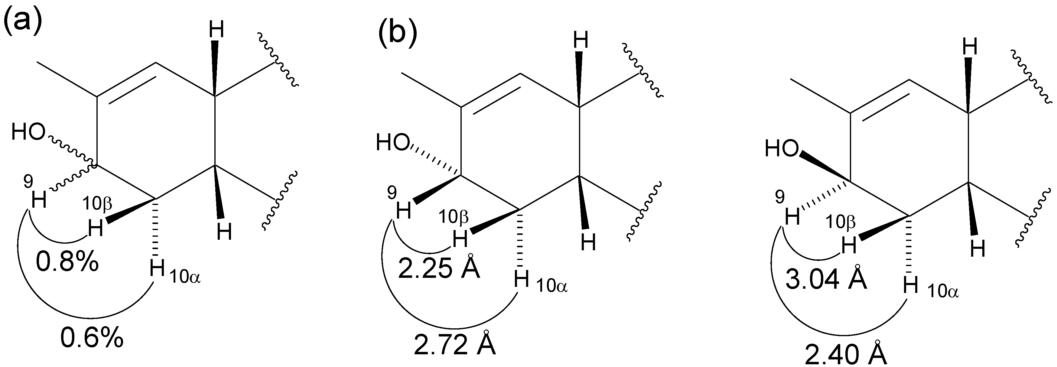
The relative stereochemistry of (4) at C9 was confirmed by selective 1D ROESY experiments, with a larger ROE enhancement observed for the H9-H10β correlation than for the H9-H10α correlation (Figure 1(a)). Molecular modeling of both the 9α-and 9β-hydroxy stereoisomers revealed the distance between H9 and H10α/β to be 2.72 Å / 2.25 Å in the 9α-hydroxy isomer and 2.40 Å / 3.04 Å in the 9β-hydroxy isomer (Figure 1(b)). Therefore, the observed difference in ROE enhancement can only be accommodated by the 9α-hydroxy isomer. This is in agreement with the relative stereochemistry assigned to the related compound, 9-hydroxyfurodysinin-O-methyl lactone, by Garson et al. [10].


| Atom | δ 1H (ppm) – J (Hz) | δ 13C (ppm) | HMBC | COSY |
|---|---|---|---|---|
| 1 | - | 173.1 | - | - |
| 2 | 5.81 (s) | 117.1 | 3, 4 | - |
| 3 | - | 169.4 | - | - |
| 4 | - | 107.1 | - | - |
| 5α | 1.42 (d; 13.7) | 38.7 | 5β, 6 | |
| 5β | 2.35 (dd; 13.7, 3.8) | 38.7 | 5α, 6 | |
| 6 | 2.81 (m) | 30.3 | 5α, 5β, 7, 11 | |
| 7 | 5.55 (dd; 5.6, 1.2) | 128.1 | 13 | 6 |
| 8 | - | 134.5 | - | - |
| 9 | 3.96 (bt; 4.0) | 68.1 | 13 | 10α, 10β |
| 10α | 1.35 (ddd; 14.7, 14.7, 4.4) | 27.8 | 9, 10β, 11 | |
| 10β | 1.77 (dm; 14.7) | 27.8 | 9, 10α, 11 | |
| 11 | 1.95 (ddd; 14.7, 4.4, 3.7) | 41.7 | 14, 15 | 6, 10α, 10β |
| 12 | - | 38.0 | - | - |
| 13 | 1.78 (s) | 20.6 | 7, 8, 9 | |
| 14 | 1.39 (s) | 25.6 | 1, 11, 12, 15 | |
| 15 | 1.25 (s) | 25.1 | 1, 11, 12, 14 | |
| 16a | 3.53 (dq; 8.9, 7.0) | 58.5 | 16b, 17 | |
| 16b | 3.22 (dq; 8.9, 7.0) | 58.5 | 16a, 17 | |
| 17 | 1.19 (t; 7.0) | 14.6 | 16 | 16a, 16b |
| OH | 1.83 (bd; 1.2) | - |
Figure 1.
(a) Percent enhancements for selected ROE correlations in (4). (b) Distances between H9 and H10α/β in the 9α and 9β isomers of (4), as determined by molecular modeling
Figure 1.
(a) Percent enhancements for selected ROE correlations in (4). (b) Distances between H9 and H10α/β in the 9α and 9β isomers of (4), as determined by molecular modeling

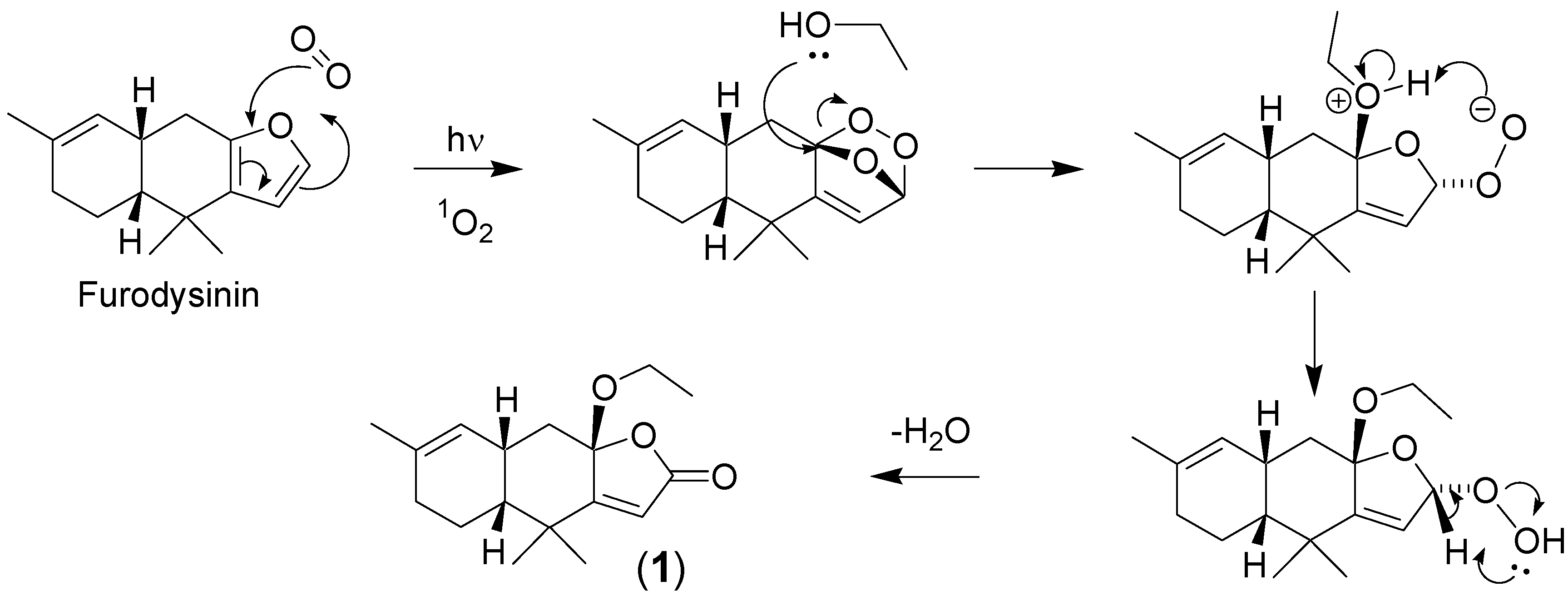
As ethyl ethers are uncommon in nature, it is likely that (1) and (4) are artifacts of the initial aqueous ethanol extraction, arising from a [4+2] cycloaddition reaction between a furan and singlet oxygen (Figure 2). The resulting ozonide can be attacked by ethanol to give an ethoxy hydroperoxide, which can then dehydrate to give a butenolide [11]. This mechanism is supported by the isolation of furodysinin hydroperoxide from the nudibranch Chromodoris funerea, which is known to feed on Dysidea spp. [12]. In addition, Faulkner and colleagues were able to synthesize furodysinin-O-methyl lactone by treating furodysinin with singlet oxygen at –78 °C and then quenching with methanol [12].
Figure 2.
Possible mechanism for the formation of furodysinin-O-ethyl lactone (1) from furodysinin via a [4+2] cycloaddition reaction with singlet oxygen.
Figure 2.
Possible mechanism for the formation of furodysinin-O-ethyl lactone (1) from furodysinin via a [4+2] cycloaddition reaction with singlet oxygen.

Conclusions
One new and three known compounds were isolated from a New Caledonian Dysidea arenaria. The appearance of ethoxy adducts highlights the advantages of using ethanol rather than methanol as an extraction solvent. Methyl ethers are common in nature, and hence it is difficult to determine whether a methoxy group in a natural product was incorporated biosynthetically or simply by reaction with the extraction solvent.
Experimental
General
A freeze-dried sample of a New Caledonian Dysidea arenaria was a kind gift from Prof. Dame Patricia Bergquist (University of Auckland). Solvents were obtained from Fronine (Australia) and were glass distilled before use. S-X3 Biobeads were obtained from Bio-Rad (USA). All other reagents were obtained from Aldrich (USA). NMR spectra were recorded on either a DPX-400 400 MHz or DRX-600K 600 MHz spectrometer (Bruker, Germany) in 5 mm Pyrex tubes (Wilmad, USA). High resolution ESI mass spectra were recorded on an APEXII FTICR spectrometer (Bruker, Germany). Low resolution ESI mass spectra were recorded on a Quattro-II triple quadrupole spectrometer (Fisons Instruments, USA). IR spectra were recorded on a Paragon PE1000 FTIR spectrometer (Perkin Elmer, USA). UV-Vis spectra were recorded on a Cary 1-Bio spectrophotometer (Varian, USA) in 1 cm matched quartz cuvettes (Selbys Scientific, Australia). HPLC was performed using a 600E solvent delivery system and a 490 programmable multi-wavelength detector (Waters, USA), on a 250 x 4.6 mm C18 column (Alltech, USA). Water was purified using a Milli-Q Ultrapure Water Purification System (Millipore, USA). Molecular modeling was performed using the Chem3D software package (Cambridgesoft, USA).
Extraction Procedure
A sample of freeze-dried Dysidea arenaria from New Caledonia (56 g) was extracted exhaustively with aqueous ethanol (70%; 6 x 100 mL), and the combined extracts were partitioned against light petroleum (3 x 200 mL) and DCM (3 x 200 mL), yielding 0.15 g and 0.32 g of residue respectively. The crude light petroleum extract was chromatographed on silica gel (gradient 0-100% ethyl acetate:light petroleum) and six fractions were collected. The 1H-NMR spectra and colors of fractions 1, 2, 3 and 5 suggested the presence of carotenoids, fatty acids, sterols and sterol endoperoxides respectively, and hence these fractions were not investigated further. Fraction 4 was further purified by reversed phase HPLC (analytical C18 column, 80:20 methanol:water), with a large UV active peak (254 nm) eluting after 11 min. The solvent was removed from this peak in vacuo, yielding furodysinin-O-ethyl lactone (1) as a white solid (1 mg, 0.002% dry weight). The crude DCM extract was subjected to gel permeation chromatography with S-X3 Biobeads (toluene), and 30 fractions were collected. On standing at room temperature overnight, a white solid precipitated from Fractions 7-11. This solid was collected at the pump and then recrystallized from toluene, yielding 9α,11α-epoxycholest-7-ene-3β,5α,6β,19-tetrol 6-acetate (2) as white crystals (20 mg, 0.04% dry weight). Fractions 14-28 were combined (TLC) and reduced to dryness in vacuo. The resulting residue was chromatographed on silica gel (gradient 0-100% ethyl acetate:light petroleum) and 30 fractions were collected. Fractions 22-25 were combined (TLC) and further purified by reversed phase HPLC (analytical C18 column, 65:35 methanol:water), with a large UV active peak (254 nm) eluting after 8 min. The solvent was removed from this peak under a stream of nitrogen, yielding 9-hydroxy-furodysinin-O-ethyl lactone (4) as a colorless oil (3 mg, 0.005% dry weight). A second UV active peak (254 nm) eluted after 16 min, which yielded furodysinin lactone (3) as a white solid (2 mg, 0.004% dry weight).
Spectral Data
Furodysinin-O-ethyl lactone (1): 1H-NMR (600 MHz, CDCl3) δ 5.78 (s, 1H, H2), 5.36 (d, 1H, H7, J = 5.4 Hz), 3.53 (dq, 1H, H16a, J = 8.9, 7.0 Hz), 3.22 (dq, 1H, H16b, J = 8.9, 7.0 Hz), 2.78 (m, 1H, H6), 2.35 (dd, 1H, H5β, J = 13.7, 3.8 Hz), 1.95 (m, 2H, H9), 1.65 (m, 2H, H10β, H11), 1.62 (s, 3H, H13), 1.57 (d, 1H, H5α, J = 13.7 Hz), 1.36 (s, 3H, H14), 1.23 (s, 3H, H15), 1.19 (t, 3H, H17, J = 7.0 Hz), 1.13 (m, 1H, 10α). MS (ESI+) m/z: 277 ([M+H]+).
9α,11α-epoxycholest-7-ene-3β,5α,6β,19-tetrol 6-acetate (2): 1H-NMR (400 MHz, CDCl3) δ 5.30 (dd, 1H, H7, J = 2.1, 2.0 Hz), 5.21 (dd, 1H, H6, J = 2.7, 2.1 Hz), 4.06 (m, 1H, H3), 3.99 (dd, 1H, H19a, J = 11.7, 2.7 Hz), 3.80 (dd, 1H, H19a, J = 11.7, 3.6 Hz), 3.40 (d, 1H, H11, J = 5.7 Hz), 2.36 (m, 1H, H14), 2.35 (s, 1H, OH), 2.20 (s, 1H, OH), 2.19 (dd, 1H, H12β, J = 15.8, 5.8 Hz), 2.16 (s, 3H, CH3C=O), 2.07 (ddd, 1H, H4α, J = 13.5, 4.7, 2.0 Hz), 2.0-1.0 (m, 17H, H1, H2, H15-H17, H20, H22-H25), 1.85 (d, 1H, H12α, J = 15.8 Hz), 1.79 (dd, 1H, H4β, J = 13.5, 4.7 Hz), 0.90 (d, 3H, H21, J = 6.3 Hz), 0.86 (d, 3H, H26, J = 6.7 Hz), 0.85 (d, 3H, H27, J = 6.6 Hz), 0.60 (s, 3H, H18). MS (ESI+) m/z: 491 ([M+H]+).
Furodysinin lactone (3): 1H-NMR (400 MHz, CDCl3) δ 5.70 (s, 1H, H2), 5.37 (dm, 1H, H7, J = 5.7 Hz), 2.82 (m, 1H, H6), 2.81 (s, 1H, OH), 2.29 (ddd, 1H, H5β, J = 13.9, 3.9, 0.9 Hz), 1.97 (m, 2H, H9), 1.75-1.62 (m, 2H, H10β, H11), 1.63 (dt, 3H, H13, J = 2.4, 1.0 Hz), 1.57 (d, 1H, H5α, J = 13.9 Hz), 1.41 (s, 3H, H14), 1.24 (s, 3H, H15), 1.13 (m, 1H, H10α). MS (ESI+) m/z: 249 ([M+H]+).
9-hydroxyfurodysinin-O-ethyl lactone (4): NMR data – See Table 1. HRMS (ESI+) m/z: 315.1568 ([M+Na]+), calcd. for C17H24O4+Na+ 315.1572. UV (MeOH) 199, 254 nm, ε 7400, 857 L·mol-1·cm-1. IR (neat film) 3364 (br), 2919 (s), 2854 (m), 1766 (s), 1712 (s), 1650 (m), 1535 (s), 1452 (m), 1366 (m), 1248 (m), 1020 (s), 924 (w), 662 (w) cm-1.
References and Notes
- Venkateswarlu, Y.; Ramesh, P.; Reddy, N.S. Nat. Prod. Sci. 1998, 4, 115–129.
- Unson, M.D.; Rose, C.B.; Faulkner, D.J.; Brinen, L.S.; Steiner, J.R.; Clardy, J. J. Org. Chem. 1993, 58, 6336–6343.
- Erickson, K.L.; Wells, R.J. Aust. J. Chem. 1982, 35, 31–38.
- Handayani, D.; Edrada, R.A.; Proksch, P.; Wray, V.; Witte, L.; Van Soest, R.W.M.; Kunzmann, A. Soedarsono. J. Nat. Prod. 1997, 60, 1313–1316. [Google Scholar] [CrossRef] [PubMed] [Green Version]
- Carte, B.; Faulkner, D.J. Tetrahedron 1981, 37, 2335–2339.
- Faulkner, D.J.; Unson, M.D.; Bewley, C.A. Pure Appl. Chem. 1994, 66, 1983–1990.
- Guella, G.; Mancini, I.; Guerriero, A.; Pietra, F. Helv. Chim. Acta 1985, 68, 1276–1282.
- Gunasekera, S.P.; Schmitz, F.J. J. Org. Chem. 1983, 48, 885–886.
- Grode, S.H.; Cardellina II, J.H. J. Nat. Prod. 1984, 47, 76–83.
- Cameron, G.M.; Stapleton, B.L.; Simonsen, S.M.; Brecknell, D.J.; Garson, M.J. Tetrahedron 2000, 56, 5247–5252.
- Gollnick, K.; Griesbeck, A. Tetrahedron 1985, 41, 2057–2068.
- Carté, B.; Kernan, M.R.; Barrabee, E.B.; Faulkner, D.J.; Matsumoto, G.K.; Clardy, J. J. Org. Chem. 1986, 51, 3528–3532.
- Sample Availability: Not available
© 2005 by MDPI (http:www.mdpi.org). Reproduction is permitted for noncommercial purposes.
Share and Cite
MDPI and ACS Style
Piggott, A.; Karuso, P. 9-Hydroxyfurodysinin-O-ethyl Lactone: A New Sesquiterpene Isolated from the Tropical Marine Sponge Dysidea arenaria. Molecules 2005, 10, 1292-1297. https://doi.org/10.3390/10101292
AMA Style
Piggott A, Karuso P. 9-Hydroxyfurodysinin-O-ethyl Lactone: A New Sesquiterpene Isolated from the Tropical Marine Sponge Dysidea arenaria. Molecules. 2005; 10(10):1292-1297. https://doi.org/10.3390/10101292
Chicago/Turabian StylePiggott, A., and P. Karuso. 2005. "9-Hydroxyfurodysinin-O-ethyl Lactone: A New Sesquiterpene Isolated from the Tropical Marine Sponge Dysidea arenaria" Molecules 10, no. 10: 1292-1297. https://doi.org/10.3390/10101292