Glycerolipids from a Sarcotragus Species Sponge

1
Guangdong Key Laboratory of Marine Materia Medica, South China Sea Institute of Oceanology, Chinese Academy of Sciences, Guangzhou 510-301, China
2
College of Pharmacy, Pusan National University, Busan 609-735, Korea
*
Author to whom correspondence should be addressed.
Molecules 2006, 11(9), 714-719; https://doi.org/10.3390/11090714
Submission received: 9 November 2005
/
Revised: 27 September 2006
/
Accepted: 28 September 2006
/
Published: 28 September 2006
Abstract
:One known and two new glycerolipids have been isolated from a Sarcotragus sp. marine sponge. The gross structures were established based on NMR and MS analysis.
Introduction
Marine sponges of the order Dictyoceratida are known to contain various linear furano-sesterpenes and their derivatives that display a wide range of interesting bioactivities [1,2]. Thirty five furano- and pyrroloterpenoids, three cyclitol derivatives and a macrolide have been isolated from Sarcotragus sp. (Dictyoceratida) collected from Korean waters [3,4,5,6,7]. During our continuing study on the cytotoxic compounds of Sarcotragus sp., three glycerolipids 1–3 have now been isolated. Compounds 2 and 3 have not been previously described. The isolation and elucidation of the gross structures of these compounds by COSY, HSQC, and HMBC experiments are described.
Results and Discussion
The methanol extract of the sponge displayed cytotoxicity against five human tumor cell lines (see Extraction and Isolation section of the Experimental) and also showed toxicity towards brine shrimp larvae (LD50, 93 μg/mL). Guided by the brine shrimp assay, the methanol extract was successively fractionated employing reversed-phase flash column chromatography and ODS HPLC to separate the causative components 1-3 (Figure 1) from the inactive fractions. Compound 1 was isolated as an amorphous solid. Its molecular formula was established as C18H38O3 based on MS and NMR spectral analyses. The FABMS of 1 showed the [M+H]+ ion at m/z 303, accompanied by the [M+Na]+ ion at m/z 325. Although this compound was previously identified in rabbit harderian glands by MS spectrometry [8], it has now been isolated for the first time from the sponge by HPLC and its spectroscopic data is herein described for the first time. The 1H-NMR spectrum exhibited signals for an isopropyl terminated aliphatic long chain (δ 0.87, d, J = 6.5 Hz, δC 22.3) and signals associated with the presence of a glycerol monoether moiety. Proton signals for three pairs of oxymethylene protons and one oxymethine were observed: a two proton AB multiplet (an apparent triplet of doublets, J = 6.5, 1.5 Hz) at δ 3.45, two AB doublets of doublets centered at δ 3.72 (J = 11.0, 4.5 Hz) and δ 3.49 (J = 11.0, 6.5 Hz), two AB doublets of doublets centered at δ 3.47 (J = 10.0, 5.0 Hz) and δ 3.40 (J = 10.0, 6.5 Hz) and a pseudo quintuplet at δ 3.74 for the methine proton. The 13C-NMR spectrum was in agreement with this structure and showed three oxymethylene carbons at δ 73.3 (C-1), δ 72.7 (C-1'), and δ 64.7 (C-3), and an oxymethine at δ 72.3 (C-2) [9]. The configuration at C-2 was established from the positive optical rotation, which is a general feature of long-chain 1-O-alkyl-sn-glycerols [10].
Figure 1.
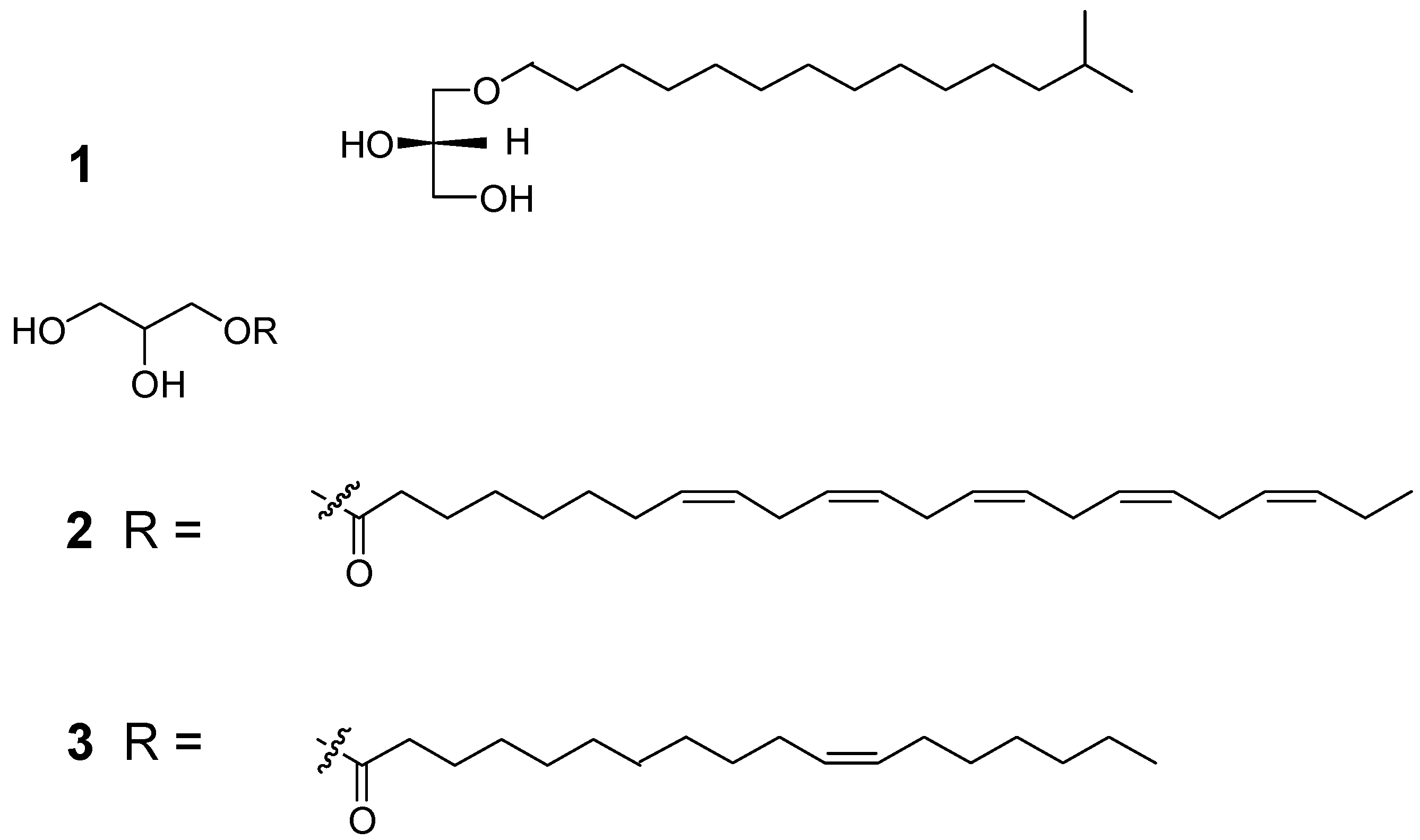
Compound 2 was isolated as a colorless oil. Its molecular formula was established as C26H44O4 based on MS and NMR spectral analyses (Table 1). The FABMS of 2 showed a [M+H]+ ion at m/z 421 accompanied by a [M+Na]+ ion at m/z 443. The 1H- and 1H-1H-COSY spectra showed signals at δ 0.97 (3H, t, J = 7.5 Hz) and δ 2.08 (4H, dq, J = 7.5, 7.5 Hz, H-7', H-22'); the latter was coupled with the signal at δ 5.36 (10H, m). In addition it displayed signals for ten olefinic carbons, eleven methylenes and a terminal methyl group. A ten proton multiplet at δ 5.36 and an eight-proton multiplet at δ 2.83 ppm indicated the presence of five non-conjugated double bonds and four doubly allylic methylenes, respectively. The Z nature of the olefinic double bonds was indicated by the allylic methylenes [5]. In addition to these signals two mutually coupled sets of ABX type protons [δ 4.14 (1H, dd, J = 11.5, 4.5 Hz), 4.05 (1H, dd, J = 11.5, 6.5), 3.81(1H, m), 3.54 (2H, p, J = 5.0, 2.5 Hz)] for a glycerol moiety were observed. Although the stereochemistry of the glycerol moiety has not been established at this time, 2 is a new monoacyl glycerol, to the best of our knowledge.

| Position | δH (mult, J) | δC |
| 1 | 4.14 (dd, 11.5, 4.5) 3.5) | 71.2 |
| 4.05(dd,11.5, 6.5) | ||
| 2 | 3.81 (m) | 71.0 |
| 3 | 3.54 (dd, 5.0, 2.5) | 73.1 |
| 1' | 175.4 | |
| 2' | 2.34 (t, 7.5) | 34.9 |
| 3' | 1.62 (quint, 8.0) | 25.9 |
| 4' | 1.28 - 1.39 (m) | 28.0 |
| 5' | 1.28 - 1.39 (m) | 29.8 |
| 6' | 1.28 - 1.39 (m) | 29.8 |
| 7' | 2.08 (m) | 28.0 |
| 8' | 5.36 (m) | 132.8 |
| 9' | 5.36 (m) | 130.9 |
| 10' | 2.83 (m) | 26.6 |
| 11' | 5.36 (m) | 129.5 |
| 12' | 5.36 (m) | 129.4 |
| 13' | 2.83 (m) | 26.6 |
| 14' | 5.36 (m) | 129.0 |
| 15' | 5.36 (m) | 129.0 |
| 16' | 2.83 (m) | 26.6 |
| 17' | 5.36 (m) | 129.0 |
| 18' | 5.36 (m) | 129.0 |
| 19' | 2.83 (m) | 26.4 |
| 20' | 5.36 (m) | 128.9 |
| 21' | 5.36 (m) | 128.2 |
| 22' | 2.08 (m) | 21.5 |
| 23' | 0.97 (t, 7.5) | 14.6 |
Compound 3 was isolated as a colorless oil. The molecular formula was established as C21H40O4, based on MS and NMR spectral analyses. The FABMS of 3 showed a [M+H]+ ion at m/z 357 accompanied by a [M+Na]+ ion at m/z 379. According to 1H- and 13C-NMR data, compound 3 displayed a typical fatty acid spectrum, with an intense peak at δ 1.26-1.36 ppm, due to the methylenes in the fatty acyl chain, a triplet at 0.87 ppm, revealing terminal methyl groups, another triplet at 2.36 ppm due to the methylene groups α to the carbonyl, and olefinic protons which were observed at δ 5.36. In addition peaks for a glycerol moiety were observed [δ 4.22 (1H, dd, J = 11.5, 3.5 Hz), 4.16 (1H, dd, J = 11.5, 6.5 Hz), 3.94 (1H, m), 3.70 (1H, dd, J = 11.0, 4.0 Hz), 3.61 (1H, dd, J = 11.0, 6.0 Hz)]. The double bond position in 3 was clearly recognized from the FAB-CID tandem mass spectrum of the [M+Na]+ ion, where a 54-mass gap between the major fragment ions of allylic cleavage at m/z 307 and 253 was observed. The Z nature of the olefinic double bonds was indicated by the allylic methylenes [5]. Glyceryl ether has strongly mutagenic properties which has potentially important implications for the etiology of colon cancer [11], while acyl glycerols are common primary metabolites, they may act as the second messengers, not as a primary cue of metamorphosis [12].
Acknowledgments
Our thanks are due to Dr. Chung Ja Sim of Hannam University for the identification of the sponge. This study was supported by a grant from the Ministry of Maritime Affairs and Fisheries (Korea Sea Grant Program) and the grant from Hundred Talents Project of Chinese Academy of Sciences.
Experimental
General
Optical rotations were obtained using a JASCO DIP-1000 digital polarimeter. 1H- and 13C‑NMR spectra were recorded on a Bruker AC200 and Varian Inova 500 instruments. Chemical shifts are reported with reference to the respective residual solvent peaks (δH 3.30 and δC 49.0 for CD3OD). FABMS data were obtained on a JEOL JMS-700 double focusing (B/E configuration) instrument. HPLC was performed with an YMC ODS-H80 column (semipreparative, 250 x 10 mm, 4 μm, 80 Å; preparative, 250 x 20 mm, 4 μm, 80 Å) using a Shodex RI-71 detector.
Animal Material
The sponge was collected in July 1998 (15-25 m depth), off the coast of Cheju Island, Korea. This specimen was identified as Sarcotragus sp. by Prof. Sim, Hannam University. A voucher specimen (J98J-5) of the sponge (registry No. Por. 33) was deposited at the Natural History Museum, Hannam University, Daejon, Korea, and has been described elsewhere [3].
Extraction and Isolation
The frozen sponge (7 kg) was extracted with MeOH at room temp. The MeOH extract of the sponge displayed cytotoxicity against five human tumor cell lines (A549, SK-OV-3, SK-MEL-2, XF498, HCT15) with ED50 values of 19.0, 20.3, 11.8, 15.5 and 12.6, respectively. The MeOH extract was partitioned between water and CH2Cl2 and the CH2Cl2 soluble fraction was further partitioned between 90% methanol and n-hexane to yield alcohol (54 g) and alkane soluble (13 g) fractions. The 90 % methanol fraction was subjected to reversed-phase flash column chromatography (YMC Gel ODS-A, 60 Å 500/400 mesh), eluting with a solvent system of 25 ° 0% H2O/MeOH, to give 20 fractions (F 1 – F 20). These fractions were evaluated for activity in the brine shrimp assay, and fractions F 6 – F 9 were found to be active. Further fractionation by ODS HPLC, eluting with 88% methanol afforded compounds 1 (1.9 mg), 2 (0.9 mg), and 3 (0.5 mg) from fractions F 10-7, F 10-4, and F 11-5, respectively.
Compound 1: A pale amorphous powder; [α]21D + 6°, (c 0.06, MeOH); 1H-NMR (500 MHz, CD3OD) δ 3.72 (1Η, dd, J = 11.0, 4.5 Hz, H-1a), 3.49 (1H, dd, J = 11.0, 6.5 Hz, H-1b), 3.74 (1H, m, H-2), 3.47 (1H, dd, J =10.0, 5.0 Hz, H-3a), 3.40 (1H, dd, J = 10.0, 6.5 Hz, H-3b), 3.45 (1H, td, J = 6.5, 1.5 Hz, H-1′), 1.56 (2H, quint, J = 6.5 Hz, H-2′), 1.26-1.34 (20H, m, H-3′-H-12′), 1.52 (2H, m, H-13′), 0.87 (6H, d. J = 6.5Hz, H-14′, H-15′); 13C-NMR (50 MHz, CD3OD) δ 73.3 (C-1), 72.3 (C-2), 64.7 (C-3), 72.7 (C-1′), 31.0 (C-2′), 27.2 (C-3′), 29.2-30.8 (C-4′-C-11′), 40.3 (C-12′), 28.5 (C-13′), 22.3 (C-14′, C-15′); FABMS m/z 325 [M + Na]+ ( 100), 303 (30), 137 (18).
Compound 2: A colorless oil; [α]21D + 10.2°, (c 0.15, MeOH); 1H- and 13C-NMR data, see Table 1; FABMS m/z 443 [M + Na]+ (5), 421 (1), 307 (30), 154 (100).
Compound 3: A colorless oil; [α]21D - 2.9°, (c 0.01, MeOH); 1H-NMR (500 MHz, CDCl3) δ 4.22 (1Η, dd, J = 11.5, 3.5 Hz, H-1a), 4.16 (1H, dd, J = 11.5, 6.5 Hz, H-1b), 3.94 (1H, m, H-2), 3.70 (1H, dd, J =11.0, 4.0 Hz, H-3a), 3.61 (1H, dd, J = 11.0, 6.0 Hz, H-3b), 2.36 (2H, t, J = 8.0 Hz, H-2′), 1.64 (2H, quint, J = 7.0 Hz, H-3′), 1.26-1.36 (22H, m, H-4′-H-9′, H-14′-H-17′), 2.01 (4H, m, H-10′,13′), 5.36 (2H, m, H-11′,12′), 0.87 (3H, t. J = 7.5Hz, H-18′); 13C-NMR (50 MHz, CDCl3) δ 65.1 (C-1), 71.8 (C-2), 63.0 (C-3), 174.3 (C-1′), 34.1(C-2′), 27.2 (C-3′), 29.2-30.8 (C-4′- 9′, C-14′, C-15′), 130.1 ( C-11′, -12′), 28.0 C-10′, C-13′), 33.2 (C-16′), 23.3 (C-17′), 14.3 (C-18′); FABMS m/z 379 [M + Na]+ (100), 357 [M + H]+ (15), 339 (28), 321 (6), 176 (49).
References and Notes
- Blunt, J.W.; Copp, B.R.; Munro, M.H.G.; Northcote, P.T.; Prinsep, M.R. Marine Natural Products. Nat. Prod. Rep. 2005, 15–41, and earlier reviews cited therein. [Google Scholar]
- Faulkner, D.J. Marine Natural Products. Nat. Prod. Rep. 2002, 1, 1–49, and earlier reviews cited therein. [Google Scholar]
- Liu, Y.; Bae, B.K.; Alam, N.; Hong, J.; Sim, C.J.; Lee, C-O.; Im, K.S.; Jung, J.H. New Cytotoxic Sesterterpenes from the Sponge Sarcotragus Species. J. Nat. Prod. 2001, 64, 1301–1304. [Google Scholar] [CrossRef]
- Liu, Y.; Hong, J.; Lee, C-O.; Im, K.S.; Kim, N.D.; Choi, J.S.; Jung, J.H. Cytotoxic Pyrrolo- and Furanoterpenoids from the Sponge Sarcotragus Species. J. Nat. Prod. 2002, 65, 1307–1314. [Google Scholar] [CrossRef]
- Liu, Y.; Lee, C-O.; Hong, J; Jung, J.H. Cyclitol Derivatives from the Sponge Sarcotragus Species. Bull. Korean Chem. Soc. 2002, 23, 1467–1469. [Google Scholar] [CrossRef]
- Liu, Y.; Mansoor, Tayyab, A; Hong, J.; Lee, C-O.; Sim, C.J.; Im, K. S.; Jung, J.H. New Cytotoxic Sesterterpenoids and Norsesterterpenoids from Two Sponges of the Genus Sarcotragus. J. Nat. Prod. 2003, 11, 1451–1456. [Google Scholar]
- Liu, Y.; Shinde, P.B.; Hong, J.; Lee, C-O.; Im, K.S.; Jung, J.H. Trisoxazole Macrolide from a Marine Sponge Sarcotragus Species. Nat. Prod. Sci. 2005, 11, 50–53. [Google Scholar]
- Harvey, D.J. Identification and Quantification of Lipids from Rabbit Harderian Glands by Gas Chromatography/Mass Spectrometry. Biomed. Chromatogr. 1991, 5, 143–147. [Google Scholar] [CrossRef]
- Quijano, L.; Cruz, F.; Navarrete, I.; Gomez, P.; Rios, T. Alkyl Glycerol Monoethers in the Marine Sponge Desmapsamma anchorata. Lipids 1994, 29, 731–734. [Google Scholar] [CrossRef]
- Costantino, V.; Fattorusso, E.; Mangoni, A.; Aknin, M.; Fall, A.; Samb, A.; Miralls, J. An Unusaual Ether Glycolipid from the Senegalese Sponge Trikentrion loeve Carter. Tetrahedron 1993, 49, 2711–2716. [Google Scholar] [CrossRef]
- Hirai, N.; Kingston, D.G. Structure Elucidation of a Potent Mutagen from Human Feces. J. Am. Chem. Soc. 1982, 104, 6149–6150. [Google Scholar] [CrossRef]
- Watanabe, N.; Watanabe, S.; Ide, J.; Watanabe, Y.; Sakata, K.; Okamoto, K. Chemical Signals involved in Larval Metamorphosis in Hydroides ezoensia (Serpulidae; Polychaeta). Part II: Isolation and Identification of a New Monacyl Glycerol from Adult Tube Clumps as a Metamorphosis – Inducing Substance. J. Mar. Biotechnol. 1998, 6, 11–15. [Google Scholar]
- Sample availability: Compounds 1-3 are available from authors.
© 2006 by MDPI (http://www.mdpi.org). Reproduction is permitted for noncommercial purposes.
Share and Cite
MDPI and ACS Style
Liu, Y.; Jung, J.H.; Ji, H.; Zhang, S. Glycerolipids from a Sarcotragus Species Sponge. Molecules 2006, 11, 714-719. https://doi.org/10.3390/11090714
AMA Style
Liu Y, Jung JH, Ji H, Zhang S. Glycerolipids from a Sarcotragus Species Sponge. Molecules. 2006; 11(9):714-719. https://doi.org/10.3390/11090714
Chicago/Turabian StyleLiu, Yonghong, Jee H. Jung, Hong Ji, and Si Zhang. 2006. "Glycerolipids from a Sarcotragus Species Sponge" Molecules 11, no. 9: 714-719. https://doi.org/10.3390/11090714