Two New Triterpenoids from Photinia serrulata

1
State Key Laboratory of Phytochemistry and Plant Resources in West China, Kunming Institute of Botany, Chinese Academy of Sciences, Kunming 650204, P. R. China
2
Institute of Medicinal Plant Development, Chinese Academy of Medical Sciences, Beijing 100094, P. R. China
3
College of Sciences, Northwest A & F University, Yangling 712100, P. R. China
*
Authors to whom correspondence should be addressed.
Molecules 2007, 12(12), 2599-2604; https://doi.org/10.3390/12122599
Submission received: 24 November 2007
/
Revised: 11 December 2007
/
Accepted: 11 December 2007
/
Published: 20 December 2007
Abstract
:Two new triterpenoids, 2α,3β,11α,13β-tetrahydroxy-12-ketooleanan-28-oic acid (1) and 3β-hydroxy-12-keto-9(11)-ursen-28,13β-olide (2) were isolated from the leaves of Photinia serrulata. Their structures were identified by spectral methods. Compounds 1 and 2 were assessed for cytotoxic activity against three human tumor cell lines (A-549, HCT-8, and BEL-7402), and they showed no cytotoxic effects at concentrations up to 5μg/mL.
Introduction
Photinia serrulata Lindl. belongs to the Rosaceae family and is found throughout the East and South of Asia [1]. Its tender leaves are used as edible vegetables in the south of China, and the matured leaves, known in China as “Shi-Nan”, are used for the treatment of nephropathy, rheumatism, and spermatorrhea [2]. Despite the wide use of the leaves of this plant in Traditional Chinese Medicine, reports on the chemical constituents of this plant are scarce. During our investigation, two new compounds including one oleanane- and one ursane-type triterpenoid 1 and 2 (Figure 1) were isolated, and their structures were characterized by means of spectroscopic methods. The cytotoxic activity of compounds 1 and 2 against three human tumor cell lines (A-549, HCT-8, and BEL-7402) were also assessed. In this paper, we report the isolation and structure determination of these new isolates.
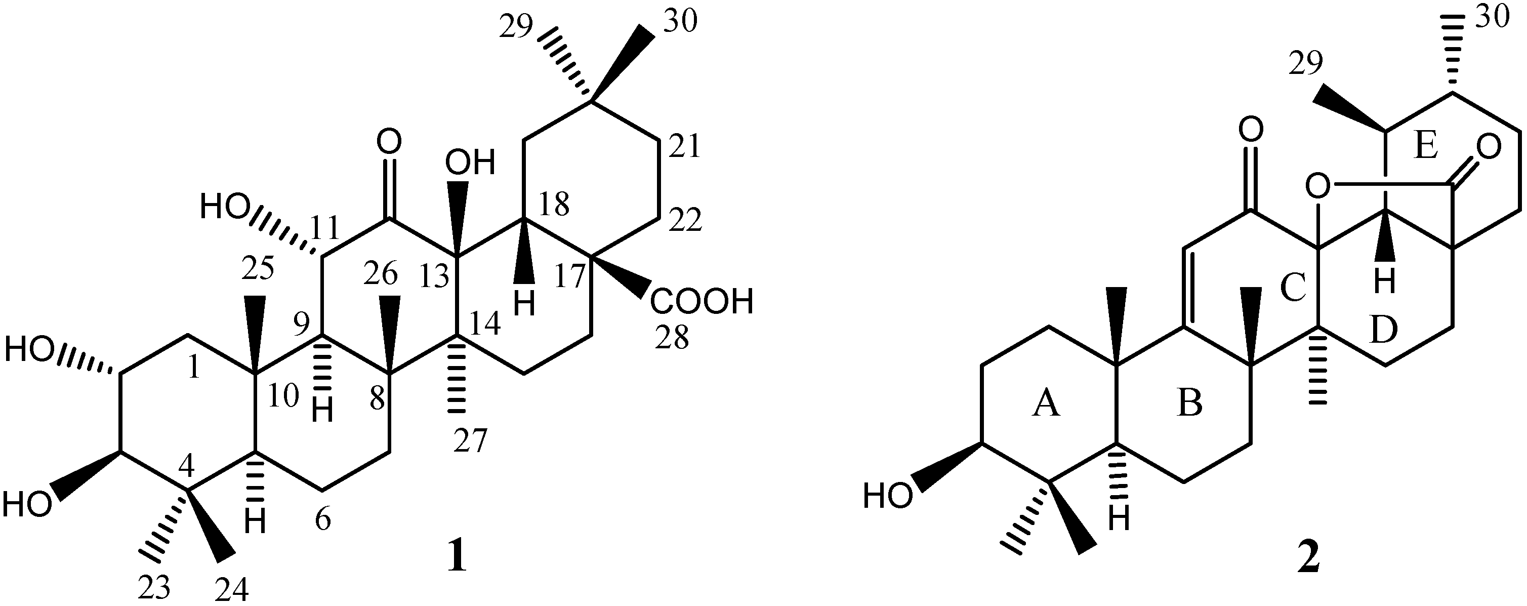
Figure 1.
Structures of compounds 1 and 2.

Results and Discussion
Compound 1 was obtained as a white powder. The HRESIMS at m/z 519.3320 [M-H]- (calcd. for C30H47O7, 519.3321) gave the molecular formula C30H48O7. The IR absorptions at 3430, 1777, and 1725 cm-1 indiated the presence of hydroxyl, ketone and carbonyl groups. The 13C-NMR (Table 1) and DEPT spectra of 1 displayed thirty signals, which included seven methyls, eight methylenes, six methines (three oxygenated) and nine quaternary signals, including a ketone and a carbonyl. These data, together with analysis of the corresponding 1H-1H COSY and HMBC spectra suggested that 1 possesses an oleanane-type skeleton [3]. Comparison of the NMR data of 1 with those of maslinic acid and 3β-acetoxy-11α,13β-dihydroxyolean-12-one [4,5] disclosed that the two olefinic carbons in maslinic acid were replaced by an oxygenated quaternary carbon at δ 90.0 and a ketone group at δ 200.2, besides, in 1 an oxygen-bearing methine at δ 56.1 was observed, instead of a methylene found in maslinic acid. The 1H-1H COSY coupling of H-9 (δ 2.16) with H-11 (δ 4.31), together with HMBC correlations from H-9 and H-11 to C-12 (δ 200.2), from H-11, Me-27 (δ 1.38), and H-19 (δ 1.82/1.42) to C-13 (δ 90.0) suggested that a ketone was positioned at C-12, and C-11 and C-13 were oxygenated, respectively. The Hax-2, Hax -3 were deduced by their coupling constants (J = 9.5 Hz), and were also evident from ROESY interactions of H-3 (δ 3.05) with Me-23 (δ 1.05). Additional ROESY correlations from Me-25 (δ 1.07) to H-2 (δ 3.71) and H-11, H-2 to Me-24 (δ 0.84), Hβ-1 (δ 2.18) to H-2 and H-11, suggested that the hydroxyl group at C-11 was α oriented, which was in agreement with the coupling constant of H-11 with H-9 (J = 7.5 Hz). It should be noted that the signal for C-11 is abnormally up-field shifted, which may be caused by the gauche effects of Hβ-11 with both 13β-OH and axial Me-25. Thus, the structure of 1 was determined to be 2α,3β,11α,13β-tetrahydroxy-12-keto- oleanan-28-oic acid.
Compound 2 was obtained as a white powder. Its molecular formula C30H44O4 was deduced from the HRESIMS at m/z 491.3129 [M+Na]+ (calcd. for C30H44O4Na 491.3137). The 13C-NMR (Table 1) and DEPT spectra of 2 showed thirty carbons, including seven methyls, eight methylenes, six methines, and nine quaternary signals. These data, together with the analysis of 1H-1H COSY and HMBC spectra suggested that 2 possessed an ursane-type skeleton, and was structurally related with ursolic acid [4]. The differences between 2 and ursolic acid were mainly in ring B. A double bond between C-9 and C-11, a ketone group at C-12, and an oxygenated carbon signal at C-13 were observed in 2, HMBC correlations of Me-25 (δ 1.28), Me-26 (δ 1.37), H-5 (δ 1.04), H-7 (δ 1.76/1.52), and H-11 (δ 6.00) all with C-9, and H-18 (δ 2.42) with C-12 confirmed the positions of the double bond and ketone groups. The UV absorption at 259 nm and IR peak at 1632 cm-1 indicated the presence of an α,β-unsaturated ketone. In addition, a lactone ring between C-13 and C-28 inferred from the molecular mass, was also confirmed by the absorption peak at 1780 cm-1in the IR spectrum. The hydroxyl at C-3 was assigned a β orientation by comparison of its 13C-NMR data with literature data [6], and ROESY correlations of H-3 (δ 3.18) with Me-23 (δ 1.04). The cis-fusion of D/E rings could be judged from the large coupling constant of Hax-18 (J=12.0 Hz). It was noted that the signal for C-9 appeared at a very abnormally down-field position, and the reasons for this phenomenon still remain unclear. Taken together, the structure of 2 was identified as 3β-hydroxy-12-keto-9(11)-ursen-28,13β-olide. The acetate of 2 has previously been described in the literature as a result of chemical transformation [7]. However, as a naturally occurring compound, 2 was isolated for the first time, the 1H- and 13C-NMR data of 2 have now been unambiguously assigned on the basis of 2D NMR experiments for the first time.

| Position | 1 | 2 | ||
|---|---|---|---|---|
| δH (J=Hz) | δC | δH (J=Hz) | δC | |
| 1 | 2.18 m; 1.30 m | 47.0 t | 2.04 m; 1.42 m | 37.0 t |
| 2 | 3.71 m | 68.6 d | 1.72 m | 28.4 t |
| 3 | 3.05 d (9.5) | 83.2 d | 3.18 m | 77.6 d |
| 4 | 39.1 s | 39.9 s | ||
| 5 | 0.91 m | 55.0 d | 1.04 m | 50.8 d |
| 6 | 1.67 m; 1.55 m | 17.4 t | 1.74 m | 18.0 t |
| 7 | 1.31 m | 31.6 t | 1.76 m; 1.52 m | 34.5 t |
| 8 | 41.5 s | 46.6 s | ||
| 9 | 2.16 d (7.5) | 57.7 d | 185.1s | |
| 10 | 39.8 s | 41.3 s | ||
| 11 | 4.31 d (7.5) | 56.1 d | 6.00 s | 122.0 d |
| 12 | 200.2 s | 193.3 s | ||
| 13 | 90.0 s | 88.4 s | ||
| 14 | 41.7 s | 42.9 s | ||
| 15 | 1.66 m | 26.3 t | 1.80 m; 1.43 m | 26.5 t |
| 16 | 2.18 m; 1.38 m | 21.3 t | 2.18 m; 1.38 m | 22.7 t |
| 17 | 44.4 s | 45.6 s | ||
| 18 | 2.71 dd (13.5, 3.0) | 48.0 d | 2.42 d (12.0) | 55.5 d |
| 19 | 1.82 m; 1.42 m | 37.0 t | 1.80 m | 37.7 d |
| 20 | 31.6 s | 1.01 m | 40.5 d | |
| 21 | 1.33 m | 34.2 t | 1.58 m | 31.2 t |
| 22 | 1.75 m; 1.31 m | 26.8 t | 1.54 m | 32.1 t |
| 23 | 1.05 s | 28.3 q | 1.04 s | 28.5 q |
| 24 | 0.84 s | 16.3 q | 0.84 s | 16.1 q |
| 25 | 1.07 s | 18.0 q | 1.28 s | 24.9 q |
| 26 | 0.98 s | 19.7 q | 1.37 s | 30.8 q |
| 27 | 1.38 s | 19.7 q | 1.21 s | 20.8 q |
| 28 | 177.6 s | 178.4 s | ||
| 29 | 0.90 s | 33.2 q | 0.76 d (6.4) | 18.7 q |
| 30 | 0.99 s | 23.3 q | 0.92 d (6.4) | 19.4 q |
a The spectra of 1 were recorded in CDCl3 and 2 in CD3COCD3 (400 MHz for 1H, 100 MHz for 13C)
The cytotoxic activity of compounds 1 and 2 against three human tumor cell lines (A-549, HCT-8, and BEL-7402) were evaluated. However, they exhibited no cytotoxic effects at concentrations up to 5 μg/mL (data not shown).
Experimental
General
Melting points were recroded on an XRC-1 micromelting apparatus. Optical rotations were determined on a JASCO-20C digital polarimeter. UV spectra were recorded on a Shimadzu UV-2401PC spectrophotometer. IR spectra were obtained with a Bruker Tensor 27 FT-IR spectrophotometer with KBr pellets. 1H-NMR (400 MHz) and 13C-NMR (100 MHz) spectra were recorded on a Bruker AM-400 spectrometer, with TMS as an internal reference. 2D NMR spectra were measured with a DRX-500 spectrometer. FABMS were recorded on a VG Auto Spec-3000 spectrometer. ESIMS and HRESIMS were carried our with an API QSTAR Pulsar 1 spectrometer. Silica gel (200-300 mesh and 10-40 μm) for column chromatography and GF254 for TLC were obtained from Qingdao Marine Chemical Factory (Qingdao, P. R. China). Sephadex LH-20 was obtained from Amersham Pharmacia Biotech (Sweden).
Plant Material
The dried and matured leaves of P. serrulata Lindl. were purchased from Nanjing Pharmaceutical Ltd. Corporation of Jiangsu Province (P. R. China) in March, 2006, and identified by Mrs. Xuedong Geng (Nanjing Pharmaceutical Ltd. Corporation of Jiangsu Province). A voucher specimen (CHYX0392) was deposited at the State Key Laboratory of Phytochemistry and Plant Resources in West China, Kunming Institute of Botany, Chinese Academy of Sciences.
Extraction and Isolation
The dried and powdered plant materials of P. serrulata (15 kg) were extracted three times with 80% EtOH under reflux. The extracts were concentrated and suspended in water followed by successive partition with petroleum ether, EtOAc and n-BuOH, respectively. The EtOAc extracts (866 g) were subjected to column chromatography (CC) over silica gel (200-300 mesh) and eluted with CHCl3-MeOH (15:1) to afford fractions 1-3. Fraction 2 (160 g) was submitted to CC over silica gel, eluted with CHCl3-MeOH-EtOAc (6:1:1) to give fractions 2.1-2.2. Fraction 2.2 (145 g) was further fractionated on silica gel eluted with a CHCl3-MeOH gradient (98:2, 96:4, 95:5) to afford fractions 2.2.1-2.2.5. Fraction 2.2.1 (16 g) was fractionated into four portions by CC over silica gel eluted with gradient CHCl3-Me2CO (50:1, 40:1, 30:1, 15:1), i.e. fractions 2.2.1.1-2.2.1.4. Repeated chromatography of fraction 2.2.1.1 (4 g) over silica gel (10-40 μm), and Sephadex LH-20 (CHCl3-MeOH, 6:4) yielded compounds 1 (3.6 mg), and 2 (3.7 mg).
2α,3β,11α,13β-tetrahydroxy-12-ketooleanan-28-oic acid (1). White powder; mp 218-219oC; [α]D27.8 -30.86o (c 0.14, CHCl3); UV (CHCl3) λmax (log ε) nm: 241 (3.24); IR (KBr) νmax: 3430, 2952, 2925, 2855, 1777, 1725, 1632, 1455, 1053 cm-1; 1H- and 13C-NMR data, see Table 1; positive ESIMS m/z: 543 [M+Na]+; negative ESIMS m/z: 555 [M+Cl]-, 519 [M-H]-.
3β-hydroxy-12-keto-9(11)-ursen-28,13β-olide (2). White powders; mp 237-238oC; [α]D26.4 +5.47o (c 0.31, Me2CO); UV (MeOH) λmax (log ε) nm: 259 (4.07), 230 (3.89); IR (KBr) νmax: 3440, 2954, 2927, 2871, 1780, 1632, 1455, 1384 cm-1; 1H- and 13C-NMR data, see Table 1; positive FABMS m/z: 469 (100) [M+H]+; negative ESIMS m/z: 503 [M+Cl]-, 467 [M-H]-; positive ESIMS m/z: 507 [M+K]+, 491 [M+Na]+, 469 [M+H]+.
Cytotoxic Assay
Compounds 1 and 2 were tested for their cytotoxic effects against human A-549, HCT-8 and Bel-7402 cell lines using the MTT [3-(4,5-dimethylthiazol-2-yl)-2,5-diphenyltetrazolium bromide] method as previously described [8], with fluorouracil (5-FU) as positive control. All the experiments were run in triplicate.
Acknowledgments
This work was financially supported by a “Talent Scholarship for the Youth of Yunnan” (No. 2007PY01-48), “Xi-Bu-Zhi-Guang” Project from the Chinese Academy of Sciences and startup grants from the State Key Laboratory of Phytochemistry and Plant Resources in West China, Kunming Institute of Botany, and Ministry of Education of the People’s Republic of China.
References
- Beijing Institute of Botany, Chinese Academy of Sciences. Flora Reipublicae Popularis Sinicae; Science Press: Beijing, 1974; Vol. 36, p. 216. [Google Scholar]
- Hou, J.; Sun, T.; Hu, J.; Chen, S. Y.; Cai, X. Q.; Zou, G. L. Chemical composition, cytotoxic and antioxidant activity of the leaf essential oil of Photinia serrulata. Food Chem. 2007, 103, 355–358. [Google Scholar] [CrossRef]
- López-Pérez, J. L.; Therón, R.; Del Olmo, E.; Diaz, D. NAPROC-13: a database for the dereplication of natural product mixtures in bioassay-guided protocols. Bioinformatics 2007, 23, 3256–3257. [Google Scholar] [CrossRef]
- Numata, A.; Yang, P. M.; Takahashi, C.; Fujiki, R.; Nabae, M.; Fujita, E. Cytotoxic triterpenes from a chinese medicine, goreishi. Chem. Pharm. Bull. 1989, 37, 648–651. [Google Scholar] [CrossRef]
- Heart, H. M.; Athukoralage, P. S.; Jamie, J. F. Two new oleanane triterpenoids from Gordonia ceylanica and their conversions to taraxarane triterpenoids. Phytochemistry 2000, 54, 823–827. [Google Scholar]
- Fang, S. Y.; He, Z. S.; Fan, G. J. Triterpenoids from Adina rubella. J. Nat. Prod. 1996, 59, 304–307. [Google Scholar] [CrossRef]
- Dreiding, J.; Jeger, O.; Ruzicka, L. Zur Kenntnis der Triterpene. Ueberfuehrung der Ursolsaeure in 2 isomere acetoxy-lactone C32H46O5. Helv. Chim. Acta 1950, 33, 1325–1334. [Google Scholar] [CrossRef]
- Réthy, B.; Kovács, A.; Zupkó, I.; Forgo, P.; Vasas, A.; Falkay, G.; Hohmann, J. Cytotoxic phenanthrenes from the rhizomes of Tamus communis. Planta Med. 2006, 72, 767–770. [Google Scholar]
- Sample Availability: Samples of compounds 1 and 2 are available from the authors.
© 2007 by MDPI (http://www.mdpi.org). Reproduction is permitted for noncommercial purposes.
Share and Cite
MDPI and ACS Style
Song, Y.; Wang, Y.; Lu, Q.; Gao, J.; Bi, M.; Cheng, Y. Two New Triterpenoids from Photinia serrulata. Molecules 2007, 12, 2599-2604. https://doi.org/10.3390/12122599
AMA Style
Song Y, Wang Y, Lu Q, Gao J, Bi M, Cheng Y. Two New Triterpenoids from Photinia serrulata. Molecules. 2007; 12(12):2599-2604. https://doi.org/10.3390/12122599
Chicago/Turabian StyleSong, Yaling, Yuehu Wang, Qing Lu, Jinming Gao, Minggang Bi, and Yongxian Cheng. 2007. "Two New Triterpenoids from Photinia serrulata" Molecules 12, no. 12: 2599-2604. https://doi.org/10.3390/12122599