Article

Synthesis and Characterization of 5,10,15,20-Tetra[3-(3-trifluoromethyl)phenoxy] Porphyrin

by
Cynthia P Tidwell
1,*,
Prakash Bharara
1,
Gretchen Rudeseal
1,
Tiffany Rudeseal
1,
Frank H Rudeseal, Jr
1,
Christine A Simmer
1,
Dugald McMillan
1,
Katherine Lanier
1,
L Dalila Fondren
1,
LaTasha L Folmar
1 and
Ken Belmore
2
1
Department of Biology, Chemistry, and Mathematics, University of Montevallo, Station 6480 Montevallo, Alabama 35115, U.S.A.
2
Department of Chemistry, University of Alabama, Box 870336 Tuscaloosa, Alabama 35487, U.S.A.
*
Author to whom correspondence should be addressed.
Molecules 2007, 12(7), 1389-1398; https://doi.org/10.3390/12071389
Submission received: 30 May 2007 / Revised: 8 July 2007 / Accepted: 9 July 2007 / Published: 11 July 2007
(This article belongs to the Special Issue Heterocycles)

Abstract

:
The newly synthesized 5,10,15,20-tetra[3-(3-trifluoromethyl)phenoxy] porphyrin, TTFMPP, has been characterized using mass spectroscopy, 1H-, 13C- and 19F- NMR, MALDI-TOF mass spectrometry, UV-Vis and fluorescence spectrophotometry, and cyclic voltammetry. The NMR confirmed the structure of the compound and the mass spectrum was in agreement with the proposed molecular formula. The UV-Vis absorption spectrum of TTFMPP shows characteristic spectral patterns similar to those of tetraphenyl porphryin, with a Soret band at 419 nm and four Q bands at 515, 550, 590, and 648 nm. Protonation of the porphyrin with TFA resulted in the expected red shift of the Soret band. Excitation at 419 nm gave an emission at 650 nm. The quantum yield of the porphyrin was determined to be 0.08. Cyclic voltammetry was used to determine the oxidation and reduction potentials of the new porphyrin. Two quasi-reversible one-electron reductions at –1.00 and –1.32 V and a quasi-reversible oxidation at 1.20 V versus the silver/silver chloride reference electrode with tetrabutylammonium tetrafluoroborate as the supporting electrolyte in methylene chloride were observed.

Introduction

Porphyrins are molecules that are being vigorously pursued in many areas of research [1,2,3,4,5,6,7,8,9,10,11,12,13]. It has been shown that these molecules are of importance in many applications including, but not limited to, sensors [1,2,3,4,5,6,7], optical memories [8,9,10], and photocatalysis [11,12,13]. Many of these applications take advantage of the ability of porphyrins to undergo fast electron transfer. For example, electron transfers have been shown to take place on a picosecond time scale in photosynthesis [14]. Also, of importance to many of these applications is the region of the spectrum in which the molecules absorb. Porphyrins are known to have intense absorptions exhibited by the high extinction coefficients of the Soret and Q-bands of these molecules between 400 nm and 700 nm. The absorptions of porphyrins are somewhat tunable by choice of substituents.
The focus of this research involves the synthesis of a new porphyrin with a trifluoromethylphenoxy group substituted in the meso position. This electron withdrawing substituent should afford the porphyrin different electronic properties than compounds previously synthesized. The synthesis of this molecule and its characterizations are reported in this paper. The molecule has been structurally characterized by mass spectrometry and NMR. The electronic structure and properties of the chromophore have been investigated using electronic absorption spectroscopy, fluorescence spectroscopy and cyclic voltammetry.

Results and Discussion

5,10,15,20-Tetra[3-(3-trifluoromethyl)-phenoxy] porphyrin, TTFMPP, was prepared from pyrrole and 3-[3-(trifluoromethyl)phenoxy]benzaldehyde using a 50:50 mixture of propionic and octanoic acids as the solvent (Scheme 1).
This solvent system was found by Lindsey and coworkers to work better than the normal acetic acid method used by Adler and many others in preparation of porphyrins [15,16]. Porphyrins are usually produced in low yields and TTFMPP was no exception. The overall yield of the reaction was 10%. The composition was verified by obtaining the exact mass using MALDI-TOF mass spectrometry. The measured mass, 1254.2 a.m.u., was in excellent agreement with the calculated value of 1255.3 a.m.u. The newly prepared TTFMPP was further characterized by 1H-, 13C- and 19F-NMR, UV-Visible spectrophotometry and fluorescence spectroscopy.

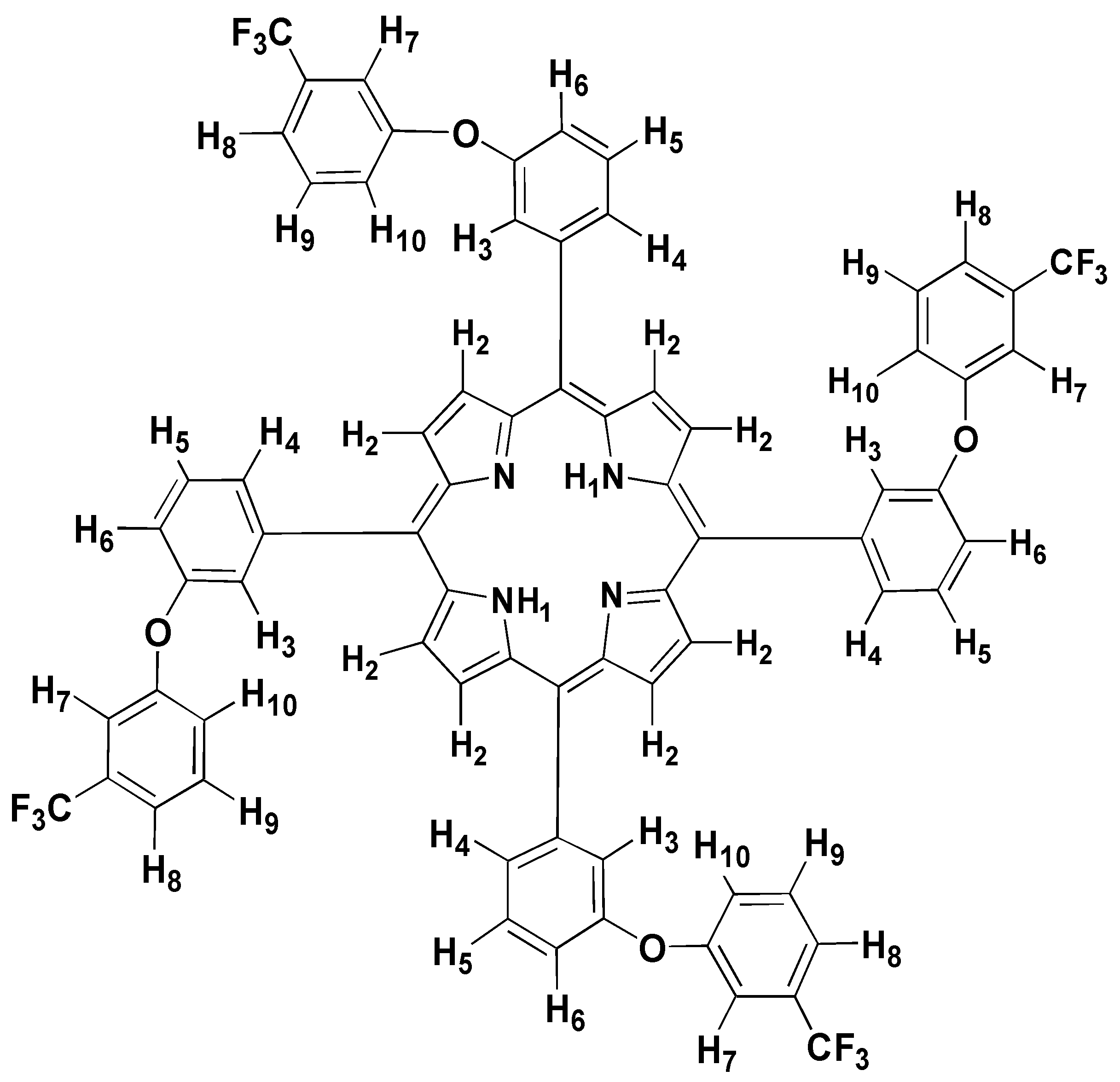
Nuclear Magnetic Resonance Spectroscopy

The sample was analyzed using 1H-, 13C- and 19F-NMR. A 13C-1H correlation was done using a heteronuclear chemical shift correlation experiment: XHCORRD.AUR. In addition, a heteronuclear multiple bond correlation was done to locate the quarternary carbons of the compound. Nuclear Overhauser Effect (NOE) results were consistent with the proposed structure.
1H-NMR studies of porphyrins reveal the aromatic nature of the porphyrin macrocycle. A diamagnetic ring current deshields protons outside the macrocycle from the external magnetic field. β-pyrrole and meso protons are included in this category of protons. The macrocycle is made up of two pyrrole units and two pyrrolenine units. The pyrrole units have an aromatic sextet of electrons. The pyrrolenine units have only five pi electrons. To compensate for this shortage of pi electrons in the pyrrolenine, an electron is pulled from the meso carbons. So, meso carbons have a tendency to be electron deficient and thus the meso protons are shifted further downfield than the β-protons. The same diamagnetic ring current that deshields the protons on the outside of the macrocycle shields the protons on the inside of the macrocycle from the external magnetic field. Thus, the N-H protons are shifted upfield, even farther than the tetramethylsilane (TMS) peak [17]. The numbering of the protons is shown in Figure 1.
The diamagnetic ring current of this porphyrin deshields the β-pyrrole protons of this porphyrin, thus, we expect to find the signal for this proton downfield of most of the other protons. Also, it is expected that it will appear as a singlet with integral of eight protons. Three singlets appear in the spectrum downfield: H2 at 9.04 ppm, H3 at 8.03 ppm, and H7 at 7.66 ppm. The integration of these peaks gives eight, four and four protons, respectively. Thus, the β-proton, H2, of this porphyrin is given the assignment of 9.04 ppm. The singlet at 8.03 ppm is assigned to H3 and the one at 7.66 ppm is assigned to H7. These assignments are consistent with the NOE data. The remaining singlet is at -2.71 ppm. Due to the shielding experienced by the N-H proton, H1, it was expected that it would be shifted far upfield and thus it was assigned to this peak. The integration of this peak is two protons, which is consistent with this assignment.
This structure contains four chemically nonequivalent sets of protons which should each appear as a doublet in the spectrum. Indeed, such doublets are observed at 8.14, 7.58, 7.51, and 7.44 ppm and are assigned to protons H4, H6, H10 and H8, respectively. According to the NOE data, there are through space interactions of the proton at 8.14 ppm with the protons exhibiting peaks at 9.04 ppm and 7.86 ppm consistent with assignment to H4. The peak at 7.51 is assigned to H10. This assignment is in agreement with NOE experiments which show that the proton with chemical shift of 7.51 ppm interacts with the protons that exhibit chemical shifts of 8.03 ppm (H3) and 9.04 ppm (H2). The remaining two doublets at 7.44 ppm and 7.58 ppm are assigned to H8 and H6, respectively. H6 is expected to be farther downfield due to the nearby electronegative oxygen atom.
One triplet is observed in the spectrum at 7.86 ppm, and is assigned to H5. There is a dd observed at 7.54 ppm which is assigned to H9. The integration of each of the peaks in this spectrum confirms these assignments. A summary of the 1H-NMR for the chemical shifts and multiplicities are given in Table 1. In Table 2, the assignments of the irradiated protons and the corresponding enhancements of the protons are listed for the NOE experiment.
13C-NMR peaks were assigned from the corresponding spectrum, from data obtained in the 13C-1H correlation and from data of a heteronuclear multiple bond correlation. The 13C assignments are given in Table 3. The expected splitting due to the fluorine atoms in the molecules was observed. A single peak in the 19F-NMR spectrum was expected and was observed at -62.08 ppm for the CF3.

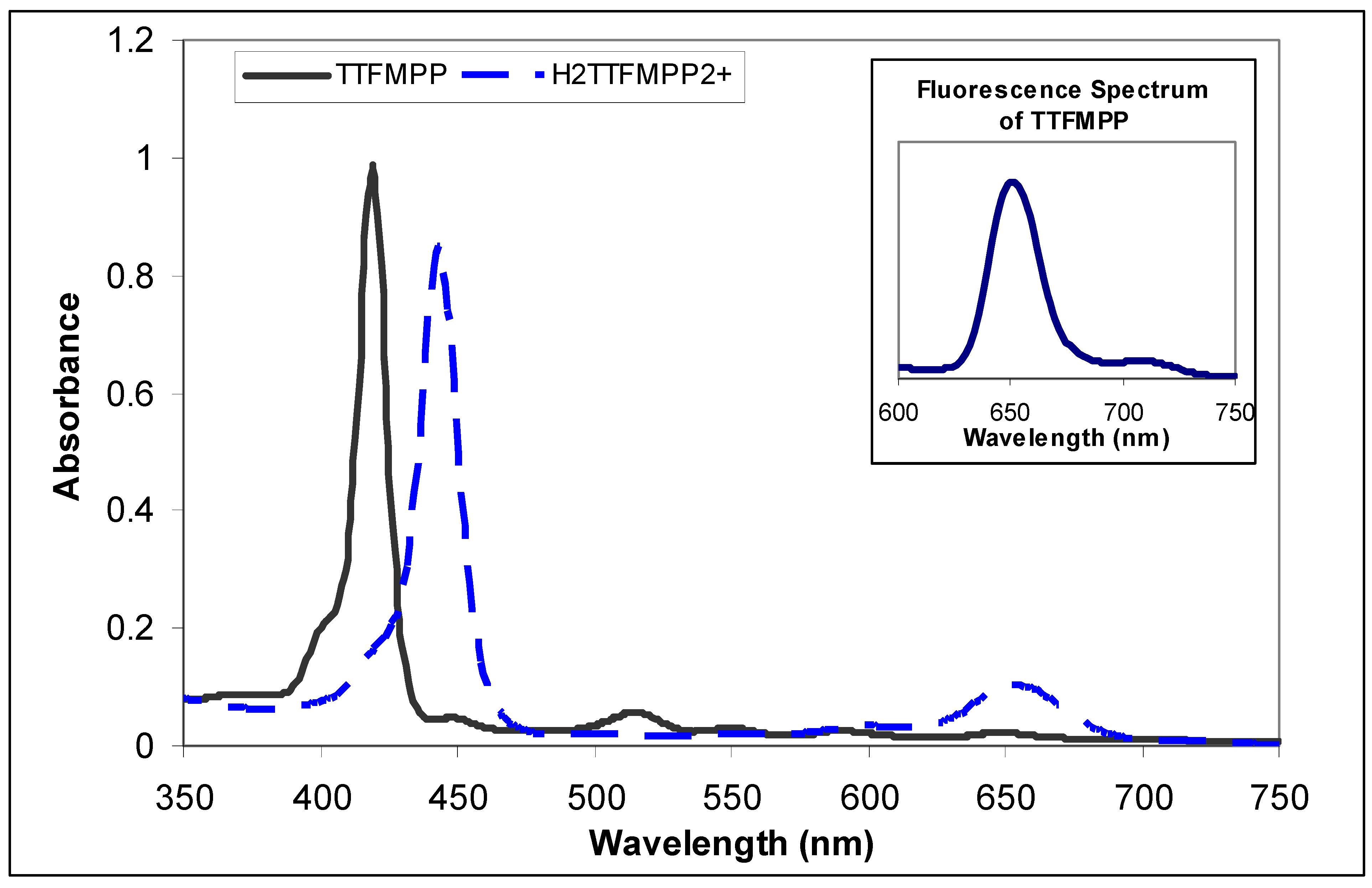
Electronic absorption spectra of 5,10,15,20-tetra[3-(3-trifluoromethyl)-phenoxy] porphyrin

The electronic absorption spectrum for TTFMPP is given in Figure 2 and is similar to that of all porphyrins. There is an intense absorption at 419 nm, the Soret band and Q bands are observed at 515, 550, 590, and 648 nm. A series of solutions were prepared of varying concentrations of TTFMPP in CHCl3. The concentrations ranged from 2.7 x 10⁻⁵ to 1.6 x 10⁻⁶M. UV-vis spectra were taken for each of the solutions. The purpose of these experiments was to elucidate the extinction coefficient of the Soret band for the porphyrin as well as the different Q bands and to determine if Beer’s Law was obeyed. Some compounds at high concentrations tend to aggregate and changes in the extinction coefficient are observed [18]. No systematic shifts in peak positions were observed and the extinction coefficients for the Soret band and the Q bands are very high. This porphyrin displays an extinction coefficient of 3 x 105 cm-1M-1 for the Soret band at 419 nm. Four Q bands are observed at 515, 550, 590, and 648 nm with extinction coefficients given as 1 x 104 cm-1M-1, 6 x 103 cm-1M-1, 5 x 103 cm-1M-1, and 7 x 103 cm-1M-1, respectively.
To investigate this porphyrin further, it was protonated using trifluoroacetic acid and the electronic absorption spectrum obtained. The formation of the dication results in a red shift in the Soret band the magnitude of which has been shown to increase with increasing electron withdrawing nature of meso substituents[19]. This shift is due to an increase in the conjugation of the molecule and hence a decrease in the energy required for the π → π* transition. The spectrum of the dication is given in Figure 2.

Fluorescence Measurements

Excitation at 419 nm of TTFMPP gave an emission line at 650 nm. The fluorescence quantum yield, ΦF, was determined to be 0.08 at room temperature for chloroform solutions of TTFMPP by a relative method using TPP in CHCl3 as a standard having ΦF=0.11 as reported in the literature [20]. The fluorescence spectrum is given in Figure 2.

Cyclic Voltammetry of 5,10,15,20-Tetra[3-(3-trifluoromethyl)phenoxy] Porphyrin

Substitution of the meso positions with an electron withdrawing group is expected to shift the reduction and oxidation potentials. The potentials are a significant attribute affecting the electron transfer and thus the usefulness of the porphyrin in many applications. Therefore, it is important to characterize the electrochemical behavior of this chromophore. Many studies have been carried out to study the oxidation and reduction of porphyrins. Often, these studies are used for comparison with new chromophores. The reduction of TTFMPP was measured in CH2Cl2 with 0.1M tetrabutylammonium tetrafluoroborate as the supporting electrolyte. Two successive, quasi-reversible one electron reductions were observed at –1.00 V and –1.32 V versus the Ag/AgCl electrode. To compare, literature values for tetraphenyl porphyrin were reported in CH2Cl2 with tetrabutylammonium hexafluorophosphate as the supporting electrolyte to be –1.11 V and –1.52 V versus the Ag/AgCl electrode [21]. As expected, with the addition of the electron withdrawing group, the reduction potentials are not as low as those for TPP without the electron withdrawing groups. The addition of the electron withdrawing groups facilitates the reduction of the porphyrin. It is also expected to make the porphyrin harder to oxidize. This was observed as the oxidation potential of TTFMPP was found to be 1.20 V versus the Ag/AgCl electrode. The corresponding oxidation of TPP was reported in the literature to be 1.03 V versus the Ag/AgCl electrode [21]. Trends observed in the oxidation and reduction potentials are consistent with the nature of the porphyrin. That is, the electron withdrawing group in TTFMPP enhanced reduction and inhibits oxidation.

Conclusions

In summary, we present the first report of the synthesis and characterization of 5,10,15,20-tetra[3-(3-trifluoromethyl)phenoxy] porphyrin. The absorption spectrum of TTFMPP showed a characteristic spectral pattern similar to those of other porphyrins. The trends in oxidation and reduction of this compound expected with introduction of electron withdrawing groups in the meso position were observed. The result of the exact mass analysis was in agreement with the proposed formulation and the 1H-, 13C- and 19F-NMR confirmed the structure. This compound and its metal complexes are being studied further for usefulness in applications involving electron transfer.

Experimental

General
Pyrrole, 3-[3-(trifluoromethyl)phenoxy]benzaldehyde, propionic acid and octanoic acid used in the synthesis of TTFMPP were purchased from Aldrich Chemical Company and used as received. Solvents used for synthesis or NMR measurements were used as received. Spectroscopic grade chloroform and anhydrous methylene chloride were obtained from Aldrich Chemical Company and was used for electronic absorption experiments and cyclic voltammetry. Tetrabutylammonium tetrafluoroborate was purchased from Fisher Scientific Company and used as received. Ultraviolet-visible (UV-vis) absorption spectra for the base TTFMPP were recorded on a Hewlett Packard 8451A spectrophotometer. UV-Vis absorption spectra for the metallated TTFMPP complexes were recorded on a Jasco V-530. Matrix assisted laser desorption ionization time of flight mass spectroscopic analysis was carried out on a VG Auto Spec instrument. All nuclear magnetic resonance (NMR) spectra for the base TTFMPP were recorded on a Bruker AM360 or Bruker AM500 spectrometer at approximately 298K. The Nuclear Overhauser Effect experiments were carried out at 313K on the Bruker AM500 spectrometer with CDCl3 as the solvent. The chemical shifts are reported in parts per million (δ) with respect to an internal reference peak of residual undeuterated solvent (that is, 7.24 ppm for CHCl3 in CDCl3) or TMS. Fluorescence spectra were obtained using a Jasco FP-6300 spectrofluorimeter. Cyclic voltammograms (CVs) were recorded using a computer linked EG&G Princeton Applied Research Potentiostat VersaStat II equipped with Power Suite 2.35.2 Electrochemical software. The CVs were obtained in a cell with a glassy carbon working electrode, a platinum wire counter electrode, and a saturated Ag/AgCl in KCl as the reference electrode. The measurements were taken in a nitrogen enriched atmosphere. The porphyrin solution was 1.3 x 10⁻³ M in a 0.1 M solution of tetrabutyl-ammonium tetrafluoroborate in methylene chloride. Potentials are reported versus a Ag/AgCl electrode and not corrected for liquid junction potentials. Deaeration of the solution was achieved by passing a stream of nitrogen through the solution.

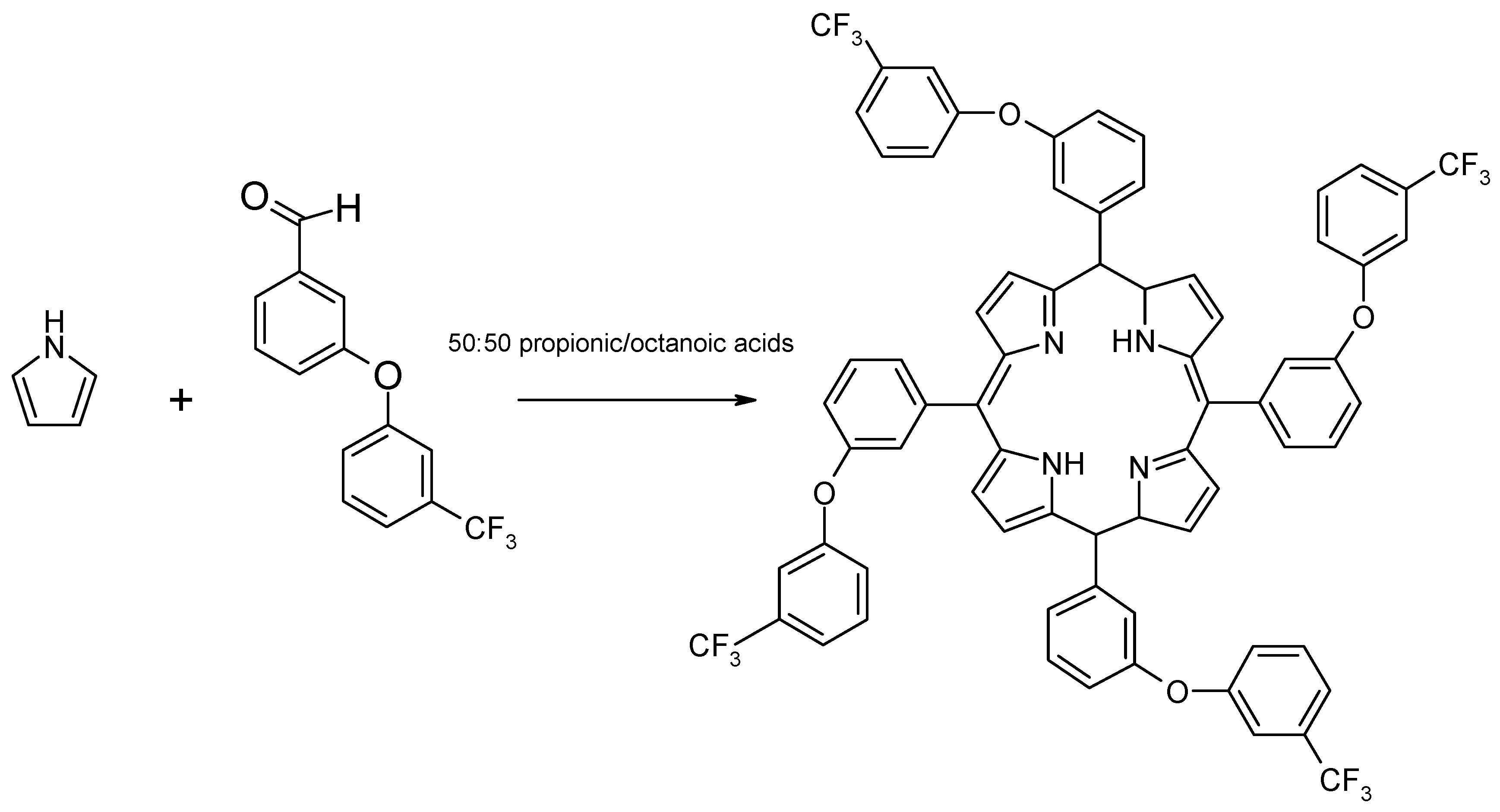
Synthesis of 5,10,15,20-tetra[3-(3-trifluoromethyl)phenoxy] porphyrin

A single necked 250 mL round bottomed flask fitted with a reflux condenser was charged with octanoic acid (60 mL) and propionic acid (60 mL). To this solvent mixture, 3-[3-(trifluoromethyl)phenoxy]benzaldehyde (2.66 g, 0.010 mol) was added. This mixture was heated with stirring. When the 3-[3-(trifluoromethyl)phenoxy]benzaldehyde was dissolved the solution was colorless. To this solution, pyrrole (0.678 g, 0.010 mol) was added, giving an amber colored solution. This was brought to reflux, and kept at this temperature for 60 minutes. This solution was allowed to cool for 30 minutes and the electronic absorption spectrum was measured. After cooling, the solution was added to a 500 mL separatory funnel followed by chloroform (120 mL) and 1M NaHCO3 (120 mL). The mixture was gently rotated until the acids were neutralized. The solution was then left in the funnel overnight to allow complete separation of the two layers. The bottom layer containing the crude porphyrin was collected, evaporated to dryness and purified using column chromatography (dry silica gel, 70-230 mesh, eluent: 13:1 mixture of chloroform and methanol) to give 2.96 g (2.4x10⁻³ moles) of pure compound. The overall reaction yield was 10%. UV (CHCl3) [λ, nm (ε, cm-1M-1)]: 419 (1.31x10⁵), 515 (7.5x10³), 550 (3.25x10³), 590 (2.61x10³); 1H-NMR (500.13 MHz): 9.04 (s, 8H, H2), 8.14 (d, 4H, H4), 8.03 (s, 4H, H3), 7.86 (t, 4H, H5), 7.66 (s, 4H, H7), 7.58 (d, 4H, H6), 7.54 (dd, 4H, H9), 7.51 (d, 4H, H10), 7.44 (d, 4H, H8), -2.71 (s, 2H, NH1) 13C-NMR: 158.02, 155.06, 146.59 (broad), 144.07, 132.64 (q, 2JCF = 33 Hz), 131.16 (broad), 130.69, 13.51, 128.28, 125.71, 124.40 (CF3, 1JCF=273 Hz), 121.89, 119.98 (3JCF = 3.4Hz), 119.25, 118.84, 115.57 (3JCF = 3.0 Hz); 19F-NMR (338.86 MHz, CDCl3): -62.08 ppm, (s, CF3); MALDI-TOF Mass Spectroscopy: calculated for C72H42N4F12O4: 1255.3 a.m.u.; observed: 1254.2 a.m.u.

Acknowledgments

This work was financially supported by the Research and Special Projects Committee of the University of Montevallo. We would like to thank Dr. Sharon Webb and Dr. Qiaoli Liang, the Mass Spectrometry Facility Managers of The University of Alabama, for running the mass spectra of our sample.

References and Notes

  1. Gupta, V.K.; Chauhan, D.K.; Saini, V.K.; Agarwal, S.; Antonijevic, M.M.; Lang, H. A Porphyrin Based Potentiometric Sensor for Zn2+ Determination. Sensors 2003, 3, 223–235. [Google Scholar]
  2. Wolfbeis, O.S. Fiber-Optic Chemical Sensors and Biosensors. Anal. Chem. 2006, 78, 3859–3874. [Google Scholar]
  3. Gupta, V.K.; Chandra, S.; Chauhan, D.K.; Mangla, R. Membranes of 5,10,15,20-tetrakis(4-Methoxyphenyl) Porphyrinatocobalt (TMOPP-Co)(I) as MoO4-2-Selective Sensors. Sensors 2002, 2, 164–173. [Google Scholar]
  4. Ozoemena, K. Anodic Oxidation and Amperometric Sensing of Hydrazine at a Glassy Carbon Electrode modified with Cobalt (II) Phthalocyanine-cobalt(II) Tetraphenylporphyrin (CoPc-(CoTPP)4) Supramolecular Complex. Sensors 2006, 6, 874–891. [Google Scholar]
  5. Furusho, Y.; Kimura, T.; Mizuno, Y.; Aida, T. Chirality-Memory Molecule: A D2-Symmetric Fully Substituted Porphyrin as a Conceptually New Chirality Sensor. J. Am. Chem. Soc. 1997, 119, 5267–5268. [Google Scholar]
  6. Zhang, Y.; Yang, R.H.; Liu, F.; Li, K. Fluorescent Sensor for Imidazole Derivatives Based on Monomer-Dimer Equilibrium of a Zinc Porphyrin Complex in a Polymeric Film. Anal. Chem. 2004, 76, 7336–7345. [Google Scholar]
  7. Ikeda, O.; Yoshinaga, K.; Lei, J. Nitric Oxide Detection with Glassy Carbon Electrodes Coated with Charge-different Polymer Films. Sensors 2005, 5, 161–170. [Google Scholar]
  8. Liu, C.Y.; Bard, A.J. Optoelectronic Properties and Memories Based on Organic Single-Crystal Thin Films. Acc. Chem. Res. 1999, 32, 235–245. [Google Scholar]
  9. Purrello, R.; Raudino, A.; Monsu Scolaro, L.; Loisi, A.; Bellacchio, E.; Laucer, R. Ternary Porphyrin Aggregates and Their Chiral Memory. J. Phys. Chem. B 2000, 104, 10900–10908. [Google Scholar]
  10. Cho, H.S.; Jeong, D.H.; Cho, S.; Kim, D.; Matsuzaki, Y.; Tanaka, K.; Tsuda, A.; Osuka, A. Photophysical Properties of Porphyrin Tapes. J. Am. Chem. Soc. 2002, 124, 14641–14654. [Google Scholar]
  11. Yu, J.; Wang, X.; Zhang, B. Prolonged Excited-State Lifetime of Porphyrin Due to the Addition of Colloidal SiO2 to Triton X-100 Micelles. Langmuir 2004, 20, 1582–1586. [Google Scholar]
  12. Tangestaninejad, S.; Habibi, M.H.; Mirkhani, V.; Moghadam, M. Mn(Br8TPPS) Supported on Amberlite IRA-400 as a Robust and Efficient Catalyst for Alkene Epoxidation and Alkane Hydroxylation. Molecules 2002, 7, 264–270. [Google Scholar]
  13. Girichev, E.G.; Bazanov, M.I.; Mamardashvili, N.Z.; Gjeyzak, A. Electrochemical and Electrocatalytical Properties of 3,7,13,17-Tetramethyl-2,8,12,18-Tetrabutylporphyrin in Alkaline Solution. Molecules 2000, 5, 767–774. [Google Scholar]
  14. Franzen, S.; Boxer, S. G.; McLendon, G. Advances in Chemistry Series 228; Bolton, J. R., Mataga, N., Eds.; American Chemical Society: Washington, DC, 1991; pp. 155–162. [Google Scholar]
  15. Ojadi, E. C. A.; Linschitz, H.; Gouterman, M.; Walter, R. I.; Lindsey, J. S.; Wagner, R. W.; Droupadi, P. R.; Wang, W. Sequential protonation of meso-[p-(dimethylamino)phenyl]porphyrins: charge-transfer excited states producing hyperporphyrins. J. Phys. Chem. 1993, 97, 13192–13197. [Google Scholar]
  16. Adler, A. D.; Longo, R. R.; Finarelli, J. D.; Goldmacher, J.; Assour, J.; Korsakoff, L. A Simplified Synthesis for Meso-tetraphenyl Porphine. J. Org Chem. 1967, 32, 476. [Google Scholar](b)Kim, J. B.; Adler, A. D.; Longo, F. R. The Porphyrins; Dolphin, D., Ed.; Academic Press: New York, 1978; Volume 1, pp. 85–100. [Google Scholar]
  17. Milgrom, L. R. The Colours of Life: An Introduction to the Chemistry of Porphyrins and Related Compounds; Oxford University Press Inc.: New York, 1997. [Google Scholar]
  18. Skoog, D. A.; West, D. M.; Holler, F. J. Fundamentals of Analytical Chemistry, 6th Ed. ed; Saunders College Publishing: Philadelphia, PA, 1992. [Google Scholar]
  19. Weinkauf, J. R.; Cooper, S. W.; Schweiger, A.; Wamser, C. C. Substituent and Solvent Effects on the Hyperporphyrin Spectra of Diprotonated Tetraphenylporphyrins. J. Phys. Chem. A. 2003, 107, 3486–3496. [Google Scholar]
  20. Gouterman, M.; Khalil, G. E. Porphyrin Free Base Phosphorescence. J. Mol. Spectrosc. 1974, 53, 88–100. [Google Scholar]
  21. Ravikanth, M.; Chandrashekar, T. K. Effect of Porphyrin Ring Distortion on the Singlet-Excited State Properties of Alkyl-Bridged, Basket-Handle Porphyrins. J. Photochem. Photobiol. A Chem. 1993, 74, 181–187. [Google Scholar]
  • Sample Availability: Samples of the compounds are available from authors.
Scheme 1. Synthesis of 5,10,15,20-tetra[3-(3-trifluoromethyl)-phenoxy] porphyrin.
Molecules 12 01389 sch001
Figure 1. Structure of TTFMPP with protons numbered for NMR analysis.
Molecules 12 01389 g001
Figure 2. Electronic Absorption spectrum of TTFMPP and the dication. Inset: Fluorescence spectrum of TTFMPP.
Molecules 12 01389 g002
Table 1. Chemical shifts and multiplicities of protons on TTFMPP in chloroform-d.
Chloroform- d
MultiplicityChemical Shift (ppm)
H1s-2.71
H2s9.04
H3 s 8.03
H4d8.14
H5 t 7.86
H6d7.58
H7 s 7.66
H8d7.44
H9dd7.54
H10d7.51
Table 2. 1H-NMR Nuclear Overhauser Effect experimental data.
Proton Irradiated (δ, ppm)# of enhanced Hydrogenppm% enhancement
H2 (9.04)H38.031.6
H48.141.4
H77.660.7
H107.510.3
H4 (8.14)H2 9.040.9
H57.861.2
H3 (8.03)H29.040.9
H77.661.0
H107.510.5
H7 (7.66)H38.031.0
Table 3. Summary of 13C-NMR assignments.
Carbon Peak (ppm)
Main data used in assigning peak
Carbon Peak (ppm)
Main data used in assigning peak
C-H2131.16 (13C at 318K)C-H10121.89 (13C-1H correlation)
C-H3125.71 (13C-1H correlation)CF3124.40 (13C)
C-H4130.69 (13C-1H correlation)C-CF3132.62 (q) (13C)
C-H5128.28 (13C-1H correlation)Meso C119.25 (HMBC)
C-H6118.84 (13C-1H correlation)C-meso C144.07 (13C)
C-H7115.57 (d) (13C-1H correlation)H3-C-C*-C-H6155.06 (HMBC)
C-H8 119.98 (d) (13C-1H correlation)H7-C-C*-C- H10158.02 (HMBC)
C-H9130.51 (13C-1H correlation)

