Review

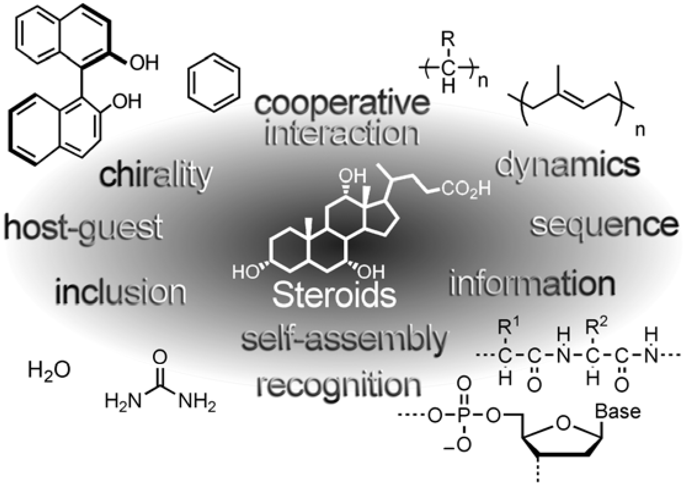
Supramolecular Chirality in Crystalline Assemblies of Bile Acids and Their Derivatives; Three-Axial, Tilt, Helical, and Bundle Chirality

by
Mikiji Miyata
*,
Norimitsu Tohnai
and
Ichiro Hisaki
Department of Material and Life Science, Graduate School of Engineering, Osaka University, 2-1 Yamadaoka, Suita, Osaka 565-0871, Japan
*
Author to whom correspondence should be addressed.
Molecules 2007, 12(8), 1973-2000; https://doi.org/10.3390/12081973
Submission received: 30 June 2007 / Revised: 20 August 2007 / Accepted: 20 August 2007 / Published: 22 August 2007
(This article belongs to the Special Issue Bile Acids)

Abstract

:
Steroidal bile acids and their derivatives exhibit characteristic inclusion behaviors in the crystalline state. Their crystals present varied assemblies due to asymmetric molecular structures, which relate to supramolecular properties through cooperative weak interactions. An overview indicates that the steroidal assemblies lie in an intermediate position among various molecules and have hierarchical structures such as primary, secondary, tertiary, and host-guest assemblies like proteins. Such an interpretation brought about the idea that the assemblies with dimensionality present supramolecular chirality such as three-axial, tilt, helical, bundle, and complementary chirality. This concept of the supramolecular chirality enables us to understand formation of chiral crystals starting from the molecular chirality of the steroidal molecules.

Table of Contents

1. Introduction1974
2.Diverse Structures of Inclusion Crystals of Bile Acids and Their Derivatives1976
3.Hierarchical Assemblies of Bile Acids and Their Derivatives1979
4.Supramolecular Chirality in Crystalline Molecular Assemblies1982
5.Supramolecular Chirality of Crystalline Assemblies of Bile Acids and Their Derivatives1988
6.Conclusion and Perspectives1991

1. Introduction

Crystalline molecular assemblies of steroidal bile acids and their derivatives (Scheme 1, Table 1) have aroused our interests for a long time, because the assemblies include various organic guest components through cooperative weak interactions [1,2,3,4]. The resulting host-guest complexes, termed as inclusion compounds [5,6], were found to have a variety of assembly structures by means of X-ray crystallographic analysis.
Scheme 1. Steroidal bile acids and their derivatives with the traditional atomic numbering.
Molecules 12 01973 g019
Table 1. Steroidal bile acids and their derivatives.
Molecules 12 01973 i001
This experimental result prompted us to consider a relationship between the molecular structures and the assembly structures. These studies led us to the fact that the assemblies lie in an intermediate among the other known molecular assemblies and biopolymers, as schematically shown in Figure 1. Thus, spherical [Figure 1(a)], axial [Figure 1(b)], and flat [Figure 1(c)] molecules make face- (or body) centered, layered, and herringbone (or stacked) assemblies, respectively, whereas biopolymers such as proteins [Figure 1(e)] form folded structures. Compared with these molecules, the steroidal molecules [Figure 1(d)] have arched skeletons with about ten chiral carbons and multiple hydrogen-bonding groups, making the intermediate assemblies which relate to supramolecular properties, such as host-guest, inclusion, reaction, chirality, recognition, dynamics, information, and so on. In other words, the steroidal assemblies serve as a fascinating entity for supramolecular chemistry in the crystalline state [7,8].
Figure 1. Relationship between molecular structures and crystal structures; (a) spherical, (b) axial, (c) flat, (d) arched, and (e) chain molecules.
Molecules 12 01973 g001
The research into the crystalline assemblies of bile acids and their derivatives can be briefly summarized from the viewpoint of the inclusion as follows. A classical host, deoxycholic acid (IIa), has been known to include various organic substances over a century [9,10,11]. Craven and DeTitta reported the first crystal structure involving acetic acid in 1972 [12], and clarified that the inclusion comes from a bilayer structure with channels. The subsequent crystallographic research by Giglio established a common occurrence of the channel-type bilayer structures [13]. In contrast, few studies of the inclusion behavior of cholic acid (Ia) have been published [9,10,11]. Johnson and Schaefer reported a cage-type structure of the inclusion crystal of 1a with ethanol in 1972 [14]. In 1988, Miyata, Miki and their coworkers reported a channel-type structure of the inclusion crystal of Ia with acetophenone [15,16,17]. Until now, such inclusion properties have been extended to their related derivatives. Several reviews are concerned with these assemblies in 1996 [1], 2004 [2,3], 2007 [4].
In this article, comprehensive studies on inclusion behaviors and crystal structures of bile acids and their derivatives are briefly summarized in Chapter 2, followed by a hierarchical interpretation of the crystalline assemblies (Chapter 3). Chapter 4 deals with an understanding of supramolecular chirality of the hierarchical assemblies, followed by illustrative description of the supramolecular chirality of the steroidal assemblies (Chapter 5). Final chapter deals with perspectives of these studies.

2. Diverse Structures of Inclusion Crystals of Bile Acids and Their Derivatives

2.1. Basic Patterns of Assembly Modes

Molecular structures of bile acids and their derivatives are generally shown in Scheme 1, and their nomenclature and abbreviations in this article are shown in Table 1. We usually searched for inclusion abilities of the steroidal derivatives by using more than one hundred organic compounds as guest candidates. It was so far confirmed that each steroidal derivative exhibits a corresponding inclusion ability and assembly mode based on a set of hydrogen bonding groups involving guest components. Figure 2 shows representative crystal structures of the steroidal derivatives. Their collected X-ray crystallographic data provides a treasure trove with respect to cooperative interactions composed of multiple non-covalent bonding groups.
Figure 2. Representative crystal structures of steroidal bile acids and their derivatives; a crossing structure with cage-like cavities for Ia (a), bilayered structures with channel-like cavities for Ia (b), IIa (c), Ib (d), IVb (e), If (f), Ig (g), and a honeycomb structure with hexagonal channels for IIIa (h).
Molecules 12 01973 g002
In order to classify the crystal structures, it is essential to understand the basic patterns of hydrogen bonding networks and molecular arrangements. Figure 3 exemplifies a typical assembly pattern of four molecules with the corresponding hydrogen bonding groups, neglecting donor-acceptor relationships. The neighboring four groups [Figure 3(a)] produce four kinds of networks; linear [Figure 3(b)], cyclic [Figure 3(c)], branched [Figure 3(d)] and arched [Figure 3(e)] ones, respectively. These networks vary with number of the groups as well as corporation of guest components. Figure 3(f)–(i) show examples of the modified networks involving guest components.
Figure 3. Association among four hydrogen bonding groups of four host molecules (a). Four kinds of hydrogen bonding networks; linear (b), cyclic (c), branched (d), and arched (e). The networks involving guest components; linear (f), cyclic (g), branched (h), and arched (i).
Molecules 12 01973 g003
The hydrophilic and lipophilic sides separately meet together to produce a bilayer structure. Figure 4 schematically depicts four kinds of molecular arrangements in the bilayers. In this Figure, we distinguish a tail-to-head direction by means of an arrow on the basis of an arched skeleton and chain parts of the steroidal molecules [Figure 4(a)]. The arrows combine together to yield two types of molecular arrangements, parallel and anti-parallel. These two arrangements theoretically combine together on both the hydrophilic and lipophilic sides to yield four types of the arrangements in Figure 4(b) to Figure 4(e).
Figure 4. Asymmetric layers with a tail-to-head alignment on their hydrophilic side (HP) and lipophilic side (LP). Four kinds of the arrangements of the layers; anti-parallel on both HP and LP (a), parallel on HP and anti-parallel on LP (b), anti-parallel on both HP and LP (c), and anti-parallel on HP and parallel on LP sides (d).
Molecules 12 01973 g004

2.2. Carboxylic Acid Hosts

Cholic acid (Ia) and deoxycholic acid (IIa) mostly make bilayered assemblies, and Ia has larger channels in size than IIa. Ia includes small aliphatic alcohols and nitriles in cage-type cavities [Figure 2(a)] with a linear hydrogen bonding network among four groups [Figure 3(b) and Figure 3(f)] [14,18,19]. On the other hand, a variety of aliphatic and aromatic compounds are included within a cyclic hydrogen bonding network [Figure 3(c)] in channel-type cavities between the bilayers [Figure 2(b)] [16,20,21,22,23,24,25]. Water molecules are also caught in three types of crystal structures [26,27,28]. Similarly, IIa forms inclusion crystals with a wide range of organic substances in the channels [Figure 2(c)] with a linear network among three groups [12,13,29]. Ia mostly arranges in an anti-parallel fashion on both hydrophilic and lipophilic sides [Figure 4(b)] [30,31,32], while IIa arranges in a parallel fashion on the hydrophilic side and in an anti-parallel fashion on the lipophilic side [Figure 4(c)]. The bilayers of Ia and IIa cause flexibilities due to conformational changes of the side-chains as well as sliding and reversion on the lipophilic sides, explaining the versatile inclusion behaviors mentioned above [13,24]. Chenodeoxycholic acid (IIIa) forms a honey-combed structure with hexagonal channels where various guest compounds are included [Figure 2(h)] [33,34,35,36]. In contrast, lithocholic acid (IVa) does not include any organic guests [37].

2.3. Amide, Ester, and Alcohol Hosts

Bile amide hosts can include many aliphatic alcohols, in contrast to the original bile acids. This is because one additional hydrogen bond donor acts as a hook for catching a guest molecule with hydrogen bond acceptor. Ib forms the same bilayer structure as Ia, with almost the same cavity dimensions [Figure 2(d) [38,39,40,41,42]. The hydroxyl groups of the guest molecules are linked between two cyclic hydrogen bonding networks [Figure 3(g)]. IIb includes alcoholic guests with an additional N-H like Ib, but forms guest-dependent bilayers, unlike Ib [43]. IIIb does not yield inclusion crystals with organic guest molecules. In contrast, IVb forms a bilayer structure with channels for including over five carbon atoms [Figure 2(e)] [44].
Esterification of the carboxylic acid decreases the number of the hydrogen bond donor groups on the steroidal side-chain, leading to a lowering of their inclusion abilities. Thus, Ic includes small aliphatic alcohols and nitriles with a linear and arched [Figure 3(i)] hydrogen bonding network, respectively [45,46,47], while IIc includes only methanol [48]. IIIc and IVc do not seem to include any organic compounds. On the other hand, reduction to alcohols of the carboxylic group causes a lowering of the inclusion ability. Id does not include organic guests at all and gives only guest-free crystals [49], while IId tends to form inclusion compounds with non-polar guests such as aromatic compounds [50]. The host arrangements and linear hydrogen bond networks of IId are similar to those of IIa. Inclusion crystal of IId with benzene takes a rare parallel arrangement on both sides [Figure 4(d)]. IIId and IVd form a bilayer structure including only a few small alcohols.

2.4. Hosts with Different Side-Chain Length and Reversed Hydroxyl Groups

Changes of the side-chain length of the hosts induce different inclusion abilities [51]. Shortening by one methylene unit brings about a change of the orientation of the carboxyl group, leading to a great change of the hydrogen bond networks. Norbile acids, which have shorter side-chain length by one methylene unit than original bile acids, form inclusion crystals with various organic substances [52,53,54]. Among them, IIe exhibits a notable inclusion ability [54]. On the other hand, the elongation of the side-chain may give rise to expansion of the host cavity. In fact, If, which has longer side-chain length by two methylene units than Ia, form inclusion compounds with larger aromatic compounds, such as 1-methylnaphthalene in a 1:1 host-to-guest ratio [Figure 2(f)] [55]. Epimerization at the 3-position alters the inclusion abilities due to a change of the direction of the hydroxyl group [56]. Ig gives inclusion crystals with various aliphatic alcohols having bilayer structures [Figure 2(g)] and branched hydrogen bond networks (Figure 3(h)). IIg has an inclusion ability similar to Ig. Epimerization at the 7- and/or 12-positions yields different molecular assemblies from the original acids. For example, 3-epiurso-deoxycholic acid forms an interesting inclusion compound [57].

2.5. Other Hosts

Apocholic acid, which has a double bond between the 8 and 14 positions in its skeleton, forms inclusion crystals with channels like IIa [58]. Hyocholic acid (IIIh) and hyodeoxycholic acid (IVh) form inclusion compounds with aliphatic alcohols and pyridine, respectively [59,60]. Bile acid salts with various primary amines serve as the hosts for including aliphatic alcohols [61,62]. Their crystals have bilayered structures with a ladder hydrogen-bonding network similar to those of Ia and IIa. N-alkyl derivatives of Ib and IIb include water [63] and small guests [64,65]. Glycine-conjugated bile acids make inclusion crystals with a three-leaved structure [66].

3. Hierarchical Assemblies of Bile Acids and Their Derivatives

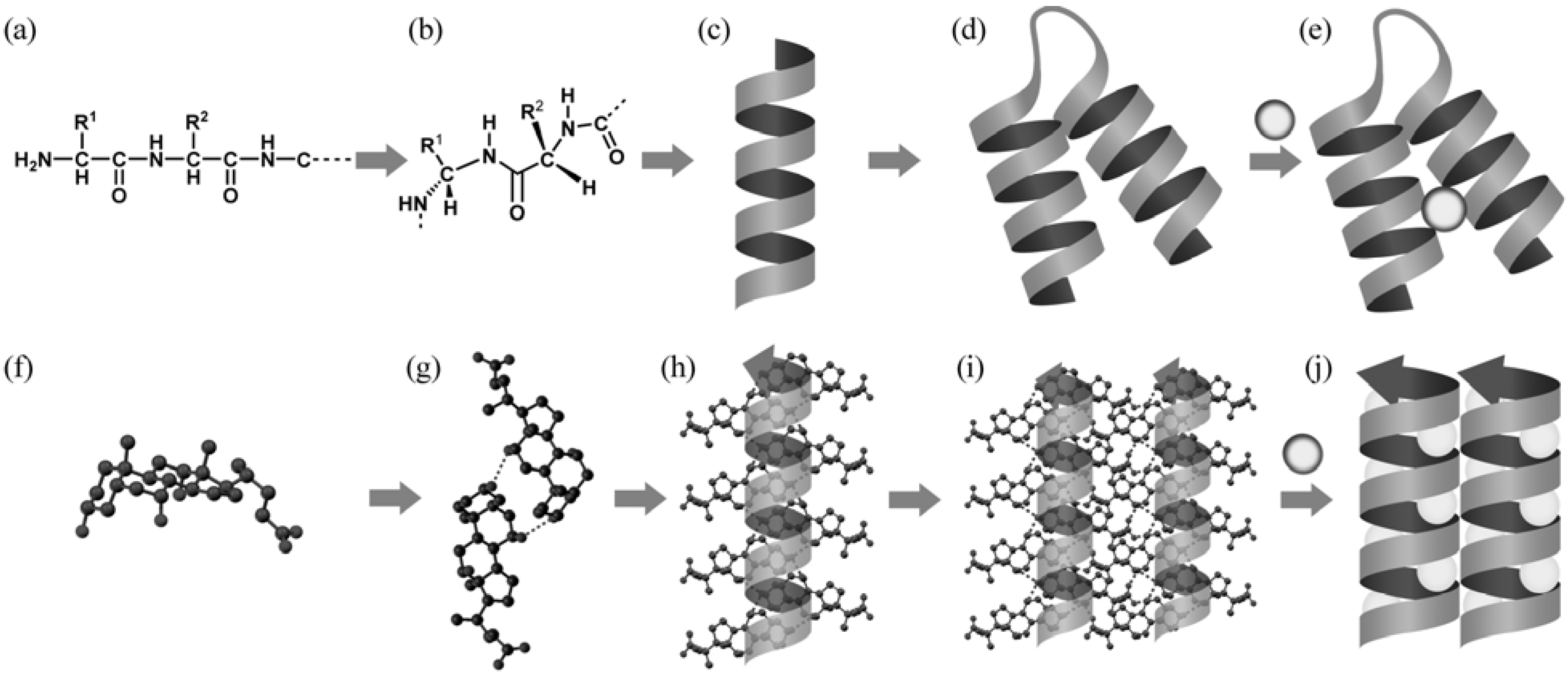
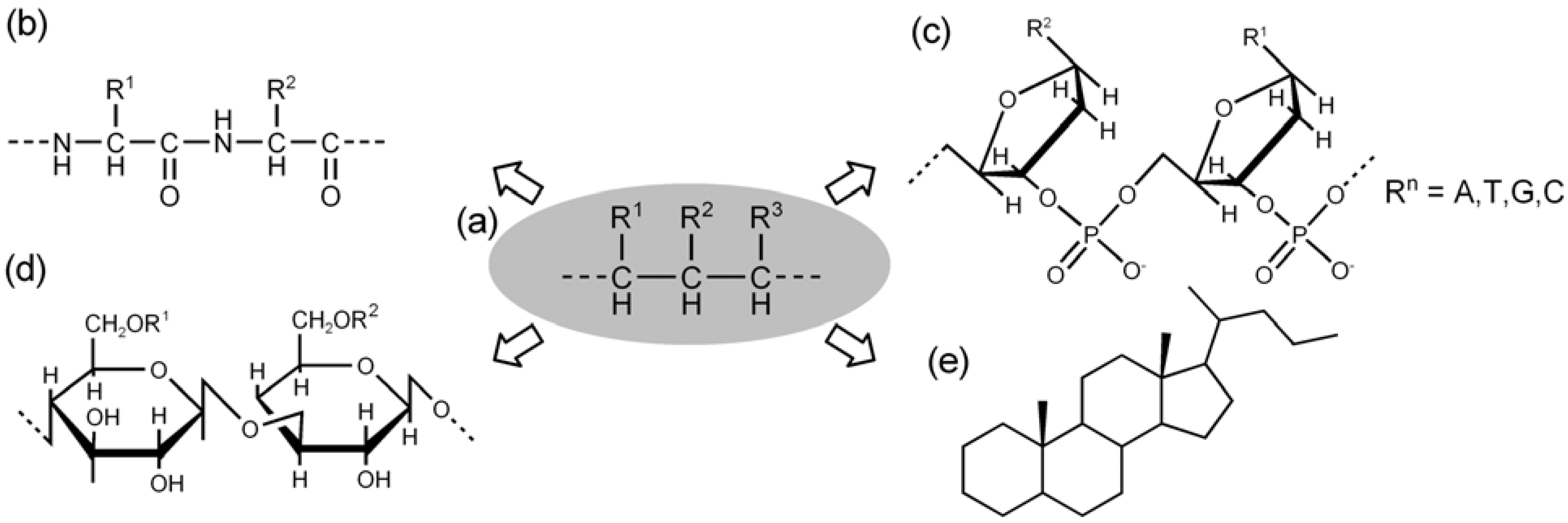
3.1. Intermediate among Various Molecules and Assemblies

As described above, we obtained comprehensive experimental results about inclusion behaviors and crystal structures of bile acids and their derivatives. Their remarkable features hinge on the following two matters. The first is that the inclusion behaviors vary from one case to another, and depend on subtle differences in donor-acceptor relationships among the hydrogen bonding groups. The other is that the steroidal molecules form diverse assemblies, such as monolayers, bilayers, helical tubes, and so on. The assembly modes are host-inherent in some cases but guest-dependent in the other cases. These results brought about the idea that the steroidal molecules are located in an intermediate among small molecules and biopolymers from various viewpoints of host-guest, inclusion, recognition, polymorphism [67,68,69,70,71,72], dynamics [17,73,74,75,76,77], polymerization [78,79,80,81,82,83], separation [3,84,85], and so on, as shown schematically in Figure 5.
Theoretically, there exist many possible combinations of intermolecular non-covalent bonds, enabling us to simulate many possible assemblies among the host and guest components. Along with this simulation study, an overview study directed us toward a research for any relationships between the molecular structures and assemblies. We expected that the relationship might be closely connected with various fundamental concepts established for proteins, since multiple non-covalent bonds play a critical role in both the steroidal molecules and proteins. In addition, it may be considered that the steroidal molecules form various crystal structures owing to environmental effects of organic solvents, while proteins form various folded structures in water.
Figure 5. Intermediate properties of crystalline assemblies of steroidal bile acids and their derivatives, which relate to supramolecular chemistry.
Molecules 12 01973 g005

3.2. Hierarchical Interpretation of the Steroidal Assemblies

The research is focused on the following question; how the steroidal host molecules assemble in corporation with guest molecules. Before going ahead with the question, we have to confirm the current situation that prediction of crystal structures still remains a challenging subject for many chemists [86,87,88].
Figure 6. Hierarchical structures in proteins (upper) and steroids (lower). The peptide chain (a) is folded with a relative position between the two units (b), yielding a helical chain (c) and a bundle of the helical chains (d), followed by a host-guest complexation (e). The steroidal molecules (f) associate to make a bimolecular assembly (g) and a helical assembly (h), followed by a bundle of the chains (i). The cavities among the helices accommodate guest components (j).
Molecules 12 01973 g006
We now tried to obtain a qualitative answer to the question on the basis of the simulation and overview studies. The first thing we noticed is that most of the steroidal assemblies consist of helical moieties, which reminds us of helical structures of proteins. This is attributed to the well-known fact that proteins express their molecular information through non-covalent bonds via the following hierarchical processes; sequential chains as primary structures, helical stereostructures as secondary ones, bundles of the helices as tertiary ones, and so on [Figure 6(a)–(e)].
Therefore, an analogy emerged between the steroidal molecules and proteins. That is, the steroidal molecules also construct a hierarchical structure involving primary, secondary, tertiary structures [Figure 6(f)–(j)] [89,90,91,92]. Thus, the steroidal molecules themselves constitute primary structures [Figure 6(f)], followed by the subsequent bimolecular [Figure 6(g)] and helical [Figure 6(h)] assemblies, as secondary structures. The helical assemblies are combined to produce their bundles as tertiary structures [Figure 6(i)]. When the bundles leave cavities for accommodating guest components, host-guest complexes are constructed as quaternary structures [Figure 6(j)].

3.3. Molecular Information and Expression of Chiral Chains

Such a hierarchical understanding of the steroidal assemblies reminded us of the concept of molecular information and expression. It is well-known that peptide chains serve as storage of the molecular information, which are expressed in the process for making three-dimensional structures and accommodating other molecules. It is also known that the steroidal molecules originate from hexamers of isoprene, suggesting that the steroidal molecules are substantially composed of the carbon-chains, which may serve as information storages for constructing any three-dimensional assemblies [1,2,3,4,17,93,94,95,96,97]. This analogy between the chains indicates that various informational molecules are theoretically created by means of chemical modifications of the carbon-chains, as shown in Figure 7.
Figure 7. A polymethylene chain with various substituents (a) serves as storage of molecular information. The sequential carbon chain can be theoretically modified to a polypeptide chain (b), a polynucleotide chain (c), a polysaccharide chain (d), and a polycyclic chain (e).
Molecules 12 01973 g007
Figure 7(a) shows a chiral carbon-chain composed of methylene units with various substituents and absolute configurations. In case of the chains, we should notice that a beginning and terminal points define directionality of the chains. The methylene units can arrange in different ways, and each carbon-chain is termed as a sequential chain with molecular information at a nanometer level. Such a chain can be chemically modified to make the best use of non-covalent bonds. Thus, the carbon-chain is converted to proteins with amide bonds [Figure 7(b)], nucleic acids with phosphate and sugar moieties [Figure 7(c)], polysaccharides with sugar moieties [Figure 7(d)], and steroidal molecules with polycycles [Figure 7(e)].
The remarkable feature of these chains is that chirality of the carbon atoms is essential for constructing definite three-dimensional structures with chirality. This concept induced the subsequent idea that the steroidal molecules must make their hierarchical assemblies with chirality in the crystalline state. In other words, the assemblies may exhibit supramolecular chirality in the crystalline state. The next chapter deals with our research directed toward the supramolecular chirality in each hierarchical assembly.

4. Supramolecular Chirality in Crystalline Molecular Assemblies

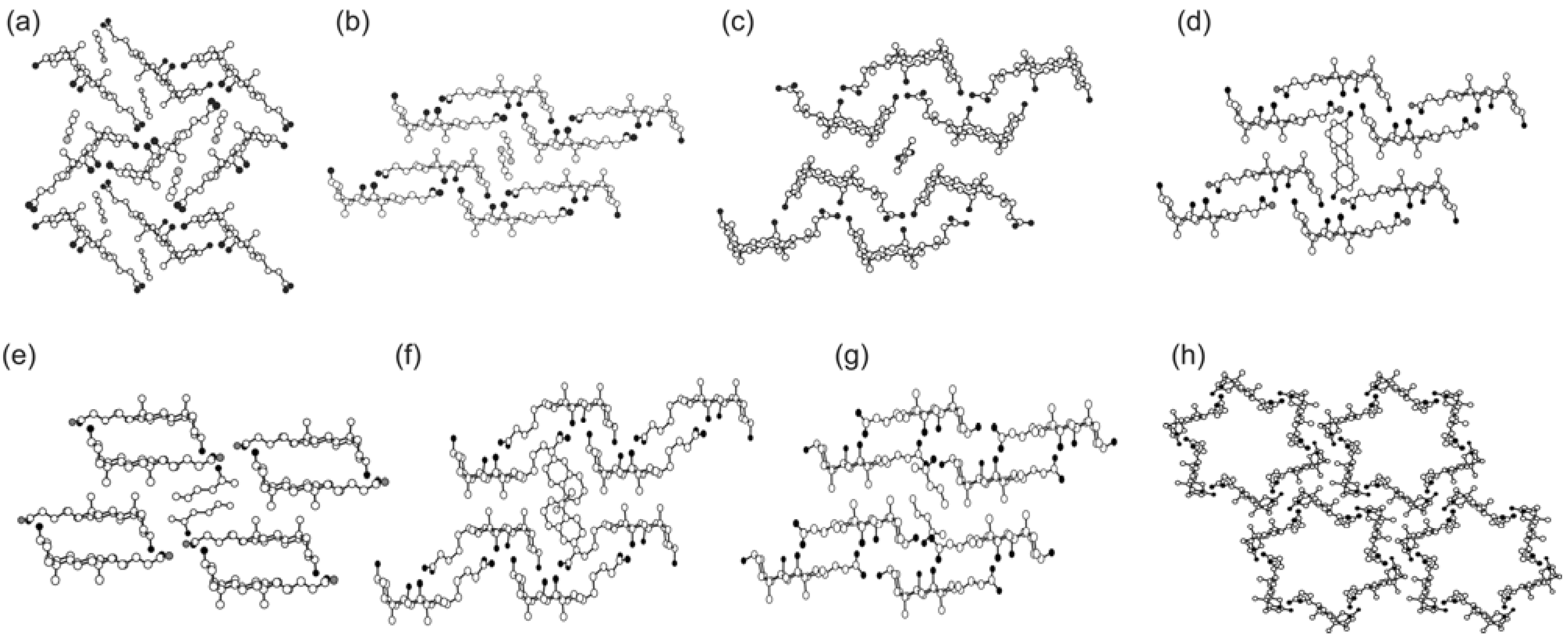
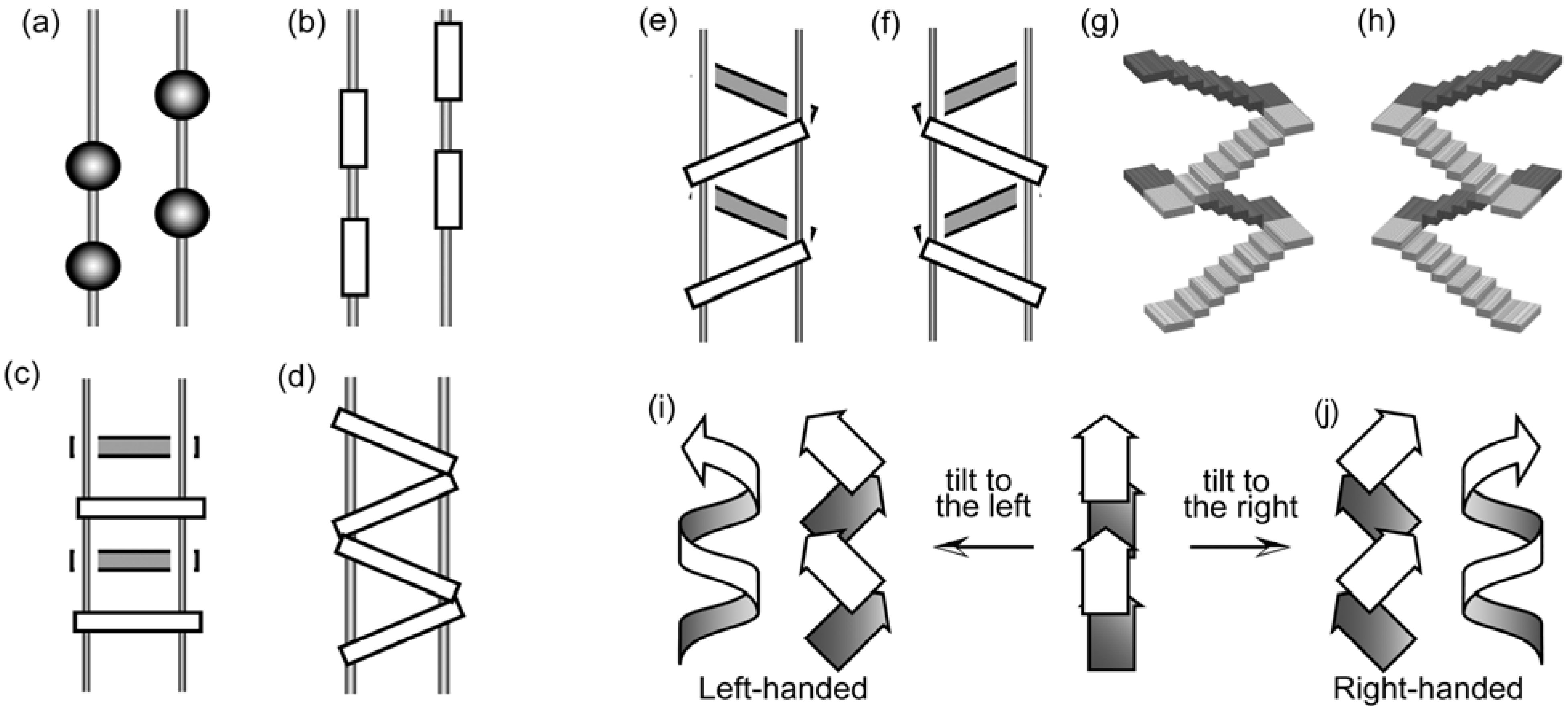
4.1. Chirality of the Hierarchical Assemblies, Especially 21 Helical Assemblies

As described above, we obtained the idea that the chiral chains may dominate chirality of the subsequent assemblies, such as helices, bundles, and host-guest complexes. Namely, each hierarchical assembly of the steroidal molecules must have its own three-dimensional structures with chirality, starting from the original molecular chirality, as shown in Figure 8 [4,92].
Figure 8. Molecular and supramolecular chirality of various molecular assemblies. Tilt chirality in a bimolecular assembly causes helical chirality in the resulting helical assembly. The helices are tied up to make various bundles as chiral crystals. The three-dimensional chirality of the molecular assemblies are interpreted in a hierarchical way.
Molecules 12 01973 g008
Such an idea is universal for proteins, but not for the steroidal molecules. To the best of our knowledge there seemed to be no studies which clarified a correlation among their molecular structures, helical assemblies and crystal space groups from the viewpoint of both molecular and supramolecular chirality. In other words, we have to search for systematic ways of describing the supramolecular chirality of the steroidal assemblies in the crystalline state.
In this manner we encountered a new subject; how to define supramolecular chirality of molecular assemblies starting from molecular chirality. The first idea is that the steroidal molecules are analogous to vertebrate animals or humans with three-axial chirality [1,2,3,4]. Secondly, as Kitaigorodskii pointed out [98], organic molecules without symmetry elements predominantly form 21 helical assemblies with chiral space groups, such as P21, P212121, and so on. Since the steroidal molecules have no symmetry elements, it may be assumed that the molecules form the 21 helical assemblies related to the corresponding chiral space groups. Thirdly, if the three-axial chirality of the starting molecules would be transferred to three-axial chirality of the 21 helical assemblies, we would be further able to interpret supramolecular chirality of the subsequent bundles and space groups. Therefore, the key for interpreting their chirality lies in handedness of the 21 helical assemblies.
We stopped due to a mysterious problem, that there are no general rules to determine handedness of the 21 helical assemblies. In the present situation, the handedness of the 21 helical assemblies is not conventionally discussed, even when we employ highly asymmetric molecules, such as steroids. In daily life, however, we have experiences of going up and down right- or left-handed stairs with 21 helical arrangements, indicating that the key structures, 21 helical assemblies, may have their handedness which can be defined in some ways. Recently, we have introduced the term, three-axial and tilt chirality for defining the handedness of the bimolecular as well as 21 helical assemblies [99,100,101]. This definition has been proven to be powerful in the elucidation of many structural problems of the assemblies.

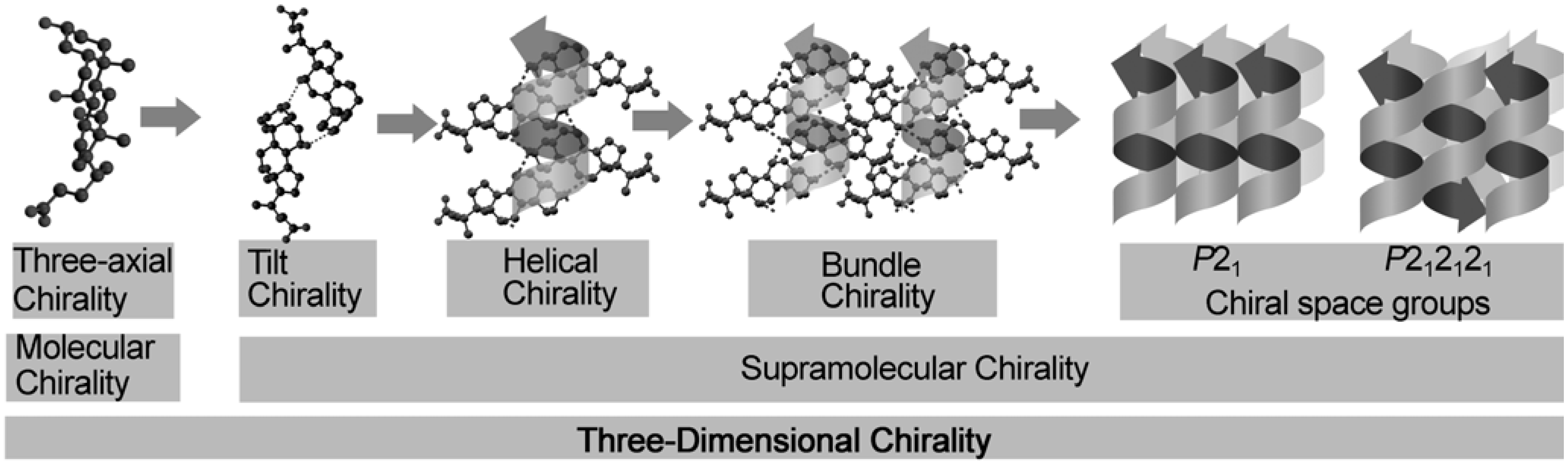
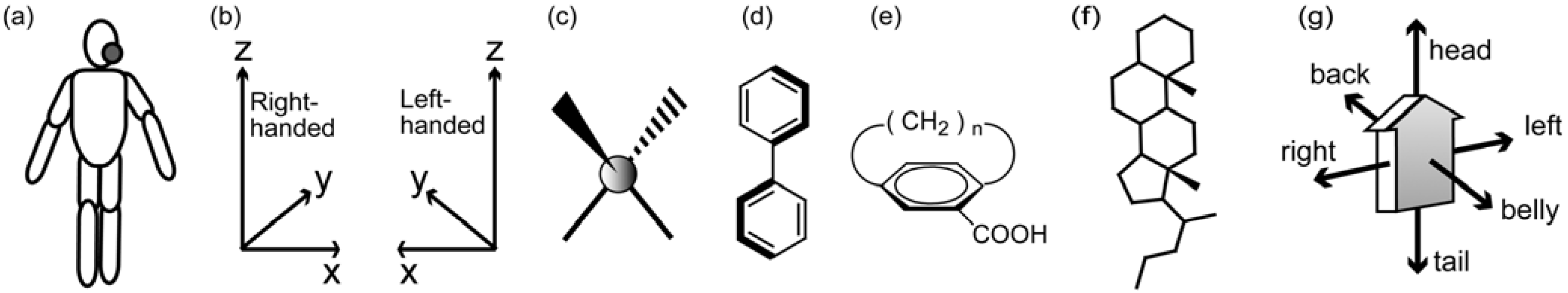
4.2. Three-axial Chirality of Molecules

We live in three-dimensional space world, and naturally understand stereostructures of materials in three dimensions. For example, we daily use a set of words for our bodies, head-and-leg, right-and-left, and belly-and-back [Figure 9(a)]. Mathematically, there is an orthogonal coordinate system with x, y, and z-axes, which accompanies right- or left-handedness [Figure 9(b)].
Figure 9. Three-axial chirality of materials. Human (a), mathematical three axes with right-handed or left-handedness (b), molecules with center (c), axial (d), and plane (e) chirality, a steroidal molecule (f), and a facial material with three-axial chirality (g).
Molecules 12 01973 g009
In the field of chemistry, molecular asymmetry has been conventionally expressed in terms of center- [Figure 9(c)], axis- [Figure 9(d)], and plane-chirality [Figure 9(e)]. These terms suggest an understanding of molecular chirality based on zero- (center), one- (axis), two- (plane) dimensional characteristics, although the molecules substantially have three-dimensional stereostructures.
However, these terms in chemistry are not suitable to express the whole chirality of arched and polycyclic molecules such as bile acids, although the terms are enough to express the local chirality of these molecules. Instead, we notice total characteristics of the steroidal molecules, that is, three-axial chirality. Since the steroidal molecules have an arched skeleton with a side chain [Figure 9(f)], the molecule is connected with a vertebrate animal, whose body is expressed by the daily words; head and tail (leg), right and left, as well as belly and back [Figure 9(a)]. In general, the facial molecule with three-axial chirality is schematically shown in Figure 9(g).

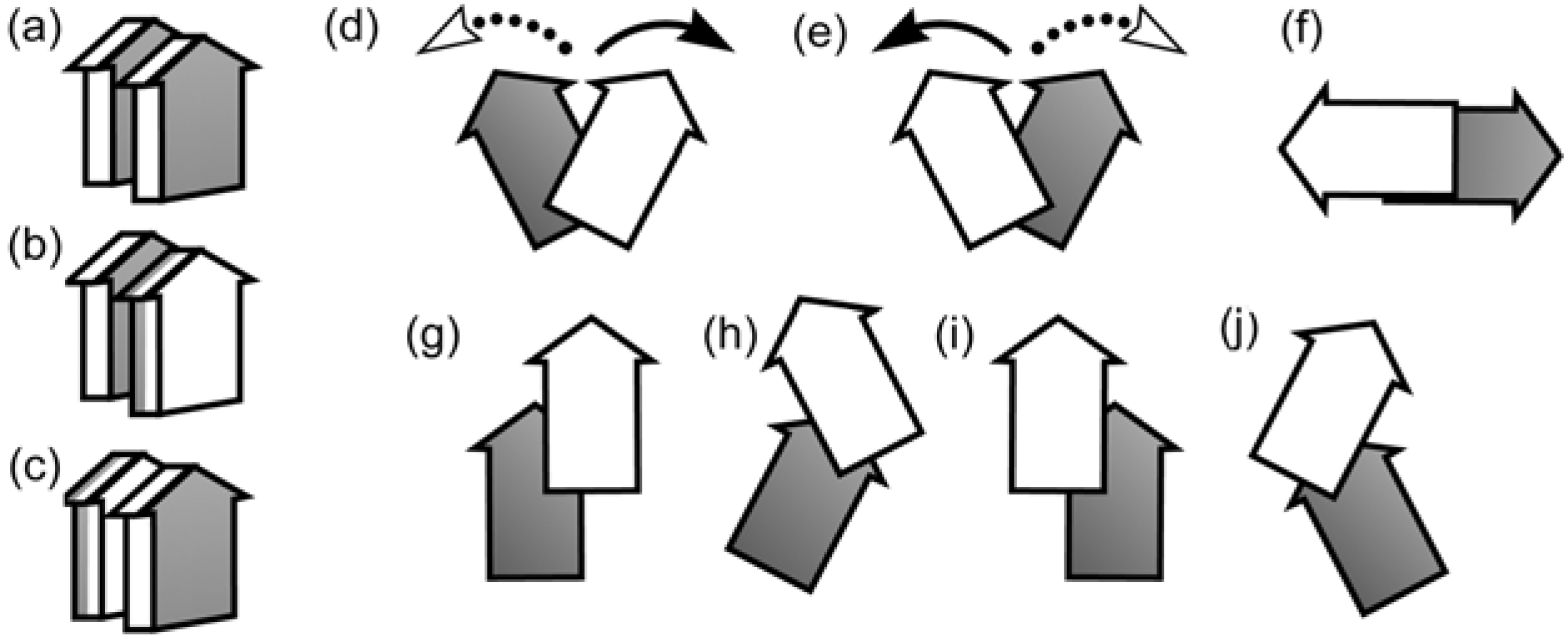
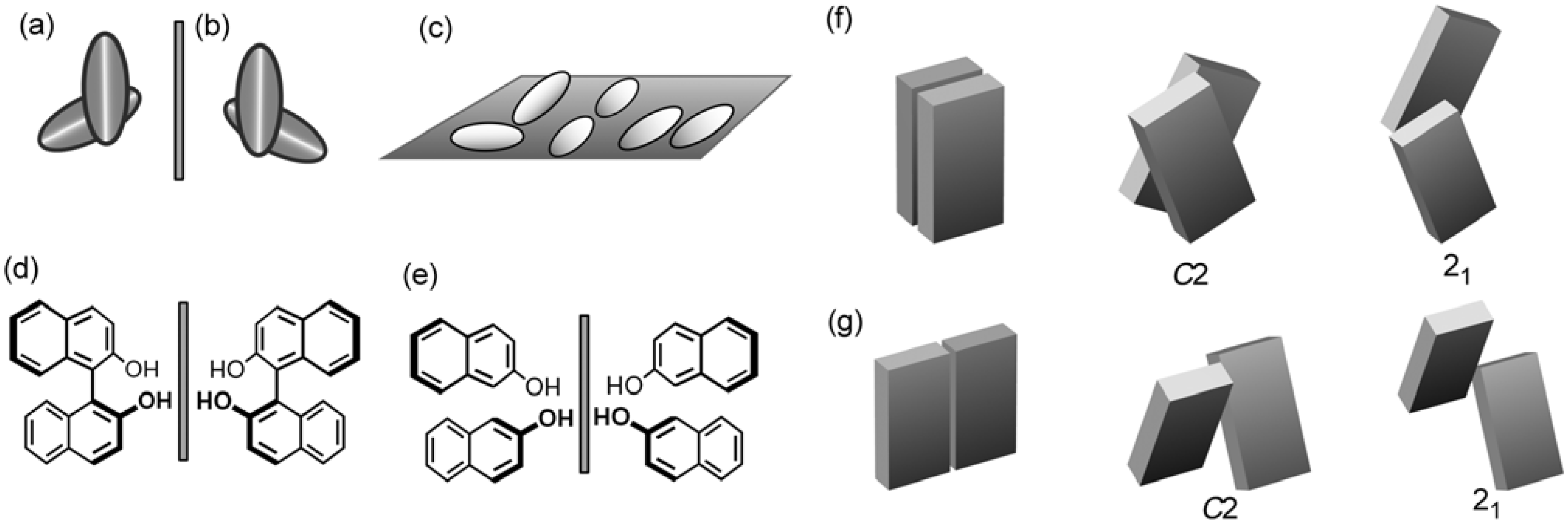
4.3. Tilt Chirality of Bimolecular Assemblies Based on Three-Axial Chirality

From a mathematical viewpoint, an assembly composed of two objects can form distinguishable enantiomorphic polyhedrons in three-dimensional world [102]. For example, the assembly of polyhedrons [Figure 10(a)] is not superimposable on its mirror image [Figure 10(b)], as in the case of right- or left-handed orthogonal coordination system [Figure 9(b)]. However, when the assembly composed of the two objects is put on an identical plane without stacking in various ways [Figure 10(c)], the assembly can be superposed on its mirror image. In general, a three-dimensional assembly with three-axial chirality cannot be superposed upon each other, while the assembly with two-axial chirality can be done. In other words, the appearance of supramolecular chirality requires a tilt between two objects, as shown in Figure 10(a) and Figure 10(b). We have proposed to use the term, supramolecular tilt chirality, for expressing such chirality [99,100].
Figure 10. Supramolecular tilt chirality of assemblies of two materials. An assembly of two polyhedrons with tilt chirality (a), its mirror image (b), their assemblies without tilt chirality (c), 1,1’-bi-2-naphthol with axial chirality (d), mirror images of hypothetical assemblies made by a bond collapse of 1,1’-bi-2-binaphthol (e), bimolecular assemblies of planar molecules with 21 or C2 symmetry in a face-to-face (f) or an edge-to-edge mode (g).
Molecules 12 01973 g010
First of all, we shall recall a single molecule with axis-chirality, such as 1,1’-bi-2-naphthol [Figure 10(d)], where two benzene rings have a tilt conformation. Hypothetical removal of the covalent bond between the rings would result in a tilt bimolecular assembly [Figure 10(e)]. This situation of the hypothetical assemblies is briefly displayed as models composed of two plates [Figure 10(f) and Figure 10 (g)]. The former model shows an assembly in a face-to-face stacked alignment, while the latter does in an edge-to-edge one. A tilt between the plates yields a chiral object with twofold axis (C2), and further sliding yields another chiral object with twofold screw axis (21). The hypothetical bimolecule in the right part of Figure 10 (e) corresponds to the object with twofold axis (C2) in Figure 10 (g).
Figure 11. Tilt chirality of assemblies of two materials with three-axial chirality. Three association modes with a head-to-head mode in addition to a belly-to-back (a), belly-to-belly (b), back-to-back (c) modes. Inclination between two materials with right-tilt (d), left-tilt (e), rotation by 90 degrees (f). Combination of sliding and tilt; right and up sliding (g), left-tilt after the right and up sliding (h), left and up sliding (i), right-tilt after the left and up sliding (j).
Molecules 12 01973 g011
The first step toward molecular assemblies is logically the formation of a bimolecular assembly. When the materials with three-axial chirality [Figure 9(g)] align in a head-to-head fashion, they have the following three association modes; belly-to-back [Figure 11(a)], belly-to-belly [Figure 11(b)] as well as back-to-back [Figure 11(c)]. Each of the bimolecular assemblies can be rotated toward reverse directions at the same time. As for belly-to-belly association [Figure 11(b)], a clockwise rotation of the upper and an anti-clockwise rotation of the lower yield a right-tilt position [Figure 11(d)]. The reverse rotations yield a left-tilt position [Figure 11(e)]. Figure 11(f) illustrates a tail-to-tail association after the rotation by 90 degrees. Moreover, the belly-to-belly assembly [Figure 11(b)] can slide toward up-to-right as well as up-to-left yield another positions [Figure 11(g) and Figure 11(i)], which further can tilt to the left [Figure 11(h)[ or to the right [Figure 11(j)].

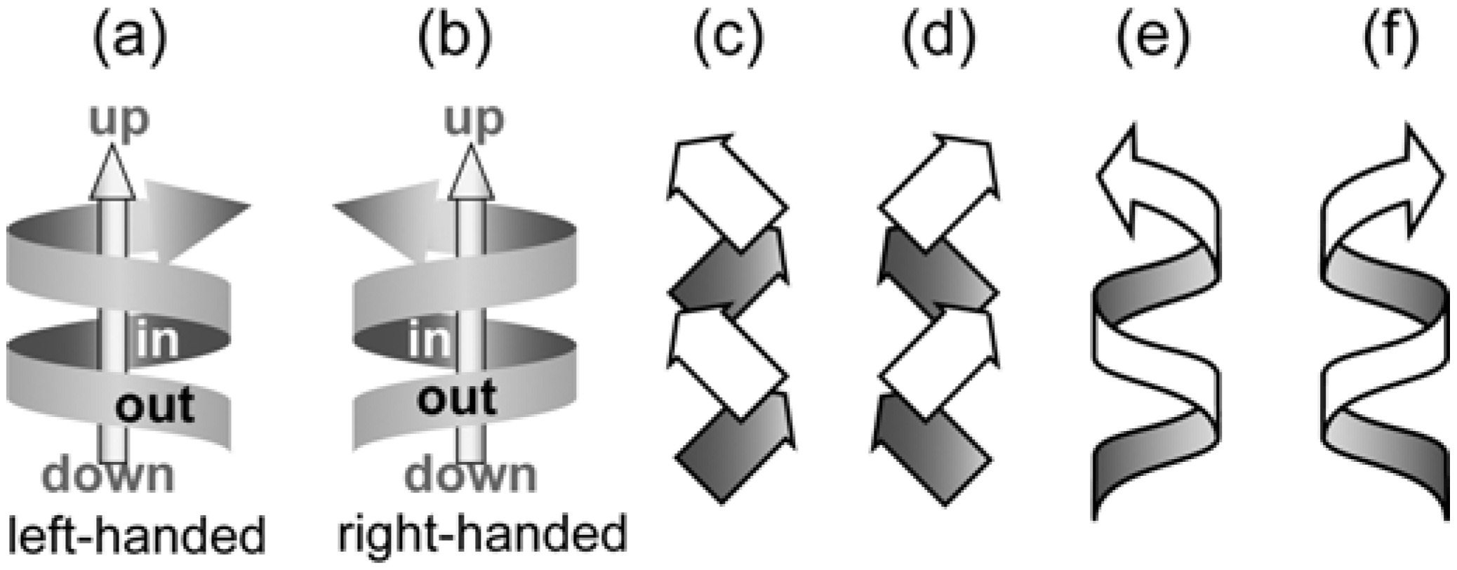
4.4. Helical Chirality of 21 Helical Assemblies Based on Three-axial and Tilt Chirality

Conventionally, it has been considered from a mathematical viewpoint that right- or left-handedness of the 21 helical assemblies cannot be distinguished, since the twofold screw axis operation includes rotation by 180 degrees and translation. This is true when we consider spherical materials [Figure 12(a)], but the situation changes when we consider other non-spherical materials. For example, when bars can be regularly arranged in a parallel [Figure 12(b)], perpendicular [Figure 12(c)], and zigzag [Figure 12(d)] fashion against an axis on the same plane, the mirror images can be superposed upon each other. However, when they are arranged in a zigzag fashion alternating on two sides of a plane [Figure 12(e)], the bars cannot be superposed upon their mirror image [Figure 12(f)]. As in the case of stairs in daily life [Figure 12(g) and Figure 12(h)], we can define handedness of the 21 helical assemblies on the basis of the supramolecular tilt chirality. As schematically shown in Figure 12(i), when the aligned bars incline to the right or left in front of a 21 screw axis, the assemblies are defined to be right- or left-handed, respectively. Graphically, the helical assemblies are designated with the corresponding helical tapes.
Figure 12. Helical chirality based on tilt chirality. 21 Helical assemblies of spheres (a), bars parallel to a 21 axis (b), bars perpendicular to a 21 axis (c), bars with a zigzag arrangement (d), bars with right-tilt arrangement (e) and a left-tilt arrangement (f), right-handed stairs with tilt chirality (g), left-handed stairs with tilt chirality (h). Definition of handedness of the 21 helical assemblies of planar molecules with left-handedness (i) and right-handedness (j).
Molecules 12 01973 g012
In general, helices with three-axial chirality are designated by the following three-axes; left and right, up and down as well as in and out [Figure 13(a) and Figure 13(b)]. When the up and in sides of the helix may correspond to the head and belly sides of the molecule with three-axial chirality, respectively, the molecules are stacked in the belly-to-belly mode on the in side of the helix. Therefore, helical chirality of the 21 helical assemblies is defined on the basis of the three-axial and tilt chirality of the bimolecular assembly as follows [101]. The sliding assembly in the belly-to-belly and left-tilt fashion [Figure 11(h)] can be expanded with a 21 symmetry operation to give its left-handed 21 helical assembly [Figure 13(c)], whereas the assembly in the right-tilt fashion [Figure 11(j)] can be expanded to give its right-handed 21 helical assembly [Figure 13(d)]. These 21 helical assemblies [Figure 13(c) and Figure 13(d)] are displayed with tape-like expression in Figure 13(e) and Figure 13(f), respectively.
Figure 13. Helices with three directions; up and down, in and out, as well as right and left. Right-handed helix (a) and left-handed helix (b). 21 Helical assemblies of molecules with three-axes; left-tilt, left-handed helix (c) and right-tilt, right-handed helix (d). Ribbon-like expression of the left-handed helix (e) and right-handed helix (f).
Molecules 12 01973 g013

4.5. Bundle Chirality Based on Three-axial, Tilt, and Helical Chirality

The 21 helical assemblies are tied up to yield bundles as tertiary structures. A basic hexagonal packing is that one helical assembly is surrounded by six other ones. Although there are many theoretical ways to assemble the helices with three-axial chirality, the representative bundles consist of helices with a parallel and anti-parallel alignment. Figure 14 shows five typical kinds of the bundles. A uniform bundle of either left- or right-handed helices gives chiral crystals with space group P21 in Figure 14(a) and Figure 14(b), respectively. Another bundle of either left- or right-handed helices with the reverse up-down directions give the corresponding chiral crystals with space group P212121 [Figure 14(c) and Figure 14(d)]. Figure 14(e) depicts a bundle of both the left- and right-handed helices with the reverse up-down directions, which corresponds to the crystal structure with space group P21/n. These space groups containing the twofold screw axes frequently appear in organic crystals [103].
Figure 14. Bundles of helices with three-axial, tilt, and helical chirality. Stacking in an identical direction with left-handed helices (a) and right-handed helices (b), in the reversed direction with left-handed helices (c) and right-handed helices (d), with both left- and right-handed helices (e).
Molecules 12 01973 g014

5. Supramolecular Chirality of Crystalline Assemblies of Bile Acids and Their Derivatives

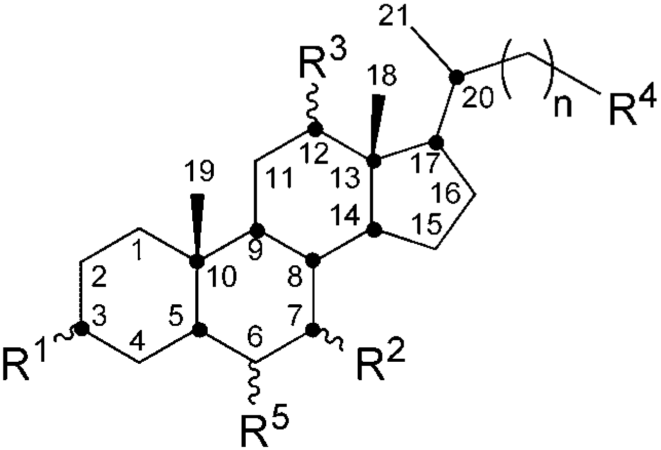
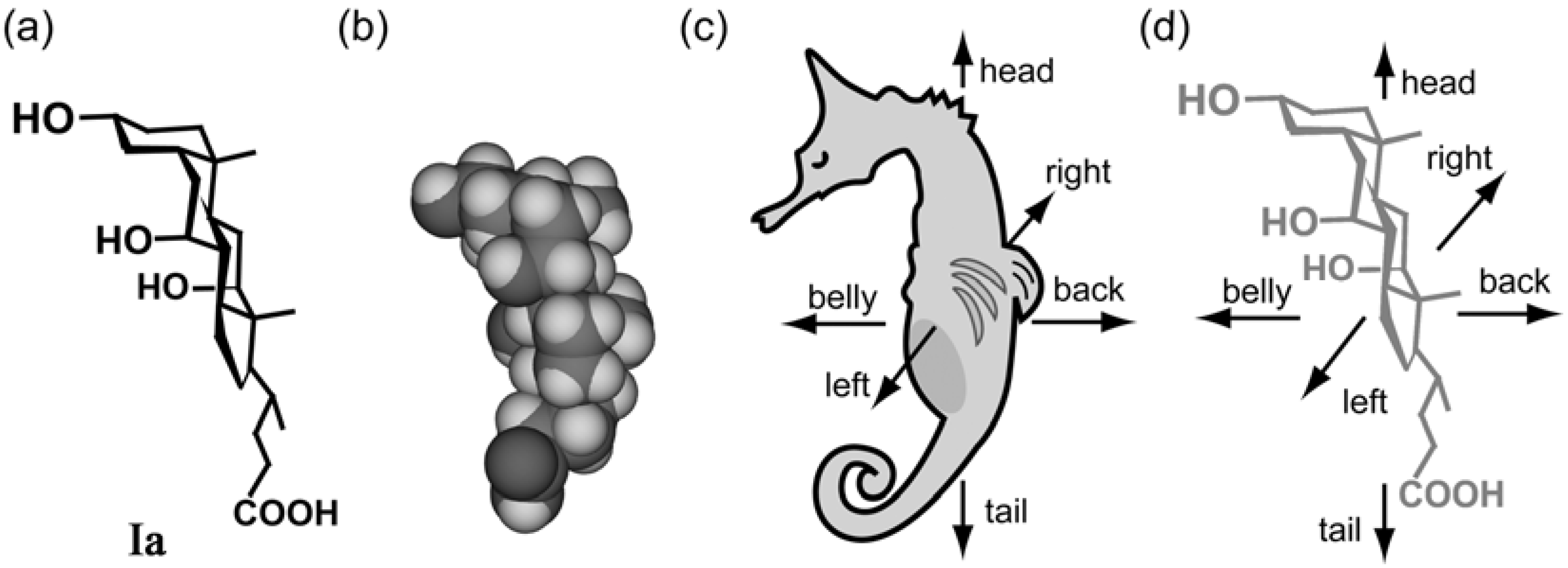
5.1. Steroidal Molecules with Three-Axial Chirality

When we saw the conventional structural formulae of Ia, for instance [Figure 15(a)], we did not get any ideas suitable for understanding its whole chirality. However, when we freely rotated a stereo model of the molecule [Figure 15(b)], we noticed that the molecule is like a vertebrate with three distinguishable directions [Figure 15(c)]. Accordingly, we proposed to use the common words head and tail (or leg), right and left, and belly and back, for defining the directions of the steroidal molecules.
The assignment of the three directions is as follows. The key is a curved steroidal skeleton and various groups attached. As shown in Figure 15(d), the molecular structure of Ia consists of a skeleton and a side-chain. The skeleton possesses three hydroxyl and two methyl groups to form a hydrophilic and a lipophilic face, respectively. This facial structure enables us to distinguish three axes as follows. First, we designate the hydrophilic and lipophilic sides as belly and back, respectively. Second, we recognize the side-chain as the tail, and the reverse arched part of the skeleton as the head. Third, we discriminate the hydroxyl groups at the 7 and 12 positions as left and right, respectively. In addition, molecule Ia is asymmetric owing to the methyl and tail groups on the right side of the skeleton, meaning that naturally-occurring bile acids and their derivatives are right-handed molecules with three-axial chirality.
Figure 15. A stereostructure of Ia (a), its space-filling model (b), a hypothetical animal (c), and a stereostructure of Ia with three-axial chirality (d).
Molecules 12 01973 g015

5.2. Steroidal Assemblies with Three-axial, Tilt, Helical, and Bundle Chirality

On the basis of the considerations described above, we can explain how to form crystals with chiral space groups starting from the steroidal molecules with three-axial chirality. Figure 16(a) to (d) show the hierarchical structure of Ia. The molecules of Ia [Figure 16(a)] are connected with a hydrogen bonding network among three hydroxyl groups at the 3-, 7-, and 12-positions. The resulting bimolecular assembly has a belly-to-belly association, an up-to-left sliding, and a right-tilt inclination [Figure 16(b)], and is expanded with 21 symmetry operation to construct a right-handed 21 helical assembly [Figure 16(c)]. The helices align in a parallel fashion with carboxylic groups to yield a crystal structure with monoclinic, P21 space group [Figure 16(d)]. This Figure corresponds to that obtained with a rotation upward by 90 degrees of Figure 2(b), which shows a parallel stacking of bilayers.
Figure 16. Hierarchical assemblies of steroidal bile acids and their derivatives. The molecules of Ia (a) and IIa (e) form right-tilt bimolecular assemblies (b) and (f), 21 helical assemblies with right handedness (c) and (g), and bundles of the helices as chiral crystals with P21 (d) and P212121 (h) space groups, respectively. The molecules of IIIa (i) form a left-handed 21 helical assembly (j), and the molecules of IVa (k) form a slightly right-handed helix (l).
Molecules 12 01973 g016
The hierarchical structure of IIa is shown in Figure 16(e) to Figure 16(h). The molecules of IIa [Figure 16(e)] are connected with the network among two groups at the 3- and 12-positions. The connection results in a right-tilt bimolecular assembly [Figure 16(f)], followed by a right-handed 21 helical assembly [Figure 16(g)]. The helices align in an anti-parallel fashion with the carboxylic groups to yield a crystal with orthogonal, P212121 space group [Figure 16(h)], which shows an anti-parallel stacking of bilayers. This Figure corresponds to that obtained by a rotation up and right by 90 degrees of Figure 2(c), which shows an anti-parallel stacking of bilayers. On the other hand, IIIa has a left-tilt [Figure 16(i)] and a left-handed 21 helical assembly [Figure 16(j)] owing to the bias of the hydroxyl groups at the 3- and 7-positions towards the left side. In contrast, IVa has a helical hydrogen bonding network with hydroxyl groups and carboxylic groups, so that the molecules lay in a perpendicular and slightly right-tilt position to the 21 axis [Figure 16(k)]. The resulting 21 helical assembly [Figure 16(l)] is right-handed. These results indicate that the tilt and handedness of the steroidal assemblies strongly depend on substituted positions of the hydroxyl groups in the steroidal skeleton.

5.3. Complementary Chirality in Host-Guest Complexes

The bundles of helices may leave cavities where the other organic substances are accommodated. Since the steroidal molecules have both hydrophilic and lipophilic sides, the resulting helices usually have lipophilic outsides where any sliding may take place. Such sliding enables the assemblies to accommodate the guest components in various ways, resulting in guest-dependent polymorphism of the host components. Key and lock mechanism is widely accepted as a principle of molecular recognition [104]. This concept is based on stereo and electronic complementarity between concave host cavities and convex guest compounds. Therefore, complementary compounds are usually included in the host cavities.
The 21 helical assemblies of the host components may leave 21 helical assemblies of the cage- or channel-like cavities, indicating that the guest components may produce the 21 helical assemblies by using the cavities. Therefore, not only chiral but also achiral guest molecules may yield handedness of the 21 helical assemblies. Indeed, inclusion crystals of Ia with benzene [Figure 17(a)] exhibit right-handed 21 helical assemblies of both Ia and benzene on the basis of supramolecular tilt chirality [Figure 17(b)] [105]. Moreover, we confirmed that about 40 kinds of benzene derivatives exhibit right-handed 21 helical assemblies in the inclusion crystals of Ia. Such a complementary relationship is compared to a right-handed bolt and nut [Figure 17(c)], and are observed for many crystalline host-guest inclusion systems. We consider that this complementary chirality should be more frequently discussed as one of supramolecular chirality in the crystalline state.
Figure 17. Complementary chirality of the 21 helical assemblies of host and guest components. The inclusion crystal of Ia with benzene (a), bundles of right-handed helical assemblies of Ia and benzene (b), and a combination of a right-handed bolt and nut (c).
Molecules 12 01973 g017

5.4. Chirality Recognition Based on Supramolecular Chirality

The steroidal assemblies form chiral spaces which function to accommodate chiral guest components. We performed systematic studies on enantioresolution of various secondary alcohols [3,106]. This is because hosts such as Ib, IIb, IVb, IIe, Ig, and IIg include aliphatic and aromatic alcohols, as described in Chapter 2. Ib prefers (R)-isomers to (S)-isomers, and the efficiency depends on alkyl groups of the alcohols in size and shape [107,108]. Most of the guest components are included in the channels, but (S)-2,2-dimethyl-3-hexanol is exclusively included in the cage-like cavities [109]. IVb includes aliphatic alcohols involving over four carbon atoms [110], and prefers (R)-isomers of the secondary alcohols to (S)-isomers. IIe is useful for 4-methyl-2-pentanol [111]. Ig and IIg display efficient chiral recognition for the aliphatic alcohols, especially 3-methyl-2-pentanol [112]. Ia functions for enantioresolution of lactones [113,114,115,116], cyclic ketones as well as epoxides with phenyl rings [117]. Dehydrocholic acid (Vi) is known to be an effective host for the enantioresolution of sulfoxides and cyclic amides [118,119,120,121]. It is noteworthy that Ia includes chiral conformers of achiral compounds [122,123,124].
We studied the detailed mechanism of chirality recognition by using bile acids and their derivatives. Unexpectedly, the mechanism for enantioresolution using inclusion crystals remains unclear. In fact, the three-point attachment model has been so far accepted for enantioselective complexation [125]. Figure 18(a) shows a simple description of the model which has a flat surface for accommodating the guest molecules. In this model, three locations (A, B, and C) of a guest component meet three locations (A’, B’, and C’) on the flat surface. However, falling of the surface may induce the following change. Four substituents at a chiral carbon attach to the wall of the dip. When the fourth substituent is the smallest atom, hydrogen, both the enantiomers may be included into a deep dip. In such a case we have to fix a location of the fourth substituent. Therefore, we should employ a four location model [126], as shown in Figure 18(b). To fix the hydrogen attached with chiral carbon, it is important that location of chiral carbon should be fixed through some weak interactions or a bulky substituent. Figure 18(c) depicts one example of the crystal structure of the inclusion crystal of Ib with 2,2-dimethyl-3-hexanol [109]. It can be seen that the cage composed of six host molecules is occupied by three substituents attached to the chiral carbon; hydroxyl, n-propyl, and tert-butyl groups. The feature is that the n-propyl group has some close contacts with a methyl group at 21-position of the host (1) and at 19-position of the host (6), and that one methyl group of the tert-butyl group has a close contact with a methyl group at 18-position of the host (6). Namely, three methyl groups at 18, 19, and 21-positions of the steroidal molecules are related to the chirality recognition. This result indicates that chirality recognition may be accomplished by cooperative work through multiple weak interactions.
Figure 18. Models for chirality recognition. A three-location model (a) and a four-location model (b). Chirality recognition in a cage of the inclusion crystal of Ia with (S)-2,2-dimethyl-3-hexanol (c).
Molecules 12 01973 g018

6. Conclusions and Perspectives

We have presented the fact that steroidal crystalline assemblies exhibit the hierarchical structures with supramolecular three-axial, tilt, helical, and bundle chirality, according to the order of weak interactions. Alternatively, we continue to study polymerization of conjugated diene monomers in the inclusion and crystalline state. The latter study aims to obtain optically active polymers with quantitative chiral centers starting from achiral monomers, forcing us to prepare chiral assemblies of the monomers as chiral crystals. During this study we noticed the research for preparing chiral crystals from achiral molecules [127], and wanted to understand a mechanism how to acquire such crystals. This research gave a chance to consider three-dimensional chirality of materials, especially steroidal assemblies with complete three-dimensional chirality, leading us to the interpretation of supramolecular chirality in the hierarchical crystal structures.
The research naturally moves in the direction of supramolecular crystal engineering. Now our study is spreading from steroidal molecules to the surrounding natural compounds, such as brucine [128], cinchona alkaloids, and further to the surrounding organic substances, such as organic salts [129,130,131]. Supramolecular chirality of assemblies of these substances fascinates us so much. Our approach would provide an efficient view for creating new chiral crystals from chiral and/or achiral compounds, finding possible application to electronics, optics, photonics, informatics, and so on. Further studies are in progress for the crystal engineering, the fabrication of new materials, and so on.

References and Notes

  1. Miyata, M.; Sada, K. Deoxycholic Acid and Related Hosts. In Solid-State Supramolecular Chemistry, Comprehensive Supramolecular Chemistry; MacNicol, D. D., Toda, F., Bishop, R., Eds.; Pergamon: Oxford, 1996; Vol. 6, pp. 147–176. [Google Scholar]
  2. Miyata, M.; Sada, K.; Yoswathananont, N. Deoxycholic, Cholic, and Apocholic Acids. In Encyclopedia of Supramolecular Chemistry; Atwood, J. L., Steed, J. W., Eds.; Marcel Dekker: New York, 2004; Vol. 1, pp. 441–451. [Google Scholar]
  3. Miyata, M.; Yoswathananont, N.; Nakano, K.; Sada, K. Separation of Isomers and Enantiomers by Bile Acid Derivatives. In Separation and Reaction in Organic Supramolecular Chemistry, Perspectives in Supramolecuar Chemistry; Toda, F., Bishop, R., Eds.; John Wiley & Sons: Chichester, U,K., 2004; Vol. 8, pp. 87–122. [Google Scholar]
  4. Miyata, M.; Tohnai, N.; Hisaki, I. Crystalline Host-Guest Assemblies of Steroidal and Related Molecules: Diversity, Hierarchy, and Supramolecular Chirality. Acc. Chem. Res. 2007, 40, 694–702. [Google Scholar] [CrossRef] [PubMed]
  5. Davies, J.E.D.; Kemula, W.; Powell, H.M.; Smith, N.O. Inclusion Compounds – Past, Present, and Future. J. Inclus. Phenom. 1983, 1, 3–44. [Google Scholar]
  6. Atwood, J.L.; Davies, J.E.D.; MacNicol, D.D. (Eds.) Inclusion Compounds; Academic Press: London, 1984; Vol. 1–3.
  7. Lehn, J.-M. Supramolecular Chemistry: Concepts and Perspectives; VCH: Weinheim, 1995. [Google Scholar]
  8. Desiraju, G.R. (Ed.) The Crystal as a Supramolecular Entity, Perspectives in Supramolecuar Chemistry”; John Wiley & Sons: Chichester, U.K., 2004; Vol. 2.
  9. Sobotka, H. The Chemistry of the Bile Acids and Related Substances. Chem. Rev. 1934, 15, 311–375. [Google Scholar] [CrossRef]
  10. Fieser, L.F.; Fieser, M. Steroids; Reinhold: New York, 1959; pp. 56–61. [Google Scholar]
  11. Herndon, W.C. The Structure of Choleic Acids. J. Chem. Educ. 1967, 44, 724–728. [Google Scholar] [CrossRef] [PubMed]
  12. Craven, B.M.; DeTitta, G.T. Crystal Structure Determination of the 1:1 Complex of Deoxycholic Acid and Acetic Acid. J. Chem. Soc., Chem. Commun. 1972, 530–531. [Google Scholar]
  13. Giglio, E. Inclusion Compounds of Deoxycholic Acid. In Inclusion Compounds; Atwood, J.L., Davies, J.E.D., MacNicol, D. D., Eds.; Academic Press: London, 1984; Vol. 2, pp. 207–229. [Google Scholar]
  14. Johnson, P.L.; Schaefer, J.P. The Crystal and Molecular Structure of an Addition Compound of Cholic Acid and Ethanol. Acta Cryst. 1972, B28, 3083–3088. [Google Scholar] [CrossRef]
  15. Miyata, M.; Shibakami, M.; Goonewardena, W.; Takemoto, K. Inclusion Compounds of Cholic Acid with a Variety of Organic Substances. Chem. Lett. 1987, 605–608. [Google Scholar] [CrossRef]
  16. Miki, K.; Masui, A.; Kasai, N.; Miyata, M.; Shibakami, M.; Takemoto, K. New Channel-Type Inclusion Compound of Steroidal Bile Acid. Structure of a 1:1 Complex between Cholic Acid and Acetophenone. J. Am. Chem. Soc. 1988, 110, 6594–6596. [Google Scholar]
  17. Miyata, M.; Shibakami, M.; Chirachanchai, S.; Takemoto, K.; Kasai, N.; Miki, K. Guest-Responsive Structural Changes in Cholic Acid Intercalation Crystals. Nature 1990, 343, 446–447. [Google Scholar] [CrossRef]
  18. Jones, E.L.; Nassimbeni, L.R. Crystal and Molecular Structures of the Inclusion Compounds of Cholic Acid with Methanol, Ethanol and 1-Propanol. Acta Cryst. 1990, B46, 399–405. [Google Scholar] [CrossRef]
  19. Miki, K.; Kasai, N.; Shibakami, M.; Chirachanchai, S.; Takemoto, K.; Miyata, M. Crystal Structure of Cholic Acid with No Guest Molecules. Acta Cryst. 1990, C46, 2442–2445. [Google Scholar]
  20. Miyata, M.; Sada, K.; Hirayama, K.; Yasuda, Y.; Miki, K. Guest-Dependent Conformations of Side Chains in Cholic Acid Inclusion Compounds. Supramol. Chem. 1993, 2, 283–288. [Google Scholar] [CrossRef]
  21. Nakano, K.; Sada, K.; Miyata, M. Inclusion Compounds of Cholic Acid with Various Hydrocarbons and the Crystal Structure of a 1:1 Complex of Cholic Acid and Benzene. Chem. Lett. 1994, 137–140. [Google Scholar] [CrossRef]
  22. Shibakami, M.; Sekiya, A. Crystal Structures of Cholic Acid-Aniline and -3-Fluoroaniline Inclusion Compound; Fluorine Atom Effect on Channel and Hydrogen Bonding Pattern. J. Chem. Soc. Chem. Commun. 1994, 429–430. [Google Scholar] [CrossRef]
  23. Caira, M.R.; Nassimbeni, L. R.; Scott, J. L. Inclusion Compounds of Cholic Acid with Aliphatic Esters. J. Chem. Soc. Perkin Trans. 2 1994, 623–628. [Google Scholar]
  24. Nakano, K.; Sada, K.; Kurozumi, Y.; Miyata, M. Importance of Packing Coefficients of Host Cavities in the Isomerization of Open Host Frameworks: Guest-Size-Dependent Isomerization in Cholic Acid Inclusion Crystals with Monosubstituted Benzenes. Chem. Eur. J. 2001, 7, 209–220. [Google Scholar] [CrossRef] [PubMed]
  25. Yoswathananont, N.; Sada, K.; Miyata, M. Inclusion Compounds of Cholic Acid with Large Aliphatic Alcohols. Mol. Cryst. Liq. Cryst. 2002, 389, 47–51. [Google Scholar] [CrossRef]
  26. Lessinger, L. Cholic Acid Monohydrate, C24H40O5·H2O. Cryst. Struc. Commun. 1982, 11, 1787–1792. [Google Scholar]
  27. Lessinger, L.; Low, B.W. Crystal Structure and Hydrogen-Bonding System of Cholic Acid Hemihydrate, C24H40O5·1/2H2O. J. Cryst. Spectrosc. Res. 1993, 23, 85–99. [Google Scholar] [CrossRef]
  28. Caira, M.R.; Nassimbeni, L.R.; Scott, J.L. Crystal Structure and Multiphase Decomposition of a Novel Cholic Acid Inclusion Compound with Mixed Guests. J. Chem. Soc. Perkin Trans. 2. 1994, 1403–1405. [Google Scholar] [CrossRef]
  29. Miki, K.; Kasai, N.; Tsutsumi, H.; Miyata, M.; Takemoto, K. Structure of a 2:1 Complex between Deoxycholic Acid and Ferrocene. J. Chem. Soc. Chem. Commun. 1987, 545–546. [Google Scholar] [CrossRef]
  30. Nakano, K.; Sada, K.; Miyata, M. Guest-Participating Reversion of Molecular Arrangements in Asymmetric Multibilayers of Cholic Acid Inclusion Crystals. J. Chem. Soc. Chem. Commun. 1995, 953–954. [Google Scholar] [CrossRef]
  31. Yoswathananont, N.; Chirachanchai, S.; Tashiro, K.; Nakano, K.; Sada, K.; Miyata, M. A Novel Host Framework of Cholic Acid Inclusion Crystals by Slide and Flip of the Layers. Cryst. Eng. Comm. 2001, 3, 74–77. [Google Scholar] [CrossRef]
  32. Nakano, K.; Sada, K.; Aburaya, K.; Nakagawa, K.; Yoswathananont, N.; Tohnai, N.; Miyata, M. Guest-Induced Inversion of an Asymmetric Host Layer in Inclusion Crystals of Cholic Acid. Cryst. Eng. Comm. 2006, 8, 461–467. [Google Scholar] [CrossRef]
  33. Lindley, P.F.; Mahmound, M.M.; Watson, F.E.; Jones, W.A. The Structure of Chenodeoxycholic Acid, C24H40O4. Acta Cryst. 1980, B36, 1893–1897. [Google Scholar] [CrossRef]
  34. Rizkallah, P.J.; Harding, M.M.; Lindley, P.F.; Aigner, A.; Bauer, A. Structure of a Low-Temperature Polymoph of Chenodeoxycholic Acid, C24H40O4, Determined with Synchrotron Radiation. Acta Cryst. 1990, B46, 262–266. [Google Scholar] [CrossRef]
  35. Sluis, P.; Schouten, A.; Kanters, J.A. Structure of Chenodeoxycholic Acid in Chenodeoxycholic Acid Ethyl Acetate Solvate. Acta Cryst. 1990, C46, 2165–2168. [Google Scholar]
  36. Chikada, M.; Sada, K.; Miyata, M. Intercalation and Polymerization in Chenodeoxycholic Acid Channels with Retention of a Crystalline State. Polym. J. 1999, 31, 1061–1064. [Google Scholar] [CrossRef]
  37. Arora, S.K.; Germain, G.; Declercq, J.P. The Crystal and Molecular Structure of Lithocholic Acid. Acta Cryst. 1976, B32, 415–419. [Google Scholar] [CrossRef]
  38. Sada, K.; Kondo, T.; Miyata, M.; Tamada, T.; Miki, K. A Novel Molecular Channel with a Hydrogen Bond ’Hook’; Inclusion Phenomena of Cholamide and the Crystal Structure of a 1:1 Complex of Cholanamide and 1,4-dioxane. J. Chem. Soc. Chem. Commun. 1993, 753–755. [Google Scholar] [CrossRef]
  39. Sada, K.; Kondo, T.; Miyata, M.; Miki, K. Inclusion Crystals of Cholic Acid and Cholanamide with Alcohols: Importance of Hydrogen Bond “Double Hooks”. Chem. Mater. 1994, 6, 1103–1105. [Google Scholar] [CrossRef]
  40. Sada, K.; Matsuura, Y.; Miyata, M. Hydrogen Bond ’Triple Hooks’ in an Inclusion Crystal of Cholanamide with Aniline. Structural Difference of Inclusion Crystals of Cholic Acid and Cholanamide with Same Guest. Mol. Cryst. Liq. Cryst. 1996, 276, 121–127. [Google Scholar]
  41. Sada, K.; Kondo, T.; Ushioda, M.; Matsuura, Y.; Nakano, K.; Miyata, M.; Miki, K. Functionalization of Inclusion Cavities of Bile Acid Hosts. Channel-Type Inclusion Compounds of Cholamide. Bull. Chem. Soc. Jpn. 1998, 71, 1931–1937. [Google Scholar]
  42. Yoswathananont, N.; Sada, K.; Nakano, K.; Aburaya, K.; Shigesato, M.; Hishikawa, Y.; Tani, K.; Tohnai, N.; Miyata, M. The Effect of a Host-guest Hydrogen Bond on the Inclusion of Alcoholic Guests in the Host Cavities of Cholamide. Eur. J. Org. Chem. 2005, 5330–5338. [Google Scholar] [CrossRef]
  43. Sada, K.; Kondo, T.; Miyata, M. Channel-Type Inclusion Crystal with Hydrogen Bond ’Hooks’ of Deoxycholanamide. Supramol. Chem. 1995, 5, 189–191. [Google Scholar] [CrossRef]
  44. Hishikawa, Y.; Aoki, Y.; Sada, K.; Miyata, M. Selective Inclusion Phenomena in Lithocholamide Crystal Lattices; Design of Bilayered Assemblies through Ladder-Type Hydrogen Bonding Network. Chem. Lett. 1998, 1289–1290. [Google Scholar] [CrossRef]
  45. Miyata, M.; Goonewardena, W.; Shibakami, M.; Takemoto, K.; Masui, A.; Miki, K.; Kasai, N. Inclusion Compounds of Methyl Esters of Cholic and Deoxycholic Acids; Structure of a 1:1 Complex between Methyl Cholate and Methanol. J. Chem. Soc., Chem. Commun. 1987, 1140–1141. [Google Scholar]
  46. Miki, K.; Masui, A.; Kasai, N.; Goonewardena, W.; Shibakami, M.; Takemoto, K.; Miyata, M. Structures of 1:1 Addition Compounds of Methyl Cholate with Methanol and with 2-Propanol. Acta Cryst. 1992, C48, 503–507. [Google Scholar]
  47. Goonewardena, W.; Miyata, M.; Takemoto, K. One-Dimensional Inclusion Polymerization of Diene and Vinyl Monomers by Using Methyl Cholate as a Host. Polym. J. 1993, 25, 731–740. [Google Scholar] [CrossRef]
  48. Miki, K.; Masui, A.; Kasai, N.; Miyata, M.; Goonewardena, W.; Shibakami, M.; Takemoto, K. Structure of a 2:1 Addition Compound of Methyl Deoxycholate with Methanol. Acta Cryst. 1989, C45, 79–83. [Google Scholar]
  49. Sada, K.; Kondo, T.; Yasuda, Y.; Miyata, M.; Miki, K. Interpretation of Variable Inclusion Abilities of Cholic Acid and Its Derivatives on the Basis of the Crystal Structure of 5β-Petromyzonol. Chem. Lett. 1994, 727–730. [Google Scholar] [CrossRef]
  50. Sada, K.; Matsuo, A.; Miyata, M. Novel Molecular Arrangement in Asymmetric Bilayered Crystals of the Inclusion Compounds of 3α, 12α, 24-Trihydroxy-5β-cholane. Chem. Lett. 1995, 877–878. [Google Scholar] [CrossRef]
  51. Sugahara, M.; Hirose, J.; Sada, K.; Miyata, M. Inclusion Abilities of Bile Acids with Different Side Chain Length. Mol. Cryst. Liq. Cryst. 2001, 356, 155–162. [Google Scholar] [CrossRef]
  52. Sada, K.; Sugahara, M.; Nakahata, Y.; Yasuda, Y.; Nishio, A.; Miyata, M. First Columnar Monolayer Structure of Bile Acids Inclusion Crystal. Inclusion Compounds of 23-Nordeoxycholic Acid. Chem. Lett. 1998, 31–32. [Google Scholar]
  53. Sugahara, M.; Sada, K.; Miyata, M. A Robust Structural Motif in Inclusion Crystals of Norbile Acids. Chem. Commun. 1999, 293–294. [Google Scholar] [CrossRef]
  54. Kato, K.; Sugahara, M.; Tohnai, N.; Sada, K.; Miyata, M. Drastic Increase in the Flexibility of Open Host Framework of a Steroidal Host Compound upon Shortening Its Spacer. Eur. J. Org. Chem. 2004, 981–994. [Google Scholar] [CrossRef]
  55. Sada, K.; Sugahara, M.; Kato, K.; Miyata, M. Controlled Expansion of a Molecular Cavity in a Steroid Host Compound. J. Am. Chem. Soc. 2001, 123, 4386–4392. [Google Scholar]
  56. Miyata, M.; Sada, K.; Miyake, Y. Combinatorial Chemistry of Inclusion Compounds By Using Steroids. Mol. Cryst. Liq. Cryst. 1998, 313, 173–178. [Google Scholar] [CrossRef]
  57. Miyake, Y.; Hirose, J.; Hasegawa, Y.; Sada, K.; Miyata, M. Novel Channel-Type Inclusion Compounds of 3-Epiursodeoxycholic Acid. Chem. Commun. 1998, 111–112. [Google Scholar] [CrossRef]
  58. Popovitz-Biro, R.; Tang, C.P.; Chang, H.C.; Lahav, M.; Leiserowitz, L. Solid-State Photochemistry of Guest Aliphatic Ketones Inside the Channels of Host Deoxycholic and Apocholic Acids. J. Am. Chem. Soc. 1985, 107, 4043–4058. [Google Scholar]
  59. Miyake, Y.; Sada, K.; Miyata, M. Inclusion Compounds of Hyocholic Acid with Specific Alcohols. Supramol. Chem. 1998, 10, 107–109. [Google Scholar] [CrossRef]
  60. Miyake, Y.; Sada, K.; Miyata, M. Inclusion Compounds of Hyodeoxycholic Acid and the Crystal Structure of a 1:1 Complex between the Acid and Pyridine. Chem. Lett. 1997, 1263–1264. [Google Scholar] [CrossRef]
  61. Sada, K.; Maeda, T.; Miyata, M. Chiral Discrimination of 1-Phenylethylamine by Diastereomeric Salt Formation with Bile Acids. Crystal Structures of Cholic Acid Salts with (R)- and (S)-1-Phenylethylamine. 1996, 837–838. [Google Scholar]
  62. Sada, K.; Shiomi, N.; Miyata, M. Nanocavities with Fine Adjustment in Channel-Type Inclusion Crystals of Alkylammonium Deoxycholates. Control of Molecular Cavities by Partial Filling of Molecular Channels. J. Am. Chem. Soc. 1998, 120, 10543–10544. [Google Scholar]
  63. Sada, K.; Hishikawa, Y.; Kondo, T.; Miyata, M. Specific Inclusion of Water by N-Methyldeoxycholanamide Revealed by the Crystal Structure of the Hemihydrate. Chem. Lett. 1994, 2113–2116. [Google Scholar] [CrossRef]
  64. Hishikawa, Y.; Watanabe, R.; Sada, K.; Miyata, M. Molecular Arrangements in Chiral Sheets of N-Alkylcholamides Bilayered Crystals. Chirality 1998, 10, 600–618. [Google Scholar] [CrossRef]
  65. Hishikawa, Y.; Sada, K.; Miyata, M. Extremely Specific Recognition of Ethylene Glycol in Three-leaved Building Blocks of N-Methyldeoxycholamide. Supramol. Chem. 1999, 11, 101–108. [Google Scholar] [CrossRef]
  66. Sada, K.; Kitamura, T.; Miyata, M. Inclusion Phenomena of Glycine-Conjugated Bile Acids and the Crystal Structure of 1:1 Complex of Glycodeoxycholic Acid and Tetrahydrofuran. J. Chem. Soc. Chem. Commun. 1994, 905–906. [Google Scholar] [CrossRef]
  67. Nakano, K.; Sada, K.; Miyata, M. Novel Additive Effect of Inclusion Crystals on Polymorphs – Cholic Acid Crystals Having Different Hydrogen-Bonded Networks with the Same Organic Guest. Chem. Commun. 1996, 989–990. [Google Scholar] [CrossRef]
  68. Nakano, K.; Tani, K.; Sada, K.; Miyata, M. Molecular Architecture of Steroidal Acids and Their Derivatives: Selective Acquisition of Polymorphic Inclusion Crystal of Cholic Acid. Progr. Colloid Polym. Sci. 1997, 106, 249–251. [Google Scholar] [CrossRef]
  69. Yoswathananont, N.; Kita, H.; Tohnai, N.; Sada, K.; Miyata, M. A Novel Three-Component Pseudo-Polymorphism in the Cholamide Inclusion Crystals Promoted by the Combination of Organic Guest and Water. Chem. Lett. 2002, 1234–1235. [Google Scholar] [CrossRef]
  70. Caira, M.R.; Nassimbeni, L.R.; Scott, J.R. Selective Inclusion by Cholic Acid. J. Chem. Soc. Chem. Commun. 1993, 612–614. [Google Scholar] [CrossRef]
  71. Nakano, K.; Mochizuki, E.; Yasui, N.; Morioka, K.; Yamauchi, Y.; Kanehisa, N.; Kai, Y.; Yoswathananont, N.; Sada, K.; Miyata, M. Mechanism of Selective and Unselective Enclathration by a Host Compound Possessing Open, Flexible Host Frameworks. Eur. J. Org. Chem 2003, 2428–2436. [Google Scholar]
  72. Nakano, K.; Katsuta, M.; Sada, K.; Miyata, M. Conformational Polymorphism in Inclusion Crystals of Cholic Acid with Ethyl Acetate. Cryst. Eng. Comm. 2001, 3, 44–45. [Google Scholar] [CrossRef]
  73. Miyata, M.; Sada, K.; Hori, S.; Miki, K. Intercalation Phenomena and Polymorphism of Cholic Acid Crystals. Mol. Cryst. Liq. Cryst. 1992, 219, 71–74. [Google Scholar] [CrossRef]
  74. Nakano, K.; Sada, K.; Miyata, M. Reversible Layer Sliding in Cholic Acid Crystals. Mol. Cryst. Liq. Cryst. 1996, 276, 129–132. [Google Scholar] [CrossRef]
  75. Nakano, K.; Sada, K.; Nakagawa, K.; Aburaya, K.; Yoswathananont, N.; Tohnai, N.; Miyata, M. Organic Intercalation Material: Reversible Change in Interlayer Distances by Guest Release and Insertion in Sandwich-Type Inclusion Crystals of Cholic Acid. Chem. Eur. J. 2005, 11, 1725–1733. [Google Scholar] [CrossRef] [PubMed]
  76. Shibakami, M.; Tamura, M.; Sekiya, A. Topological and Nontopological Rearrangement in Crystal Lattice of Cholic Acid Inclusion Complex Induced by Guest Exchange. J. Am. Chem. Soc. 1995, 117, 4499–4505. [Google Scholar] [CrossRef]
  77. Scott, J.L. Solid-Vapour Reactions of Cholic Acid and Methyl Cholate with Acetonitrile: Structures and Reaction Kinetics. J. Chem. Soc. Perkin Trans. 2. 1995, 495–502. [Google Scholar] [CrossRef]
  78. Takemoto, K.; Miyata, M. Polymerization of Vinyl and Diene Monomers in Canal Complexes. J. Macromol. Sci. -Rev. Macromol. Chem. 1980, C18, 83–107. [Google Scholar]
  79. Miyata, M. Polymerization in Constrained Media. In Polymerization in Organized Media; Paleos, C. M., Ed.; Gordon and Breach Science Publishers: New York, 1992; pp. 327–367. [Google Scholar]
  80. Takemoto, K.; Miyata, M. Inclusion Polymerization in Steroidal Canal Complexes. In Advances in Supramolecular Chemistry; Gokel, G. W., Ed.; JAI Press Inc.: Greenwich, CT, 1993; Vol.3, pp. 37–63. [Google Scholar]
  81. Miyata, M. Inclusion Polymerization. In Special Topics Related to Supramolecular Chemistry, Comprehensive Supramolecular Chemistry; Reinhoudt, N.D., Ed.; Pergamon Press: Oxford, 1996; Vol.10, pp. 557–582. [Google Scholar]
  82. Miyata, M.; Sada, K. Inclusion Polymerization – Low-Dimensional and Space-Dependent Polymerization. In Soft Chemistry leading to Novel Materials; Agarwala, R. P., Ed.; Scitec Publications: Switzerland, 2001; pp. 99–123. [Google Scholar]
  83. Miyata, M. Inclusion Polymerization. In Encyclopedia of Supramolecular Chemistry; Atwood, J. L., Steed, J. W., Eds.; Marcel Dekker: New York, 2004; Vol. 1, pp. 705–711. [Google Scholar]
  84. Yoswathananont, N.; Sada, K.; Miyata, M.; Akita, S.; Nakano, K. Dependence of Selective Enclathration on Types of Cholic Acid Crystals. Org. Biomol. Chem. 2003, 1, 210–214. [Google Scholar] [CrossRef] [PubMed]
  85. Nakano, K.; Akita, S.; Yoswathananont, N.; Sada, K.; Miyata, M. Relationship between Separation Behavior and Structural Flexibility in Inclusion Crystal of Cholic Acid. J. Incl. Phenom. Macrocyclic Chem. 2004, 48, 181–184. [Google Scholar] [CrossRef]
  86. Desiraju, G.R. Crystal Engineering: The Design of Organic Solids; Elsevier: New York, 1989. [Google Scholar]
  87. Gavezzotti, A. Are Crystal Structures Predictable? Acc. Chem. Res. 1994, 27, 309–314. [Google Scholar] [CrossRef]
  88. Dunitz, J.D. Are Crystal Structures Predictable? Chem. Commun. 2003, 545–548. [Google Scholar]
  89. Kato, K.; Inoue, K.; Tohnai, N.; Miyata, M. Helical Tape Assemblies in Inclusion Crystals of Bile Acids and Their Derivatives. J. Incl. Phenom. Macrocyclic Chem. 2004, 48, 61–67. [Google Scholar] [CrossRef]
  90. Kato, K.; Sugahara, M.; Tohnai, N.; Sada, K.; Miyata, M. Systematic Structural Study on Asymmetric Supramolecular Assembly by a Series of Bile Acid Derivatives with Different Side-Chain Length. Cryst. Growth Des. 2004, 4, 263–272. [Google Scholar] [CrossRef]
  91. Kato, K.; Tohnai, N.; Miyata, M. Hierarchical Prediction Process of Cholic Acid Crystal Structures Based on Characteristic Helical Assemblies. Mol. Cryst. Liq. Cryst. 2005, 440, 125–132. [Google Scholar] [CrossRef]
  92. Watabe, T.; Kato, K.; Tohnai, N.; Miyata, M. An approach to the Qualitative Prediction of Crystal Structures; Hierarchical Crystal Structures of Inclusion Compounds of Steroids and Alkaloids. In Structure and Dynamics in Macromolecular Systems with Specific Interactions; Adachi, K., Sato, T., Eds.; Osaka University Press: Osaka, 2005; pp. 157–167. [Google Scholar]
  93. Miyata, M. Expression of Molecular Information of Organic Molecules through Flexible Bilayered Assemblies. Biophysics 1992, 32, 169–173. (in Japanese). [Google Scholar]
  94. Miyata, M. Expression of Molecular Information of Cholic Acid and Its Derivatives. J. Chem. Soc. Jpn. Chem. Ind. Chem. 1993, 128–138. (in Japanese). [Google Scholar]
  95. Miyata, M.; Miki, K. Reversible Intercalation of Guest Molecules in Crystals of Cholic Acid. In Reactivity in Molecular Crystals; Ohashi, Y., Ed.; VCH: Weinheim, 1993; pp. 153–164. [Google Scholar]
  96. Miyata, M. Kamachi, M., Nakamura, A., Eds.; Springer Verlag: Berlin, 1995; pp. 21–30.
  97. Sada, K.; Miyata, M. Inclusion Compounds of Bile Acids and Their Derivatives. J. Synth. Org. Chem. Jpn. 1996, 54, 113–121. (in Japanese). [Google Scholar]
  98. Kitaigorodskii, A. I. Molecular Crystals and Molecules; Academic Press: New York, 1973. [Google Scholar]
  99. Hisaki, I.; Watabe, T.; Kogami, Y.; Tohnai, N.; Miyata, M. 21 Helical Assemblies of Cinchona Alkaloids in Crystals: Definition of Their Handedness Based on the Molecular Tilt. Chem. Lett. 2006, 35, 1274–1275. [Google Scholar] [CrossRef]
  100. Hisaki, I.; Tohnai, N.; Miyata, M. Supramolecular Tilt Chirality in Crystals of Steroids and Alkaloids. Chirality. Published Online: 5 Jul 2007.
  101. Watabe, T.; Hisaki, I.; Tohnai, N.; Miyata, M. Four Kinds of 21 Helical Assemblies with the Molecular Tilt as Well as Three-Directional and Facial Chirality. Chem. Lett. 2007, 36, 234–235. [Google Scholar] [CrossRef]
  102. Gardner, M. The New Ambidextrous Universe; W. H. Freeman: New York, 1999. [Google Scholar]
  103. Yao, J.W.; Cole, J.C.; Pidcock, E.; Allen, F.H.; Howard, J.A.K.; Motherwell, W.D.S. CSD Symmetry: the Definitive Database of Point-Group and Space-Group Symmetry Relationships in Small-Molecule Crystal Structures. Acta Cryst., Sect B 2002, 58, 640–646. [Google Scholar]
  104. Fischer, E. Influence of Configuration on the Action of Enzymes. Ber. Dtsch. Chem. Ges. 1894, 27, 2985–2995. [Google Scholar]
  105. Tanaka, A.; Hisaki, I.; Tohnai, N.; Miyata, M. Supramolecular Tilt-chirality Derived from Symmetric Benzene Molecules: Handedness of the 21 Helical Assembly. Chem. Asian J. 2007, 2, 230–238. [Google Scholar] [CrossRef] [PubMed]
  106. Miyata, M.; Sada, K.; Miyake, Y. Chiral Recognition of Organic Substances by Using Bile Acids/Salts and Their Derivatives. In Organized Assemblies in Chemical Analysis; Hinze, W., Ed.; JAI Press: Greenwich, CT, 2000; Vol.2, Chap. 7; pp. 205–228. [Google Scholar]
  107. Sada, K.; Kondo, T.; Miyata, M. Resolution of Aliphatic Alcohols by Hydrogen Bond ”Double Hooks” of Cholanamide Inclusion Compounds. Tetrahedron: Asymmetry 1995, 6, 2655–2656. [Google Scholar]
  108. Briozzo, P.; Kondo, T.; Sada, K.; Miyata, M.; Miki, K. Structures of Three Inclusion Compounds of Cholanamide with Either (S)-Enantiomer, (R)-Enantiomer or an Optically Resolved Mixture of Butan-2-ol. Acta Cryst. 1996, B52, 728–733. [Google Scholar] [CrossRef]
  109. Aburaya, K.; Hisaki, I.; Tohnai, N.; Miyata, M. Dependence of the Enantioselectivity on Reversion of Layer Directions in Cholamide Inclusion Compounds. Chem. Commun. Published Online 9 Aug 2007.
  110. Aoki, Y.; Hishikawa, Y.; Sada, K.; Miyata, M. Enantioresolution of Aliphatic Alcohols by Lithocholamide. Enantiomer 2000, 5, 95–104. [Google Scholar] [PubMed]
  111. Kato, K.; Aoki., Y.; Sugahara, M.; Tohnai, N.; Sada, K.; Miyata, M. Interpretation of Enantioresolution in Nordeoxycholic Acid Channels Based on the Four-Location Model. Chirality 2003, 15, 53–59. [Google Scholar] [CrossRef] [PubMed]
  112. Kato, K.; Aburaya, K.; Miyake, Y.; Sada, K.; Tohnai, N.; Miyata, M. Excellent Enantio-Selective Enclathration of (2R, 3S)-3-Methyl-2-pentanol in Channel-like Cavity of 3-Epideoxycholic Acid, Interpreted by the Four-location Model for Chiral Recognition. Chem. Commun. 2003, 2872–2873. [Google Scholar] [CrossRef]
  113. Miyata, M.; Shibakami, M.; Takemoto, K. Optical Resolution of Lactones by an Inclusion Method Using Cholic Acid as the Host. J. Chem. Soc. Chem. Commun. 1988, 655–656. [Google Scholar] [CrossRef]
  114. Miki, K.; Kasai, N.; Shibakami, M.; Takemoto, K.; Miyata, M. New Chiral Recognition Ability of a Steroidal Bile Acid; Direct Evidence fro Efficient Optical Resolution of Racemic Lactones by Cholic Acid Inclusion Crystals. J. Chem. Soc. Chem. Commun. 1991, 1757–1759. [Google Scholar] [CrossRef]
  115. Sada, K.; Nakano, K.; Hirayama, K.; Miyata, M.; Sasaki, S.; Takemoto, K.; Kasai, N.; Kato., K.; Shigesato, M.; Miki, K. Supramol. Chem. 2001, 13, 35–44.
  116. Nakamura, S.; Imashiro, F.; Takegoshi, K.; Terao, T. Sequential Arrangement of Gamma-Valerolactone Enantiomers Enclathrated in Cholic Acid Channels as Studied by 13C Solid-State NMR: Elucidation of the Optical Resolution Mechanism. J. Am. Chem. Soc. 2004, 126, 8769–8776. [Google Scholar] [CrossRef] [PubMed]
  117. Bortolini, O.; Fantin, G.; Fogagnolo, M.; Medici, Z.; Pedrini, P. Resolution of Unfunctionalized Epoxides by Cholic Acid Inclusion Compounds. Chem. Lett. 2000, 1246–1247. [Google Scholar] [CrossRef]
  118. Bortolini, O.; Fantin, G.; Fogagnolo, M.; Medici, A.; Pedrini, P. Optical Resolution of Sulfoxides by Inclusion in Host Dehydrocholic Acid. Chem. Commun. 2000, 365–366. [Google Scholar] [CrossRef]
  119. Bertolasi, V.; Bortolini, O.; Fogagnolo, M.; Fantin, G.; Pedrini, P. Enantioselective Inclusion in Bile Acids: Resolution of Cyclic Ketones. Tetrahedron: Asymmetry 2001, 12, 1479–1483. [Google Scholar]
  120. Bortolini, O.; Fogagnolo, M.; Fantin, G.; Medici, A. Optical Resolution of Cyclic Amides by Inclusion in Dehydrocholic Acid. Chem. Lett. 2003, 32, 206–207. [Google Scholar] [CrossRef]
  121. Bortolini, O.; Fantin, G.; Fogagnolo, M. Bile Acid Derivatives as Enantiodifferentiating Host Molecules in Inclusion Processes. Chirality 2005, 17, 121–130. [Google Scholar]
  122. Gdaniec, M.; Polonski, T. Generation of Chirality in Guest Aromatic Ketones Included in the Crystals of Steroidal Bile Acids. J. Am. Chem. Soc. 1998, 120, 7353–7354. [Google Scholar]
  123. Gdanice, M.; Milewska, M.; Polonski, T. Enantioselective Inclusion Complexation of N-Nitrosopiperidines by Steroidal Bile Acids. Angew. Chem. Int. Ed. 1999, 38, 392–395. [Google Scholar] [CrossRef]
  124. Polonski, T.; Szyrszyng, M.; Gdanice, M.; Nowak, E.; Herman, A. Absolute Sense of Twist of the Benzil Molecule in Its Enantiomorphous Crystals and Inclusion Complexes with Optically Active Hosts. Tetrahedron: Asymmetry 2001, 12, 797–800. [Google Scholar]
  125. Ogston, A. G. Interpretation of Experiments on Metabolic Processes, Using Isotopic Tracer Elements. Nature 1948, 162, 963. [Google Scholar] [CrossRef] [PubMed]
  126. Mesecar, A. D.; Koshland, D. E., Jr. Structural Biology: A New Model for Protein Stereospecificity. Nature 2000, 403, 614–615. [Google Scholar] [PubMed]
  127. Matsuura, T.; Koshima, H. Introduction to Chiral Crystallization of Achiral Organic Compounds. J. Photochem. Photobio. C: Photo. Chem. Rev 2005, 6, 7–24. [Google Scholar]
  128. Watabe, T.; Kobayashi, K.; Hisaki, I.; Tohnai, N.; Miyata, M. Guest-induced Supramolecular Isomerism and Chirality of Brucine Inclusion Crystals with Aliphatic Alcohols: A Hierarchical Interpretation. Bull. Chem. Soc. Jpn. 2007, 80, 464–475. [Google Scholar] [CrossRef]
  129. Tanaka, A.; Inoue, K.; Hisaki, I.; Tohnai, N.; Miyata, M.; Matsumoto, A. Supramolecular Chirality in Layered Crystals of Achiral Ammonium Salts and Fatty Acids: A Hierarchical Interpretation. Angew. Chem. Int. Ed. 2006, 45, 4142–4145. [Google Scholar] [CrossRef]
  130. Tohnai, N.; Mizobe, Y.; Doi, M.; Sukata, S.; Hinoue, T.; Yuge, T.; Hisaki, I.; Matsukawa, Y.; Miyata, M. Well-Designed Supramolecular Clusters Comprising Triphenylmethylamine and Various Sulfonic Acids. Angew. Chem. Int. Ed. 2007, 46, 2220–2223. [Google Scholar] [CrossRef]
  131. Yuge, T.; Tohnai, N.; Fukuda, T.; Hisaki, I.; Miyata, M. Topological Study of Pseudo-Cubic Hydrogen-Bond Networks in a Binary System Composed of Primary Ammonium Carboxylates: An Analogue of an Ice Cube. Chem. Eur. J. 2007, 13, 4163–4168. [Google Scholar] [CrossRef] [PubMed]
  • Sample Availability: not available.

