Isolation, Synthesis and Structures of Cytotoxic Ginsenoside Derivatives

1
College of Chinese Medicinal Materials, Jilin Agricultural University, Changchun 130118, P. R. China
2
Pharmaceutical College, Dalian University, Dalian 116622, P. R. China
*
Author to whom correspondence should be addressed.
Molecules 2007, 12(9), 2140-2150; https://doi.org/10.3390/12092140
Submission received: 30 July 2007
/
Revised: 24 August 2007
/
Accepted: 26 August 2007
/
Published: 5 September 2007
Abstract
:Four known ginsenosides: ginsenoside-Rb1 (1), Rb3 (2), Rd (3) and Re (4) were isolated from the methanolic extract of the traditional Chinese medicine Panax ginseng C. A. Meyer. Further enzyme reactions and chemical modifications led us to obtain ginsenoside-M1 (5) and synthesize three novel mono-esters of ginsenoside-M1, ginsenoside-DM1 (6), PM1 (7) and SM1 (8) 30 - 50% of yield via a facile and green synthetic strategy. The structures were elucidated on the basis of extensive 1D- and 2D-NMR, as well as high resolution ESI-TOF mass spectroscopic analyses. The isolated and synthetic compounds were tested in an anti-tumor bioassay, and compounds 5-8 showed considerable cytotoxicity (SRB) against several human cancer cell lines (breast cancer MCF-7, skin melanoma SK-MEL-2 and human ovarian carcinoma B16), but moderate effects on lung carcinoma COR-L23. The other ginsenosides showed no effects.
Introduction
Ginseng (Panax ginseng C. A. Meyer) has been used in China for thousands of years as a traditional medicine and proven to possess a wide range of pharmacological properties, such as anti-fatigue, memory stimulating and anti-diabetes, as well as in the prevention of cancer and the ageing process [1,2,3,4,5,6,7]. Recent publications also indicated some rare ginsenosides such as Rh2, Rg3 and M1 showed significant in vivo anti-tumor activities [8,9,10,11,12]. Pharmaceutical studies have shown that orally ingested ginsenoside passes through the stomach and small intestine without decomposition, but the colonic bacteria cleave the oligosaccharide connected to the aglycone stepwise from the terminal sugar to afford the major metabolite, ginsenoside-M1 [13,14]. Previous studies also showed that ginsenoside-M1 was further esterified with fatty acids which could be sustained longer in the body; this result indicated that fatty acid ester of the M1 might be the real anti-tumor active species in vivo [15].
In our continued studies [16,17,18] on bioactive ginsenosides from Panax ginseng, four known ginsenosides were isolated, a ginsenoside was obtained from enzyme reaction and three novel fatty acid esterified ginsenosides derivatives were synthesized. Their structures were elucidated on the basis of high resolution ESI-TOF-MS, 1H- and 13C-NMR, together with 2D-NMR spectroscopic analyses. The isolated and synthetic compounds were tested in an anti-tumor bioassay, and compounds 5-8 showed considerable cytotoxicity (SRB) against several human cancer cell lines: breast cancer MCF-7, skin melanoma SK-MEL-2 and human ovarian carcinoma B16, but moderate effects on lung carcinoma COR-L23, while the other ginsenosides showed no effects. To our knowledge, this is first time these fatty acid esterified ginsenosides have been synthesized. In this paper, we describe the isolation and structure elucidation of the novel ginsenoside derivatives and their bioactivity results.
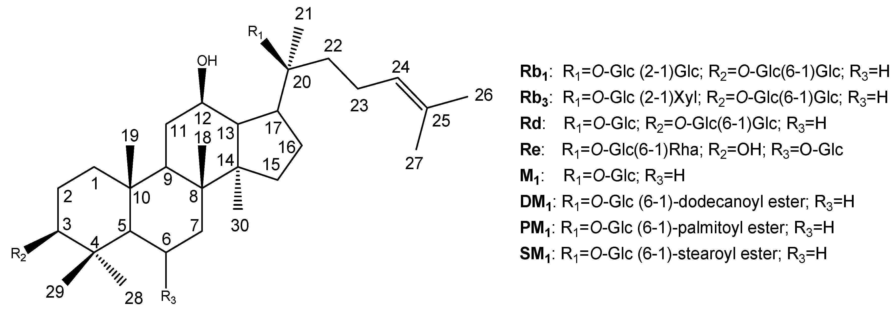
Figure 1.
Isolated, enzyme produced and synthetic ginsenosides.

Results and Discussion
Characterizations of compounds 1 – 8
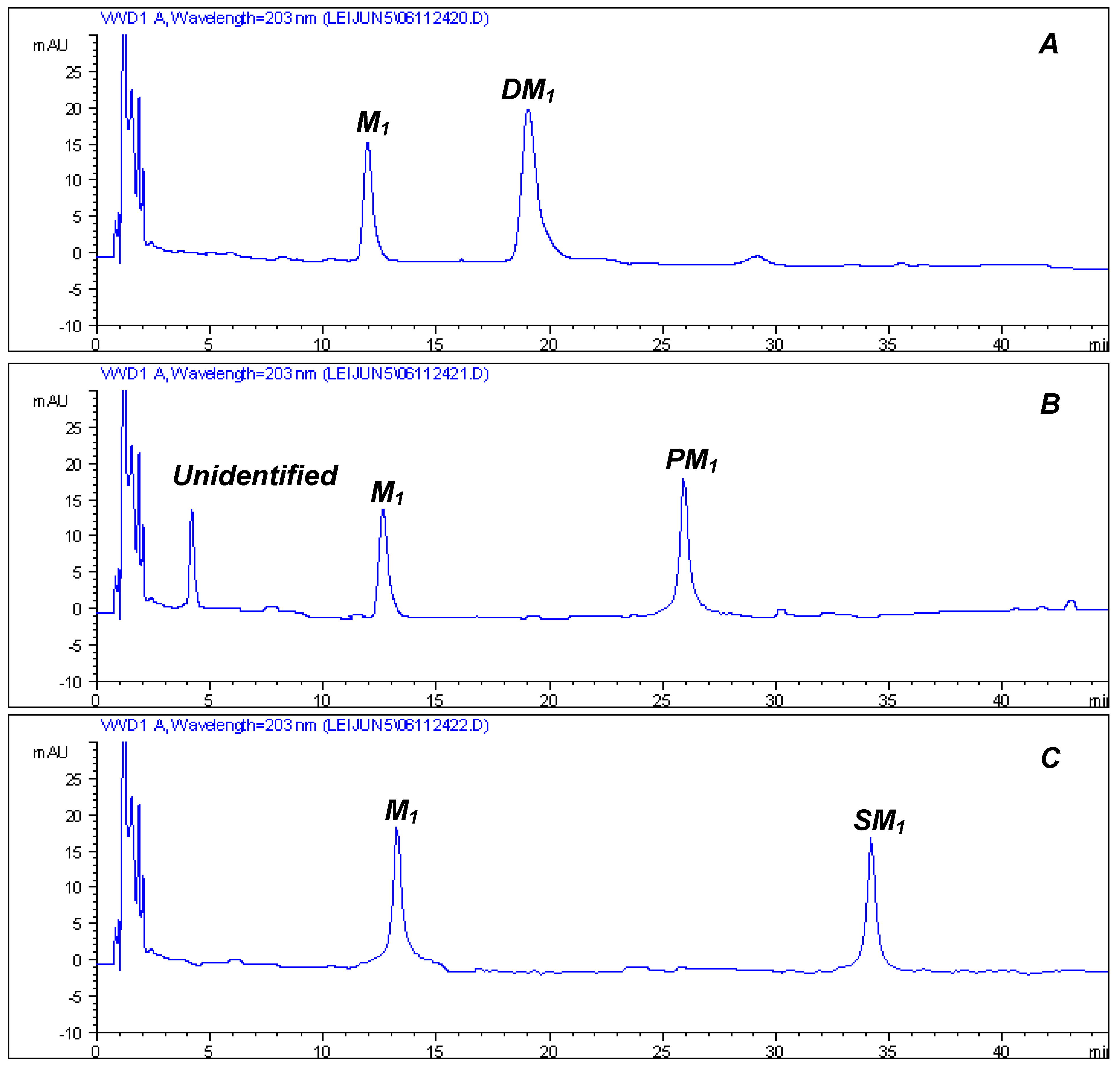
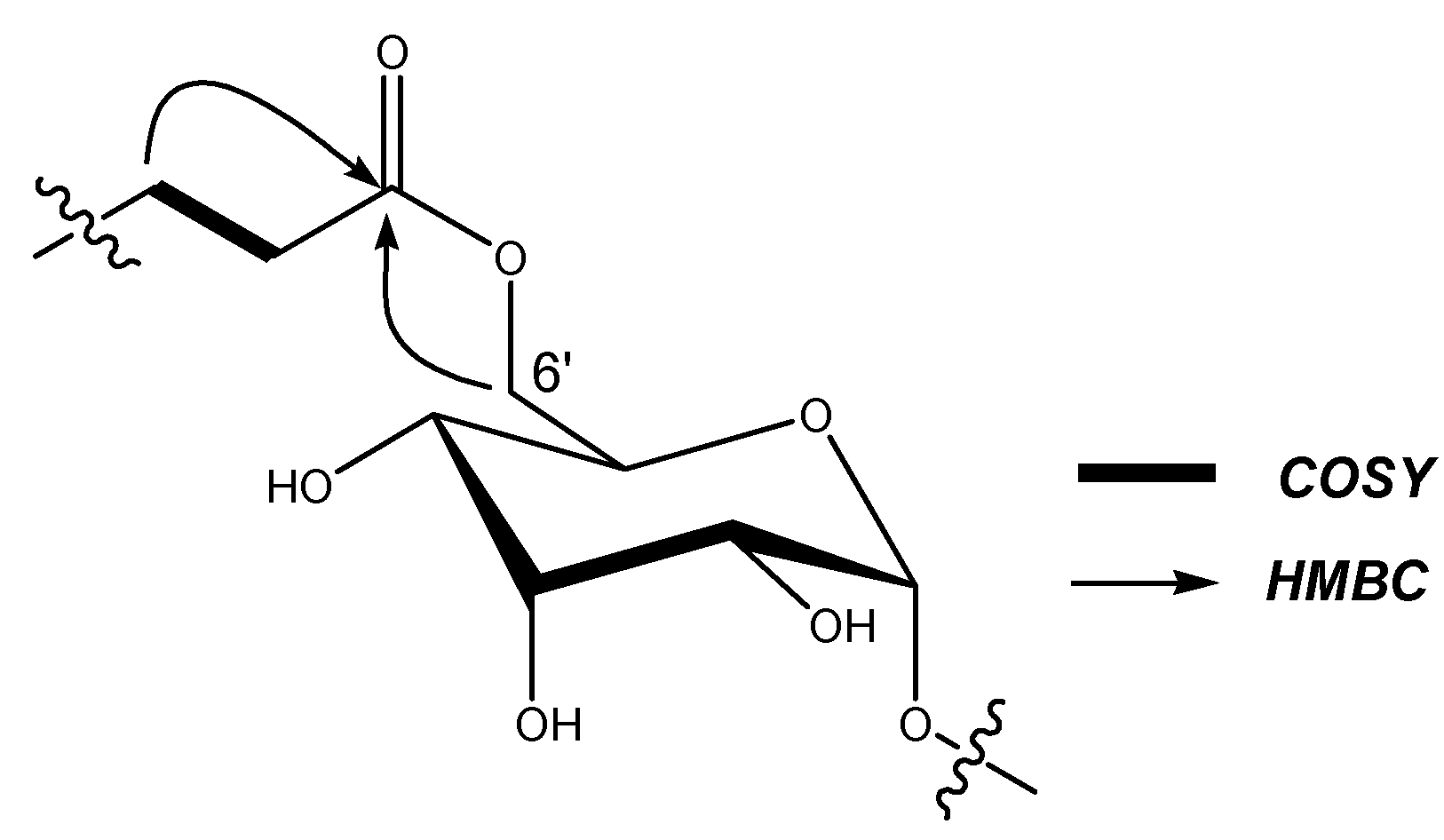
The known compounds were identified on the basis of comparison of their NMR data (see Table 1 and Table 2) with those of the literature [19]. Four compounds have been characterized as ginsenoside-Rb1 (1), Rb3 (2), Rd (3) and Re (4). Compound 1 had been used as a substrate in an enzymatic reaction to produce ginsenoside-M1 (5), whose structure was elucidated by MS, 1D- and 2D-NMR data analysis (not shown). The process whereby ginsenoside M1 was esterified by dodecanoyl chloride, palmitoyl chloride and stearoyl chloride, respectively, was monitored by HPLC. As shown in Figure 2, compounds 6-8 showed lower polarities compared with M1 (5). Comparison of the 13C-NMR data of 6-8 with those of 5, indicated no significant chemical shifts changes in the main skeleton, but the C-6’ of the 20-O-Glc group was shifted upfield to δ 63.3, 64.6, 64.4, respectively, suggesting that the fatty acid ester substituent was connected to the C-6’ position. This assumption had been verified by HMBC (see Figure 3) which showed a cross-peak between H-6’ to the carboxyl carbon.
Figure 2.
HPLC spectra of the reaction product of DM1 (6), PM1 (7) and SM1 (8); A: M1 reacted with dodecanoyl chloride; B: M1 reacted with palmitoyl chloride; C: M1 reacted with stearoyl chloride. HPLC conditions: gradient MeOH-H2O from 45% to 90% for 45 min.
Figure 2.
HPLC spectra of the reaction product of DM1 (6), PM1 (7) and SM1 (8); A: M1 reacted with dodecanoyl chloride; B: M1 reacted with palmitoyl chloride; C: M1 reacted with stearoyl chloride. HPLC conditions: gradient MeOH-H2O from 45% to 90% for 45 min.

Figure 3.
Partial HMBC and COSY correlation of the synthetic compound.


| Position | Rb1 (1) | Rb3 (2) | Rd (3) | Re (4) | M1 (5) | DM1 (6) | PM1 (7) | SM1 (8) |
|---|---|---|---|---|---|---|---|---|
| 1 | 39.2 | 39.3 | 39.2 | 39.7 | 39.3 | 38.9 | 39.5 | 39.1 |
| 2 | 26.3 | 26.3 | 26.7 | 26.6 | 28.1 | 27.3 | 28.2 | 28.0 |
| 3 | 88.9 | 89.0 | 89.0 | 78.1 | 77.9 | 78.8 | 78.0 | 77.8 |
| 4 | 39.6 | 39.6 | 39.1 | 39.9 | 39.4 | 38.9 | 39.4 | 39.3 |
| 5 | 56.1 | 56.7 | 56.3 | 60.7 | 56.2 | 55.8 | 56.4 | 56.1 |
| 6 | 18.6 | 18.6 | 18.5 | 74.2 | 18.6 | 18.2 | 18.7 | 18.5 |
| 7 | 35.1 | 35.2 | 35.1 | 45.8 | 35.0 | 34.7 | 35.2 | 34.9 |
| 8 | 39.8 | 39.9 | 40.4 | 41.3 | 39.9 | 39.7 | 39.5 | 39.8 |
| 9 | 50.0 | 50.2 | 50.2 | 49.5 | 50.2 | 49.8 | 50.3 | 50.0 |
| 10 | 36.7 | 37.0 | 37.1 | 39.6 | 37.2 | 37.0 | 37.3 | 37.1 |
| 11 | 30.8 | 31.0 | 31.1 | 30.7 | 30.7 | 30.3 | 30.7 | 30.7 |
| 12 | 70.1 | 70.1 | 70.2 | 70.1 | 70.1 | 70.6 | 70.1 | 69.8 |
| 13 | 49.3 | 49.5 | 49.6 | 49.4 | 49.3 | 47.8 | 49.5 | 49.3 |
| 14 | 51.3 | 51.4 | 51.5 | 51.3 | 51.3 | 51.7 | 51.4 | 51.3 |
| 15 | 30.8 | 30.8 | 30.7 | 30.9 | 30.7 | 30.6 | 31.0 | 30.5 |
| 16 | 26.6 | 26.8 | 26.6 | 26.6 | 26.5 | 26.6 | 26.7 | 26.5 |
| 17 | 51.5 | 51.6 | 51.6 | 51.6 | 51.5 | 51.3 | 51.6 | 51.2 |
| 18 | 16.0 | 16.0 | 17.7 | 17.7 | 16.2 | 16.1 | 16.3 | 16.1 |
| 19 | 16.1 | 16.2 | 16.4 | 17.8 | 15.9 | 15.7 | 16.0 | 15.8 |
| 20 | 83.1 | 83.5 | 83.1 | 83.3 | 83.2 | 84.3 | 83.4 | 83.1 |
| 21 | 22.3 | 22.3 | 22.4 | 22.4 | 22.3 | 21.2 | 22.9 | 22.7 |
| 22 | 36.2 | 36.1 | 36.2 | 35.8 | 36.0 | 35.4 | 36.1 | 35.9 |
| 23 | 23.0 | 23.0 | 23.2 | 23.3 | 23.1 | 22.0 | 23.0 | 22.8 |
| 24 | 125.8 | 126.0 | 125.8 | 126.0 | 125.8 | 124.5 | 126.0 | 125.8 |
| 25 | 131.1 | 131.0 | 131.0 | 130.9 | 130.8 | 131.5 | 130.9 | 130.7 |
| 26 | 25.8 | 25.8 | 25.7 | 25.8 | 25.7 | 25.7 | 25.8 | 25.5 |
| 27 | 17.9 | 17.9 | 18.0 | 17.6 | 17.7 | 17.6 | 17.8 | 17.6 |
| 28 | 28.0 | 28.3 | 28.1 | 32.3 | 28.6 | 28.0 | 28.6 | 28.4 |
| 29 | 16.6 | 16.5 | 16.3 | 17.3 | 16.2 | 15.3 | 16.3 | 16.1 |
| 30 | 17.1 | 17.4 | 17.7 | 17.3 | 17.3 | 16.9 | 17.4 | 17.2 |
a Compound 6 was measured in CDCl3 and the other compounds were measured in pyridine-d5 and chemical shifts are expressed in ppm.
| Position | M1 (5) | DM1 (6) | PM1 (7) | SM1 (8) | ||||
|---|---|---|---|---|---|---|---|---|
| δC | δH | δC | δH | δC | δH | δC | δH | |
| 20-O-Glc | ||||||||
| 1’ | 98.1 | 5.14 (d, 7.5) | 96.8 | 4.51 (d, 7.6) | 98.0 | 5.11 (d, 7.5) | 97.8 | 5.14 (d, 7.5) |
| 2’ | 75.0 | 3.96 (t, 8.0) | 73.3 | 3.36 (m) | 75.0 | 3.95 (d. 8.0) | 74.7 | 3.92 (d. 8.0) |
| 3’ | 79.2 | 4.19 (m) | 76.7 | 3.55 (m) | 79.2 | 4.18 (m) | 79.0 | 4.19 (m) |
| 4’ | 71.5 | 4.12 (dd, 9.0, 8.5) | 70.0 | 3.43 (m) | 71.6 | 4.16 (m) | 71.4 | 4.17 (m) |
| 5’ | 78.2 | 3.88 (m) | 73.4 | 3.44 (m) | 78.0 | 3.98 (m) | 77.8 | 3.98 (m) |
| 6’ | 62.7 | 4.44, 4.27 (m) | 63.3 | 4.30, 4.38 (m) | 64.6 | 4.63, 5.02 (m) | 64.4 | 4.65, 5.05 (m) |
| Glc-6’-O-ester | ||||||||
| 1’’ | 174.1 | 173.5 | 173.3 | |||||
| 2’’ | 34.2 | 2.27, 2.35 (m) | 34.4 | 2.43, 2.48 (m) | 34.2 | 2.42, 2.49 (m) | ||
| 3’’ | 24.8 | 1.27c | 25.3 | 1.42 | 25.0 | 1.45 | ||
| 4’’ | 29.6 c | 1.27c | 29.4 | 1.27c | 30.7 | 1.27c | ||
| 5’’ | 29.1 c | 1.27c | 28.2 | 1.27c | 29.2 | 1.27c | ||
| 6’’ | 29.2 c | 1.27c | 29.6 | 1.27c | 29.3 | 1.27c | ||
| 7’’ | 29.3 c | 1.27c | 30.0c | 1.27c | 29.4 | 1.27c | ||
| 8’’ | 29.4 c | 1.27c | 30.0 c | 1.27c | 29.7 c | 1.27c | ||
| 9’’ | 29.6 c | 1.27c | 30.0 c | 1.27c | 29.7 c | 1.27c | ||
| 10’’ | 31.9 | 1.36 (m) | 30.0 c | 1.27c | 29.7 c | 1.27c | ||
| 11’’ | 22.6 | 1.56 (m) | 30.0 c | 1.27c | 29.7 c | 1.27c | ||
| 12’’ | 14.1 | 0.85 (t, 6.0) | 29.6 | 1.27c | 29.7 c | 1.27c | ||
| 13’’ | 29.8 | 1.27c | 29.5 | 1.27c | ||||
| 14’’ | 32.1 | 1.30 (m) | 30.5 | 1.27c | ||||
| 15’’ | 22.1 | 1.48 (m) | 29.7 | 1.27c | ||||
| 16’’ | 14.2 | 0.83 (t, 5.6) | 31.9 | 1.36 (m) | ||||
| 17’’ | 22.7 | 1.48 (m) | ||||||
| 18’’ | 14.0 | 0.85 (t, 0.56) | ||||||
a Compound 6 was measured in CDCl3 and compounds 5, 7-8 were measured in pyridine-d5; chemical shifts are expressed in ppm; b Multiplicity and J values in Hz are given in parentheses; c Signals may be exchanged.
Bioactivity Results
The isolated and synthetic compounds were tested in an anti-tumor bioassay. Compounds 1-4 showed no cytotoxicity effects, while compounds 5-8 showed significant cytotoxicity (SRB) against several human cancer cell lines: breast cancer MCF-7, skin melanoma SK-MEL-2 and human ovarian carcinoma B16, but moderate effects on lung carcinoma COR-L23. The results are shown in Table 3. Compound 8 showed highest cytotoxic effect on skin melanoma SK-MEL-2 and human ovarian carcinoma B16; 6 showed highest cytotoxic effects on breast cancer cell lines MCF-7; Compounds 6-8 showed moderate effects on lung carcinoma COR-L23. All the tested compounds showed no cytotoxic activities towards normal cells at the concentration of 300 μg/mL.
| M1 (5) | DM1 (6) | PM1 (7) | SM1 (8) | |
|---|---|---|---|---|
| MCF-7 | 8.48 | 0.50 | 2.31 | 1.65 |
| SK-MEL-2 | 14.71 | 1.46 | 1.88 | 0.17 |
| B16 | 6.10 | 6.13 | 5.73 | 0.33 |
| COR-L23 | 33.0 | 5.68 | 4.86 | 7.76 |
Conclusions
The effects of some ginsenosides have been demonstrated on various models of tumor and endothelial cells. For example Rg3 inhibited the proliferation and induced of apoptosis in bladder cancer cells [20], and Rh2 shows effects on human lung adenocarcinoma A549 cells [21]. Most of the reported antitumor ginsengosides belongs to the protopanaxodiol-type saponins. In the present study, we isolated and transformed the protopanaxodiol-type ginsenosides Rb1 into M1, and further chemical modification led to the synthesis of three ginsenoside mono- fatty acid esters. Comparison of the cytotoxic activity results suggested that the fatty acid esterified ginsenoside were more active against human tumor cells. The most probable reason was that when cells acquire the molecules and ions they need from their surrounding extracellular fluid (ECF), the membrane transportation of small molecules depends on their lipophilic abilities [22]. In this study, we synthesized ginsenoside-DM1, PM1 and SM1 used fatty acid acyl-chlorides, so these reaction products showed significant lower polarities compared with the ginsenosides as shown in Figure 2. These lipophilic compounds which could pass through the membrane easily led to their increased cytotoxic activities on tumor cells.
Experimental
General
The 1H- and 13C-NMR spectra were measured on a Bruker Avance DRX 500 NMR spectrometer in C5D5N or CDCl3, using TMS as an internal standard. Chemical shifts (δ) are expressed in parts per million (ppm), with the coupling constants (J) reported in Hertz (Hz). The HR-ESI-TOF mass spectra were obtained from a MDS SCIEX API QSTAR-MS instrument. Column chromatographies were carried out with silica gel 60M (200-300 mesh), MCI and ODS RP-18 (20 μm); TLC was performed with silica gel plates (Macherey -Nagel, SilG / UV254, 0.20mm); HPLC were carried out on an Agilent 1100 system.
Chemicals and reagents
Dodecanoyl chloride, palmitoyl chloride and stearoyl chloride were purchased from ABCR GmbH & Co. KG. Sulforhodamine B (SRB) was purchased from Sigma (St. Louis, MO, USA). Enzyme NS 37040 was purchased from Novozymes Co. Ltd. (Holland). Other chemicals and reagents were purchased from the Chinese Chemical Group (Beijing, China).
Extraction and isolation
Crude powder of Panax ginseng (16 Kg) was extracted with 70% MeOH at room temperature, and the extract was concentrated to give a brown oily residue (3,700 g). The extract was directly subjected to chromatography on a silica column, eluted with a gradient mixture of CHCl3-MeOH-H2O (50: 10: 1, 7: 3: 0.5, 13: 7: 2), to yield eight fractions (FA - FH). The seventh fraction FG was subjected to silica gel, eluted with CHCl3-MeOH-H2O = 7: 3: 0.5 to afford compound 1 (2.1 g) and 2 (1.7 g). The fifth fraction FE was subjected to ODS RP-18 column eluted with 70% MeOH to afford compound 3 (1.3 g). The fourth fraction FD was subjected to further chromatography on MCI column, with 55% MeOH elution, to afford compound 4 (2.6 g).
Enzymatic reaction
Ginsenoside-Rb1 (1,500 mg) was dissolved in H2O (100 mL), enzyme NS37040 (30 mL) was added and the mixture was shaken at 45 ºC for 3 days with control of the pH at a value of 4.8. The reaction was terminated by heating to 80 ºC, then the enzyme reaction mixture was centrifuged under 10,000 rpm for 5 min and the supernatant was subjected to silica gel eluted with CHCl3-MeOH (10:1) to give purified compound 5 (581 mg).
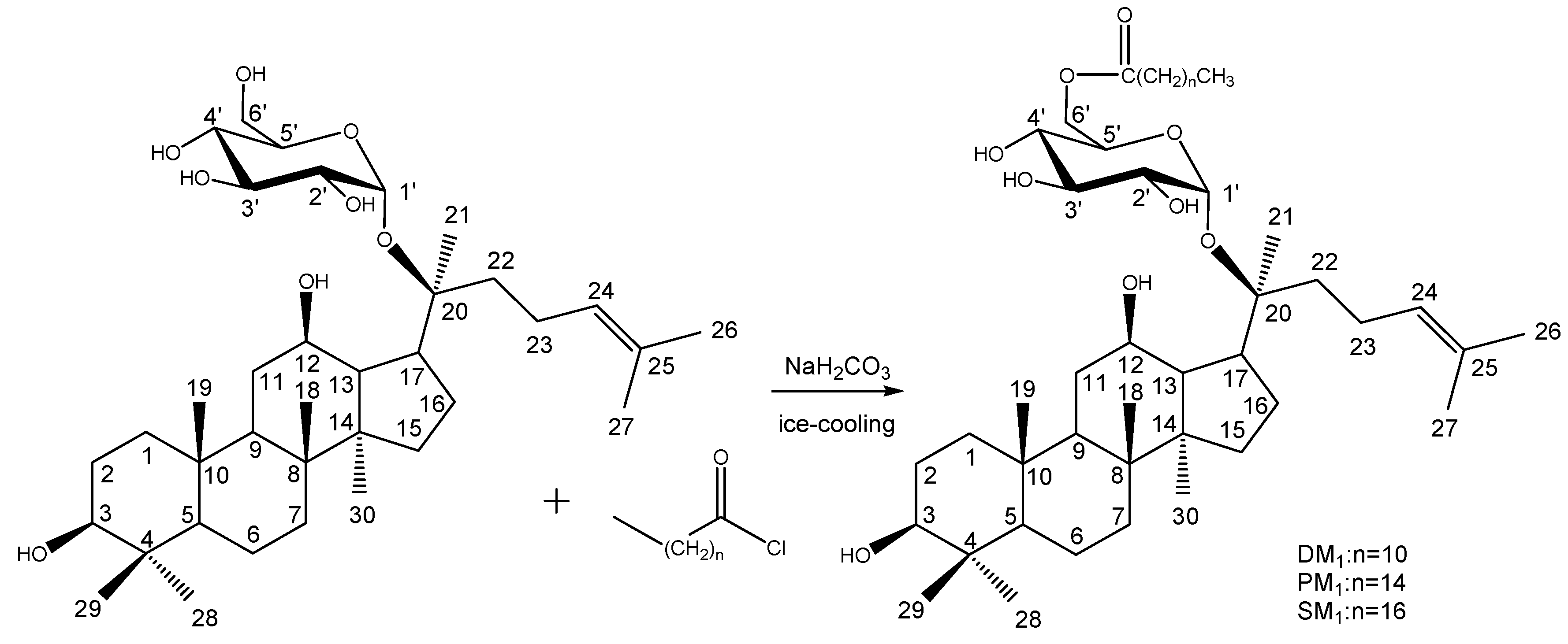
Synthesis of DM1, PM1 and SM1
Compound M1 (1.0 mmol) was dissolved in CHCl3 (200 mL) under ice-cooling, then aqueous saturated NaH2CO3 (10 mL) was added slowly. Dodecanoyl chloride (2.0 mmol) was added to the aqueous CHCl3 mixture with a measuring pipette under stirring, which was continued for 2 h. The aqueous layer was washed five times with CHCl3 (100 mL). The CHCl3 solution was concentrated under reduced pressure, then analyzed by TLC with CHCl3-MeOH = 15:1 as the developing solvent. Crude DM1 (6, 62 mg) were purified by silica gel column chromatography, eluted by CHCl3-MeOH = 18:1, then the purity was analyzed by HPLC (80% MeOH, Hypersil C18, 40ºC, 0.5 mL/min). The same experimental procedure (see Scheme 1) was followed using palmitoyl chloride and stearoyl chloride for the production of the corresponding compounds 7 (PM1, 77 mg) and 8 (SM1, 58 mg), respectively.
Scheme 1.
Preparation of ginsenosides DM1 (6), PM1 (7) and SM1 (8).

Ginsenoside Rb1 (1). White amorphous powder, mp 199 - 202 ºC; ESI-MS [+]: m/z = 1131.3 [M+Na]+; 407 [M-4xGlc]+; 1H-NMR (pyridine-d5): δ 0.78 (3H, s, H-19), 0.93 (3H, s, H-30), 0.94 (3H, s, H-18), 1.08 (3H, s, H-29), 1.25 (3H, s, H-28), 1.57 (3H, s, H-21), 1.63 (3H, s, H-26), 1.64 (3H, s, H-27), 5.45 (1H, m, H-24), 13C-NMR data, see Table 1.
Ginsenoside Rb3 (2). White amorphous powder, mp 196 - 198 ºC; IR (KBr) νmax cm-1: 3322, 2930, 2893, 1651; ESI-MS [+]: m/z = 1079.0 [M+H]+; 1H NMR (pyridine-d5): δ 0.79 (3H, s, H-19), 0.93 (3H, s, H-30), 0.95 (3H, s, H-18), 1.09 (3H, s, H-29), 1.37 (3H, s, H-28), 1.53 (3H, s, H-21), 1.59 (3H, s, H-26), 1.62 (3H, s, H-27), 3.83 (1H, m, H-12), 5.40 (1H, brs, H-24), 13C-NMR data, see Table 1.
Ginsenoside Rd (3). White amorphous powder, mp 205 - 206 ºC; ESI-MS [+]: m/z = 947.6 [M+H]+; 1H NMR (pyridine-d5): δ 0.73 (3H, s, H-19), 0.89 (3H, s, H-30), 0.91 (3H, s, H-18), 1.05 (3H, s, H-29), 1.23 (3H, s, H-28), 1.56 (6H, s, H-26, 27), 13C-NMR data, see Table 1.
Ginsenoside Re (4). White amorphous powder, mp 201 - 203 ºC; ESI-MS [+]: m/z = 969.3 [M+Na]+; 13C-NMR data, see Table 1.
Ginsenoside M1 (5). White amorphous powder, mp 192 - 193 ºC; IR (KBr) νmax cm-1: 3423, 2943, 2875, 1715, 1639; ESI-MS [+]: m/z = 645.2 [M+Na]+; 1H-NMR (pyridine-d5): δ 0.85 (3H, s, H-19), 0.90 (3H, s, H-30), 0.94 (3H, s, H-18), 1.00 (3H, s, H-29), 1.19 (3H, s, H-28), 1.56 (6H, s, H-26, 27), 1.58 (3H, s, H-21), 5.21 (1H, brs, H-24); other NMR data, see Table 1 and Table 2.
Ginsenoside DM1 (6). Colorless oil; [α]D25- 107.1 (c 0.70, MeOH); ESI-MS [+]: m/z = 805.3 [M+H]+; HR-ESI-TOF-MS [+]: m/z = 805.6186 [M+H]+; 1H-NMR (CDCl3): δ 5.13 (1H, t, J = 6.0, H-24), 3.22 (1H, dd, J = 5.0, 11.0 ,H-3), 2.20 (1H, m, H-17), 2.18 (1H, m, H-23b), 1.98 (1H, m, H-23a), 1.82 (1H, m, H-11a), 1.80 (1H, m, H-13), 1.70 (3H,s, H-26), 1.73 (1H,s, H-1a), 1.66 (1H, m, H-16b), 1.63 (1H, m, H-2b), 1.62 (3H, s, H-27), 1.63 (1H, m, H-22b), 1.58 (1H, m, H-2a), 1.56 (1H, m, H-6b), 1.50(1H, m, H-7b), 1.47 (1H, m, H-6a), 1.43(1H, m, H-22a), 1.40 (1H, m, H-9a), 1.37 (3H, s, H-21), 1.28 (2H, m, H-7a, H-15b), 1.27 (2H, m, H-11a, H-16a), 1.00 (6H, s, H-18, 28), 0.99 (1H, m, H-2b), 0.91 (3H, s, H-30), 0.89 (3H, s, H-19), 0.88 (1H, m, H-15a), 0.80 (3H, s, H-29), 0.74 (1H, d, J = 11.0, H-5); other NMR data, see Table 1 and Table 2.
Ginsenoside PM1 (7). Colorless oil; ESI-MS [+]: m/z = 861.1 [M+H]+; HR-ESI-TOF-MS [+]: m/z = 861.6455 [M+H]+; 1H-NMR (C5D5N): δ 5.32 (1H, t, J = 6.5, H-24), 3.39 (1H, dd, J = 5.0, 11.0 ,H-3), 2.59 (1H, m, H-17), 2.55 (1H, m, H-23b), 2.43 (1H, m, H-22a), 2.31 (1H, m, H-23a), 2.02 (1H, m, H-11a), 2.00 (1H, m, H-13), 1.67 (3H,s, H-26), 1.73 (1H,s, H-1a), 1.69 (1H, m, H-1b), 1.85 (1H, m, H-16b), 1.88 (1H, m, H-2b), 1.64 (3H, s, H-27), 1.82 (1H, m, H-22b), 1.80 (1H, m, H-2a), 1.59 (1H, m, H-6b), 1.50(1H, m, H-7b), 1.47 (1H, m, H-6a), 1.43(1H, m, H-22a), 1.40 (1H, m, H-9a), 1.64 (3H, s, H-21), 1.53 (1H, m, H-15b), 1.31 (1H, m, H-7a), 1.55 (1H, m, H-11a), 1.40 (1H, m, H-16a), 1.20 (3H, s, H-28), 0.87 (3H, s, H-18), 0.89 (1H, m, H-2b), 0.94 (3H, s, H-30), 0.98 (3H, s, H-19), 0.88 (1H, m, H-15a), 1.02 (3H, s, H-29), 0.79 (1H, d, J = 11.0, H-5); other NMR data, see Table 1 and Table 2.
Ginsenoside SM1 (8). Colorless oil; ESI-MS [+]: m/z = 889.3 [M+H]+; HR-ESI-TOF-MS [+]: m/z = 889.5203 [M+H]+; 1H-NMR (C5D5N): δ 5.33 (1H, t, J = 6.5, H-24), 3.40 (1H, dd, J = 5.0, 11.0 ,H-3), 2.58 (1H, m, H-17), 2.56 (1H, m, H-23b), 2.41 (1H, m, H-22a), 2.32 (1H, m, H-23a), 2.01 (1H, m, H-11a), 2.00 (1H, m, H-13), 1.67 (3H,s, H-26), 1.73 (1H,s, H-1a), 1.69 (1H, m, H-1b), 1.85 (1H, m, H-16b), 1.64 (3H, s, H-27), 1.82 (1H, m, H-22b), 1.80 (1H, m, H-2a), 1.89 (1H, m, H-2b), 1.58 (1H, m, H-6b), 1.50 (1H, m, H-7b), 1.47 (1H, m, H-6a), 1.43(1H, m, H-22a), 1.42 (1H, m, H-9a), 1.66 (3H, s, H-21), 1.53 (1H, m, H-15b), 1.33 (1H, m, H-7a), 1.53 (1H, m, H-11a), 1.39 (1H, m, H-16a), 1.20 (3H, s, H-28), 0.87 (3H, s, H-18), 0.89 (1H, m, H-2b), 0.94 (3H, s, H-30), 0.98 (3H, s, H-19), 0.88 (1H, m, H-15a), 1.02 (3H, s, H-29), 0.77 (1H, d, J = 11.0, H-5); other NMR data, see Table 1 and Table 2.
In vitro cytotoxicity assays
Five-day in vitro SRB cytotoxicity tests against human tumors cell lines were carried out at the Cell Culture Laboratory, Pharmaceutical College, Jilin University, using modified protocols for MCF-7 (breast cancer), SK-MEL-2 (skin melanoma,) B16 (human ovarian carcinoma) and COR-L23 (lung carcinoma), the normal cells were used as control [23]. Generally, 5x10³/mL cells were placed in a 24-well plate and treated with obtained compounds. The plate was incubated at 37 ºC for 5 days. Then the medium was removed from the 24-well plate, and 10% ice-cold TCA (trichloroacetic acid, 1 mL) was added. The plate was kept at 4°C for two hours after which was washed four times with cold water, then stained with SRB (Sulforhodamine B, Sigma St. Louis, MO, USA). After washing with 1% acetic acid, the bound dye was solubilized with Tris base A (Sigma) and 100 μL of each sample were transferred into a 96-well plate, and then read at 492 nm.
Acknowledgments
We are grateful to Professor Zhiwei Deng (BNU) for measuring the NMR spectra and Professor Shijie Yang (JLU) for the bioactivity tests. This research project has been supported by Research Fund for the Doctoral Program of Higher Education of China; Project No. 20050193006.
References
- Ligor, T.; Ludwiczuk, A.; Wolski, T.; Buszewski, B. Isolation and determination of ginsenosides in American ginseng leaves and root extracts by LC-MS. Anal. Bioanal. Chem. 2005, 383, 1098–1105. [Google Scholar] [CrossRef]
- Liu, W.; Xu, S.; Che, C. Anti-proliferative efect of ginseng saponins on human prostate cancer cell line. Life Sci. 2000, 67, 1297–130. [Google Scholar] [CrossRef]
- Kim, S.; Lee, Y.; Park, J.; Lee, S. Ginsenoside-Rs4, a new type of ginseng saponin concurrently induces apoptosis and selectively elevates protein levels of p53 and p21WAF1 in human hepotoma SK-HEP-1 cells. Eur. J. Cancer 1999, 35, 507–511. [Google Scholar] [CrossRef]
- Lee, K.; Lee, Y.; Kim, S.; Park, J.; Lee, S. Ginsenoside-Rg5 suppresses cyclin E-dependent protein kinase activity via up-regulation of p21 Cip/WAF1 and down-regulating cyclin E in SK-HEP-1 cells. Anticancer Res. 1997, 17, 1067–1072. [Google Scholar]
- Lee, Y.; Lee, H.; Lee, Y.; Chung, H.; Kim, S.; Lee, S.; Park, B.; Kim, K. Involvement of glucocorticoid receptor in the induction of differentiation by ginsenoside in F9 teratocarcinoma cells. J Steroid Biochem. Mol. Biol. 1998, 67, 105–111. [Google Scholar]
- Cho, W.; Chung, W.; Lee, S.; Leung, A.; Cheng, C.; Yue, K. Ginsenoside Re of Panax ginseng possesses significant antioxidant and antihyperlipidemic efficacies in streptozotocin-induced diabetic rats. Eur. J. Pharmacol. 2006, 550, 173–179. [Google Scholar] [CrossRef]
- Lee, W.; Kao, S.; Liu, I.; Cheng, J. Ginsenoside Rh2 is one of the active principles of Panax ginseng root to improve insulin sensitivity in fructose-rich chow-fed rats. Horm. Metab. Res. 2007, 39, 347–354. [Google Scholar] [CrossRef]
- Akao, T.; Kida, H.; Kanaoka, M.; Hattori, M.; Kobashi, K. Intestinal bacterial hydrolysis is required for the appearance of compound K in rat plasma after oral administration of ginsenoside Rb1 from Panax ginseng. J. Pharm. Pharmacol. 1998, 50, 1155–1160. [Google Scholar] [CrossRef]
- Wakabayashi, C.; Hasegawa, H.; Murata, J.; Saiki, I. In vivo anti-metastatic action of ginseng proto-panaxadiol saponins is based on their intestinal metabolites after oral administration. Oncol. Res. 1997, 9, 411–417. [Google Scholar]
- Wakabayashi, C.; Hasegawa, H.; Murata, J. The expression of in vivo anti-metastatic effect of ginseng protopanaxadiol saponins is mediated by their intestinal metabolites after oral administration. J. Trad. Med. 1997, 14, 180–185. [Google Scholar]
- Hasegawa, H.; Sung, J.; Huh, J. Ginseng intestinal bacterial metabolite IH901 as a new anti-metastatic agent. Arch. Pharm. Res. 1997, 20, 539–544. [Google Scholar]
- Hasegawa, H. Proof of the mysterious efficacy of ginseng: basic and clinical trials: metabolic activation of ginsenoside: deglycosylation by intestinal bacteria and esterification with Fatty Acid. J. Pharmacol. Sci. 2004, 95, 153–157. [Google Scholar]
- Tawab, M.; Bahr, U.; Karas, M.; Wurglics, M. Degeneration of ginsenosides in humans after oral administration. Drug Metab. Dispos. 2003, 31, 1065–1071. [Google Scholar] [CrossRef]
- Lee, J.; Shin, J.; Chun, K.; Park, K.; Chung, W.; Bang, Y.; Sung, J.; Surh, Y. Antitumor promotional effects of a novel intestinal bacterial metabolite (IH-901) derived from the protopanaxadiol-type ginsenosides in mouse skin. Carcinogenesis 2005, 26, 359–367. [Google Scholar]
- Hasegawa, H.; Lee, K.; Nagaoka, T.; Tezuka, Y.; Uchiyama, M.; Kadota, S.; Saiki, I. Pharmacokinetics of ginsenoside deglycosylated by intestinal bacteria and its transformation to biologically active fatty acid esters. Biol. Pharm. Bull. 2000, 23, 298–304. [Google Scholar]
- Li, X.; Li, X.; Lei, J. Studies on chemical components and their pharmacological activities of Panax ginseng root. Drug Discovery and Traditional Chinese Medicine: Science, Regulation, and Globalization; Kluwer Academic Publishers: Maryland, 2000; pp. 95–109. [Google Scholar]
- Wang, J.; Li, X.; Zheng, Y.; Yang, X. Isoginsenoside-Rh3, a new triterpenoid saponin from the fruits of Panax ginseng C. A. Mey. J. Asian Nat. Prod. Res. 2004, 6, 289–293. [Google Scholar] [CrossRef]
- Sun, G.; Liu, Z.; Li, X.; Zheng, Y.; Wang, J. Isolation and identification of two malonyl-ginsenosides from the fresh root of Panax Ginseng. Chin. J. Anal. Chem. 2005, 33, 1783–1786. [Google Scholar]
- Gong, X. Study on chemical modification of the enzyme of the enzyme metabolites and its anti-tumor activities. PHD dissertation of Jilin Agricultural University, 2004; pp. 96–103. [Google Scholar]
- Chen, J.; Peng, H.; Pu, S.; Guo, Y. Apoptosis induced by ginsenoside Rg3 in a human bladder carcinoma cell line. Chin. J. Clin. Oncol. 2006, 4, 283–287. [Google Scholar] [CrossRef]
- Cheng, C.; Yang, S.; Huang, C.; Chen, J.; Chang, W.; Hsu, S. Molecular mechanisms of ginsenoside Rh2-mediated G1 growth arrest and apoptosis in human lung adenocarcinoma A549 cells. Cancer Chemother. Pharmacol. 2005, 55, 531–540. [Google Scholar] [CrossRef]
- Connolly, T.; Carruthers, A.; Melchior, D. Effects of bilayer cholesterol on human erythrocyte hexose transport protein activity in synthetic lecithin bilayers. Biochemistry 1985, 24, 2865–2873. [Google Scholar] [CrossRef]
- Baruch, B.; Lina, W.; Einat, B.; Jardena, N.; Jacob, S.; Haim, G.; Eyal, F. Variable cytotoxicity of amifostine in malignant and non-malignant cell lines. Oncol. Rep. 2003, 10, 1609–1613. [Google Scholar]
- Sample availability: From the author.
© 2007 by MDPI (http://www.mdpi.org). Reproduction is permitted for noncommercial purposes.
Share and Cite
MDPI and ACS Style
Lei, J.; Li, X.; Gong, X.-j.; Zheng, Y.-n. Isolation, Synthesis and Structures of Cytotoxic Ginsenoside Derivatives. Molecules 2007, 12, 2140-2150. https://doi.org/10.3390/12092140
AMA Style
Lei J, Li X, Gong X-j, Zheng Y-n. Isolation, Synthesis and Structures of Cytotoxic Ginsenoside Derivatives. Molecules. 2007; 12(9):2140-2150. https://doi.org/10.3390/12092140
Chicago/Turabian StyleLei, Jun, Xiang Li, Xiao-jie Gong, and Yi-nan Zheng. 2007. "Isolation, Synthesis and Structures of Cytotoxic Ginsenoside Derivatives" Molecules 12, no. 9: 2140-2150. https://doi.org/10.3390/12092140