NMR Spectra of Sparteine N1-oxide and α-Isosparteine N-oxide

Faculty of Chemistry, A. Mickiewicz University, Grunwaldzka 6, 60-780 Poznań, Poland
Molecules 2008, 13(1), 3-10; https://doi.org/10.3390/molecules13010003
Submission received: 4 December 2007
/
Accepted: 30 December 2007
/
Published: 9 January 2008
(This article belongs to the Special Issue Alkaloids: Novel Therapeutic Perspectives)
Abstract
:Sparteine N1-oxide and α-isosparteine N-oxide were prepared and their structures determined for the first time by 1H- and 13C-NMR spectroscopy using two-dimensional techniques. The N-oxide effects were also calculated.
Introduction
The wide use of quinolizidine alkaloids in chemistry is related first of all with a possibility of configurational-conformational changes that could take place in the bis-quinolizidine skeleton. Naturally occurring (–)-sparteine is an equilibrium mixture in which the conformer possessing a boat ring C and trans junction of rings C/D predominates [1,2,3,4]. On the other hand, the less stable all-chair conformer participates in complex formation [5,6,7]. The compound α-isosparteine, consisting of two trans-quinolizidine systems, exists solely in an all-chair conformation, and similarly in the free base form [8] and in metal complexes [5,6,7,9,10]. As a continuation of our study on the complex forming ability of bis-quinolizidine alkaloids [6,7,9,10], the choice of N-oxides as ligands was made. We have already obtained the complexes of sparteine N16-oxides with lithium [11] and zinc [12] salts. This time the subject of our study was the synthesis of the complexes of sparteine N1-oxide, sparteine epi-N-oxide and α-isosparteine N-oxide.
NMR spectroscopy is known to permit observation of conformational changes taking place in the structure of the ligands during complexation reactions. Moreover, a comparison of the chemical shifts of carbon atoms and protons of the initial alkaloid and the complex formed, enables determination of the effects of complexation. The present work is a continuation of our studies on the structural investigation of bis-quinolizidine alkaloids [13,14,15,16,17]. The NMR data of sparteine N16-oxide and sparteine epi-N-oxide have been presented before [11,18]. In this paper we present the NMR spectra of sparteine N1-oxide and α-isosparteine N-oxide. For each of them the N-oxide effects were determined.
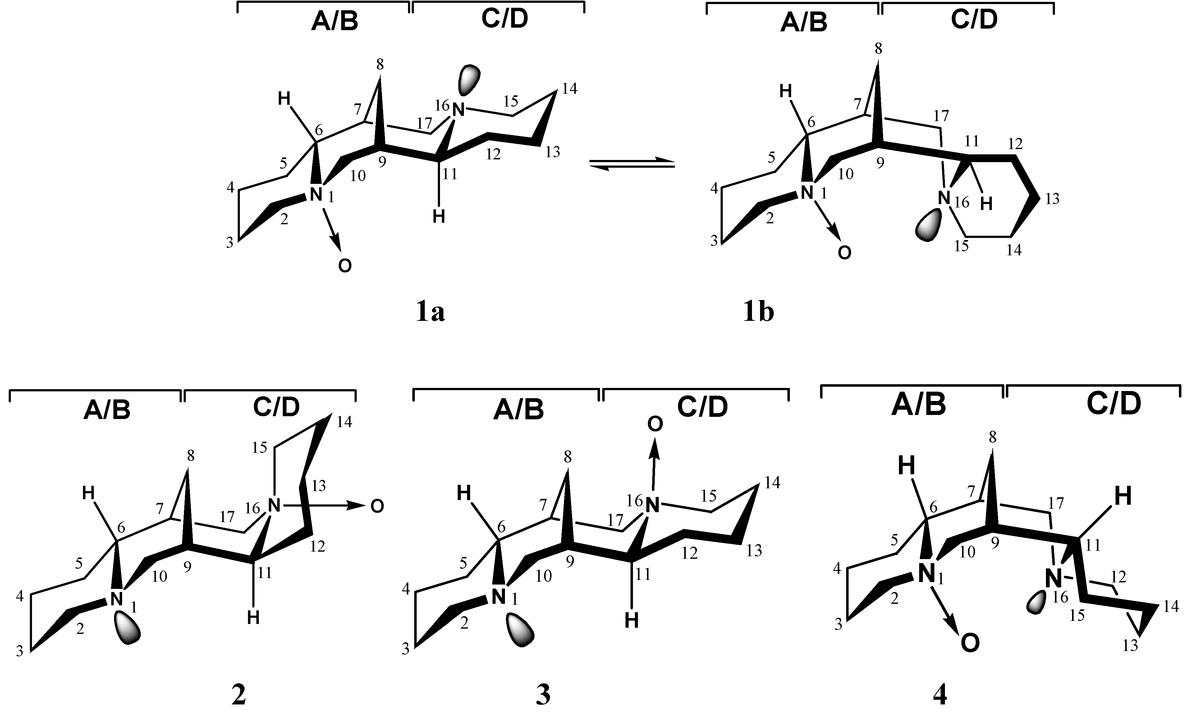
The two conformers of sparteine form three isomeric mono N-oxides: sparteine N1-oxide (1), sparteine N16-oxide (2) and sparteine epi-N-oxide (3). In the reactions of sparteine with H2O2 a 1:3 mixture of the two sparteine N-oxides: sparteine N1-oxide and sparteine N16-oxide is obtained [19,20]. Sparteine N1-oxide (1) occurs in a chair-boat type equilibrium involving inversion of the lone pair on the N16 atom [20]; the N16-oxide of sparteine (2), previously thought to adopt the all chair conformation, has been found recently to have ring C in a boat conformation and a cis C/D ring junction [21]. Sparteine epi-N-oxide (3) has the C ring boat conformation and must be obtained by NaBH4 reduction of lupanine N-oxide [20]. α-Isosparteine N-oxide (4) has a conformation identical to that of the free base (Figure 1) [19].
Figure 1.
Chemical structures of sparteine N-oxides 1-3 and α-isosparteine N-oxide (4).

Results and Discussion
In order to obtain the NMR spectra of the two conformers of the N1-oxide of sparteine (1a and 1b), the NMR spectra were measured in two solvents: CDCl3 and DMSO-d6. The NMR spectrum of sparteine N1-oxide recorded in DMSO-d6 solution is typical of that expected for pure 1a conformer, while the spectrum recorded in CDCl3 seems to be that of sparteine N1-oxide hydrochloride (all-chair conformation) since in its 1H-NMR spectrum the signal of the “acid” proton appears at 17.5 ppm.
The 1H- and 13C-NMR data for sparteine N1-oxide (conformer 1a), sparteine N1-oxide hydro-chloride (1-HCl) and free base of sparteine are collected in Table 1. The N-oxide effect can be derived as a difference in the chemical shifts of the appropriate carbon atoms in N-oxide and its basic amine. This effect is superimposed by the solvent effect as the solvent is changed from CDCl3 (free base of sparteine) into DMSO-d6 (sparteine N1-oxide). The 13C chemical shifts for C7, C8, C9, C12, C13, C14 and C15 carbon atoms of conformer 1a approximate the analogous δC values of sparteine [22]. This result corroborates the presence of chemically unchanged C and D rings preserving the trans-quinolizidine form in the N-oxide. The deshielding influence of the N-oxide function, generated on the N1 nitrogen of the alkaloid considered, causes a down-field shift of the α carbons, i.e. C2 (ΔδC = 13.4 ppm), C6 (ΔδC = 4.3 ppm) and C10 (ΔδC = 8.7 ppm), as compared with respective sparteine δC values.

Table 1.
13C- and 1H-NMR chemical shifts of sparteine, sparteine N1-oxide (conformer 1a) and sparteine N1-oxide hydrochloride (1-HCl) (δ in ppm).
| C | Sparteine [see ref. 22] CDCl3 | Sparteine N1-oxide (1a) DMSO-d6 | N-oxide effects | Sparteine N1-oxide hydrochloride (1-HCl) CDCl3 | N-oxide and protonation effects | |||
|---|---|---|---|---|---|---|---|---|
| δC | δH | δC | δH | Δ δC | δC | δH | Δ δC | |
| 2 | 56.0 | 1.79 2.53 | 69.4 | 4.80 3.10 | +13.4 | 66.9 | 5.00 3.41 | +10.9 |
| 3 | 25.6 | 1.38 1.38 | 19.9 | 1.35 2.35 | –5.7 | 20.3 | 1.65 2.18 | –5.3 |
| 4 | 24.5 | 1.05 1.55 | 23.1 | 1.30 1.60 | –1.4 | 21.8 | 1.85 1.85 | –2.7 |
| 5 | 29.1 | 1.24 1.12 | 26.0 | 1.35 2.25 | –3.1 | 24.3 | 1.45 2.20 | –4.8 |
| 6 | 66.3 | 1.58 | 70.6 | 3.20 | +4.3 | 72.9 | 4.70 | +6.6 |
| 7 | 32.9 | 1.69 | 32.1 | 1.95 | –0.8 | 33.8 | 2.10 | +0.9 |
| 8 | 27.4 | 0.90 1.91 | 26.1 | 1.25 2.15 | –1.3 | 24.0 | 1.45 2.15 | –3.4 |
| 9 | 35.9 | 1.32 | 35.5 | 1.60 | –0.4 | 34.2 | 2.10 | –1.7 |
| 10 | 61.8 | 1.84 2.38 | 70.5 | 4.15 3.10 | +8.7 | 70.9 | 4.50 3.30 | +9.1 |
| 11 | 64.2 | 1.83 | 57.8 | 3.70 | –6.4 | 58.4 | 3.05 | –5.8 |
| 12 | 34.5 | 1.35 1.21 | 35.2 | 1.10 1.40 | +0.7 | 24.5 | 1.45 1.90 | –10.0 |
| 13 | 24.6 | 1.15 1.55 | 24.3 | 2.30 1.15 | –0.3 | 23.2 | 2.50 1.45 | –1.4 |
| 14 | 25.8 | 1.43 1.43 | 24.8 | 1.50 1.60 | –1.0 | 18.4 | 1.50 2.18 | –7.4 |
| 15 | 55.2 | 1.86 2.63 | 53.9 | 2.60 2.00 | –1.3 | 51.8 | 2.90 2.00 | –3.4 |
| 17 | 53.4 | 2.20 2.54 | 48.4 | 2.40 3.80 | –5.0 | 43.9 | 2.60 3.20 | –9.5 |
Analysis of the chemical shifts given in Table 1 shows that the conformational changes are observed for the bis-quinolizidine skeleton in sparteine N1-oxide hydrochloride on passing from the free base to the N-oxide salt. The γ-gauche effects which usually accompany a conformational change from boat-chair to all-chair [23] are observed at carbon atoms C12 (ΔδC = 10.0 ppm), C14 (ΔδC = 7.4 ppm) and C17 (ΔδC = 9.5 ppm). The conformational changes are superimposed by the N-oxide effect assuming the greatest values for the carbon atoms in the αposition with respect to the N-oxide group: C2 (+10.9 ppm), C6 (+6.6 ppm) and C10 (+9.1 ppm).
The β–effect influencing the secondary carbon atoms in the outer ring (A) is negative, amounting to –5.7 and –3.1 ppm for 1a and –5.3 and –4.8 ppm for 1-HCl. In the inner ring (B), the oxidation effects on on the tertiary carbon atoms differ only slightly from 0. The γ-effect in the outer ring amounts to ca. –1.4 ppm (1a) and –2.7 ppm (1-HCl), in the inner rings (B and C), it amounts ca. –1.3, –5.0, –6.4 ppm for the boat conformer 1a and –3.4, –5.8 and –9.5 ppm for the hydrochloride salt. In the proton spectra, the greatest N-oxidation effect is observed for C2, C6 and C10 (Table 1). The protons connected with these carbons appear within the range 5.00 > δH > 3.10. The NMR spectra of α-isosparteine N-oxide and the free base of α-isosparteine are shown in Table 2. The spectra of both compounds display the 13C chemical shift of C8 characteristic of the α-isosparteine skeleton structure (32.8 ppm for α-isosparteine and 31.7 ppm for the N-oxide).
| C | α-isosparteine DMSO-d6 | α-isosparteine N-oxide (4) DMSO-d6 | N-oxide effects | ||
|---|---|---|---|---|---|
| δC | δH | δC | δH | Δ δC | |
| 2 | 55.5 | 1.76 2.74 | 66.5 | 3.42 3.50 | +11.0 |
| 3 | 23.8 | 1.75 1.40 | 19.9 | 1.60 2.02, | –3.9 |
| 4 | 22.9 | 1.24 1.70 | 21.8 | 1.44 1.76 | –1.1 |
| 5 | 28.0 | 1.69 1.19 | 23.8 | 1.70 1.40 | –4.2 |
| 6 | 65.7 | 1.86 | 72.9 | 3.74 | +7.2 |
| 7 | 32.1 | 1.45 | 32.4 | 2.07 | +0.3 |
| 8 | 32.8 | 1.31 1.52 | 31.7 | 1.80 2.00 | –1.1 |
| 9 | 32.1 | 1.31 | 33.0 | 2.18 | +0.9 |
| 10 | 55.7 | 2.04 2.96 | 65.5 | 3.74 3.74 | +9.8 |
| 11 | 65.7 | 1.86 | 60.6 | 2.68 | –5.1 |
| 12 | 28.0 | 1.69 1.19 | 27.4 | 1.60 1.74 | –0.6 |
| 13 | 22.9 | 1.24 1.70 | 22.6 | 1.44 1.76 | –0.3 |
| 14 | 23.8 | 1.75 1.40 | 23.3 | 2.16 1.40 | –0.5 |
| 15 | 55.5 | 1.76 2.74 | 52.4 | 2.48 3.08 | –3.1 |
| 17 | 55.7 | 2.04 2.96 | 50.7 | 2.58 3.00 | –5.0 |
The relatively rigid skeleton of α-isosparteine allows us to determine the N-oxide effects more precisely than in the flexible sparteine. The α N-oxide effect for the metine carbon atom (C6) is +7.2 ppm, and for methylene carbon atoms (C2, C10) +11.0 and + 9.8 ppm, respectively, while the value of the N-oxide β effect on carbon atoms C3, C5, C7 and C9 range from –4.2 to +0.9 ppm. The γ-effect in the A ring amounts to ca. –1.1 ppm. In the rings B and C, it is generally greater and amounts to ca. –5.1 ppm. Chemical shift changes (from –0.6 to –3.1 ppm) are noted on the carbon atoms of ring D. These changes follow mainly from the presence of the N-O group. It seems probable that the effect of a slight change in the geometry of the molecule involving greater deformation of rings B and C than that of ring A (following an elongation of the N-C bonds as a result of introducing the N+−O- function) is imposed on the direct N-oxidation effect in the inner rings. In the proton spectra, the greatest N-oxidation effect (Table 2) is for H6 (ΔδH = 1.88 ppm). Other large effects are those on the α-axial protons (above 1.70 ppm). Also significant are the effects on α-equatorial protons (0.80 ppm).
The conformational assignment for compounds 1a, 1-HCl and 2 was also carried out by comparison of the experimental 13C-NMR chemical shifts with those predicted by DFT/CSGT shielding calculations. The results of these calculations for the optimized structures, together with the experimental values are listed in Table 3. The correlation coefficient (R2) for carbon chemical shifts is 0.97 for 1a, 0.98 for 1-HCl and 0.98 for 2. The results suggest that the structures of the N-oxides investigated in solution and under vacuum are the same.
Table 3.
Comparison of CSGT chemical shifts (δ, in ppm) calculated at the DFT level of theory for sparteine N1-oxide (conformer 1a), sparteine N1-oxide hydrochloride (1-HCl) and α-isosparteine N-oxide (2).
| C | δ exper. (1a) | δ theor. (1a) | ΔδC (1a) | δ exper. (1-HCl) | δ theor. (1-HCl) | ΔδC (1-HCl) | δ exper. (2) | δ theor. (2) | ΔδC (2) |
|---|---|---|---|---|---|---|---|---|---|
| C2 | 69.4 | 72.2 | 2.8 | 66.9 | 66.3 | –0.6 | 52.4 | 48.9 | –3.8 |
| C3 | 19.9 | 21.3 | 1.4 | 20.3 | 17.9 | –2.4 | 23.3 | 23.4 | 0.1 |
| C4 | 23.1 | 23.0 | –0.1 | 21.8 | 21.4 | –0.4 | 22.6 | 23.4 | 0.8 |
| C5 | 26.0 | 27.1 | 1.1 | 24.3 | 26.2 | 1.9 | 27.4 | 28.9 | 1.5 |
| C6 | 70.6 | 70.3 | –0.3 | 72.9 | 70.9 | –2.0 | 60.6 | 57.5 | –3.1 |
| C7 | 32.1 | 33.3 | 1.2 | 33.8 | 31.2 | –2.6 | 33.0 | 34.0 | 1.0 |
| C8 | 26.1 | 23.4 | –2.7 | 24.0 | 22.3 | –1.7 | 31.7 | 33.3 | 1.6 |
| C9 | 35.5 | 35.0 | –0.5 | 34.2 | 32.5 | –1.7 | 32.4 | 33.4 | 1.0 |
| C10 | 70.5 | 71.8 | 1.3 | 70.9 | 67.3 | –3.6 | 50.7 | 45.5 | –5.2 |
| C11 | 57.8 | 60.8 | 3.0 | 58.4 | 62.9 | 4.5 | 72.9 | 71.8 | –1.1 |
| C12 | 35.2 | 34.4 | –0.8 | 24.5 | 25.4 | 0.9 | 23.8 | 23.5 | –0.3 |
| C13 | 24.3 | 26.6 | 2.3 | 23.2 | 22.9 | –0.3 | 21.8 | 23.5 | 1.7 |
| C14 | 24.8 | 21.5 | –3.3 | 18.4 | 17.6 | –0.8 | 19.9 | 19.6 | –0.3 |
| C15 | 53.9 | 53.2 | –0.7 | 51.8 | 53.1 | 1.3 | 66.5 | 69.4 | 2.9 |
| C17 | 48.4 | 52.2 | 3.8 | 43.9 | 45.9 | 2.0 | 65.5 | 65.9 | 0.4 |
Conclusions
The NMR data of sparteine N1-oxide and α-isosparteine N-oxide have been presented. The spectrum of sparteine N1-oxide recorded in DMSO-d6 solution is typical of that expected for the boat conformer 1a, while the spectrum recorded in CDCl3 solution turned out to be a spectrum of sparteine N1-oxide hydrochloride (in the all chair conformation 1a). The similarity of the conformation of 1a and 4 to that of their free bases allowed us to determine the N-oxidation effect better than previously possible.
Experimental
General
The 1H- and 13C-NMR spectra (including 1H-1H COSY, 13C-1H COSY) were measured on a Varian 300 Mercury spectrometer operating at 300.13 and 75.462 MHz, respectively, and at ambient temperature, using ~0.5 M solutions in CDCl3 and DMSO-d6 with TMS as internal reference. The conditions of the spectra recording were: 13C NMR: acquisition time 1.5 s; spectral width 23 000 Hz; number of points 69 000. 1H NMR: acquisition time 3.0 s; spectral width 9000 Hz; number of points 45 000 (1a, 2), 70 000 (1-HCl). 1H-1H Cosy 90-90: relax. delay 1.0 s; acquisition time 0.170 s (1a, 2), 0.250 s (1-HCl); spectral width and 2D width 6 330 Hz (1a), 4160 Hz (1-HCl), 5700 Hz (2); 16 repetitions (1a), 32 repetitions (1-HCl, 2); 256 increments (1-HCl), 512 increments (1a, 2). 13C-1H COSY: relax. delay 0.9 s; acquisition time 0.180 s (1a, 2), 0.250 s (1-HCl); spectral width 4160 Hz (1-HCl), 5700 (2), 6300 (1a); 2D width 2 2630 Hz; 32 repetitions (1a), 64 repetitions (1-HCl, 2); 2 x 256 increments (1-HCl), 2 x 512 increments (1a, 2).
Syntheses
Sparteine N1-oxide (1) was prepared by oxidation with 30% aqueous H2O2 in methanol of the alkaloid freed from commercial (–)-sparteine sulphate pentahydrate (Aldrich), according to a previously described method [20].
α-Isosparteine N-oxide (4). α-Isosparteine (234 mg) was dissolved in methanol (6 mL) and 30% aqueous hydrogen peroxide (4 mL) was added. The reaction was complete after 1 day (TLC). A small amount of palladium on asbestos was added to decompose excess H2O2 and filtered off after 12 h. The solvents were evaporated under reduced pressure giving a white crystalline product. Yield: 64%. Elemental analysis: calcd. (%) for C15H28N2O: C, 67.16; H, 10.45; N, 10.45. Found: C, 67.24; H, 10.38; N, 10.46.
DFT calculations
The 13C-NMR absolute shielding constants (σ values) were calculated at the B3LYP/DFT level with the continuous set of gauge transformations (CSGT) method using the (6)6-311+G basis set. The calculated magnetic shieldings were converted into the δ chemical shifts using the calculated 13C absolute shieldings in TMS (177.3) at the same level of theory [3].
References
- Wiewiórowski, M.; Edwards, O.E.; Bratek-Wiewiórowska, M.D. Conformation of the C15 lupine alkaloids. Can. J. Chem. 1967, 45, 1447–1457. [Google Scholar] [CrossRef]
- Wysocka, W.; Brukwicki, T. Conformational equilibria in quinolizidine alkaloids. J. Mol. Struct. 1996, 385, 23–33. [Google Scholar] [CrossRef]
- Galasso, V.; Asaro, F.; Berti, F.; Kovač, B.; Habuš, I.; Sacchetti, A. On the structure and spectroscopic properties of sparteine and its diastereoisomers. Chem. Phys. 2003, 294, 155–169. [Google Scholar]
- Haasnoot, C.A.G. Conformational analysis of six-membered rings in solution: Ring puckering coordinates derived from vicinal NMR proton-proton coupling constants. J. Am. Chem. Soc. 1993, 115, 1460–1468. [Google Scholar] [CrossRef]
- Boschmann, E.; Weinstock, L.M.; Carmack, M. Metal Complexes of the Three Sparteine Diastereoisomers. Properties and Reactivities of the Copper(II) Derivatives. Inorg. Chem. 1974, 13, 1297–1300. [Google Scholar] [CrossRef]
- Jasiewicz, B.; Boczoń, W.; Warżajtis, B.; Rychlewska, U.; Rafałowicz, T. Synthesis, spactral and structural characterization of zinc(II) methacrylate complexes with sparteine and α-isosparteine; The role of hydrogen bonds and dipolar interactions in stabilizing the molecular structure. J. Mol. Struct. 2005, 753, 45–52. [Google Scholar] [CrossRef]
- Jasiewicz, B.; Boczoń, W.; Kowalczyk, A. Synthesis and spectral characterization of sparteine and α-isosparteine complexes with copper(II) sulfate. J. Coord. Chem. 2007, 60, 2441–2448. [Google Scholar] [CrossRef]
- Przybylska, M.; Barnes, W.H. The crystal and molecular structure of α-isosparteine monohydrate. Acta Cryst. 1953, 6, 377–384. [Google Scholar] [CrossRef]
- Jasiewicz, B.; Boczoń, W.; Mumot, A.; Warżajtis, B.; Rychlewska, U. Synthesis, spectroscopy and crystal structure of α-isosparteine complexes with ZnX2 (X = Br, CN, Cl). J. Mol. Struct. 2005, 737, 239–244. [Google Scholar] [CrossRef]
- Jasiewicz, B.; Boczoń, W.; Borowiak, T.; Wolska, I. Synthesis and structural characterization of zinc(II) acetate complexes with α-isosparteine. J. Mol. Struct. 2007. [Google Scholar] [CrossRef]
- Jasiewicz, B. NMR and DFT studies of bis-quinolizidine complexes with LiClO4 and LiCl. J. Mol. Struct. 2007. [Google Scholar] [CrossRef]
- Jasiewicz, B. N16-oxides of sparteine, 2-methylsparteine and 2-phenylsparteine as ligands; spectroscopic and DFT studies of complexes with ZnX2 (X=Cl, Br). J. Coord. Chem. Accepted manuscript..
- Boczoń, Wł.; Kozioł, B. Synthesis and Structure of 17β-Isopropyllupanine N-Oxide and Its Perchlorate Salt. Pol. J. Chem. 2000, 74, 1249–1258. [Google Scholar]
- Boczoń, Wł.; Jasiewicz, B. Synthesis and conformational analysis of di-substituted sparteine derivatives. Collect. Czech. Chem. Commun. 2003, 68, 696–710. [Google Scholar] [CrossRef]
- Jasiewicz, B.; Boczoń, Wł.; Warżajtis, B.; Rychlewska, U. rac-2-Cyano-2-(p-tolyl)sparteine and its perchlorate salt. J. Mol. Struct. 2004, 688, 111–119. [Google Scholar] [CrossRef]
- Jasiewicz, B.; Boczoń, Wł.; Kurek, J. Synthesis and conformational analysis of new 17-alkyl derivatives of lupanine and their perchlorate salts. Coll. Czech. Chem. Commun. 2004, 69, 2068–2080. [Google Scholar] [CrossRef]
- Jasiewicz, B.; Boczoń, Wł. Oxidation Reaction of bis-quinolizidine System: Synthesis of new 2-Methyl-17-oxosparteine and 2-Methyl-α-isosparteine. Lett. Org. Chem. 2007, 4, 27–31. [Google Scholar] [CrossRef]
- Brukwicki, T.; Wysocka, W. NMR spectra and geometry of epi N-oxides of sparteine and some of its derivatives in solution. J. Mol. Struct. 2003, 647, 275–286. [Google Scholar] [CrossRef]
- Wiewiórowski, M.; Baranowski, P. Amine Oxides of Lupin Alkaloids. I. The Structure of the N-oxides of N1 and N16-Sparteine, α-Isosparteine and Lupanine. Bull. Acad. Pol.: Chim. 1962, 10, 537–542. [Google Scholar]
- Bratek-Wiewiórowska, M.D.; Skolik, J.; Łangowska, K.; Wiewiórowski, M. Further Studies on the Chemistry and Structure of N-oxides of Sparteine and its derivatives. IV. Synthesis and Structure of a New Sparteine-N(16)-oxide. Conformation of Sparteine-N(1)-oxide. Bull. Acad. Pol.: Chim. 1974, 12, 1025–1036. [Google Scholar]
- Thiel, J.; Boczoń, Wł.; Fiedorow, P.; Jasiewicz, B.; Knychała, M. Conformation of some sparteine N-16 oxides revisited. J. Mol. Struct. 2002, 642, 15–23. [Google Scholar] [CrossRef]
- Jasiewicz, B.; Boczoń, Wł. A comparative study of sparteine, α-isosparteine and 2-methylsparteine monoperchlorate salts and zinc(II) complexes by NMR chemical shifts. J. Mol. Struct. 2005, 752, 115–123. [Google Scholar]
- Boczoń, Wł.; Skolik, J.; Kozioł, B. Further studies on the stereochemistry of sparteine, its isomers and derivatives: Part XXII. 13C NMR analysis of sparteine derivatives substituted in external rings – monosalts. J. Mol. Struct. 1994, 328, 1–10. [Google Scholar] [CrossRef]
- Sample Availability: Contact the author.
© 2008 by MDPI (http://www.mdpi.org). Reproduction is permitted for noncommercial purposes.
Share and Cite
MDPI and ACS Style
Jasiewicz, B. NMR Spectra of Sparteine N1-oxide and α-Isosparteine N-oxide. Molecules 2008, 13, 3-10. https://doi.org/10.3390/molecules13010003
AMA Style
Jasiewicz B. NMR Spectra of Sparteine N1-oxide and α-Isosparteine N-oxide. Molecules. 2008; 13(1):3-10. https://doi.org/10.3390/molecules13010003
Chicago/Turabian StyleJasiewicz, Beata. 2008. "NMR Spectra of Sparteine N1-oxide and α-Isosparteine N-oxide" Molecules 13, no. 1: 3-10. https://doi.org/10.3390/molecules13010003