Review

Radiation- and Photo-induced Activation of 5-Fluorouracil Prodrugs as a Strategy for the Selective Treatment of Solid Tumors

by
Takeo Ito
,
Kazuhito Tanabe
,
Hisatsugu Yamada
,
Hiroshi Hatta
and
Sei-ichi Nishimoto
*
Department of Energy and Hydrocarbon Chemistry, Graduate School of Engineering, Kyoto University, Kyoto 615-8510, Japan
*
Author to whom correspondence should be addressed.
Molecules 2008, 13(10), 2370-2384; https://doi.org/10.3390/molecules13102370
Submission received: 8 September 2008 / Revised: 22 September 2008 / Accepted: 22 September 2008 / Published: 1 October 2008
(This article belongs to the Special Issue 5-Fluorouracil)

Abstract

:
5-Fluorouracil (5-FU) is used widely as an anticancer drug to treat solid cancers, such as colon, breast, rectal, and pancreatic cancers, although its clinical application is limited because 5-FU has gastrointestinal and hematological toxicity. Many groups are searching for prodrugs with functions that are tumor selective in their delivery and can be activated to improve the clinical utility of 5-FU as an important cancer chemotherapeutic agent. UV and ionizing radiation can cause chemical reactions in a localized area of the body, and these have been applied in the development of site-specific drug activation and sensitization. In this review, we describe recent progress in the development of novel 5-FU prodrugs that are activated site specifically by UV light and ionizing radiation in the tumor microenvironment. We also discuss the chemical mechanisms underlying this activation.

Graphical Abstract

Introduction

5-Fluorouracil (5-FU) is the first class of compounds that has been subjected to intensive research as a chemotherapeutic agent. Although 5-FU is a potent radiosensitizer in colon and rectal cancers, and acts in a similar manner as 5-bromouracil, its main biochemical action is as an antimetabolite of the uracil anabolic pathways [1,2]. The cytotoxic effect of 5-FU in most systems is attributed primarily to its anabolism to 5-fluoro-2’-deoxyuridine monophosphate (FdUMP), a potent inhibitor of thymidylate synthase [3,4], a pivotal enzyme in pyrimidine biosynthesis [5,6,7]. Thymidylate synthase is a vital enzyme for the growth of tumors, and the expression of this gene is dependent on the cell cycle. 5-FU shows greater selectivity against solid tumors with multiresistance to drugs than do other antitumor agents. However, 5-FU is particularly toxic to dividing tissues, and its clinical use is limited by its severe side effects on normal cells.
The term “prodrug”, first introduced by Albert, refers to a chemically modified form of a drug [8] that is devoid of pharmacological activity, but that can be converted to the active form of the drug in a biological system, where it exerts the desired action. This strategy can improve the limitations associated with the effective transport into tumor cells, catabolic inactivation before the cytotoxic entity can reach the tumor, and short plasma half-1ife [9,10,11,12]. The prodrug strategy for a site-specific or tumor-targeting delivery has been employed, and much effort has been expended in searching for prodrugs that might improve the clinical utility of 5-FU as an important cancer chemotherapeutic agent. Examples include (1) prodrug forms of 5-FU such as tegafur (Ftorafur) [1-(2-tetrahydro-furanyl)-5-fluorouracil] derivatives [13,14,15], 1-alkylcarbamoyl-5-fluorouracils [16,17], 5-fluoro-2’-deoxyuridine (5-FdUrd) derivatives [18,19], and polymeric matrix systems for the controlled release of 5-FU [20,21,22]; (2) the recently advanced tumor-specific targeting of 5-FU prodrugs using tumor-specific gene expression such as antibody-directed enzyme prodrug therapy [23,24,25] and targeting carcinoembryonic antigen-promoted activity [26,27]; and (3) intratumoral prodrug activation in which a nontoxic drug is converted into 5-FU by intratumorally expressed enzymes [28,29].
Figure 1. Activation of prodrugs by external triggers.
Molecules 13 02370 g001
Most antitumor prodrugs including 5-FU have been designed to release the parent drug by either chemical hydrolysis or enzymatic hydrolysis. However, both processes have their drawbacks: chemical hydrolysis is not restricted to tumor tissue, and enzymatic release requires the identification of suitable tumor-specific enzymes or the targeted delivery of enzymes to the tumor tissue. To provide an alternative prodrug design for overcoming these problems, attention has focused on the external and nonenzymatic activation of prodrugs to achieve more direct control of drug release in a targeted area of the body. Among the various external triggers, ionizing radiation and UV-visible (UV-vis) light are particularly attractive because they can be controlled precisely in terms of the energy and irradiation site (Figure 1). The isolation and characterization of N1-C5’-linked pyrimidine dimer hydrates with a 5-FU component has been reported [30,31]. 5-FU dimer hydrates have also been identified as a new class of radiation-activated prodrugs that are not activated by enzymatic or spontaneous reaction processes and show no antitumor activity, but these prodrugs show selective toxicity against hypoxic tumor cells after the release of the antitumor agent of 5-FU by exposure to ionizing radiation under hypoxic  conditions. Photoinduced activation of prodrugs is achieved by photocleavage of the protecting groups in the prodrugs via direct absorption of UV-vis light. Although short-wavelength light does not penetrate through the human body, endoscopic delivery of light of a specific wavelength can activate this type of prodrug. In the current review, we summarize recent efforts to develop novel 5-FU prodrugs that are activatable by external physical agents such as ionizing radiation and UV–vis light under tumor-specific conditions.

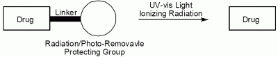
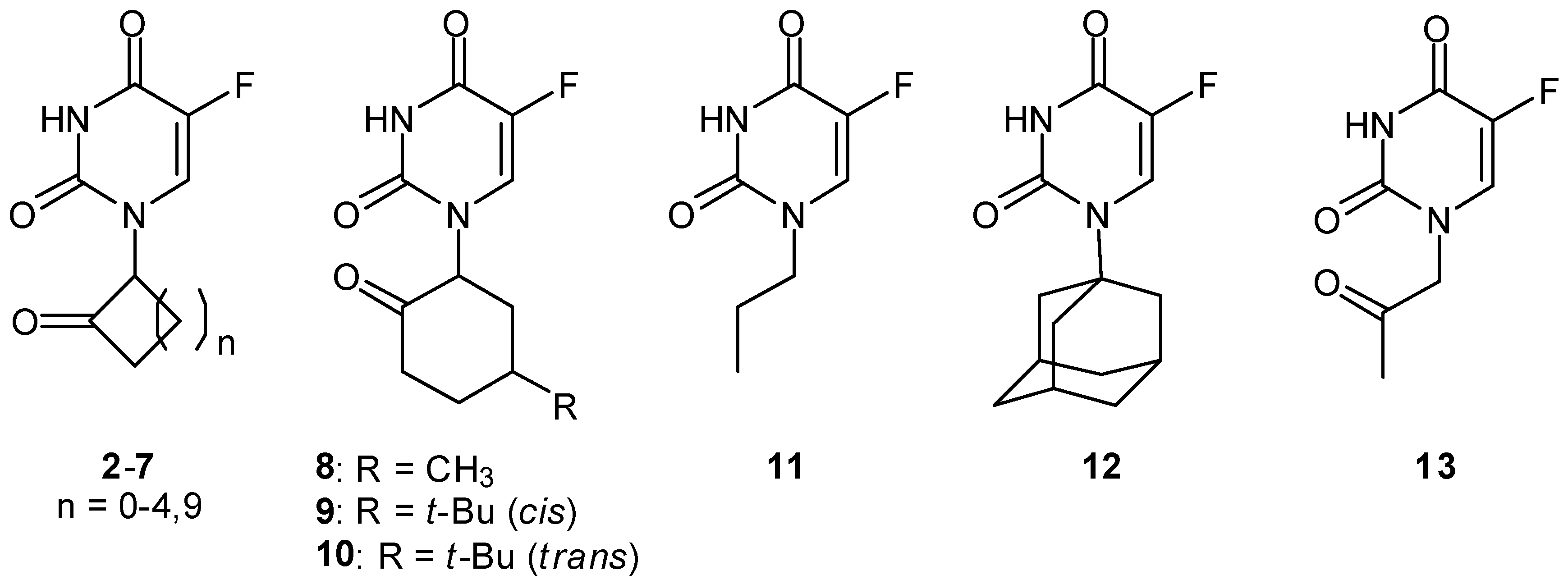
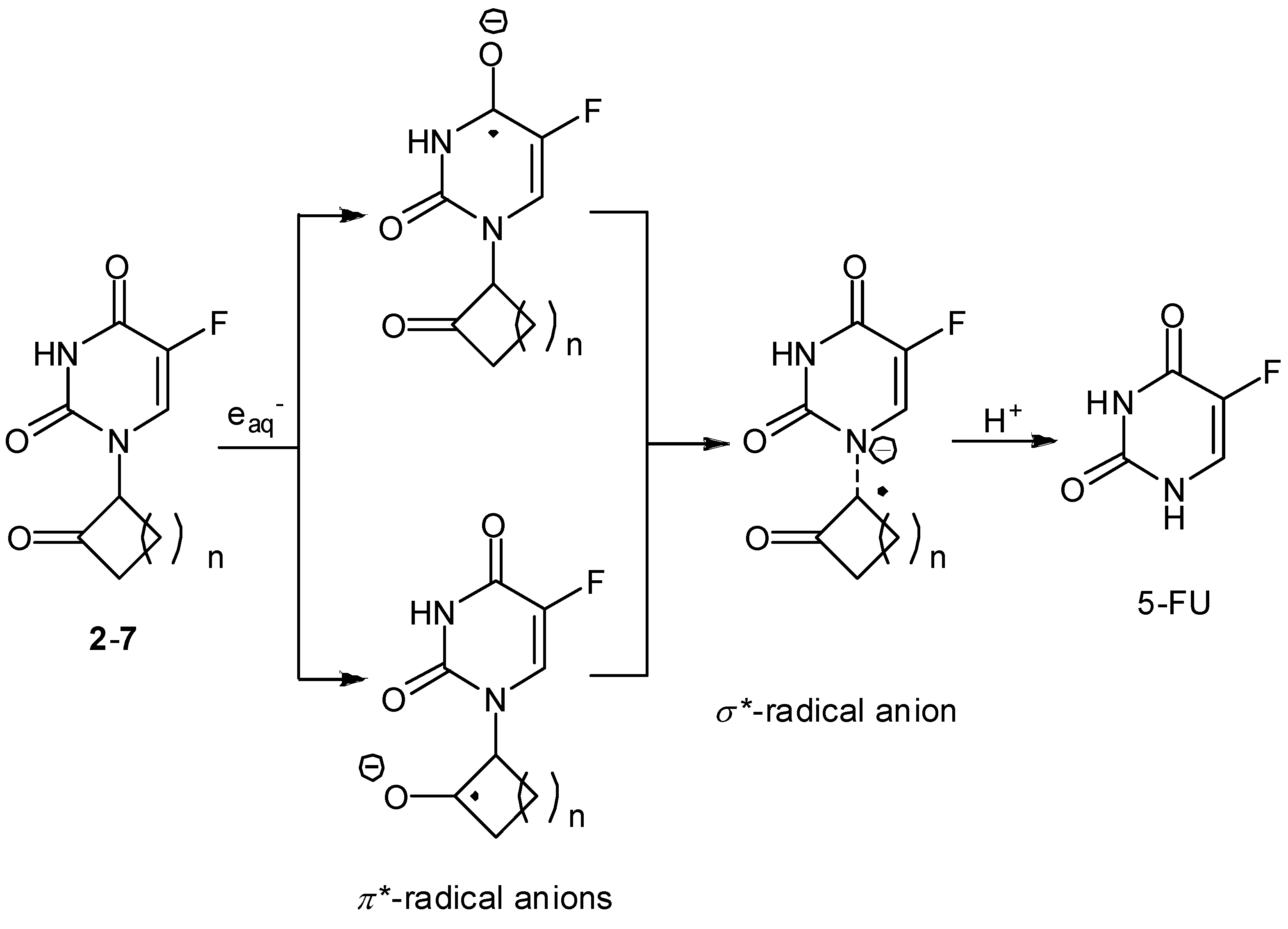
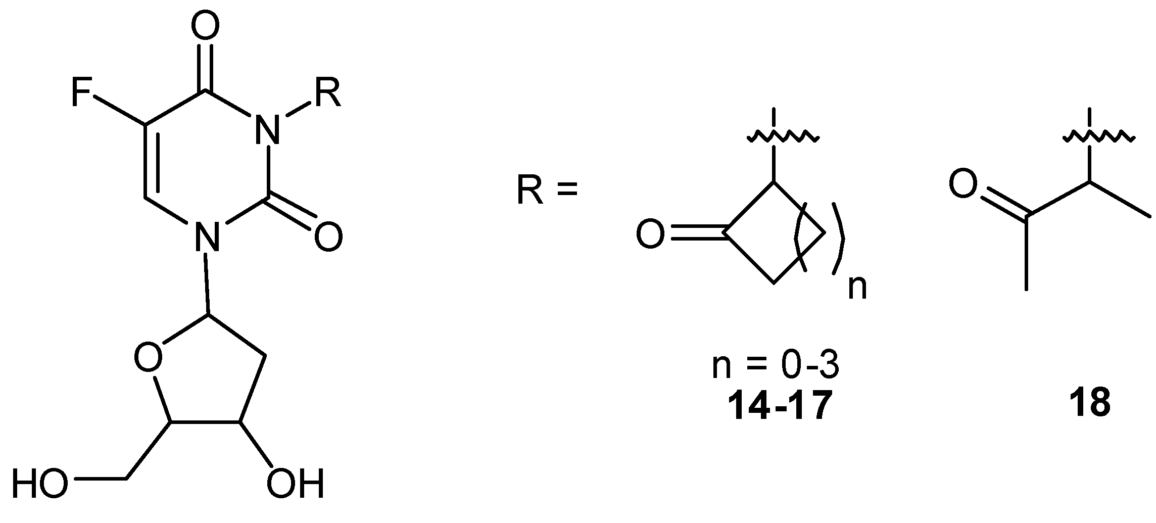
Radiation-activatable 5-FU Prodrugs

It is well known that various electrophilic compounds can enhance the sensitivity of hypoxic cells toward ionizing radiation [32,33]. Because of the inefficient supply of oxygen to the core of tumor tissue by poorly developed blood vessels, hypoxic cells in the solid tumors are resistant to radiation therapy and may be likewise refractory to certain types of chemotherapeutic agents operative through a variety of mechanisms [34].
Wide varieties of bioreduction-activatable prodrugs have been developed as anticancer agents [35,36,37]. Bioreductive prodrugs can be activated into their radical intermediates, which are oxidized to the prodrug under aerobic conditions. However, under hypoxic conditions (e.g., in solid tumors), this oxidation is much slower and usually increases the levels of toxic radical intermediates, resulting in a solid tumor-selective therapy. In general, hypoxic cells in low oxygen tension regions are more resistant to treatment with radiotherapy and require a two- to three-fold higher radiation dose, indicating the importance of bioreductive drugs.
Radiation generates high concentrations of molecular free radicals, hydrated electrons (eaq), hydrogen atoms (H) and hydroxyl radicals (OH) in irradiated tissues [38]. Under aerobic conditions, hydrated electrons react with O2 to produce superoxide radical anion (O2–•) (Scheme 1). In contrast, hydrated electrons can initiate reductive activation of the prodrugs under hypoxic conditions.
Scheme 1. Primary water radicals generated from water radiolysis.
Molecules 13 02370 g012
We have reported that galvanostatic electrolysis or radiation-induced oxidation of aqueous 5-FU solution yields the N1–C5’-linked dimer hydrate of 5-FU (1) via head-to-tail coupling between the N1- centered radical and C5’-centered radical of 5-FU (Figure 2) [30,31]. An interesting finding is that the N1-C5’-dimer hydrate 1 undergoes radiation-induced reduction under anaerobic conditions and releases the parent 5-FU with a 16% yield (Figure 3). Radiolysis of the dimer hydrate 1 under N2O-saturated conditions, where the primary species of hydrated electrons are scavenged by N2O, induced no such release of 5-FU, suggesting the reductive splitting of the dimer 1 is operative under anaerobic irradiation. To evaluate the relationship between the molecular structure and the reactivity of the one-electron reductive release of 5-FU in anoxic aqueous solution, a series of 5-fluoro-1-(2’-oxocycloalkyl)uracils (2–10) were synthesized (Figure 4) [39].
Figure 2. Electrochemical synthesis of N1-C5’-linked 5-FU dimer hydrate 1.
Molecules 13 02370 g002
Figure 3. Reductive splitting of N1-C5’-linked 5-FU dimer hydrate 1 and release of 5-FU.
Molecules 13 02370 g003
Figure 4. Structures of N1-substituted 5-FU prodrugs 2-13.
Molecules 13 02370 g004
All the 5-FU derivatives 2-10 bearing the 2’-oxo group underwent one-electron reduction by the addition of eaq and thereby released 5-FU in sufficient yields of 47-96% upon radiolysis of the anoxic aqueous solution. Release of fluoride ion from the 5-FU derivatives was not observed during the radiolysis suggesting that eaq is captured primarily by the 2’-oxo group. In fact, the control compounds without the 2’-oxo substituent (11, 12) had no reactivity toward such a reductive C1’-N1 bond splitting. Comparing the reactivities of the stereoisomeric derivatives 9 and 10 showed that the efficiency of 5-FU release was strongly dependent on their structural flexibility. X-ray crystallographic studies of representative compounds revealed that the C1’-N1 bond possesses normal geometry and bond length in the ground state. Molecular orbital (MO) calculations by the AM1 method for optimized structures demonstrated that the lowest unoccupied molecular orbital (LUMO) is localized primarily at the π* orbital of the C5–C6 double bond of the 5-FU moiety and that the LUMO + 1 is delocalized between the π* orbital of the 2’-oxo substituent and the σ* orbital of the adjacent C1’–N1 bond (Figure 5). It is presumable that the one-electron reductive release of 5-FU in anoxic aqueous solution occurs from the LUMO + 1 of the radical anion intermediates possessing a partial mixing of the antibonding C(2’)=O π* and C1’–N1 σ* MOs, and that dynamic conformational changes may achieve a higher degree of (π* + σ*) MO mixing.
Figure 5. Proposed mechanism for reductive release of 5-FU from 2-7.
Molecules 13 02370 g005
Based on these observations, our group has developed the simplest prototypes of the 5-FU prodrugs 13, and we have investigated the radiation chemical reactivity, biological effects, in vivo efficacy, pharmacokinetics, and toxicity [40,41]. The prodrug 13 dissolved in phosphate buffer released 5-FU with a G-value (mol number of molecules that are decomposed or produced by 1 J of absorbed radiation energy) of 1.9 × 10⁻⁷ mol/ J (26%, based on total amount of primary species generated from water) following hypoxic irradiation.
The G-value for 5-FU release was 1.0 × 10⁻⁸ mol/J following aerobic irradiation. Adding hypoxically irradiated (7.5–30 Gy) 13 to murine SCCVII cells for 1–24 h showed a significant cytotoxic effect. In contrast, cytotoxicity was minimal in cells treated with aerobically irradiated or unirradiated 13. This compound had no radiosensitizing effect against SCCVII cells under either aerobic or hypoxic conditions when the drug was removed immediately after irradiation. Following administration of 13 and irradiation at 30 Gy, the average 5-FU levels in the tumor and serum were 179ng/g and 83ng/mL, respectively. However, a TCD-50 tumor growth delay assay of 13 demonstrated an enhancement ratio of only 1.2. We recently synthesized a similar family of radiation-activated prodrug of 5-FdUrd [42], which is generally more toxic than 5-FU [43]. 5-FdUrd derivatives 14-18 possessing a 2-oxoalkyl group at the N3 position were synthesized, and their radiolytic one-electron reduction product was characterized (Figure 6).
Figure 6. Structures of 5-fluoro-3-(2’-oxoalkyl)-2’-deoxyuridines.
Molecules 13 02370 g006
The prodrugs released 5-FdUrd efficiently upon hypoxic irradiation by reacting with eaq as in the case of 5-FU. A biological assay using P388 T cells and EMT6/KU cells revealed that hypoxic X-irradiation enhanced cytotoxicity of the prodrugs dramatically. However, we observed no marked effects in vivo even when 100 or 300 mg/kg of 16 was combined with 20 Gy of irradiation [44]. Although the in vivo effect of both prodrugs of 5-FU and 5-FdUrd was not enough for clinical efficacy, this prototype agent suggests that radiation-activated prodrugs exhibit a certain level of cytotoxicity toward hypoxic tumor cells. As an extension of our work on the development of radiation-activated antitumor prodrugs, we designed another 5-FdUrd prodrug possessing indolequinone structure 19 (Figure 7) [45]. Indolequinone has the following unique reaction characteristics [46,47,48,49]. First, both enzymatic reduction and radiolytic reduction can activate this substituent. Second, the reductive activation of indolequinone derivatives to release drugs is accompanied by the concomitant formation of electrophilic iminium cations like 20, which may invoke DNA alkylation or other cellular damage [50]. In view of these reaction characteristics, a 5-FdUrd prodrug possessing an indolequinone structural unit may result in synergic cytotoxicity that originates from both the parent drug and electrophilic iminium species released upon radiolytic reduction and bioreduction in hypoxic cells. Hypoxic irradiation of 19 significantly increased the cytotoxicity against hypoxic tumor cells and caused higher cytotoxicity toward EMT6/KU cells than did the hypoxic irradiation with the original 5- FdUrd alone. Our group is currently investigating the in vivo effect of 19.
Figure 7. Reductive release of 5-FdUrd from 5-FdUrd-indolequinone prodrug 19.
Molecules 13 02370 g007

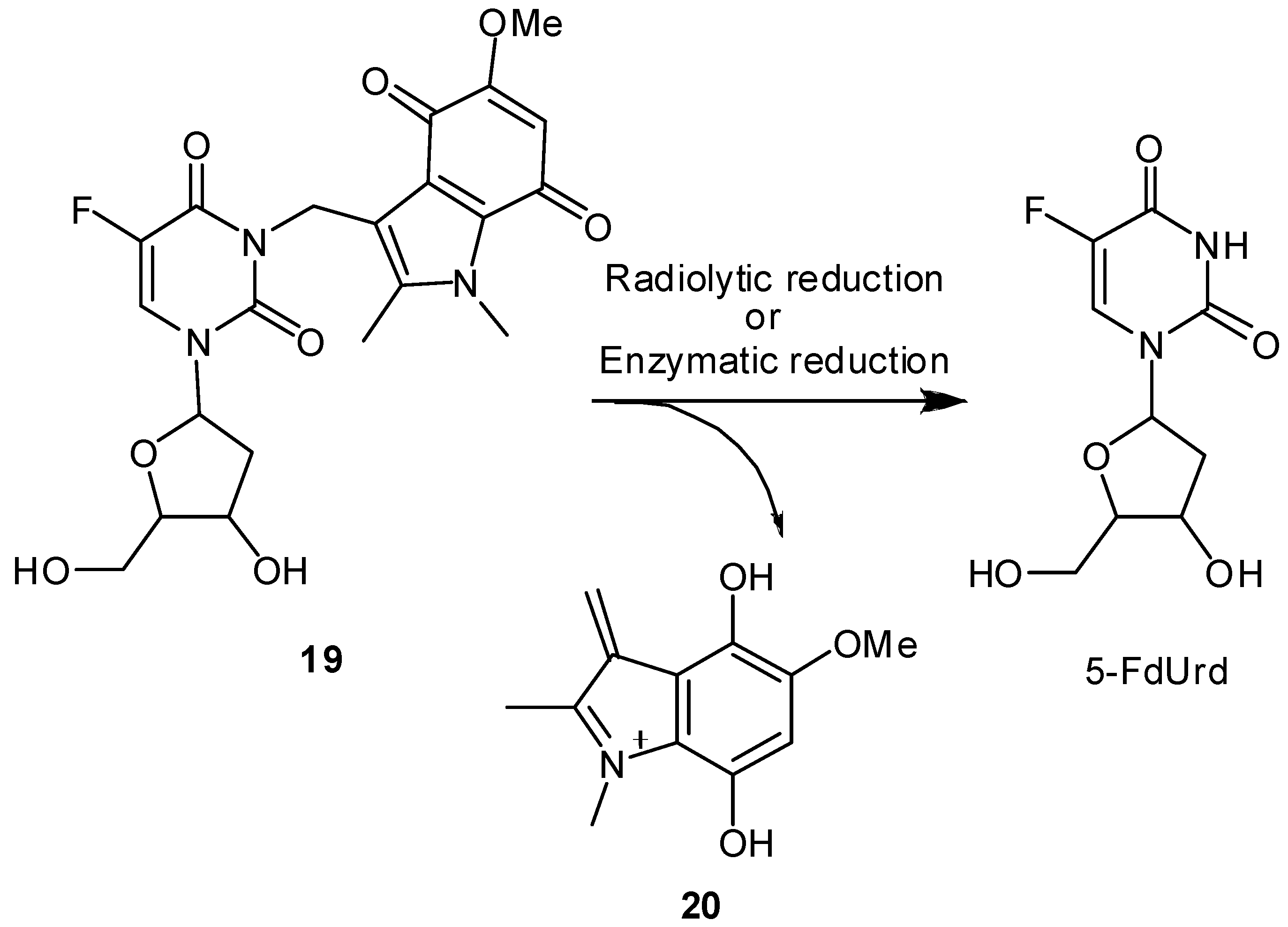
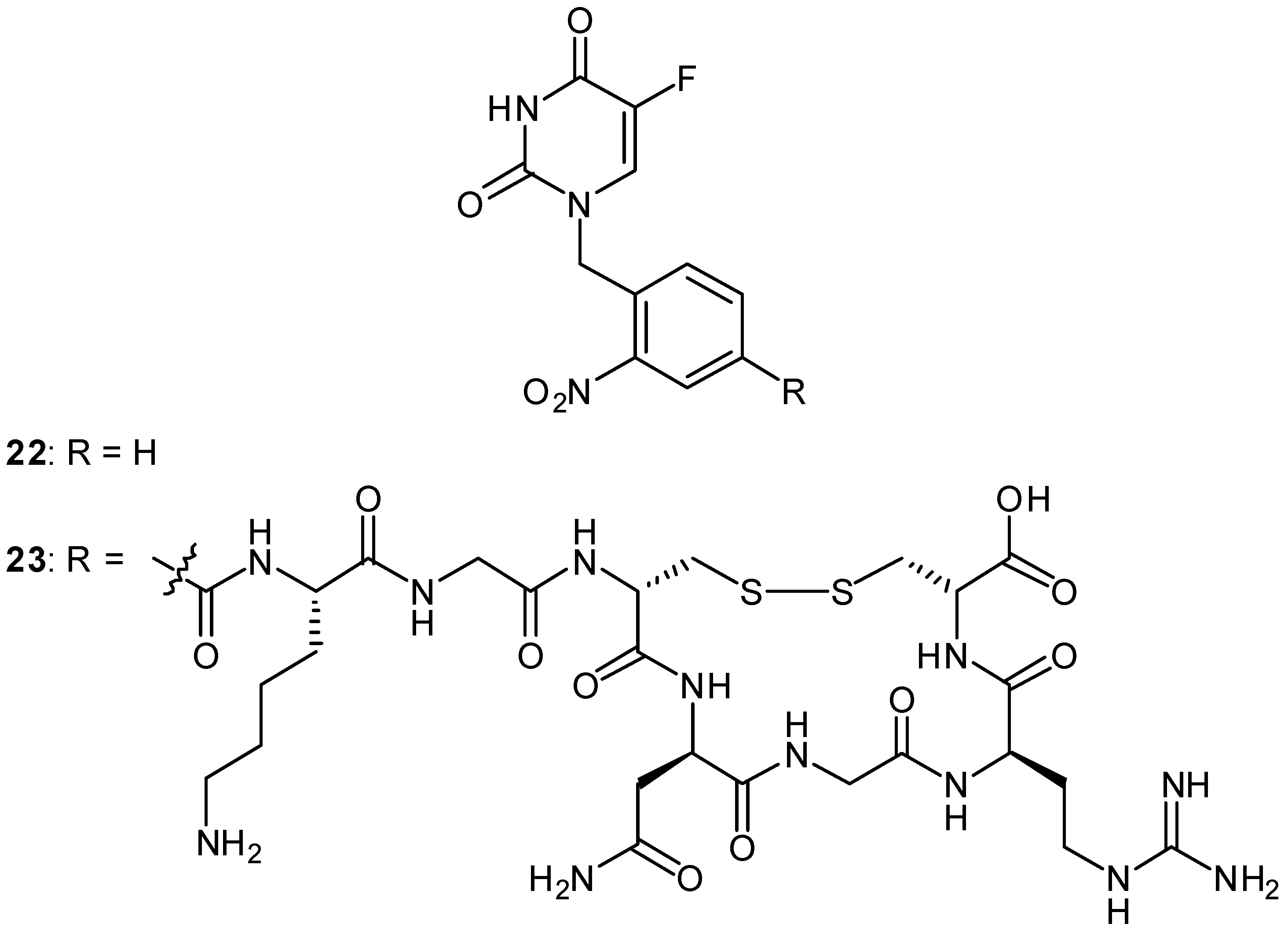
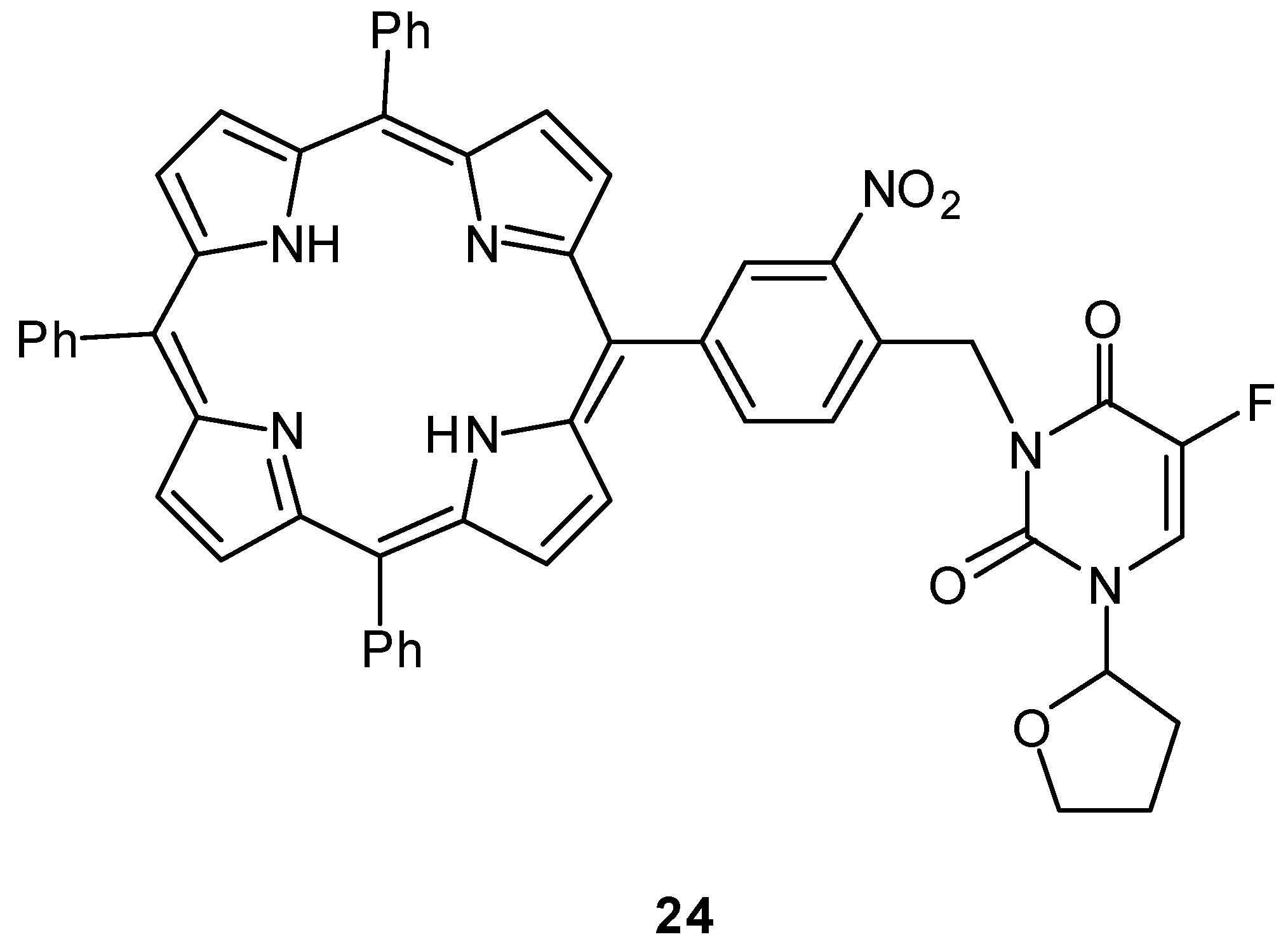
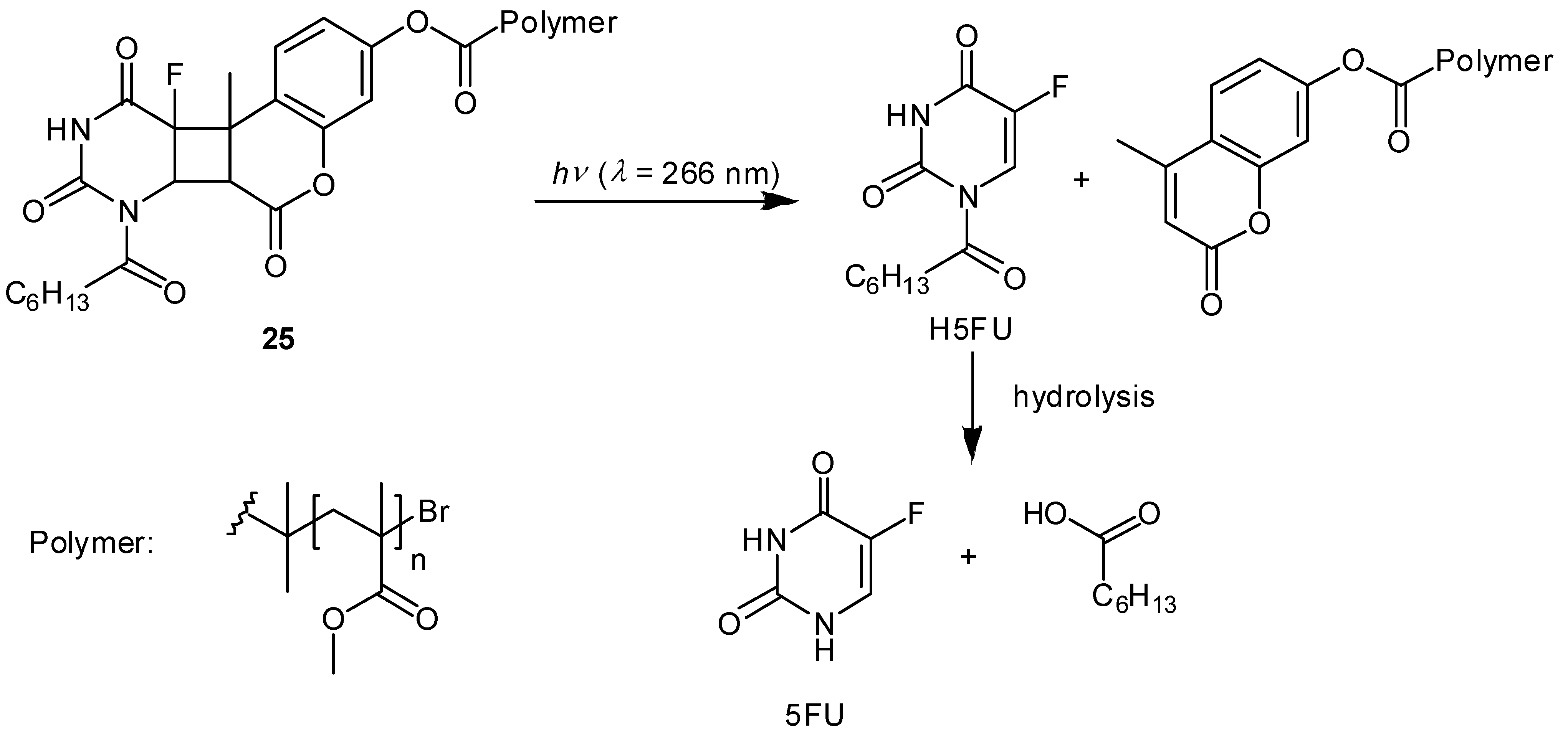
Photoactivatable 5-FU Prodrugs

Much research effort has focused on the development of antitumor prodrugs with functions that are activated upon controlled photoirradiation [51]. One general strategy to achieve photoinduced activation of the prodrugs is the initial deactivation of a parent drug by introduction of a photolabile protecting group at a critical position to reduce the cytotoxicity of the chemically modified parent drug under unirradiated conditions. Under photoirradiation with appropriate wavelength, the photolabile protecting group is removed from the prodrug constitution to release a parent drug, which regenerates the original cytotoxicity. Various photoactivated prodrugs with effective photolabile protecting groups (e.g., o-nitrobenzyl chromophores, 3,5-dimethoxybenzoin derivatives, and coumarin derivatives) have been developed that can be activated efficiently by irradiation with UV-A (320-400 nm) or visible light to release the parent drugs, including phosphoramidite mustard [52], L-leucyl-L-leucine methyl ester [53,54], aspirin [55,56], cyclic enediyne [57], and paclitaxel [58,59]. Photoactivated prodrugs can be applied in surface cancer treatment and can even be adapted for deep-seated cancer therapy employing endoscopes or optical fibers, as used in photodynamic therapy.
Among the various photosensitive protective groups, o-nitrobenzyl chromophore [60] is used most commonly for providing photoactivated prodrugs. o-Nitrobenzyl is a general photosensitive protective group for various chemical functionalities, such as alcohols, aldehydes, carboxylic acids, amines, and phosphates. Pei and Gong reported recently on a 5-FdUrd prodrug possessing the photolabile 4,5- dimethoxy-2-nitrobenzyl group 21 (Figure 8) [61]. They demonstrated the efficient photoinduced release of 5-FdUrd and 4,5-dimethoxy-2-nitrosobenzaldehyde via a spontaneous decarboxylation reaction from the prodrug 21 by first-order kinetics with a t1/2 = 6 min. They also showed inhibition of cell growth by photoirradiation. Without photoirradiation, prodrug 21 only slightly inhibited growth, whereas cell growth was inhibited completely when the cell culture containing prodrug 21 was irradiated with a 350 nm lamp.
Figure 8. Structure of 5-FdUrd prodrug with the photolabile 4,5-dimethoxy-2-nitrobenzyl group.
Molecules 13 02370 g008
We recently designed a photoactivated prodrug of 5-FU conjugated with a tumor-homing cyclic peptide to generate the first prototype compound of a tumor-targeting photoactivated antitumor prodrug 23 (Figure 9) [62]. The tumor vasculature is morphologically abnormal and carries various types of tumor molecular markers that can be used to discriminate tumor vessels from the normal vasculature. We employed chemical conjugation of a 5-FU prodrug possessing an o-nitrobenzyl group with cyclic peptide Cys-Asn-Gly-Arg-Cys (CNGRC), which should target one of the tumor molecular markers, a specific APN/CD13 aminopeptidase N. Photoirradiation of the photoactivated prodrug 23 readily removed the photolabile o-nitrobenzyl chromophore to give quantitative amounts of 5-FU, whereas the prodrug was quite stable under dark conditions. We also clarified the photoactivation mechanism of the simple model compound 22 using nanosecond laser flash photolysis studies. Although the in vitro and in vivo evaluation is in progress, the prodrug 23 is expected to show selective cytotoxicity toward tumor tissue upon photoirradiation.
Figure 9. Structures of photoactivated prodrugs containing 5-FU.
Molecules 13 02370 g009
Lin and coworkers reported another example of a photoactivated prodrug containing 5-fluoro-1- (tetrahydro-2-furanyl)-2,4(1H,3H)-pyrimidinedione (tegafur/Ftorafur) 24 (Figure 10) [63]. Tegafur, an orally active prodrug of 5-FU, is used widely in the treatment of gastrointestinal malignancies and has modest efficacy [64]. Upon photoirradiation at 350 nm, the tegafur prodrug incorporating a triphenylporphyrin and photolabile o-nitrobenzyl group is converted into the anticancer drug tegafur with the photochemical quantum yield Ф = 0.032. An MTT [3-(4,5-dimethylthiazole-2-yl)-2,5- diphenyl-tetrazolium bromide] assay demonstrated that the porphyrin–nitrobenzene–tegafur conjugate shows photoinduced cytotoxicity for MCF-7 mammary cancer cells because of the efficient release of tegafur upon photoirradiation at 350 nm, whereas the prodrug 24 is significantly less toxic than its parent anticancer drug tegafur in the absence of UV irradiation.
Figure 10. Structures of a photoactivated prodrug containing 5-FU analog 24.
Molecules 13 02370 g010
Other interesting examples have been reported. Agarwal and coworkers demonstrated a photoinduced 5-FU release system from methylcoumarin end-functionalized acrylic polymer conjugated with the prodrug 1-heptanoyl-5-fluorouracil (H5FU), which was developed for intraocular lenses equipped with a multidose drug depot used in cataract surgery (Figure 11) [65]. The coumarin-functionalized polymer-H5FU conjugate 25 was synthesized photochemically via a [2+2] cycloaddition reaction between the C3-C4 double bond of methylcoumarin and the C5-C6 double bond of H5FU upon photoirradiation at 350 nm. In contrast to the [2+2] cycloaddition reaction, the [2+2] cycloreversion reaction on the cyclobutane ring proceeds under photoirradiation using a shorter wavelength. Consequently, photoirradiation of 25 at 266 nm induces the cleavage of the cyclobutane linker between the methylcoumarin moiety and the prodrug H5FU, leading to the efficient release of H5FU. Subsequent hydrolysis of H5FU in aqueous solution produces 4.76 μg of 5-FU from 1 mg of the polymer–drug conjugate 25 after photoirradiation for 150 min, indicating that LD50 of 5-FU for rabbit lens epithelial cells can be realized even with small amounts of polymer-drug conjugate 25, although the photoinduced drug release system has not been evaluated in vitro and in vivo. Further studies to avoid the risk of DNA being damaged by UV-C radiation would be necessary for its clinical use.
Figure 11. Photo-induced drug release from coumarin-functionalized polymer-H5FU conjugate 25 and subsequent hydrolysis of the prodrug H5FU to 5-FU.
Molecules 13 02370 g011

Conclusions

In the past decade, varieties of photoremovable protecting groups have been developed to control the activity of small molecules, including bioimaging molecules and several kinds of pharmaceutical drugs, by irradiating them with light of specific wavelength and intensity. On the other hand, the ionizing radiation-activated system is not ideal for diagnostic use in the medical field but should be applicable to activating antitumor drugs for sensitizing the drugs to increase the cytotoxic effect of ionizing radiation. In this review, we have summarized the properties of 5-FU prodrugs, which are radiation-chemically or photochemically activatable under the microenvironment of tumor cells. Irradiated prodrugs have high cytotoxicity that is dependent on both the drug contact time and radiation dose, whereas unirradiated prodrugs show minimal cytotoxicity against tumor cells. Disappointingly, the effects of these prototypes, including N1- or N3-(2’-oxoalkyl)-5-FU derivatives, are not strong enough for clinical efficacy, partly due to low efficiency (26% or less) of 5-FU-release from the prodrugs. Although there is limit in clinically permitted does of radiation, we expect that further research on the synthesis and evaluation of more potent anticancer drugs bearing both 5-FU and alkylating agents, such as indolequinone derivatives, will lead to the development of more promising prodrugs that are activatable by external signals.

References

  1. Rominger, C.J.; Gelber, R.D.; Gunderson, L.L.; Conner, N. Radiation-therapy alone or in combination with chemotherapy in the treatment of residual or inoperable carcinoma of the rectum and rectosigmoid or pelvic recurrence following colorectal surgery - Radiation-therapy oncology group-study. Am. J. Clin. Oncol. 1985, 8, 118–127. [Google Scholar] [CrossRef] [PubMed]
  2. Wolmark, N.; Fisher, B.; Rockette, H.; Redmond, C.; Wickerham, D.L.; Fisher, E.R.; Jones, J.; Glass, A.; Lerner, H.; Lawrence, W.; Prager, D.; Wexler, M.; Evans, J.; Cruz, A.; Dimitrov, N.; Jochimsen, P. Postoperative adjuvant chemotherapy or BCG for colon cancer - Results from NSABP protocol-C-01. J. Natl. Cancer Inst. 1988, 80, 30–36. [Google Scholar] [CrossRef] [PubMed]
  3. Schwartz, E.L.; Baptiste, N.; Wadler, S.; Makower, D. Thymidine phosphorylase mediates the sensitivity of human colon-carcinoma cells to 5-fluorouracil. J. Biol. Chem. 1995, 270, 19073–19077. [Google Scholar] [PubMed]
  4. Evrard, A.; Cuq, P.; Robert, B.; Vian, L.; Pelegrin, A.; Cano, J.P. Enhancement of 5-fluorouracil cytotoxicity by human thymidine-phosphorylase expression in cancer cells: In vitro and in vivo study. Int. J. Cancer 1999, 80, 465–470. [Google Scholar] [CrossRef]
  5. Hartmann, K.U.; Heidelberger, C. Studies on fluorinated pyrimidines. 13. Inhibition of thymidylate synthetase. J. Biol. Chem. 1961, 236, 3006–3013. [Google Scholar] [PubMed]
  6. Peters, G.J.; Vanderwilt, C.L.; Vantriest, B.; Codaccipisanelli, G.; Johnston, P.G.; Vangroeningen, C.J.; Pinedo, H.M. Thymidylate synthase and drug-resistance. Eur. J. Cancer 1995, 31A, 1299–1305. [Google Scholar] [CrossRef]
  7. Yeh, K.H.; Shun, C.T.; Chen, C.L.; Lin, J.T.; Lee, W.J.; Lee, P.H.; Chen, Y.C.; Cheng, A.L. High expression of thymidylate synthase is associated with the drug resistance of gastric carcinoma to high dose 5-fluorouracil-based systemic chemotherapy. Cancer 1998, 82, 1626–1631. [Google Scholar] [CrossRef]
  8. Albert, A. Chemical aspects of selective toxicity. Nature 1958, 182, 421–423. [Google Scholar] [CrossRef] [PubMed]
  9. Stella, V.J.; Himmelstein, K.J. Prodrugs and site-specific drug delivery. J. Med. Chem. 1980, 23, 1275–1282. [Google Scholar] [CrossRef] [PubMed]
  10. Kratz, F.; Muller, I.A.; Ryppa, C.; Warnecke, A. Prodrug strategies in anticancer chemotherapy. ChemMedChem 2008, 3, 20–53. [Google Scholar] [CrossRef] [PubMed]
  11. Rautio, J.; Kumpulainen, H.; Heimbach, T.; Oliyai, R.; Oh, D.; Järvinen, T.; Savolainen, J. Prodrugs: Design and clinical applications. Nat. Rev. Drug Discov. 2008, 7, 255–270. [Google Scholar] [CrossRef] [PubMed]
  12. Rooseboom, M.; Commandeur, J.N.M.; Vermeulen, N.P.E. Enzyme-catalyzed activation of anticancer prodrugs. Pharmacol. Rev. 2004, 56, 53–102. [Google Scholar] [CrossRef] [PubMed]
  13. Yasumoto, M.; Yamawaki, I.; Marunaka, T.; Hashimoto, S. Studies on anti-tumor agents.2. Syntheses and anti-tumor activities of 1-(tetrahydro-2-furanyl)-5-fluorouracil and 1,3- bis(tetrahydro-2-furanyl)-5-fluorouracil. J. Med. Chem. 1978, 21, 738–741. [Google Scholar] [CrossRef] [PubMed]
  14. Meyer, R.B.; Levenson, C.H. Structure of two hydroxylated metabolites of ftorafur. Biochem. Pharmacol. 1980, 29, 665–668. [Google Scholar] [CrossRef]
  15. Au, J.L.; Sadee, W. The pharmacology of ftorafur (R, S-1-(tetrahydro-2-furanyl)-5-fluorouracil). Recent Results Cancer Res. 1981, 76, 100–114. [Google Scholar]
  16. Iigo, M.; Hoshi, A.; Nakamura, A.; Kuretani, K. Anti-tumor activity of 1-alkylcarbamoyl derivatives of 5-fluorouracil in a variety of mouse tumors. Cancer Chemother. Pharmacol. 1978, 1, 203–208. [Google Scholar] [PubMed]
  17. Sasaki, H.; Takahashi, T.; Mori, Y.; Nakamura, J.; Shibasaki, J. Transdermal delivery of 5- fluorouracil and its alkylcarbamoyl derivatives. Int. J. Pharm. 1990, 60, 1–9. [Google Scholar] [CrossRef]
  18. Grem, J.L. 5-Fluorouracil: forty-plus and still ticking. A review of its preclinical and clinical development. Invest. New Drugs 2000, 18, 299–313. [Google Scholar] [CrossRef] [PubMed]
  19. van Laar, J.A.M.; Rustum, Y.M.; Ackland, S.P.; van Groeningen, C.J.; Peters, G.J. Comparison of 5-fluoro-2'-deoxyuridine with 5-fluorouracil and their role in the treatment of colorectal cancer. Eur. J. Cancer 1998, 34, 296–306. [Google Scholar] [CrossRef]
  20. Denizli, A.; Kiremitci, M.; Piskin, E. Subcutaneous polymeric matrix system p(hema-bga) for controlled release of an anticancer drug (5-fluorouracil). 2. Release kinetics. Biomaterials 1988, 9, 363–366. [Google Scholar] [CrossRef]
  21. Endoh, H.; Kawaguchi, T.; Seki, T.; Hasegawa, T.; Juni, K. Controlled release of 5-fluoro-2'-deoxyuridine by the combination of prodrug and polymer matrix. Chem. Pharm. Bull. 1991, 39, 458–464. [Google Scholar] [CrossRef] [PubMed]
  22. Kim, K.S.; Kim, T.K.; Graham, N.B. Controlled release behavior of prodrugs based on the biodegradable poly(L-glutamic acid) microspheres. Polym. J. 1999, 31, 813–816. [Google Scholar] [CrossRef]
  23. Michael, N.P.; Chester, K.A.; Melton, R.G.; Robson, L.; Nicholas, W.; Boden, J.A.; Pedley, R.B.; Begent, R.H.; Sherwood, R.F.; Minton, N.P. In vitro and in vivo characterisation of a recombinant carboxypeptidase G(2)::anti-CEA scFv fusion protein. Immunotechnology 1996, 2, 47–57. [Google Scholar] [CrossRef]
  24. Coelho, V.; Dernedde, J.; Petrausch, U.; Panjideh, H.; Fuchs, H.; Menzel, C.; Dubel, S.; Keilholz, U.; Thiel, E.; Deckert, P.M. Design, construction, and in vitro analysis of A33scFv::CDy, a recombinant fusion protein for antibody-directed enzyme prodrug therapy in colon cancer. Int. J. Oncol. 2007, 31, 951–957. [Google Scholar] [CrossRef] [PubMed]
  25. Madec-Lougerstay, R.; Florent, J.C.; Monneret, C. Synthesis of self-immolative glucuronide spacers based on aminomethylcarbamate. Application to 5-fluorouracil prodrugs for antibody-directed enzyme prodrug therapy. J. Chem. Soc. Perkin Trans. 1 1999, 1369–1375. [Google Scholar] [CrossRef]
  26. Fichera, A.; Michelassi, F.; Arenas, R.B. Selective expression of carcinoembryonic antigen promoter in cancer cell lines- Targeting strategy for gene therapy in colorectal cancer. Dis. Colon Rectum. 1998, 41, 747–754. [Google Scholar] [CrossRef] [PubMed]
  27. Aquino, A.; Formica, V.; Prete, S.P.; Correale, P.P.; Massara, M.C.; Turriziani, M.; De Vecchis, L.; Bonmassar, E. Drug-induced increase of carcinoembryonic antigen expression in cancer cells. Pharmacol. Res. 2004, 49, 383–396. [Google Scholar] [CrossRef] [PubMed]
  28. Topf, N.; Worgall, S.; Hackett, N.R. Regional 'pro-drug' gene therapy: intravenous administration of an adenoviral Vector expressing the E-coli cytosine deaminase gene and systemic administration of 5-fluorocytosine suppresses growth of hepatic metastasis of colon carcinoma. Gene Ther. 1998, 5, 507–513. [Google Scholar] [CrossRef] [PubMed]
  29. Denny, W.A. Prodrugs for gene-directed enzyme-prodrug therapy (suicide gene therapy). J. Biomed. Biotechnol. 2003, 48–70. [Google Scholar] [CrossRef] [PubMed]
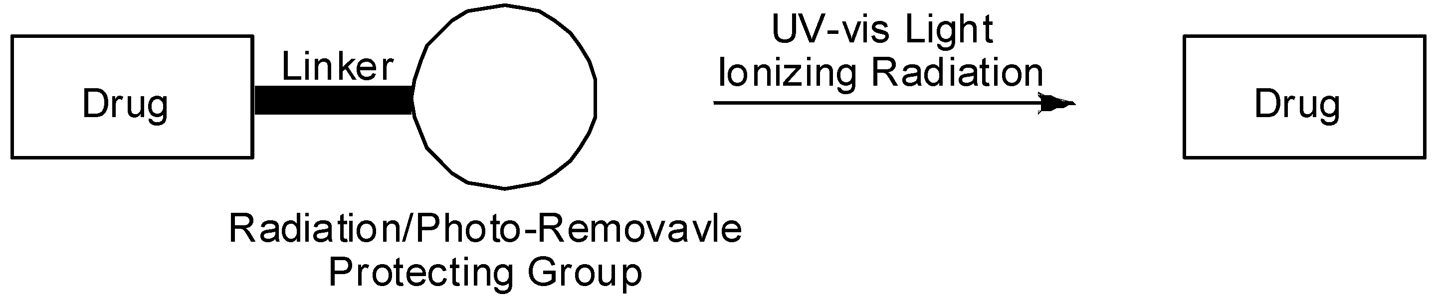
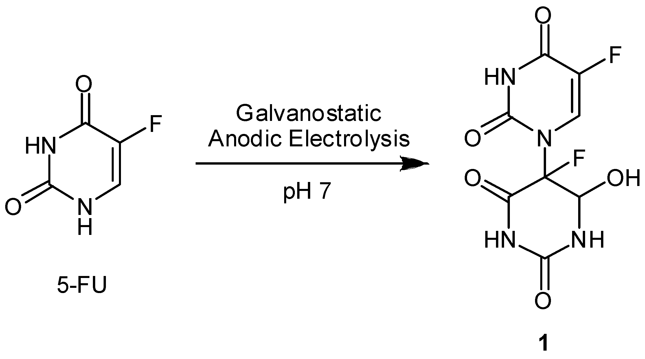
  30. Hatta, H.; Zhou, L.; Mori, M.; Teshima, S.; Nishimoto, S. N(1)-C(5')-linked dimer hydrates of 5-substituted uracils produced by anodic oxidation in aqueous solution. J. Org. Chem. 2001, 66, 2232–2239. [Google Scholar] [CrossRef] [PubMed]
  31. Nishimoto, S.; Hatta, H.; Ueshima, H.; Kagiya, T. 1-(5'-Fluoro-6'-hydroxy-5',6'-dihydrouracil-5'- yl)-5-fluorouracil, a novel N(1)-C(5’)-linked dimer that releases 5-fluorouracil by radiation activation under hypoxic conditions. J. Med. Chem. 1992, 35, 2711–2712. [Google Scholar] [CrossRef] [PubMed]
  32. Vaupel, P.; Kallinowski, F.; Okunieff, P. Blood-flow, oxygen and nutrient supply, and metabolic microenvironment of human-tumors- A review. Cancer Res. 1986, 49, 6449–6465. [Google Scholar]
  33. Vaupel, P.; Schlenger, K.; Knoop, C.; Höckel, M. Oxygenation of human tumors - Evaluation of tissue oxygen distribution in breast cancers by computerized O2 tension measurements. Cancer Res. 1991, 51, 3316–3322. [Google Scholar] [PubMed]
  34. Denny, W.A. The design of drugs that target tumour hypoxia. Aust. J. Chem. 2004, 57, 821–828. [Google Scholar] [CrossRef]
  35. Bruno, R.D.; Njar, V.C.O. Targeting cytochrome P450 enzymes: A new approach in anti-cancer drug development. Bioorg. Med. Chem. 2007, 15, 5047–5060. [Google Scholar] [CrossRef] [PubMed]
  36. Denny, W.A. Hypoxia-activated anticancer drugs. Expert Opin. Ther. Pat. 2005, 15, 635–646. [Google Scholar] [CrossRef]
  37. Tanabe, K.; Zhang, Z.; Ito, T.; Hatta, H.; Nishimoto, S. Current molecular design of intelligent drugs and imaging probes targeting tumor-specic microenvironments. Org. Biomol. Chem. 2007, 5, 3745–3757. [Google Scholar] [CrossRef] [PubMed]
  38. von Sonntag, C. Free-Radical-Induced DNA Damage and Its Repair. A Chemical Perspective; Springer-Verlag: Berlin, Heidelberg, Germany, 2006. [Google Scholar]
  39. Mori, M.; Hatta, H.; Nishimoto, S. Stereoelectronic effect on one-electron reductive release of 5- fluorouracil from 5-fluoro-1-(2'-oxocycloalkyl)uracils as a new class of radiation-activated antitumor prodrugs. J. Org. Chem. 2000, 65, 4641–4647. [Google Scholar] [CrossRef] [PubMed]
  40. Shibamoto, Y.; Zhou, L.; Hatta, H.; Mori, M.; Nishimoto, S. A novel class of antitumor prodrug, 1-(2 '-oxopropyl)-5-fluorouracil (OFU001), which releases 5-fluorouracil upon hypoxic irradiation. Jpn. J. Cancer Res. 2000, 91, 433–438. [Google Scholar] [CrossRef] [PubMed]
  41. Shibamoto, Y.; Zhou, L.; Hatta, H.; Mori, M.; Nishimoto, S. In vivo evaluation of a novel antitumor prodrug, 1-(2 '-oxopropyl)-5-fluorouracil (OFU001), which releases 5-fluorouracil upon hypoxic irradiation. Int. J. Radiat. Oncol. Biol. Phys. 2001, 49, 407–413. [Google Scholar] [CrossRef]
  42. Tanabe, K.; Mimasu, Y.; Eto, A.; Tachi, Y.; Sakakibara, S.; Mori, M.; Hatta, H.; Nishimoto, S. One-electron reduction characteristics of N(3)-substituted 5-fluorodeoxyuridines synthesized as radiation-activated prodrugs. Bioorg. Med. Chem. 2003, 11, 4551–4556. [Google Scholar] [CrossRef] [PubMed]
  43. Shibamoto, Y.; Mimasu, Y.; Tachi, Y.; Hatta, H.; Nishimoto, S. Comparison of 5-fluorouracil and 5-fluoro-2'-deoxyuridine as an effector in radiation-activated prodrugs. J. Chemother. 2002, 14, 390–396. [Google Scholar] [CrossRef] [PubMed]
  44. Shibamoto, Y.; Tachi, Y.; Tanabe, K.; Hatta, H.; Nishimoto, S. In vitro and in vivo evaluation of novel antitumor prodrugs of 5-fluoro-2'-deoxyuridine activated by hypoxic irradiation. Int. J. Radiat. Oncol. Biol. Phys. 2004, 58, 397–402. [Google Scholar] [CrossRef] [PubMed]
  45. Tanabe, K.; Makimura, Y.; Tachi, Y.; Imagawa-Sato, A.; Nishimoto, S. Hypoxia-selective activation of 5-fluorodeoxyuridine prodrug possessing indolequinone structure: Radiolytic reduction and cytotoxicity characteristics. Bioorg. Med. Chem. Lett. 2005, 15, 2321–2324. [Google Scholar] [CrossRef] [PubMed]
  46. Naylor, M.A.; Swann, E.; Everett, S.A.; Jaffar, M.; Nolan, J.; Robertson, N.; Lockyer, S.D.; Patel, K.B.; Dennis, M.F.; Stratford, M.R.L.; Wardman, P.; Adams, G.E.; Moody, C.J.; Stratford, I.J. Indolequinone antitumor agents: reductive activation and elimination from (5- methoxy-1-methyl-4,7-dioxoindol-3-yl)methyl derivatives and hypoxia-selective cytotoxicity in vitro. J. Med. Chem. 1998, 41, 2720–2731. [Google Scholar] [CrossRef] [PubMed]
  47. Swann, E.; Barraja, P.; Oberlander, A.M.; Gardipee, W.T.; Hudnott, A.R.; Beall, H.D.; Moody, C.J. Indolequinone antitumor agents: correlation between quinine structure and rate of metabolism by recombinant human NAD(P)H: quinine oxidoreductase. Part 2. J. Med. Chem. 2001, 44, 3311–3319. [Google Scholar] [CrossRef] [PubMed]
  48. Hernick, M.; Flader, C.; Borch, R.F. Design, synthesis, and biological evaluation of indolequinone phosphoramidate prodrugs targeted to DT-diaphorase. J. Med. Chem. 2002, 45, 3540–3548. [Google Scholar] [CrossRef] [PubMed]
  49. Everett, S.A.; Swann, E.; Naylor, M.A.; Stratford, M.R.L.; Patel, K.B.; Tian, A.; Newman, R.G.; Vojnovic, B.; Moody, C.J.; Wardman, P. Modifying rates of reductive elimination of leaving groups from indolequinone prodrugs: A key factor in controlling hypoxia-selective drug release. Biochem. Pharmacol. 2002, 63, 1629–1639. [Google Scholar] [CrossRef]
  50. Skibo, E.B.; Xing, C.; Groy, T. Recognition and cleavage at the DNA major groove. Bioorg. Med. Chem. 2001, 9, 2445–2459. [Google Scholar] [CrossRef]
  51. Dormán, G.; Prestwich, G.D. Using photolabile ligands in drug discovery and development. Trends Biotech. 2000, 18, 64–77. [Google Scholar]
  52. Reinhard, R.; Schmidt, B.F. Nitrobenzyl-based photosensitive phosphoramide mustards: Synthesis and photochemical properties of potential prodrugs for cancer therapy. J. Org. Chem. 1998, 63, 2434–2441. [Google Scholar] [CrossRef] [PubMed]
  53. Watanabe, S.; Sato, M.; Sakamoto, S.; Yamaguchi, K.; Iwamura, M. New dendritic caged compounds: synthesis, mass spectrometric characterization, and photochemical properties of dentrimers with a-carboxy-2-nitrobenzyl caged compounds at their periphery. J. Am. Chem. Soc. 2000, 122, 12588–12589. [Google Scholar] [CrossRef]
  54. Mizuta, H.; Watanabe, S.; Sakurai, Y.; Nishiyama, K.; Furuta, T.; Kobayashi, Y.; Iwamura, M. Design, synthesis, photochemical properties and cytotoxic activities of water-soluble caged L-leucyl-L-leucine methyl esters that control apoptosis of immune cells. Bioorg. Med. Chem. 2002, 10, 675–683. [Google Scholar] [CrossRef]
  55. McCoy, C.P.; Rooney, C.; Edwards, C.R.; Jones, D.S.; Gorman, S.P. Light-triggered molecule-scale drug dosing devices. J. Am. Chem. Soc. 2007, 129, 9572–9573. [Google Scholar] [CrossRef] [PubMed]
  56. McCoy, C.P.; Rooney, C.; Jones, D.S.; Gorman, S.P.; Nieuwenhuyzen, M. Rational design of a dual-mode optical and chemical prodrug. Pharm. Res. 2007, 24, 194–200. [Google Scholar] [CrossRef] [PubMed]
  57. Tachi, Y.; Dai, W.-M.; Tanabe, K.; Nishimoto, S. Synthesis and DNA cleavage reaction characteristics of enediyne prodrugs activated via an allylic rearrangement by base or UV irradiation. Bioorg. Med. Chem. 2006, 14, 3199–3209. [Google Scholar] [CrossRef] [PubMed]
  58. Skwarczynski, M.; Noguchi, M.; Hirota, S.; Sohma, Y.; Kimura, T.; Hayashi, Y.; Kiso, Y. Development of first photoresponsive prodrug of paclitaxel. Bioorg. Med. Chem. Lett. 2006, 16, 4492–4496. [Google Scholar] [CrossRef] [PubMed]
  59. Noguchi, M.; Skwarczynski, M.; Prakash, H.; Hirota, S.; Kimura, T.; Hayashi, Y.; Kiso, Y. Development of novel water-soluble photocleavable protecting group and its application for design of photoresponsive paclitaxel prodrugs. Bioorg. Med. Chem. 2008, 16, 5389–5397. [Google Scholar] [CrossRef] [PubMed]
  60. Willner, I.; Willner, B. Biological Applications of Photochemical Switches; Morrison, H., Ed.; John Wiley & Sons, Inc: New York, USA, 1993. [Google Scholar]
  61. Wei, Y.; Yan, Y.; Pei, D.; Gong, B. A photoactivated prodrug. Bioorg. Med. Chem. Lett. 1998, 8, 2419–2422. [Google Scholar] [CrossRef]
  62. Zhang, Z.; Hatta, H.; Ito, T.; Nishimoto, S. Synthesis and photochemical properties of photoactivated antitumor prodrugs releasing 5-fluorouracil. Org. Biomol. Chem. 2005, 3, 592–596. [Google Scholar] [CrossRef] [PubMed]
  63. Lin, W.; Peng, D.; Wang, B.; Long, L.; Guo, C.; Yuan, J. A model for light-triggered porphyrin anticancer prodrugs based on an o-nitrobenzyl photolabile group. Eur. J. Org. Chem. 2008, 793, 796. [Google Scholar] [CrossRef]
  64. Takiuchi, H.; Ajani, J.A. Uracil-tegafur in gastric carcinoma: a comprehensive review. J. Clin. Oncol. 1998, 16, 2877–2885. [Google Scholar] [PubMed]
  65. Sinkel, C.; Greiner, A.; Agarwal, S. Synthesis, characterization, and properties evaluation of methylcoumarin end-functionalized poly(methyl methacrylate) for photoinduced drug release. Macromolecules 2008, 41, 3460–3467. [Google Scholar] [CrossRef]
  • Sample Availability: Samples are not available.

