An Emulsion System Based on a Chip Polymerase Chain Reaction

Abstract
:Introduction
Results
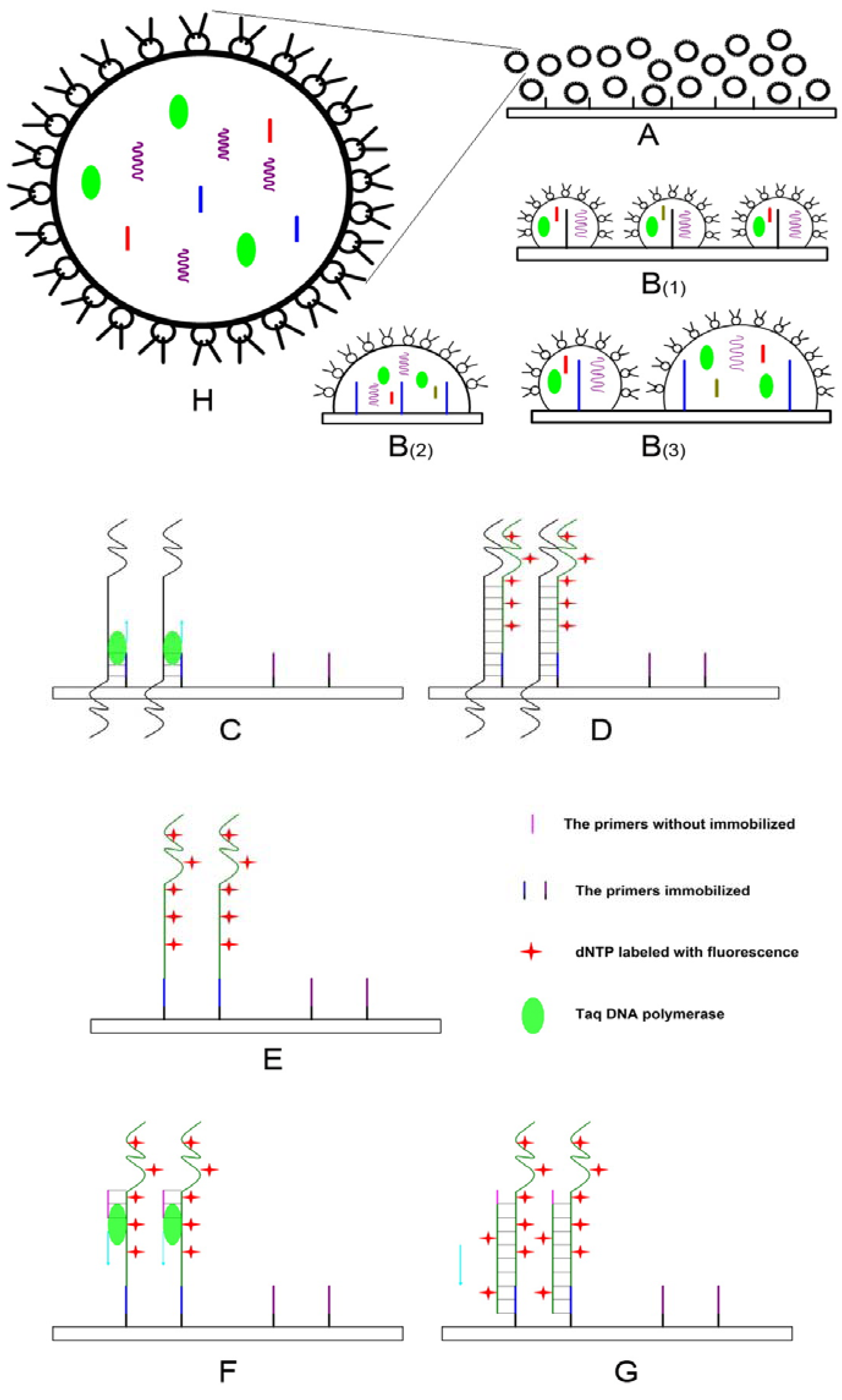
Strategy of EC-PCR

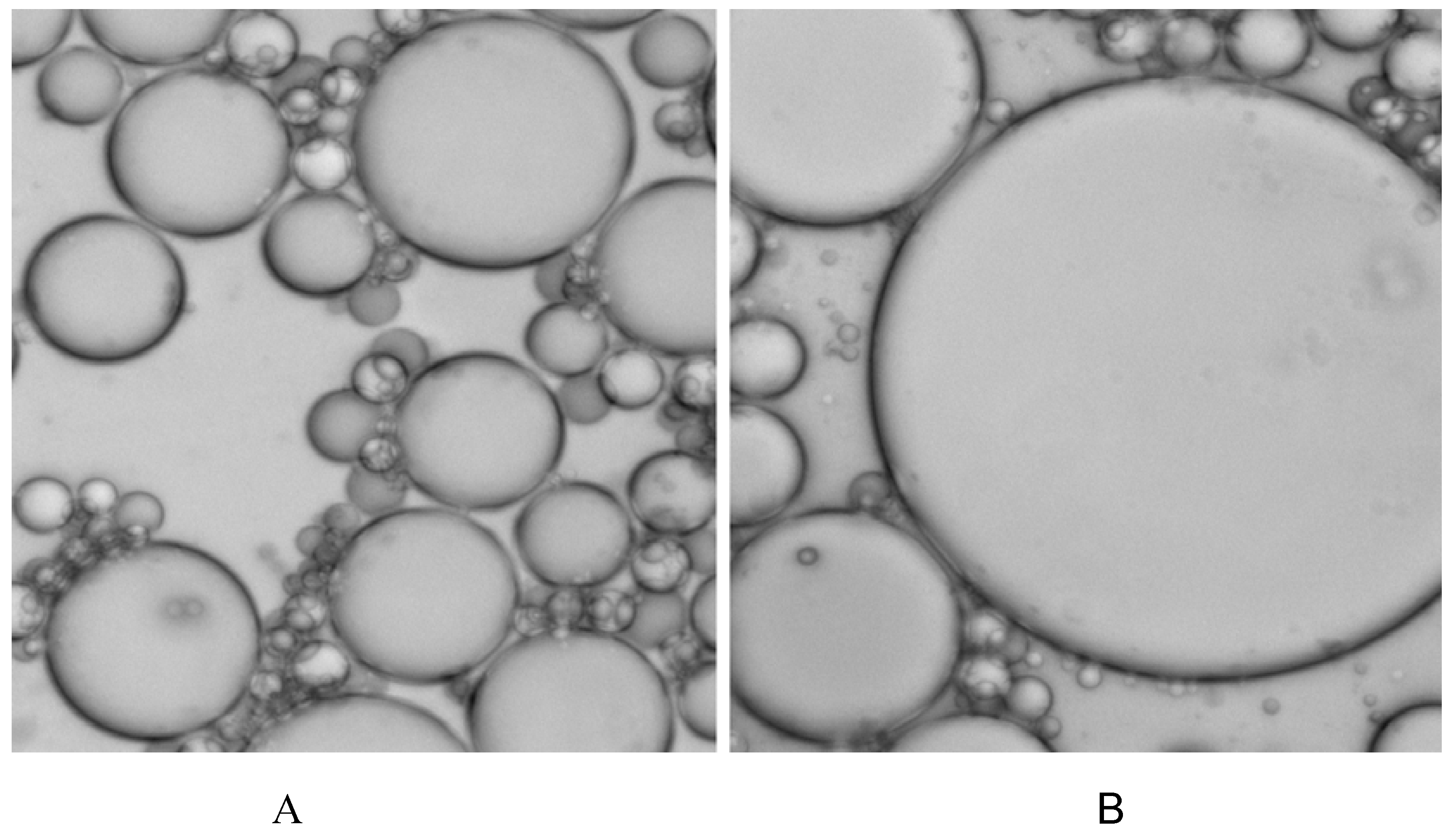
Thermal instability of certain emulsion system

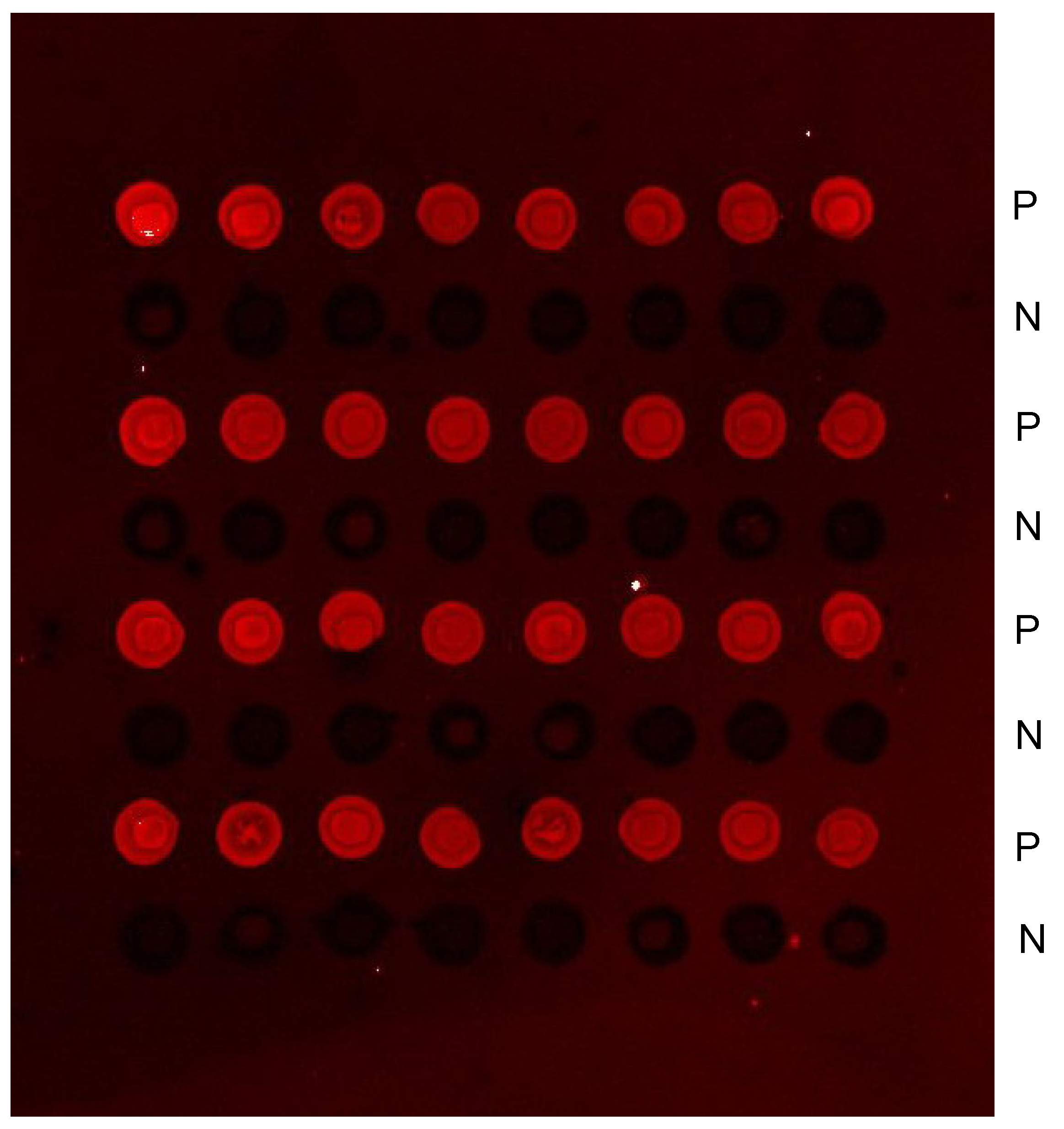
EC-PCR from maternal plasma DNA

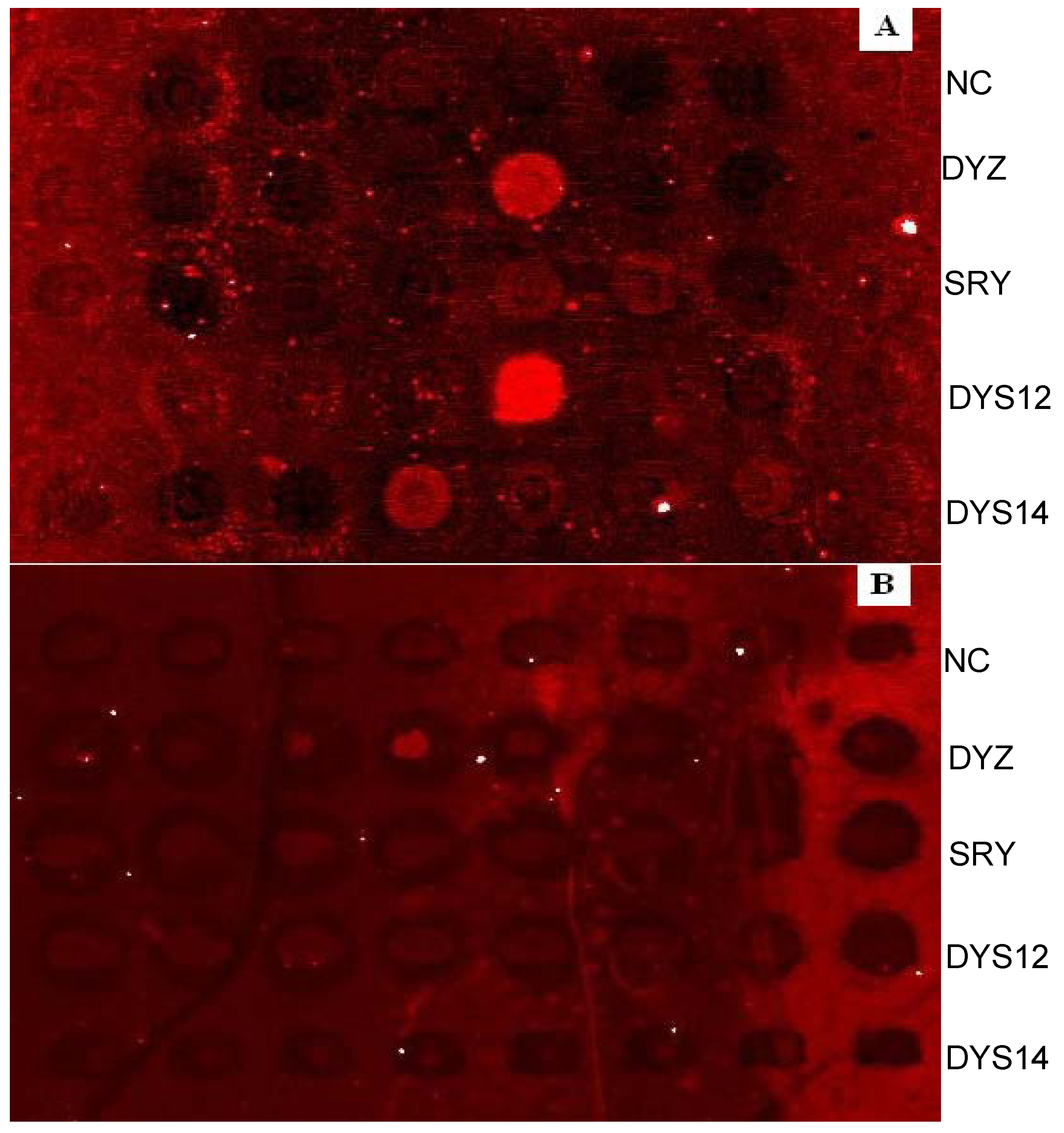
Multiplexed EC-PCR

| Name | DNA Sequences (5’ to 3’) | 5’Modification | GenBank No. | |
|---|---|---|---|---|
| SRY | 462F | (T)6GCAGGGTACCGAAGAGGGA | amido | gi:785026 |
| 210R | (T)6GTCTCGCGATCAGAGGCGCAAGA | |||
| DYS12 | 423F | (T)6ACTTCCCTCTGACATTACCTGATAATTG | amido | gi:245973 |
| 172R | GTCATAGAAGAGTCAAGTCAGTCA | |||
| DYS14 | 637F | (T)6GCCAGGAAGGCCTTTTCTCGGCA | amido | gi:38003 |
| 880R | TTCCCCTTTGTTCCCCAAA | |||
| DYZ | 33642F | (T)6GTGGATTCATCTCACAGAGTTAAA | amido | gi:29824712 |
| 33259R | ACACATCACAAAGAACTATG |
| Samples | Gestation Ages | Signals obtained in the sites detected | |||
|---|---|---|---|---|---|
| SRY | DYS12 | DYS14 | DYZ | ||
| 1 | 54 Days | √ | √ | √ | √ |
| 2 | 53 Days | √ | √ | √ | √ |
| 3 | 42 Days | √ | √ | √ | √ |
| 4 | 61 Days | √ | √ | √ | √ |
| 5 | 44 Days | √ | √ | √ | √ |
| 6 | 44 Days | √ | √ | √ | √ |
| 7 | 57 Days | √ | √ | √ | √ |
| 8 | 55 Days | √ | √ | √ | √ |
| 9 | 45 Days | √ | √ | √ | √ |
| 10 | 56 Days | √ | √ | √ | √ |
| 11* | 52 Days | √ | × | √ | √ |
| 12 | 55 Days | √ | √ | √ | √ |
| 13 | 61 Days | √ | √ | √ | √ |
| 14 | 43 Days | √ | √ | √ | √ |
| 15 | 48 Days | √ | √ | √ | √ |
| 16** | 35 Days | × | × | × | × |
| 17 | 47 Days | √ | √ | √ | √ |
| 18 | 65 Days | √ | √ | √ | √ |
| 19 | 74 Days | √ | √ | √ | √ |
| 20* | 40 Days | √ | √ | √ | × |
| 21 | 60 Days | √ | √ | √ | √ |
| 22 | 51 Days | √ | √ | √ | √ |
| 23 | 46 Days | √ | √ | √ | √ |

Non-emulsion control
Discussion

Conclusions
Experimental
General
Sample preparation
Preparation of slides and EC-PCR
Multiplex detection and scanning
Acknowledgements
References and Notes
- Ding, C.; Cantor, C. R. A high-throughput gene expression analysis technique using competitive PCR and matrix-assisted laser desorption ionization time-of-flight MS. Proc. Natl. Acad. Sci. U. S. A. 2003, 100, 3059–3064. [Google Scholar] [CrossRef]
- Tettelin, H.; Radune, D.; Kasif, S.; Khouri, H.; Salzberg, S. L. Optimized multiplex PCR: efficiently closing a whole-genome shotgun sequencing project. Genomics 1999, 62, 500–507. [Google Scholar] [CrossRef]
- Elnifro, E. M.; Ashshi, A. M.; Cooper, R. J.; Klapper, P. E. Multiplex PCR: optimization and application in diagnostic virology. Clin. Microbiol. Rev. 2000, 13, 559–570. [Google Scholar] [CrossRef]
- Inagaki, S.; Yamamoto, Y.; Doi, Y. A new 39-plex analysis method for SNPs including 15 blood group loci. Forensic. Sci. Int. 2004, 144, 45–57. [Google Scholar] [CrossRef]
- Adessi, C.; Matton, G.; Ayala, G. Solid phase DNA amplification: characterisation of primer attachment and amplification mechanisms. Nucleic Acids Res. 2000, 28, E87. [Google Scholar] [CrossRef]
- Khan, Z.; Poetter, K.; Park, D. J. Enhanced solid phase PCR: mechanisms to increase priming by solid support primers. Anal. Biochem. 2008, 375, 391–393. [Google Scholar] [CrossRef]
- Carmon, A.; Vision, T. J.; Mitchell, S. E.; Thannhauser, T. W.; Muller, U.; Kresovich, S. Solid-phase PCR in microwells: effects of linker length and composition on tethering, hybridization, and extension. BioTechniques 2002, 32, 410, 412, 414-418, 420. [Google Scholar]
- Liu, H.; Li, S.; Wang, Z.; Ji, M.; Nie, L.; He, N. High-throughput SNP genotyping based on solid-phase PCR on magnetic nanoparticles with dual-color hybridization. J. Biotechnol. 2007, 131, 217–222. [Google Scholar] [CrossRef]
- Williams, R.; Peisajovich, S. G.; Miller, O. J.; Magdassi, S.; Tawfik, D. S.; Griffiths, A. D. Amplification of complex gene libraries by emulsion PCR. Nat. Methods 2006, 3, 545–550. [Google Scholar] [CrossRef]
- Diehl, F.; Li, M.; He, Y.; Kinzler, K. W.; Vogelstein, B.; Dressman, D. BEAMing: single-molecule PCR on microparticles in water-in-oil emulsions. Nat. Methods 2006, 3, 551–559. [Google Scholar] [CrossRef]
- Li, M.; Diehl, F.; Dressman, D.; Vogelstein, B.; Kinzler, K. W. BEAMing up for detection and quantification of rare sequence variants. Nat. Methods 2006, 3, 95–97. [Google Scholar] [CrossRef]
- Kojima, T.; Takei, Y.; Ohtsuka, M.; Kawarasaki, Y.; Yamane, T.; Nakano, H. PCR amplification from single DNA molecules on magnetic beads in emulsion: application for high-throughput screening of transcription factor targets. Nucleic Acids Res. 2005, 33, e150. [Google Scholar] [CrossRef]
- Wetmur, J. G.; Kumar, M.; Zhang, L.; Palomeque, C.; Wallenstein, S.; Chen, J. Molecular haplotyping by linking emulsion PCR: analysis of paraoxonase 1 haplotypes and phenotypes. Nucleic Acids Res. 2005, 33, 2615–2619. [Google Scholar] [CrossRef]
- Lo, Y. M.; Corbetta, N.; Chamberlain, P. F. Presence of fetal DNA in maternal plasma and serum. Lancet 1997, 350, 485–487. [Google Scholar] [CrossRef]
- Caramelli, E.; Rizzo, N.; Concu, M. Cell-free fetal DNA concentration in plasma of patients with abnormal uterine artery Doppler waveform and intrauterine growth restriction--a pilot study. Prenat. Diagn. 2003, 23, 367–371. [Google Scholar] [CrossRef]
- Lo, Y. M.; Poon, L. L. The ins and outs of fetal DNA in maternal plasma. Lancet 2003, 361, 193–194. [Google Scholar] [CrossRef]
- Gomes, A. L.; Melo, F. L.; Werkhauser, R. P.; Abath, F. G. Development of a real time polymerase chain reaction for quantitation of Schistosoma mansoni DNA. Mem. Inst. Oswaldo Cruz 2006, 101 Suppl 1, 133–136. [Google Scholar]
- Wataganara, T.; Chen, A. Y.; LeShane, E. S. Cell-free fetal DNA levels in maternal plasma after elective first-trimester termination of pregnancy. Fertil. Steril. 2004, 81, 638–644. [Google Scholar] [CrossRef]
- Ge, Q.; Liu, Q.; Bai, Y.; Wen, T.; Lu, Z. A semi-quantitative microarray method to detect fetal RNAs in maternal plasma. Prenat. Diagn. 2005, 25, 912–918. [Google Scholar] [CrossRef]
- Dressman, D.; Yan, H.; Traverso, G.; Kinzler, K. W.; Vogelstein, B. Transforming single DNA molecules into fluorescent magnetic particles for detection and enumeration of genetic variations. PNAS 2003, 100, 8817–8822. [Google Scholar] [CrossRef]
- Sample availability: DNA samples mentioned in this paper are available from the corresponding author.
© 2008 by the authors. Licensee Molecular Diversity Preservation International, Basel, Switzerland. This article is an open-access article distributed under the terms and conditions of the Creative Commons Attribution license ( http://creativecommons.org/licenses/by/3.0/).
Share and Cite
Ge, Q.; Yu, P.; Bai, Y.; Lu, Z. An Emulsion System Based on a Chip Polymerase Chain Reaction. Molecules 2008, 13, 3057-3068. https://doi.org/10.3390/molecules13123057
Ge Q, Yu P, Bai Y, Lu Z. An Emulsion System Based on a Chip Polymerase Chain Reaction. Molecules. 2008; 13(12):3057-3068. https://doi.org/10.3390/molecules13123057
Chicago/Turabian StyleGe, Qinyu, Pinfei Yu, Yunfei Bai, and Zuhong Lu. 2008. "An Emulsion System Based on a Chip Polymerase Chain Reaction" Molecules 13, no. 12: 3057-3068. https://doi.org/10.3390/molecules13123057