Progress in the Studies on Rutaecarpine
by
Seung Ho Lee
,
Jong-Keun Son
,
Byeong Seon Jeong
,
Tae-Cheon Jeong
,
Hyeon Wook Chang
,
Eung-Seok Lee
and
Yurngdong Jahng
* 
College of Pharmacy, Yeungnam University, Gyeongsan 712-749 Korea
*
Author to whom correspondence should be addressed.
Molecules 2008, 13(2), 272-300; https://doi.org/10.3390/molecules13020272
Submission received: 1 January 2008
/
Revised: 1 February 2008
/
Accepted: 2 February 2008
/
Published: 6 February 2008
(This article belongs to the Special Issue Alkaloids: Novel Therapeutic Perspectives)
Abstract
:Rutaecarpine is an indolopyridoquinazolinone alkaloid isolated from Evodia rutaecarpa and related herbs, which has shown a variety of intriguing biological properties such as anti-thrombotic, anticancer, anti-inflammatory and analgesic, anti-obesity and thermoregulatory, vasorelaxing activity, as well as effects on the cardiovascular and endocrine systems. Recent progress in the studies on the isolation, synthesis, structure-activity relationship studies, biological activities and metabolism of rutaecarpine are reviewed.
1. Introduction
Herbs have been widely employed as important remedies all over the world [1]. Progress in science and technology in recent decades has made possible not only to isolate and characterize the biologically active constituents of herbs, but also to evaluate their biological activities. Rutaceous plants, especially Evodia rutaecarpa (whose dried fruit is named ‘Wu-Chu-Yu’ in China), have long been used for the treatment of gastrointestinal disorders, headache, amenorrhea, and postpartum hemorrhage in traditional oriental medicine [2]. Rutaecarpine (8,13-dihydroindolo-[2',3':3,4]pyrido[2,1-b]quinazolin-5(7H)-one, 1a), first isolated by Asahina and Kashiwaki from acetone extracts of Evodia rutaecarpa after basic treatment [3] in 1915 and later from ‘Wu-Chu-Yu’ [4] is one of the intriguing indolopyridoquinazoline alkaloids of the Rutaceae plants.
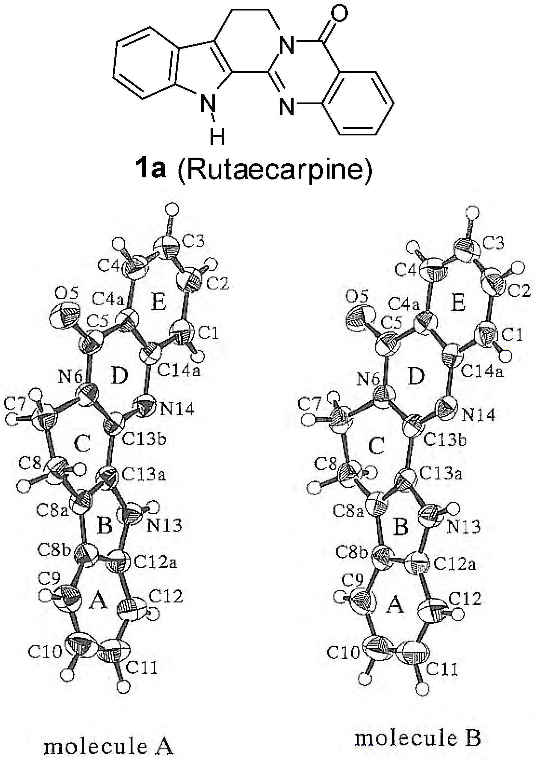
The structure of rutaecarpine was determined by degradation methods [3]. Hydrolysis of rutaecarpine with KOH in amyl alcohol afforded anthranilic acid, while fusion with KOH yielded aniline, CO2, NH3 and indole-2-carboxylic acid [3]. Later infrared [5], ultraviolet spectroscopy [6], mass spectrometry [7], 1H-NMR [8] and 13C-NMR [8] spectroscopy were employed to confirm the structure. Finally, the molecular structure was confirmed by X-ray analysis by Fujii, Kobayashi and Hirayama [9], who reported that rutaecarpine (C18H13N3O) gave monoclinic crystals, P21/a, a = 26.909(1) Å, b = 7.398(1) Å, c = 14.468(1) Å, β = 98.672(5)o, V = 2847.3(4) Å3, Z = 8. It should be noted that rutaecarpine exists in two forms. The X-ray crystallographic structure of rutaecarpine is an essentially planar structure in which rings A and B as well as rings D and E are coplanar and ring C has a half-chair conformation, which can undergo a ring flip, thus having dihedral angles between the two coplanar rings (co-plane A-B vs D-E) of 6.20° and 6.45°, for forms A and B, respectively (Figure 1).
Figure 1.
Structure and two forms of rutaecarpine molecules with numbering [9].
Figure 1.
Structure and two forms of rutaecarpine molecules with numbering [9].

A search if the SCIFinder database (provided by the American Chemical Society) afforded 320 references, including 22 patents, covered isolation, biological activity, synthesis, metabolism, toxicology, etc, since the first report in 1915 (Table 2). Examination of these references provided three noticeable trends: references covering pharmacological properties, the annual numbers of references and number of references originating from China, are increasing (data not shown) (Table 1).

| Period | 1915-2002 | 2003 | 2004 | 2005 | 2006 | 2007 | Total |
|---|---|---|---|---|---|---|---|
| Numbers | 192 | 18 | 24 | 31 | 31 | 24 | 320 |
Among the 10 review papers written so far, five were focused on synthesis [10], three on pharmacology [11], one on the modulation of cytochrome P450 [12], and one on detection methods [13]. A review written in 1983 by Bergman [10e] is a well organized one covering nomenclature, structure, synthesis and pharmacological properties of rutaecarpine, as well as related quinazolinone alkaloids. In addition, a review [11c], written in 1999 by Sheu, covered in vitro as well as vivo pharmacology of rutaecarpine. Although a recent review [10a], written in Chinese in 2006, provided good coverage of the syntheses of rutaecarpine based on the construction patterns for the five-ring system, since the present work will emphasize the progress in the isolation, synthesis, studies on additional pharmacological properties and the metabolism of rutaecarpine, some overlap between parts of this review and prior publications is inevitable.
| Topic | Number | Topic | Number |
|---|---|---|---|
| Pharmacology | 81 | Biochemical Methods | 5 |
| Alkaloid | 67 | Agrochemical Bioregulators | 2 |
| Plant Biochemistry | 47 | Analytical Chemistry | 2 |
| Pharmaceuticals & Cosmetics | 39 | Radiation Biochemistry | 2 |
| Pharmaceutical analysis | 29 | Toxicology | 2 |
| Organic Chemistry | 19 | Biological Chemistry | 2 |
| Heterocyclic Compounds (more than one heteroatom) | 9 | Miscellaneous | 14 |
| Total | 320 |
2. Sources of Rutaecarpine – their Distribution and Systematic Significance
As the name of the compound implies, the major sources of the quinazoline alkaloid rutaecarpine are the Rutaceae. The Rutaceae is a predominantly tropical family of trees and shrubs containing genera of importance in horticulure (Citrus), silviculture (Chloroxylon, Flindersia, Zanthoxylum) and medicine (Pilocarpus, Agathosma) [14]. It is considered to consist of about sixteen hundred species, distributed between approximately one hundred and fifty genera [15]. Among the genera of the Rutaceae, Evodia, Hortia, Zanthoxylum, Phellodendron, Tetradium, Spiranthera, Vepris, Metrodorea, Bouchardatia, and Fagara are a few species so far reported to produce rutaecarpine (Table 3).
| Family | Name | Ref. | Family | Name | Ref. |
|---|---|---|---|---|---|
| Rutaceae | Evodia rutaecarpa | [3,16,17,18] | Rutaceae | Z. pistaciiflorum | [32] |
| E. ailanthifolia | [19] | Metrodorea flavida | [33] | ||
| E. prerparata | [20] | Bouchardatia neurococca | [34] | ||
| E. officinalis | [21] | Phellodendron amurense | [35,36,37] | ||
| Hortia arborea | [22a] | P. japonicum | [38] | ||
| H. regia | [23] | Spiranthera odoratissima | [39] | ||
| H. colombiana | [24] | Tetradium sambucinum | [40] | ||
| H. badinii | [22b] | T .glabrifolium | [41,42] | ||
| Zanthoxylum rhetsa | [25] | T. trichotomum | [43] | ||
| Z. pluviatile | [26] | Fagara rhetza | [44] | ||
| Z. intergrifoliolum | [27,28,29,30] | Texaceae | Taxus chinensis | [45] | |
| Z. budrunga | [31] | Apocynaceae | Winchia calophylla | [46] |
From the 10 rutaecarpine-generated genera of the Rutaceae listed above, Zanthoxylum is the only genus that has produced rutaecarpine and its derivatives such as 1-methoxyrutaecarpine (1f), 1-hydroxyrutaecarpine (1h) and 1-methoxy-7,8-dehydrorutaecarpine (2e) [27,28] together. Although such distribution of rutaecarpine and its possible metabolites implies something in the oxidative metabolism of rutaecarpine in the Rutaceae, the distribution of rutaecarpine in the Rutaceae does not appear to agree with presently accepted classification as Watermann pointed out [47]. In deed, three genera of the Rutaceae such as Euxylophora [48], Vepris [49], and Leptothyrsa [50] produced substituted rutaecarpines. Euxylophora produced 6 substituted rutaecarpines such as euxylophoricine A-F (1c-e and 2b-d), Vepris two derivatives, 1-hydroxyrutaecarpine (1h) and 1-hydroxy-7,8-dehydro-rutaecarpine (2f), and Leptothyrsa 3-hydroxyrutaecarpine (1j) without any trace of rutaecarpine.

3. Studies on the Derivatives of Rutaecarpine
In this review, we only focused on the compounds with the 8,13-dihydroindolo[2’,3’:3,4]pyrido-[2,1-b]quinazolin-5(7H)-one rutaecarpine skeleton. The first derivative of rutaecarpine was isolated from Hortia aborea and named as hortiacine (1b), is known to be 10-methoxyrutaecarpine [22]. Additional methoxyrutaecarpines were isolated from Euxylphora paraënsis and named as euxylophoricine A-F (1c-e and 2b-d) [48]. Among them, euxylophoricine B, E, and F are derivatives of 7,8-dehydrorutaecarpine (2a), which were isolated from Phellodendron amurense [35,36,37] three decades after the first isolation of the corresponding methoxy derivative, euxylophoricine B (2b) in 1968. Additionally, 1-methoxyrutaecarpine (1f) [27] and 1-methoxy-7,8-dehydrorutaecarpine (2e) [28] were isolated from Zanthoxylum integrifoliolum in 1996 and 2005, respectively, and 2-methoxy-rutaecarpine (1g) [51] was isolated from Araliopsis tabouensis (Rutaceae).

| Compound | Plant | Reference(s) |
|---|---|---|
| 1-Hydroxyrutaecarpine (1h) | Euxylophora paraënsis | [52] |
| Vepris louisii | [49] | |
| Bouchardatia neurococca | [34] | |
| Tetradium glabrifolium | [41,42] | |
| Zanthoxylum integrifoliolum | [27,28,29] | |
| Zanthoxylum pistaciiflorum | [32] | |
| Spiranthera odoratissima | [39] | |
| 1,2-Dihydroxyrutaecarpine (1i) | Bouchardatia neurococca | [34] |
| 3-Hydroxyrutaecarpine (1j) | Leptothyrsa sprucei | [50] |
| 14-N-Formyrutaecarpine (1k) | Zanthoxylum intergrifoliolum | [29] |
| 7-Hydroxyrutaecarpine (1l) | Tetradium glabrifolium T. ruticarpum Phellodendron amurense Evodia rutaecarpa Evodia officinalis | [53] [53] [36,37] [54] [55] |
| 7,8-Dihydroxyrutaecarpine (1m) | Phellodendron amurense | [36,42] |
| 7,8-Dehydrorutaecarpine (2a) | Phellodendron amurense | [35,36,41] |
| 1-Hydroxy-7,8-dehydrorutaecarpine (2f) | Vepris louisii | [49] |
A series of hydroxylated derivatives of rutaecarpine have also been isolated from the Rutaceae family, which are summarized in Table 4. Danieli, et al. isolated 1-hydroxyrutaecarpine (1h) from Euxylophora paraënsis in 1974 [52] as a first hydroxyrutaecarpine. The aromatic region of the 1H-NMR spectrum of 1h was too complex either to assign all the seven proton resonances or to determine the position where the OH group was attached. The structure was therefore confirmed by comparing the data of 1-hydroxyrutaecarpine that was prepared from 1,2,3,4-tetrahydronorharman-1-one and 3-hydroxyanthanilic acid. Although assigniment of H4 of the E ring was possible in the 100 MHz 1H- NMR spectrum, the other six aromatic proton resonances were not [49], until the HMBC experiment as well as studies on 3J correlations with the carbons allowed the assignment of all the protons and confirmed the present structure [34]. Since then, nine additional hydroxyrutaecarpines have been isolated from various genera of Rutaceae plants and the 1H- and 13C-NMR data of selected rutaecarpine derivatives are summarized in Table 5.
| Position | 1a | 1h | 1i | 1j | 1l | 1m | 2a | 2f | |||
|---|---|---|---|---|---|---|---|---|---|---|---|
| δH | δC | δH | δH | δH | δC(N) | δH | δH | δH | δC | δH | |
| 1 | 7.64 | 125.3 | 8.60a | 7.56 | 128.2 | 7.58 | 7.74 | 7.81 | 126.0 | ||
a OH resonance. bOverlapped.
Recently, two indolopyridoquinazoline alkaloidal glycosides, 11-O-β-D-glucopyranosyl rutaecarpine (ternatoside C) and 11-O-α-L-rhamnosyl-(1-6)-β-D-glucopyranosyl rutaecarpine (ternatoside D) were isolated from the roots of Ranunculus ternatus (Ranunculaceae) [56]. However, to our surprise, the corresponding hydrolyzed product of ternatoside C and/or D, 11-hydroxyrutaecarpine, has never been isolated as yet from the natural sources even though it is an isolated metabolite of cytochrome P450-catalyzed oxidative metabolism of rutaecarpine (vide infra).

4. Synthesis
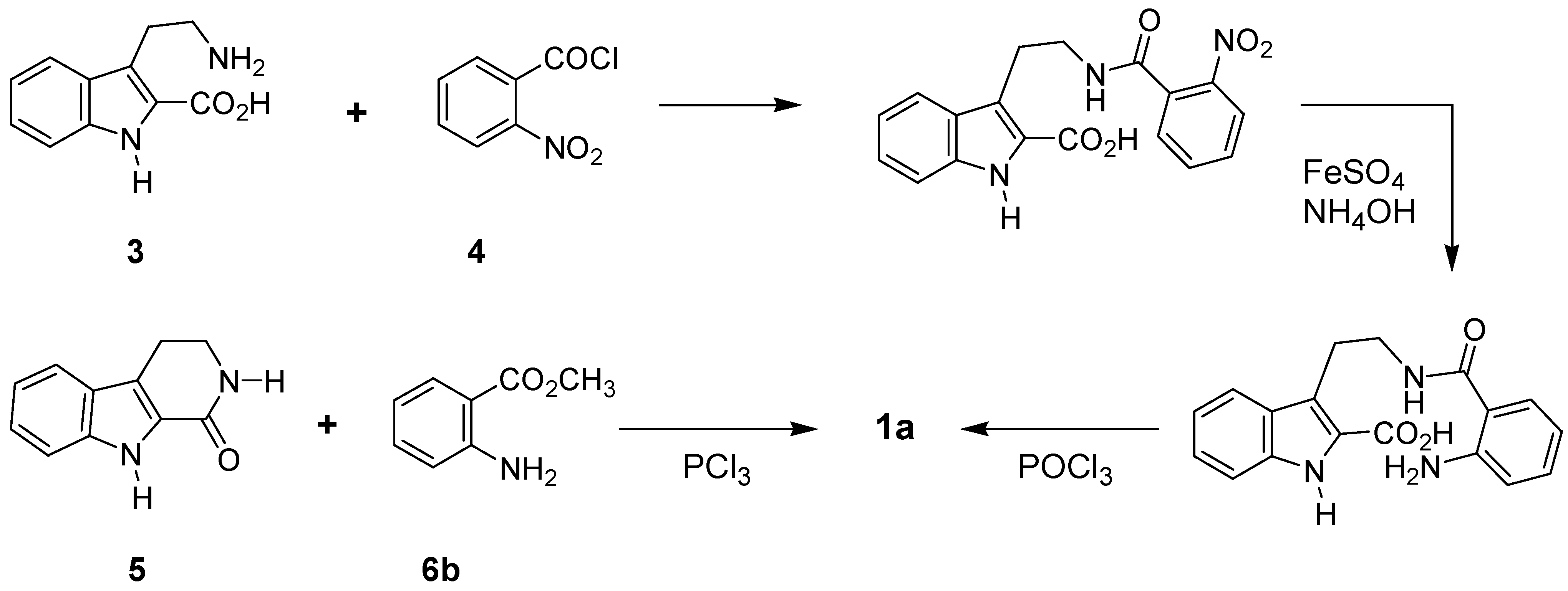
Asahina et al. reported two papers for the synthesis of rutaecarpine in 1927: one covered three-step synthesis from 3-(2-aminoethyl)indole-2-carboxylic acid (yield not given) (3) [57] and the other one-pot synthesis (24%) from ketotetrahydrocarboline (5), methyl anthranilate (6b), and PCl3 [58] (Scheme 1). Later, Patcher et al. claimed that PCl3 was inefficient and POCl3 should be preferred reagent [22a].
Scheme 1.

These two papers provided a good starting point for the future syntheses of rutaecarpine as these two sets of starting materials, 3-(2-aminoethyl)indole-2-carboxylic acid (3) and 2-nitrobenzoyl chloride (4) as well as 2,3,4,9-tetrahydro-1H-pyrido[3,4-b]indol-1-one (5) and methyl anthranilate (6b), have been employed as starting materials for the most of consequent rutaecarpine synthesis.
1) 2,3,4,9-Tetrahydro-1H-pyrido[3,4-b]indol-1-one - Derived Synthesis.
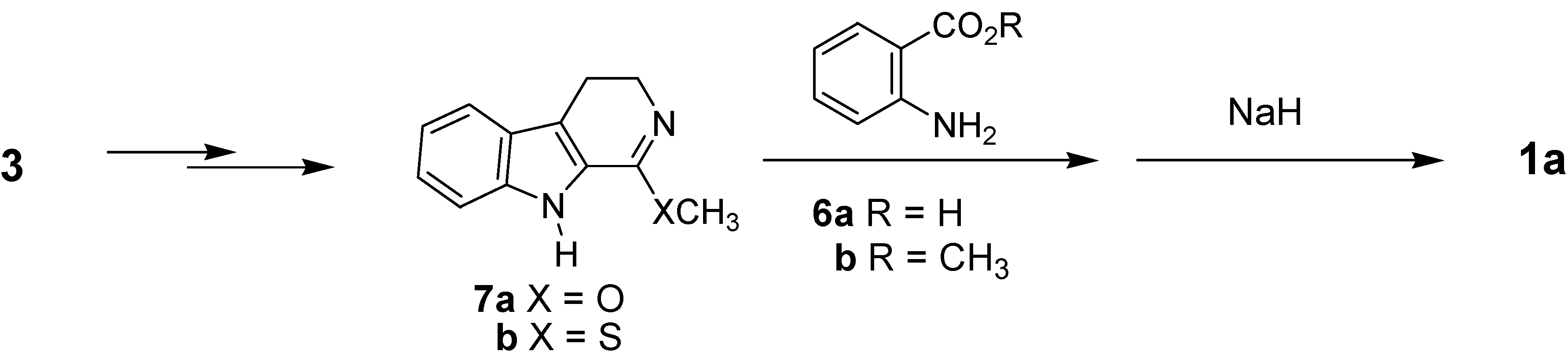
Since the first synthesis [57], several attempts to synthesize rutaecarpine and its derivatives used 5 as a starting material [59]; some of them employed structurally modified derivatives of 5 which required consturction of the D-ring at the final stage. It should be noted that compound 5, a degradation product of rutaecarpine and related quinazolinone alkaloids, was isolated from the extract of E. rutaecarpa [60] as an alkaloid.
When the corresponding O-methyllactim 7a, prepared from 5 by reacting with trialkyloxonium ion, was employed, the yield was improved up to 66% [61]. Recently, methylthiolactim (7b) was replaced for 7a, which somewhat improved the yield (89%) [62]. The starting 7b could be readily prepared in two steps from compound 5 [63] (Scheme 2).
Scheme 2.

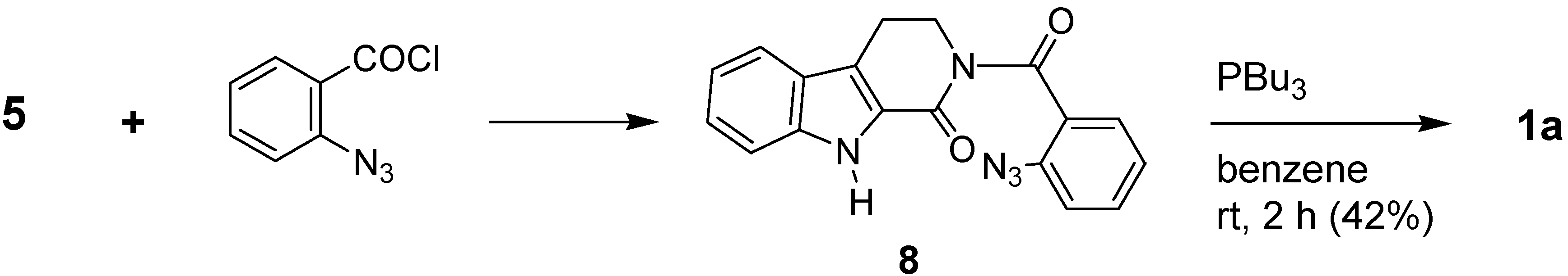
Eguchi et al. used an aza-Wittig reaction in the presence of tributylphosphine for the construction of ring D from 2-(2-azidobenzoyl)-2,3,4,9-tetrahydro-1H-pyrido[3,4-b]indol-1-one (8) [64] (Scheme 3).
Scheme 3.

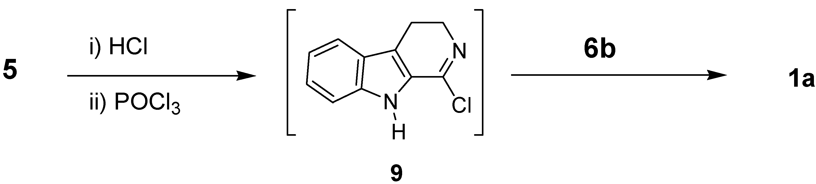
The imino chloride 9, generated from the HCl salt of 5 and POCl3, was condensed with methyl anthranilate to afford rutaecarpine in excellent yield [65a]. In addition, a one-pot synthesis of rutaecarpine from 5, anthranilic acid (6a), and SOCl2 in refluxing pyridine has also been established [65b] (Scheme 4).
Scheme 4.

Although the procedures described above have advantages for introducing substituent(s) on the 4(3H)-quinazolinone skeleton, the relative unavailability of 5 and its derivatives limits the general utility of this procedure.
2) Tryptamine-Derived Synthesis
Tryptamine-derive syntheses of rutaecarpine are basically one step or stepwise constructions of the C- and D-rings of rutaecarpine (Scheme 5).
Scheme 5.

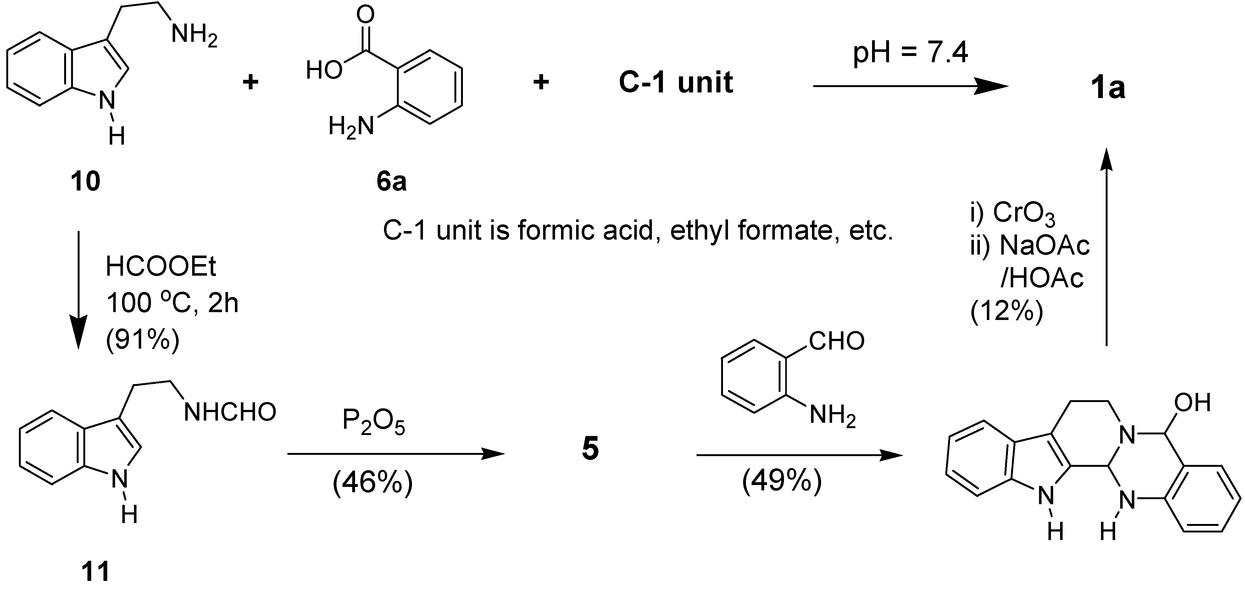
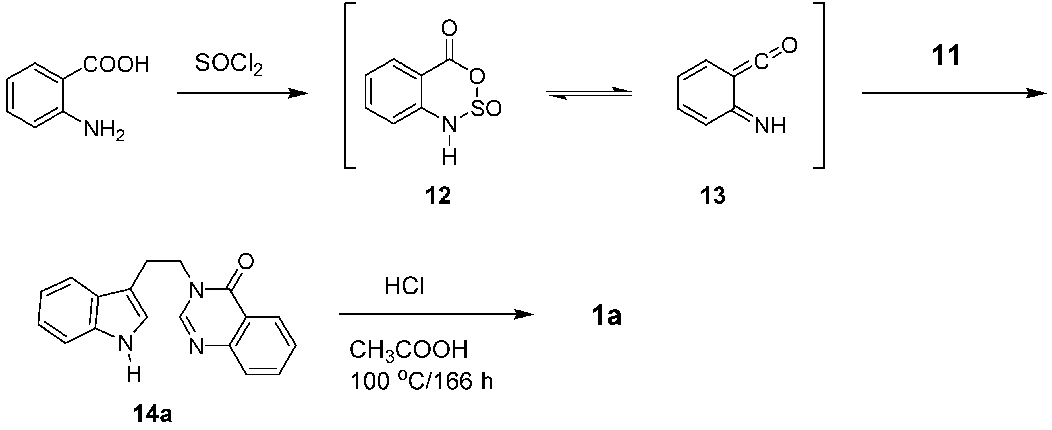
A decade after its discovery, Schöf and Steuer reported a synthesis of rutaecarpine under so-called physiological conditions by condensing anthranilic acid with 5,6-dihydro-4-carboline [66], which suggested the possible utility of tryptamine (10). They supported their biochemical studies by a chemical synthesis of rutaecarpine from 10 via 5 [67]. The carbon added to the N of tryptamine that would be the C2 of the quinazolinone ring, is generally afforded by formic acid, alkyl formate, triethyl orthformate, alkyloxycarbonyl halide, etc. Tryptamine (10) and/or its equivalents (i.e. 11) can thus be readily available starting materials for the synthesis of rutaecarpine. First, N-formyltryptamine (11) was used as a starting material, which was reacted with sulfonamide anhydride 12 or iminoketene 13, generated from anthranilic acid and SOCl2, to afford N-[2-(indol-2-yl)ethyl]-4(3H)-quinazolinone, which was then cyclized by HCl in HOAc to afford rutaecarpine in 45% yield [68] (Scheme 6). The harsh reaction temperature (110 oC) and long time (166 h) as well as the relatively low yield may be the drawbacks to be overcome.
Scheme 6.

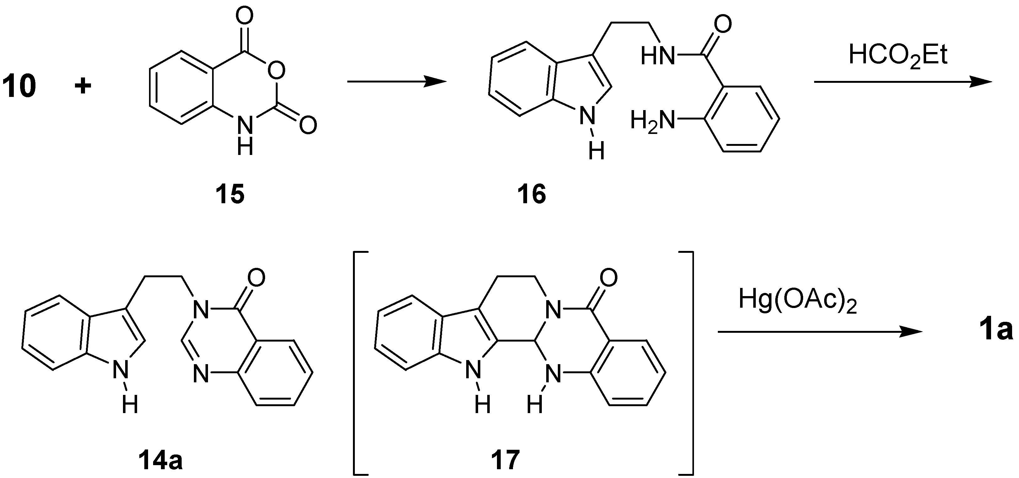
Condensation of tryptamine with isatoic anhydride (15) led to N-(2-aminobenzoyl)tryptamine (16). Although Horvath-Dora and Clauder claimed that reaction of 16 with triethyl orthoformate afforded 13b,14-dihydrorutaecarpine (17) [69b], the real structure of the product was corrected by Bergman and Bergman [8b,70] to be 14a (Scheme 7). The structure of 17 was further confirmed by isolation from Zanthoxylum flavum [71] and Evodia rutaecarpa [72], as well as chemical synthesis [72].
Scheme 7.

Bergman and Bergman additionally introduced a modified procedure for the preparation of 14a by reacting N-(2-aminobenzoyl)tryptamine (16) with triethyl orthoformate in 89% yield [8b]. Attempts to convert 17 directly to rutaecarpine (1a) using oxidative coupling reagents such as Hg(OAc)2, FeCl3, and Pb(OAc)2 were rather indiscriminate and resulted complex reaction mixtures (Scheme 8).
Scheme 8.

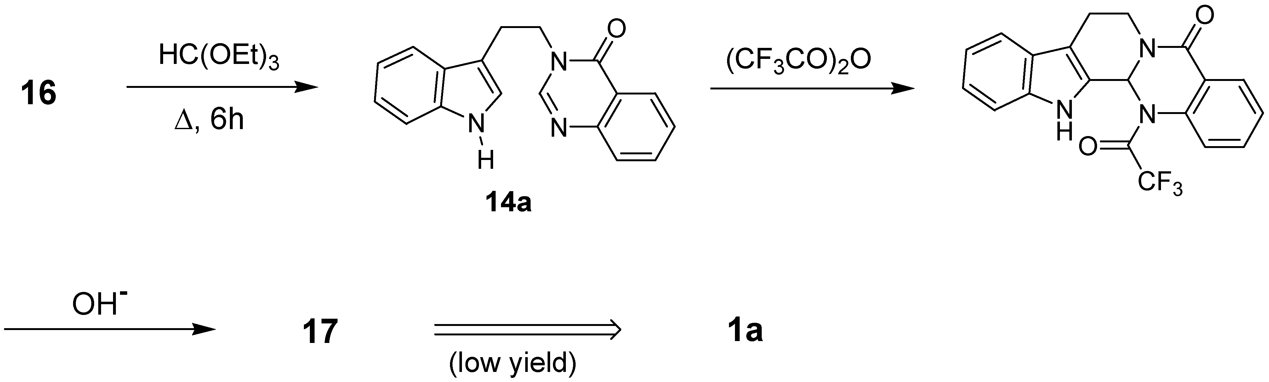
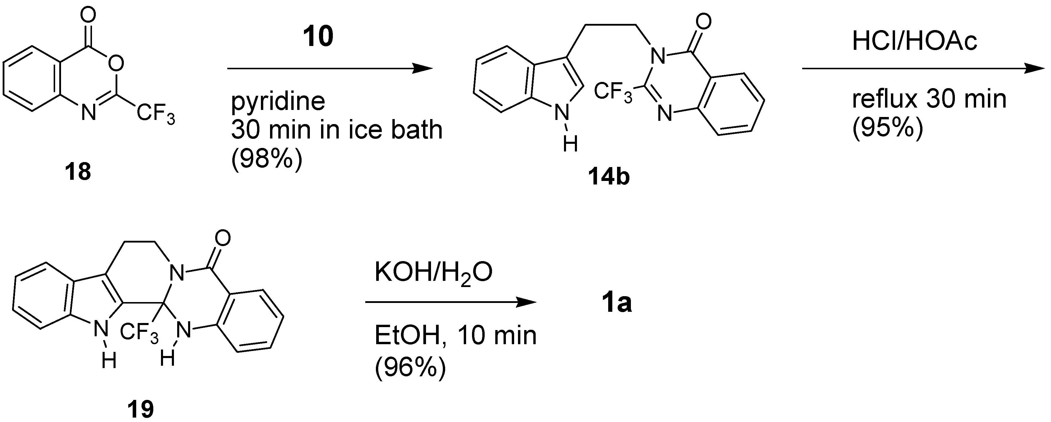
A rationale to improve the conversion of 14a to rutaecarpine could be to introduce a good leaving group at C2 of either the 4(3H)-quinazolinone or the indole ring. The introduction of a good leaving group such as CF3 at C2 of quinazolinone ring would not only facilitate the acid-catalyzed ring closure of 14b to the pentacyclic intermediate 19, but would also act as a leaving group in the final step [70]. Reaction may proceed by protonation of the 4(3H)-quinazolinone moiety of 14b, followed by electrophilic attack on the indole ring leads to 19, which has an angular CF3 group in the 13b-position. The reaction time required for the three steps is less than 1 h, the reaction temperature is approximately 150 oC, and the yield for each step is over 95%. This procedure has been applied for the introduction of various substituent(s) on ring A to pursue structure-activity relationship [73]. Prerequisite compound 18 could be readily prepared by reacting isatoic anhydride (15) with trifluoroacetic anhydride [70].
Scheme 9.

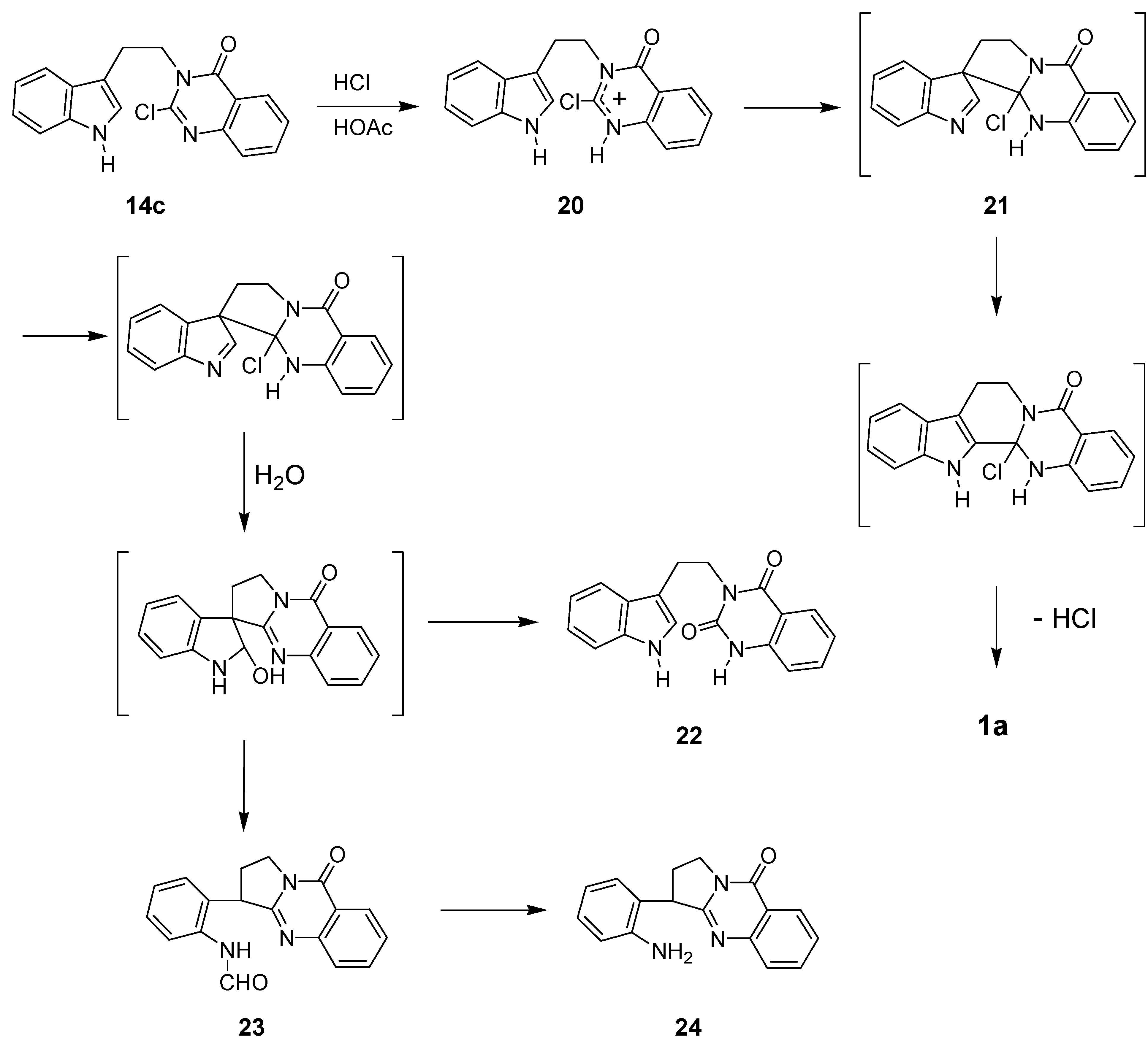
It should be noted that the reactions attempted with a relatively poor leaving Cl moiety at C2 of the quinazolinone ring afforded a mixture of products [74]. The distributions of the products 1a, 22, 23, and 24 were highly dependent on the reaction conditions (Table 6). Such information and additional experimentation gave enough information to explore the mechanism of this-type of reaction as shown below (Scheme 10).
Scheme 10.

| Entry | Reaction Conditions | Product (yield in %) | |||||
|---|---|---|---|---|---|---|---|
| Solvent | Time | Temp. | 1a | 22 | 23 | 24 | |
| 1 | CHCl3 containing a small amount of HCl (g) | 1 h | Reflux | 44 | - | 26 | - |
| 2 | CHCl3 saturated with HCl (g) | 1 h | r.t. | 42 | - | 40 | - |
| 3 | Conc. HCl-H2O-MeOH (1:20:180) | 17 h | r.t. | 9 | 20 | - | 70 |
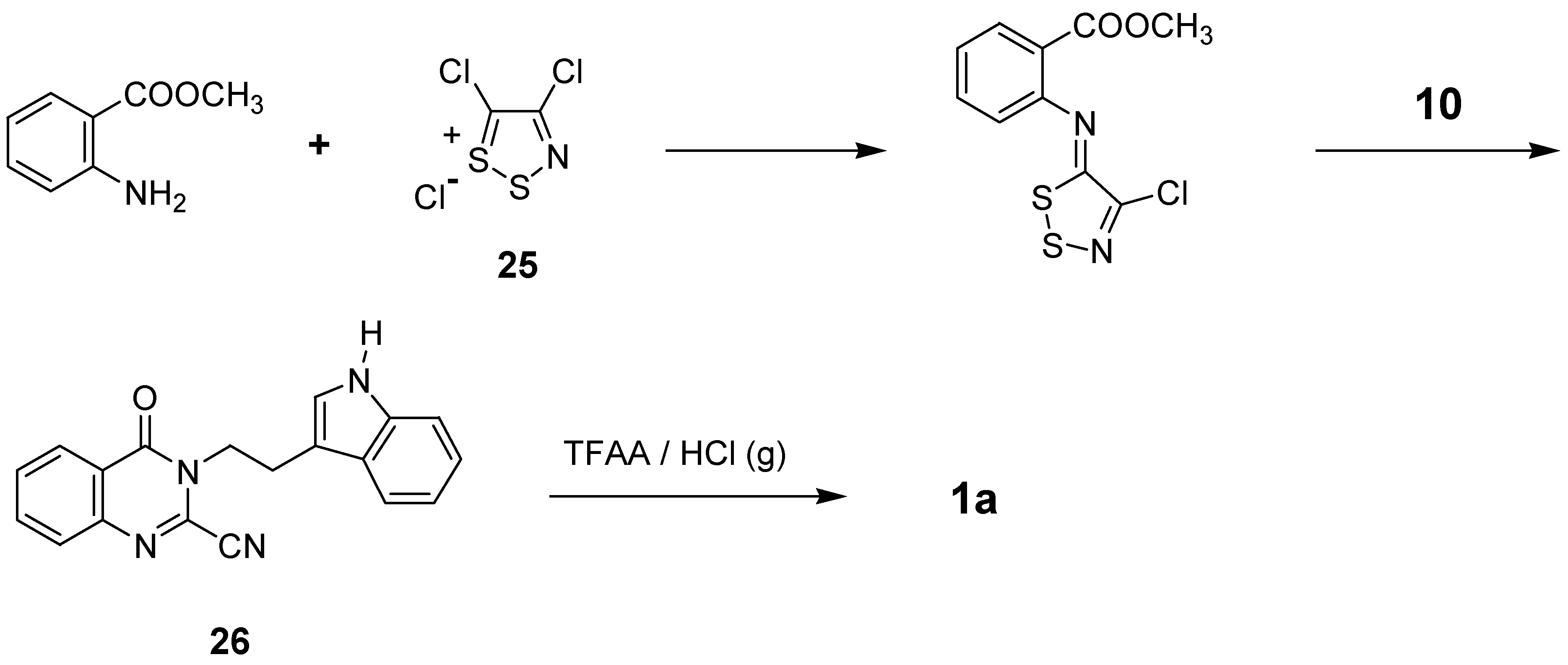
Tryptamine was further condensed with methyl N-(4-chloro-5H-1,2,3-dithiazol-5-ylidene)anthranilate, prepared from methyl anthranilate and 4,5-dichloro-1,2,3-dithiazolium chloride (Appel’s salt, 25), to give a cyano compound 26, which then underwent cyclization on heating with TFAA/HCl(g) to afford rutaecarpine in 37% overall yields [75] (Scheme 11).
Scheme 11.

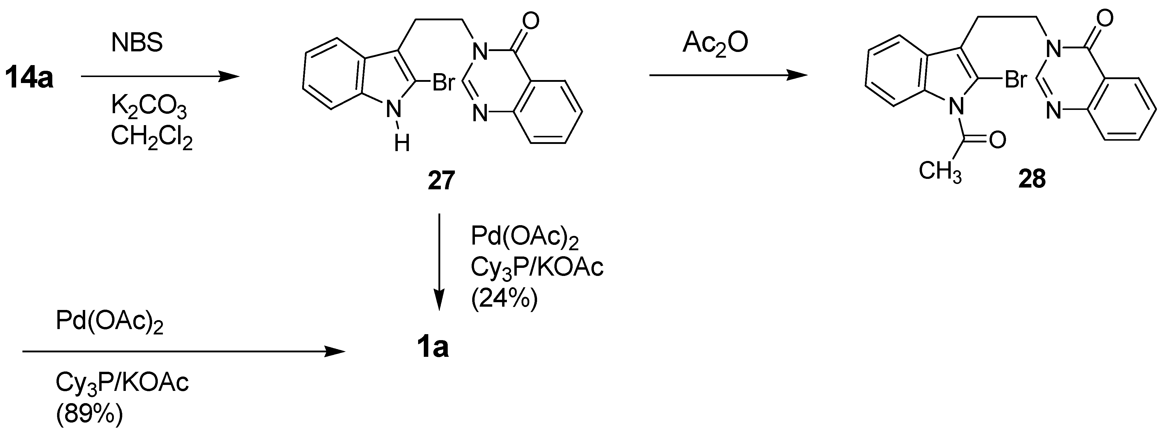
A second modification was pursued by introducing a good leaving group at C2 of the indole moiety, which then subjected to Pd-catalyzed coupling. Only the N1-acetyl derivative 28 of 3-[2-(2-bromoindol-3-yl)ethyl]-4(3H)-quinazolinone (27), smoothly underwent a coupling reaction, while direct conversion gave poor yield [76] (Scheme 12).
Scheme 12.

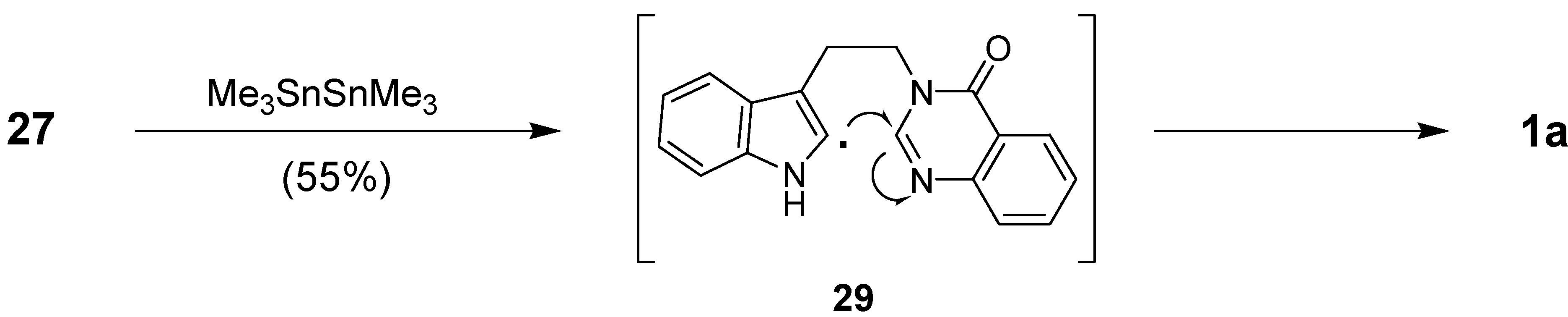
Recently a radical reaction was employed for the preparation of rutaecarpine. Bowman et al. used indol-2-yl radical cyclization onto the quinazolinone motif. Previously reported 27 [76] was treated with (Me3Sn)2 to afford an indole radical 29 which would undergo 6-exo cyclization to lead to rutaecarpine in 55% yield [77] (Scheme 13).
Scheme 13.

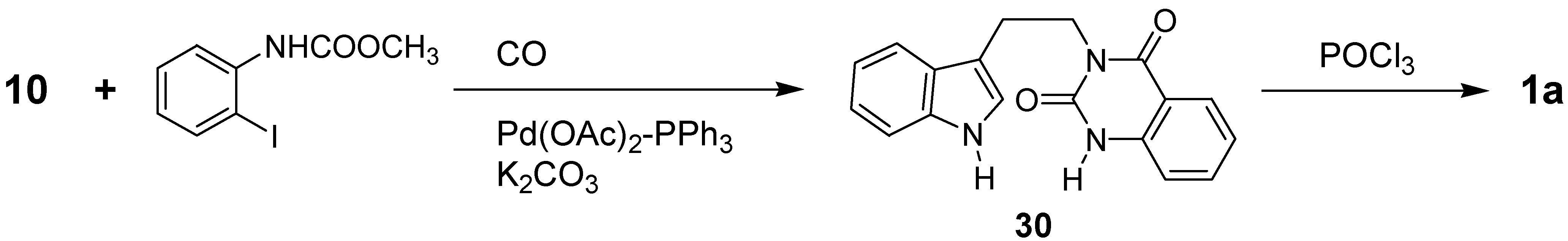
Tryptamine-derived palladium-catalyzed carbonylation of N-carbomethoxy-2-iodoaniline under carbon monoxide (1 atm) afforded 3-[2-(1H-indol-3-yl)ethyl]-(1H,3H)-quinazoline-2,4-dione (30), which could be readily cyclized with POCl3 to lead rutaecarpine [78].
Scheme 14.

3) 3,4-Dihydro-β-carboline-Derived Synthesis
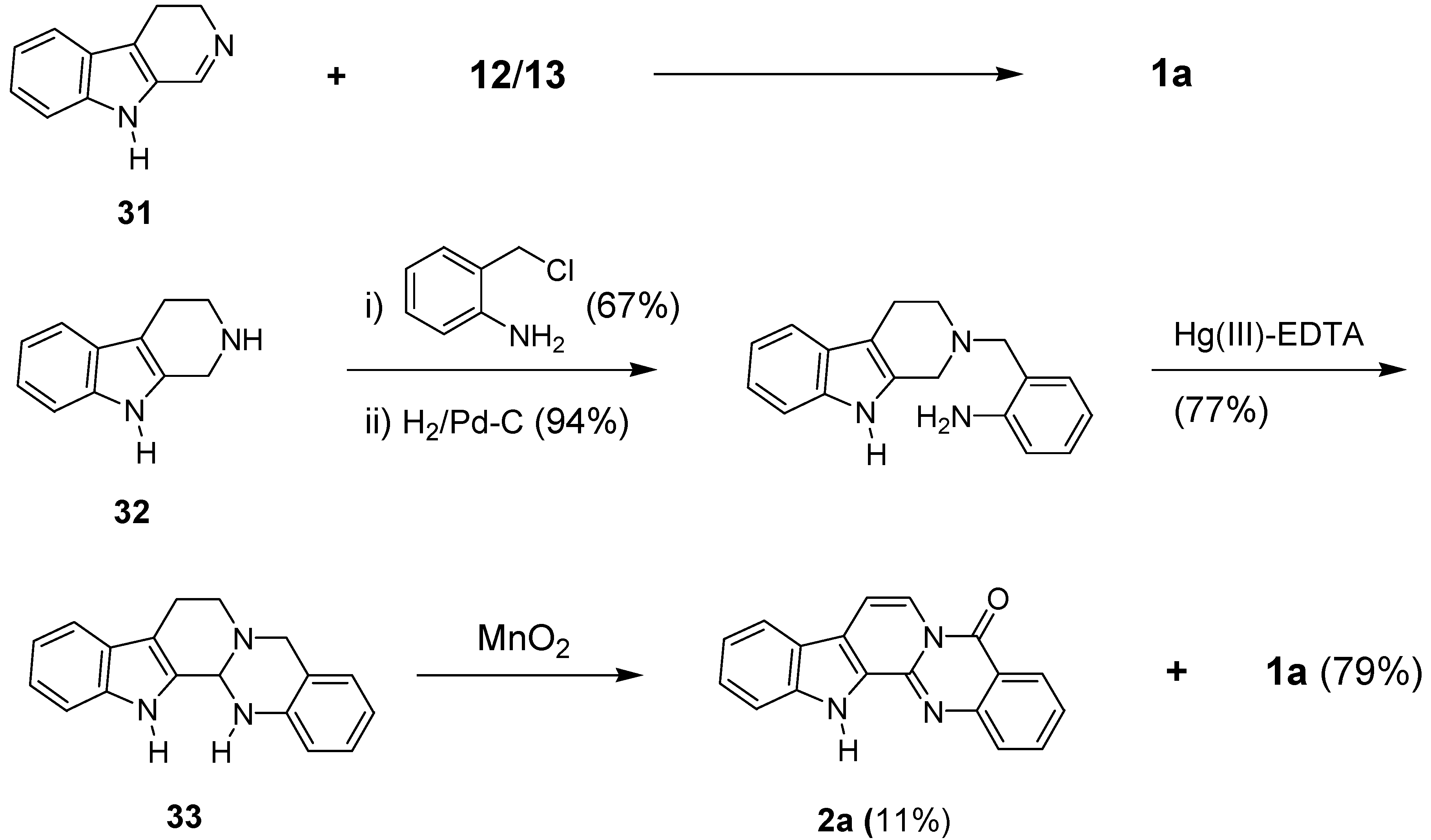
Several synthetic methods, reported later, employed 3,4-dihydro-β-carboline (5,6-dihydro-4-carboline, 31) or its hydrogenated analog 1,2,3-tetrahydro-β-carboline (32) as key starting materials. Cycloaddition reaction of 31 to anthranilic acid-derived iminoketene 12 or 13 would lead 1a in 85% yield [79] (Scheme 15).
Scheme 15.

A four-step synthesis of rutaecarpine from 32 was additionally reported. Although the keto group at C5 and dehydrogenation at C15a and N16 position onto 33 could be accomplished with MnO2, over-dehydrogenation of 1a led 7,8-dehydrorutaecarpine (2a) in 11% yield [80]. This method has, however, somewhat limited general applicability due to the unavailability of the staring carboline and/or its precursor.
4) Mackinazolinone-Derived Synthesis
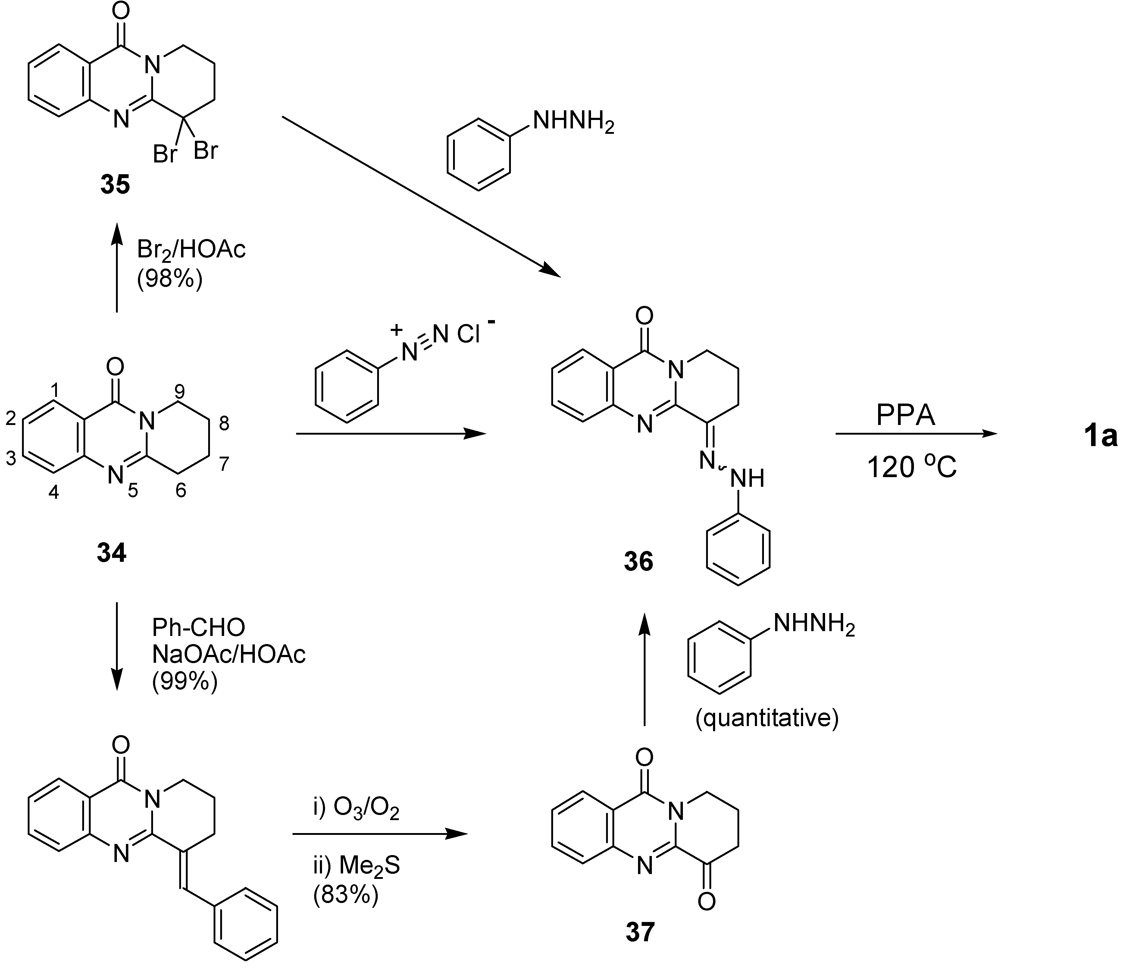
The construction of B ring of rutaecarpine at the final stage was also achieved by employing 9,10,11,12-tetrahydro-4H-pyrido[2,1-b]-quinazolin-4-one (mackinazolinone, 34) as a starting material. (Scheme 16). In fact, mackinazolinone was chemically prepared [81] some 30 years before its first isolation as an alkaloid from Mackinlaya species [82]. The Fischer indole synthesis was applied to the key intermediate 9-phenylhyrazono-9,10,11,12-tetrahydro-4H-pyrido[2,1-b]quinazolin-4-one (36), which could be prepared by three different routes: a condensation of the corresponding 9,9-dibromo compound 35 with phenylhydrazine [83a], a direct condensation of 34 with phenyldiazonium chloride [83b,c], and three-step conversion via 37 [84]. The intermediate diketone 37 could also be prepared 5 steps from isatoic anhydride in 32-49% overall yields [85].
Scheme 16.

5) Miscellaneous
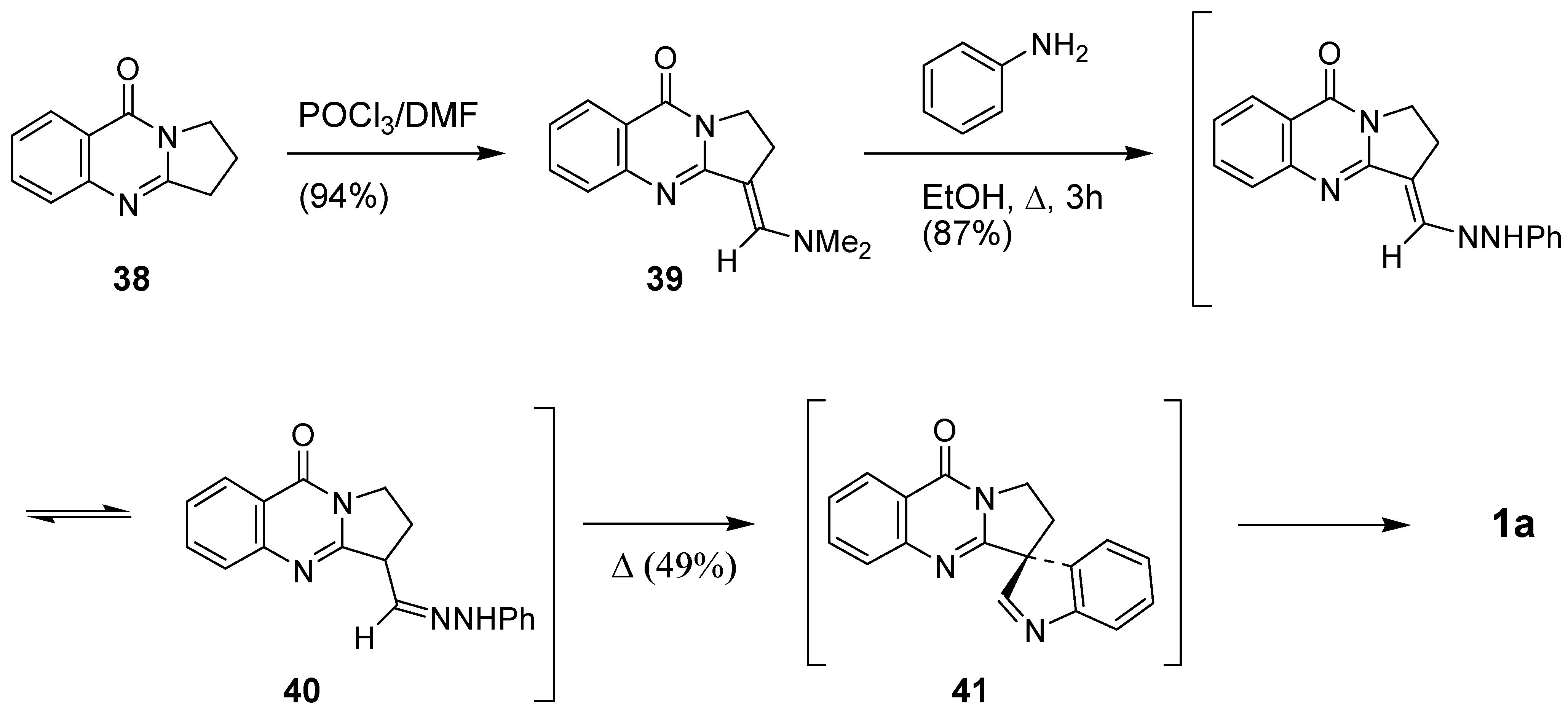
Use of 2,3-polymethylene-4(3H)-quinazolinone was further pursued by Kökösi et al. by employing deoxyvasicinone (38) as a starting material [87]. The Vilsmeier-Haack reaction on 38 yielded corresponding N,N-dimethylmethylidene compound 39, which was treated with aniline in refluxing EtOH to give hydrazone 40 in 87% yield. Fischer indole synthesis was then applied to 40 to yield rutaecarpine in 40% over three-step yields via spiro compound 41 (Scheme 17) [74].
Scheme 17.

Although a lot of synthetic procedures have been reported for the synthesis of rutaecarpine, many of them were limited to for the introduction of substituent(s) only on a part of the 5 rings in the molecules. For the further pursuing the structure-activity relationship study on rutaecarpine derivatives, it definitely needs either to develop more practical methods to introduce substituents all over the 5 rings or to design (a) new procedure(s) combining methods reported so far.
5. Biological Properties
A comprehensive review written in 1999 by Sheu [11c], covered very well the in vitro as well as the in vivo pharmacology of rutaecarpine, whose pharmacological actions were reviewed under several subheadings: (1) cardiovascular effects, (2) antithrombotic activity, (3) anticancer effects, (4) antiinflammatory and analgesic effects, (5) effects on endocrine system, (6) antiobesity and thermoregulatory effects, (7) effects on smooth muscle except cardiovascular, and (8) others. Herein we cover only studies on the cytotoxicity and inhibitory activity on COX. Studies on the cytotoxicity of rutaecarpine and its derivatives are summarized in Table 7. In general, a substitution on the ring resulted in selectivity towards specific cell lines. 11-Methoxyrutaecarpine showed selective cytotoxicity against the lung and renal cancer subpanel at 1.38 and 0.31 μM level, respectively, while the 10,11-methylenedioxy analogue showed selective cytotoxicity for the ovarian cancer subpanel [73a]. 10-Bromo- and 10-methylthiorutaecarpine showed high selectivity on a renal carcinoma cell line and strong cytotoxicity at 0.3 and 0.08 μM level, respectively [73b,88] while 12-fluororutaecarpine showed strong cytotoxicity with selectivity on the HT-29 human cell line [89]. Introduction of substitutions on ring E affected the cytotoxicity more significantly. 2-Chlororutaecarpine showed strong inhibitory activity against topoisomerase I and II, with potency comparable to camptothecin [89]. It should be noted that the inhibitory activities on topo I and II seemed to be affected by a substitution on the E-ring but not by substitutions on ring A and C [55,89]. In addition, rutaecarpine inhibited tumor cell migration approximately 30-40% level compared to control at 100 μg/mL [90], which would be a new vista of the further studies on rutaecarpine.
| CNS U251 | HT-29 | A549/ ATCC | NCI- H460 | OVCAR-4 | 786-0 | Renal ACHN | HS- 578T | |
|---|---|---|---|---|---|---|---|---|
| Rutaecarpine | 0.02 [73b] | 31.6 [31] | 14.5 [73a] | - | 18.9 [73a] | - | - | 22.6 |
| 11-OCH3 [73a] | - | - | 0.75 | 1.38 | >25.0 | 0.31 | - | 1.59 |
| 10,11-OCH2O- | - | - | >25.0 | 1.55 | 1.50 | 1.08 | - | 5.05 |
| 10-Br [88,73b] | 5 | 33b | 59c | - | 3d | - | >100 | - |
| 2-CH3,10-Br | 0.3 | 14b | 18c | - | 3d | - | 0.3 | - |
| 10-SCH3[73b] | 3 | - | - | - | 13d | - | 0.08 | - |
| 1-OH [31] | - | 7.39 | 10.43 | - | - | - | - | - |
| 2-Cl [89] | - | 5.62 | 22.4 | - | - | - | - | 21.6e |
| 12-F [89] | - | 1.26 | 8.4 | - | - | - | - | 3.18e |
Tumor subpanels: human lung adenocarcinoma (A549), human colon carcinoma (HT-29), leukemia (CCRF-CEM), nonsmall cell lung cancer (A549/ATCC and NCI-11460), renal cancer (786-0), ovarian cancer (OVCAR-4), and breast cancer (HS-578T).aThe cytotoxicity GI50 values are the concentrations corresponding to 50% growth inhibition, and they are the averages of at least two determinations. bfor colon SW620 cell line. cfor lung H522 cell line. dfor ovarian SKOV3 cell line. efor human breast cancer carcinoma MCF-7.
Strong and selective inhibitory activity on COX-2 has been claimed as the origin of the anti-inflammatory activity of rutaecarpine [91]. A series of substituted rutaecarpines were therefore prepared by employing Fischer indole synthesis as the key step and their inhibitory activities on COX-1 and 2 as well as selectivity on COX-2 were evaluated. The compounds 10-methanesulfonyl-rutaecarpine and 10-bromorutaecarpine showed promising inhibitory activity (IC50 = 0.27 and 0.35 μM, respectively) with selectivity (39 and 62, respectively) [92].
It should be noted that more compounds with various substituents on each ring as well as both A- and E-ring together are definitely required to lead better and more decisive conclusion for the structure-activity relationship and to pursue further studies in the process of drug development for specified biological activity.
6. Metabolism
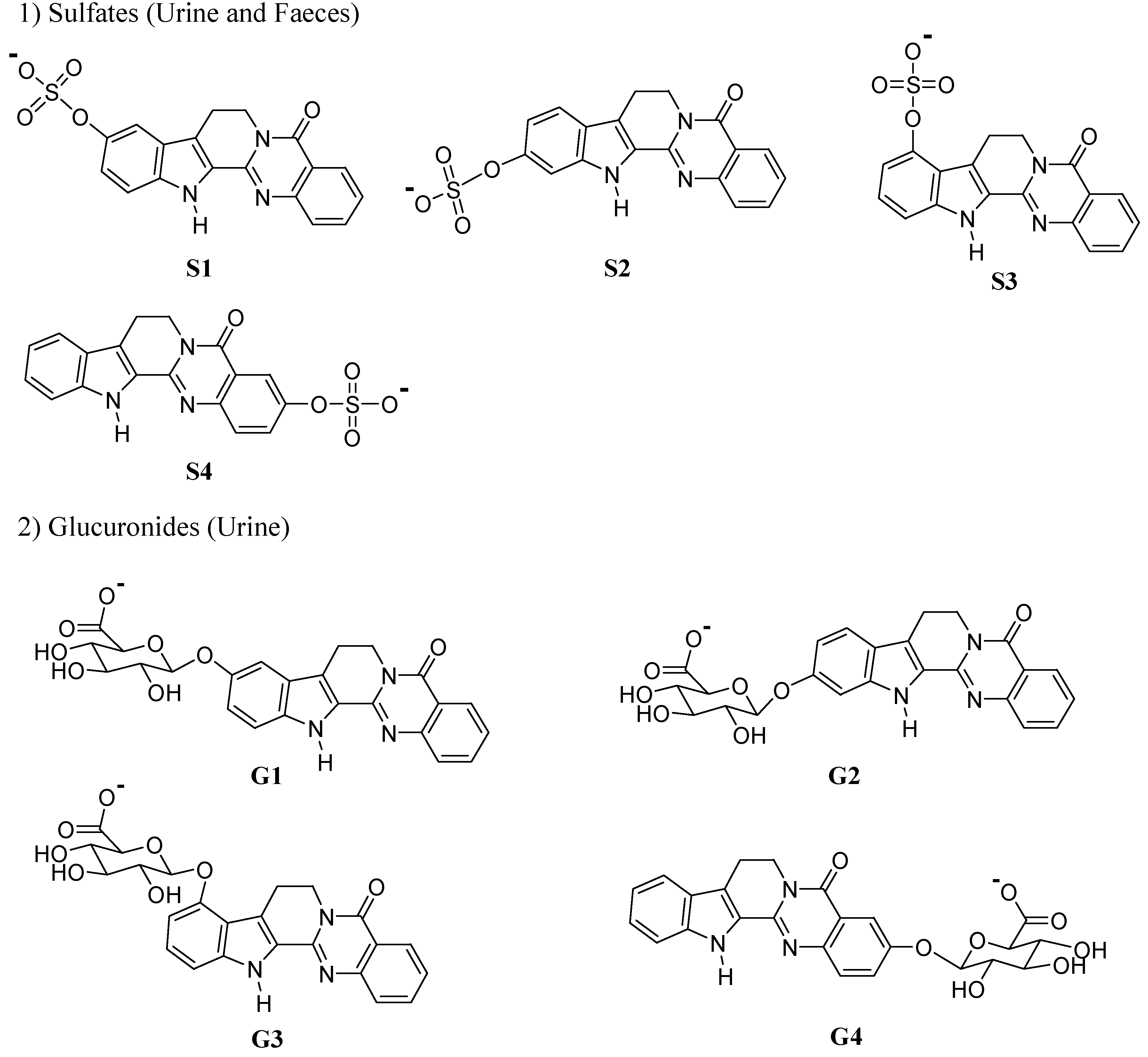
Understanding of metabolic profiles of biologically active compounds in human body would be a critical step in the process of developing new candidates of drugs. In this regard, systematic studies on the metabolic biotransformations of a plant-derived biologically active rutaecarpine have been pursued. We have summarized herein the in vitro (rat and human liver microsomes) and in vivo (rat) studies done to identify the phase I and phase II metabolites of rutaecarpine.
1) Phase I Studies
In vitro phase I metabolites of rutaecarpine were determined by collision-induced dissociation (CID) fragmentation spectra of MS2, MS3 and retention times by LC/ESI-MS [93]. Incubation of rutaecarpine with rat liver microsomes in the presence of NADPH afforded five mono-hydroxylated rutaecarpines (M1-M5), of which three isomers were hydroxylated on ring E, and one on ring C, and one on ring A, as well as 4 di-hydroxylated rutaecarpines (Figure 2). Ueng et al. confirmed the structures of four mono-hydroxylated metabolites as 3-, 10-, 11-, and 12-hydroxyrutaecarpines, by synthesis and comparison of their spectral data such as mass, UV absorbance and 1H-NMR spectra with those of authentic synthetic standards [94]. A later in vitro study with human liver microsomes afforded six mono-hydroxylated rutaecarpines of which an additional mono-hydroxylated one (M6) was found to be 9-hydroxyrutaecarpine [95]. In addition, four dihydroxylated rutaecarpines could be divided to three di-hydroxylated metabolites on the A- and C-rings and one on C- and D-rings [95]. By using combinations of chemical inhibition, immunoinhibition and metabolism by cDNA expressed cytochrome P450 (CYP) enzymes, CYP isozyme responsible for each rutaecarpine metabolite was specified as shown (Figure 1) [94,95,96].
| Metabolites | In vitro | In vivo | |
|---|---|---|---|
| Microsomes | Urine | Faeces | |
| 7/8-Hydroxyrutaecrpinea) | 47.0 ± 1.3 | N.D.b) | N.D.b) |
| 3-Hydroxyrutaecarpine | 4.9 ± 0.1 | 10.1 ± 0.9 | 9.9 ± 1.4 |
| 9-Hydroxyrutaecarpine | 2.9 ± 0.1 | 7.2 ± 0.5 | 26.9 ± 4.2 |
| 10-Hydroxyrutaecarpine | 9.5 ± 0.2 | 24.6 ± 2.5 | 15.6 ± 0.8 |
| 11-Hydroxyrutaecarpine | 33.9 ± 0.7 | 58.1 ± 2.2 | 47.6 ± 2.6 |
Figure 2.
Metabolites of rutaecarpine.

In vivo phase I studies on male Sprague-Dawley rats were also pursued to give four mono-hydroxylated metabolites such as 3-hydroxyrutaecarpine (M5, 10.1%), 9-hydroxyrutaecarpine (M4, 7.2%), 10-hydroxyrutaecarpine (M2, 24.6%) and 11-hydroxyrutaecarpine (M3, 58.1%) and 4 isobaric di-hydroxylated metabolites (M7-M10) (Figure 1) in urine [97], which were identical to the in vitro metabolites except one (M1) that was hydroxylated in the aliphatic moiety. On the other hand, in faeces, the distribution of monohydroxylated metabolites were somewhat different from those in urine. Additionally, M9 was not detected in faeces. It should be noted that the formation of M1 in the rat liver microsomes accounted for 47% the total metabolites, which was not detected in the in vivo system. Such result could be explained by the relative amount of CYP2B enzymes in control liver vs in rat in which amount of CYP2B is usually very low or does not exist [98].
2) Phase II Studies
From the urine of male Sprague-Dawley rats, pretreated intravenously with rutaecarpine, 16 different phase I and II metabolites were identified including four sulfate and four glucuronide conjugates (Figure 3) [97]. Phase I metabolites of rutaecarpine were identified as four mono-hydroxylated rutaecarpines and four isobaric di-hydroxylated rutaecarpines as metabolites (vide ante). In addition, eight phase II metabolites were identified as conjugated with 4-sulfate (S1-S4) and 4-glucuronide (G1-G4). On the other hand, in faeces, 11 different metabolites were identified, in which the di-hydroxylated metabolite M9 and 4 glucuronides G1-G4 were not detected.
Figure 3.
In vivo Phase II metabolites of rutaecarpine.

7. Conclusions
Herbal medicines have been employed as the main source for the development of new agents for treating diseases as well as disorders. Although many of them are still used as mixtures and/or extracts, without identification of active ingredient(s), or with unknown biological action mechanisms, many drugs of the currently used in clinics as a single identity are originally derived from herbs and other plants. Rutaecarpine is one of the important alkaloids isolated from the Rutaceae, exhibits a variety of interesting biological properties, thus continuously attract the scientists in academy as well as industry. Recent years have witnessed the steady progress in the chemistry and biology of rutaecarpine. In the present review, we focused only at the area of natural sources, derivatives, synthesis, biological activities and metabolism of rutaecarpine.
We anticipate that recent progress in science and technology will permit the isolation and identification of new minor rutaecarpine-related components, as well as their biological activities. In addition, more efficient and/or practical methods for the synthesis of rutaecarpine derivatives are still required to pursue structure-activity relationship study and to find more potent compounds for drug development.
Acknowledgements
Financial support from the Korean Research Foundation Grant (KRF-2006–005-J01101) and Research Team for Nonsteroidal Antiinflammatory Drugs is gratefully acknowledged.
References and Notes
- Li, S. C. Pentsao Kang Mu 1596. Li, S. Bencao Gangmu: Compendium of Materia Medica; Foreign Language Press: China, 2003. [Google Scholar] Pedanius, D. De materia Medica; Beck, L. Y., Translator; Georg Olms Verlag: Hildesheim, Germany, 2005. [Google Scholar]
- Chen, A. L.; Chen, K. K. The Constituents of Wuchuyin (Evodia rutaecarpa). J. Am. Pharm. Assoc. 1933, 22, 716–719. [Google Scholar] Liao, J. F.; Chen, C. F.; Chow, S. Y. Pharmacological Studies of Chinese Herbs (9). Pharmacological Effects of Evodia fructus. J. Formosan Med. Assoc. 1981, 79, 30–38. [Google Scholar]
- Asahina, Y.; Kashiwaki, K. Chemical Constituents of the Fruits of Evodia rutaecarpa. J. Pharm. Soc. Jpn. 1915, 1293. [Google Scholar] Asahina, Y.; Mayeda, S. Evodiamine and Rutaecarpine, Alkaloids of Evodia rutaecarpa. J. Pharm. Soc. Jpn. 1916, 871. [Google Scholar] Asahina, Y.; Fujita, A. Constitution of Rutaecarpine. J. Pharm. Soc. Jpn. 1921, 863–869. [Google Scholar]
- Chu, J. H. Constituents of the Chinese Drug Wu-Chu-Yu, Evodia rutaecarpa. Science Record (China) 1951, 4, 279–284, [Chem. Abst. 1952, 46, 11589b]. [Google Scholar]
- Marion, L.; Ramsay, D. A.; Jones, R. N. The Infrared Absorption Spectra of Alkaloids. J. Am. Chem. Soc. 1951, 73, 305–308. [Google Scholar] [CrossRef]
- Raymond-Hamet. Ketoyobyrine. Compt. Rend. 1948, 226, 1379–1381. [Google Scholar]
- Tames, J. ; Bujtas, G. ; Horvath-Dora, K. ; Clauder, O. Alkaloids Containing the Indolo[2,3-c]-quinazolino[3,2-a]pyridine skeleton, IV. The Mass Spectra of Rutaecarpine, Evodiamine, and 3,14-Dihydrorutaecarpine. Acta Chim. Acad. Sci. Hung. 1976, 89, 85–89. [Google Scholar]
- Toth, G.; Horvath-Dora, K.; Clauder, O.; Duddeck, H. Alkaloids with Indolo[2’,3’;3,4]pyrido-[2,1-b]quinazoline Structure, VII. Synthesis and Structure of cis- and trans-Hexahydro-rutaecarpine. Liebigs Ann. Chem. 1977, 529–536. [Google Scholar] Bergman, J.; Bergman, S. Studies of Rutaecarpine and Related Quinazolinocarboline Alkaloids. J. Org. Chem. 1985, 50, 1246–1255. [Google Scholar]
- Fujii, I.; Kobayashi, Y.; Hirayama, N. Molecular Structure of Two Alkaloids, Evodiamine and rutaecarpine, from Evodia Fruit. Z. Kristallogr. 2000, 215, 762–765, Crystallographic data for rutaecarpine can be obtained, free of charge, on application to the Cambridge Crystallographic Data Centre, 12 Union Road, Cambridge CB2 1EZ, UK (fax: +44-(0) 1223-336033 or e-mail: [email protected]). Deposition number: 146432.. [Google Scholar]
- Wang, C.-L.; Liu, J.-L.; Ling, Y.-P. Progress in the Synthesis of Rutaecarpine. Chin. J. Org. Chem. 2006, 26, 1437–1443. [Google Scholar] Kollenz, G. Product Class 10: Imidoylketenes. Sci. Synthesis 2006, 23, 351–380. [Google Scholar] Kikelj, D. Product Class 13: Quinazolines. Sci. Synthesis 2004, 16, 573–749. [Google Scholar] Witt, A.; Bergman, J. Recent Developments in the Field of Quinazoline Chemistry. Curr. Org. Chem. 2003, 7, 659–677. [Google Scholar] Bergman, J. The Quinazolinocarboline Alkaloids. In Alkaloids – Chemistry and Pharmacology; Brossi, A., Ed.; Academic Press: New York, 1983; Vol. XXI; pp. 29–54. [Google Scholar] See also Mhaske, S. B.; Agrade, N. P. The Chemistry of Recently Isolated Naturally Occurring Quinazolinone Alkaloids. Tetrahedron 2006, 62, 9787–9826. [Google Scholar]
- Hu, C.; Li, Y. Research Progress in Pharmacological Actions of Evodiamine and Rutaecarpine. Chin. Pharmacol. Bull. 2003, 19, 1084–1087. [Google Scholar] Chen, C.-F.; Chiou, W.-F.; Chou, C.-J.; Liao, J.-F.; Lin, L.-C.; Wang, G.-J.; Ueng, Y.-F. Pharmacological Effects of Evodia rutaecarpa and its Bioactive Components. Chin. Pharmaceutical J. (Taipei). 2002, 54, 419–435. [Google Scholar] Sheu, J.-R. Pharmacological Effects of Rutaecarpine, an Alkaloid Isolated from Evodia rutaecarpa. Cardiovasc. Drug Rev. 1999, 17, 237–245. [Google Scholar]
- Wang, Y.; Gao, Y. Advances in Modulation of Cytochrome P-450 by Chinese Herbal Medicine. Chin. Tradit. Herb. Drugs (Zhongcaoyao) 2003, 34, 477–478, s1.. [Google Scholar]
- Ueda, J.; Ohsawa, K. Determination of Main Components in Oriental Pharmaceutical Decoctions and Extract Preparations by Ion-Pair High-Performance Liquid Chromatography. J. Tohoku Pharmaceut. Univ. 2002, 49, 13–25. [Google Scholar]
- Hutchnson, J. The Families of Flowering Plants, 2nd Ed. ed; Oxford University Press: Oxford, 1956; Vol. 1; p. 353. [Google Scholar]
- Engler, A. Syllabus der Pflanzenfamilien, 12th Ed.; H. Melchior, H., Ed.; Borntrager: Berlin, 1964; p. 262. [Google Scholar]
- Kamikado, T.; Murakoshi, S.; Tamura, S. Structure Elucidation and Synthesis of Alkaloids from Fruits of Evodia rutaecarpa. Agric. Biol. Chem. 1978, 42, 1515–1519. [Google Scholar] [CrossRef]
- Li, M.-T.; Huang, H.-I. Studies on the Chemical Constituents of the Chinese Drug, Shih-Hu (Evodia rutaecarpa var. officinalis). Acta Pharmaceut. Sin.(Yaoxue Xuebao) 1966, 13, 265–272, [Chem. Abst. 65: 20995]. [Google Scholar] Tschfche, R.; Werner, W. Evocarpin, ein Neues Alkaloids aus Evodia rutaecarpa. Tetrahedron 1967, 23, 1873–1881. [Google Scholar]
- Matsuda, H.; Wu, J.-x.; Tanaka, T.; Iinuma, M.; Kudo, M. Antinociceptive Activities of 70% Methanol Extract of Evodia Fructus (fruit of Evodia rutaecarpa var. bodinaieri) and Its Alkaloidal Components. Biol. Pharm. Bull. 1997, 20, 243–248. [Google Scholar] Yang, X.-W.; Zhang, H.; Li, M.; Du, L.-J.; Yang, Z.; Xiao, S.-Y. Studies on the Alkaloid Constituents of Evodia rutaecarpa (Juss) Benth var. bodinaieri (Dode) Huang and Their Acute Toxicity in Mice. J. Asian Nat. Prod. Res. 2006, 8, 697–703. [Google Scholar]
- Zhao, C.; Zhu, H.; Hao, X.; Yang, X. Study on Chemical Constituents of Evodia ailanthifolia. Tianran Chanwu Yanjiu Yu Kaifa 2006, 18, 418–419, [Chem. Abst. 2007, 147, 230708]. [Google Scholar]
- Bao, T.-d.; Dong, Y.; Yang, Q.; Zhu, X.-x. Determination of Evodiamine, Rutaecarpine, and Evodin in Fructus Evodiae preparata and Its Extract by HPLC. Zhongguo Shiyan Fangjixue Zazhi 2007, 13, 1–3, [Chem. Abst. 2007, 147, 372031]. [Google Scholar]
- Lee, S. W.; Hwang, G. Y.; Kin, S. E.; Kim, H. M.; Kim, Y. H.; Lee, K. S.; Lee, J. J.; Ro, J.-S. Isolation of Modulators for Multidrug Resistance from the Fruits of Evodia officinalis. Saengyak Hakhoechi 1995, 26, 344–348. [Google Scholar] Shin, H.-K.; Do, J.-C.; Son, J.-K.; Lee, C.-S.; Lee, C.-H.; Cheong, C.-J. Quinoline Alkaloids from the Fruits of Evodia officinalis. Planta Medica 1998, 64, 764–765. [Google Scholar] Jin, H.-Z.; Du, J.-L.; Zhang, W.-D.; Chen, H.-S.; Lee, J.-H.; Lee, J.-J. A Novel Alkaloid from the Fruits of Evodia officinalis. J. Asian Nat. Prod. Res. 2007, 9, 685–688. [Google Scholar]
- Pachter, I. J.; Raffauf, F.; Ullyot, G. E.; Ribeiro, O. The Alkaloids of Hortia arborea Eng1. J. Am. Chem. Soc. 1960, 5187–5193. [Google Scholar] Correa, D. de B.; Gottlieb, O. R.; Pimenta de Padua, A. Chemistry of Brazilian Rutaceae. I. Dihydrocinnamic Acids from Hortia badinii. Phytochemistry 1975, 14, 2059–2060. [Google Scholar] Correa, D. de B.; Gottlieb, O. R.; De Padua, A. P.; Da Rocha, A. I. The Chemistry of Brazilian Rutaceae. II. Constituents of Hortia longifolia. Rev. Latinoam. Quim. 1976, 7, 43, [Chem. Abst. 1976, 84, 161790]. [Google Scholar]
- Jacobs, H.; Ramadayal, F.; McLean, S.; Perpick-Dumont, M.; Puzzuoli, F.; Reynolds, W. F. Constituents of Hortia regia: 6,7-Dimethoxycoumarin, Rutaecarpine, Skimmianine, and (+) -Methyl (E,E)-10,11-Dihydroxy-3,7,11-trimethyl-2,6-dodecadienoate. J. Nat. Prod. 1987, 50, 507–509. [Google Scholar] [CrossRef]
- Cuca, S. L. E.; Martinez V., J. C.; Delle Monache, F. Alkaloids Present in Hortia colombiana. Revista Colomb. Quim. 1998, 27, 23–29, [Chem. Abst. 1998, 129, 313387]. [Google Scholar]
- Chatterjee, A.; Mitra, J. Chemistry of Rhetine and Synthesis of Rhetsine. The Alkaloids of Zanthoxylum rhetsa. Sci. Culture 1960, 25, 493–494, [Chem. Abst. 1960, 54, 129271]. [Google Scholar]
- Corrie, J. E. T.; Green, G. H.; Ritchie, E.; Taylor, W. C. Chemical Constituents of Australian Zanthoxylum species. V. Constituents of Z. [Zanthoxylum] pluviatile; the Structures of Two New Lignans. Aust. J. Chem. 1970, 23, 133–145. [Google Scholar] [CrossRef]
- Sheen, W.-S.; Tsai, I.-L.; Teng, C.-M.; Ko, F.-N.; Chen, I.-S. Indolopyridoquinazoline Alkaloids with Antiplatelet Aggregation activity from Zanthoxylum integrifoliolum. Planta Medica 1996, 62, 175–176. [Google Scholar] [CrossRef]
- Chen, J.-J.; Fang, H.-Y.; Duh, C.-Y.; Chen, I.-S. New Indolopyridoquinazoline, Benzo[c]phenanthridines and Cytotoxic Constituents from Zanthoxylum integrifoliolum. Planta Medica 2005, 71, 470–475. [Google Scholar] [CrossRef]
- Chen, I.-S.; Chen, T.-L.; Chang, Y.-L.; Teng, C.-M.; Lin, W.-Y. Chemical Constituents and Biological Activities of the Fruit of Zanthoxylum integrifoliolum. J. Nat. Prod. 1999, 62, 833–837. [Google Scholar] [CrossRef]
- Ishii, H.; Chen, I.-S.; Akaike, M.; Ishikawa, T.; Lu, S. T. Studies on the Chemical Constituents of Rutaceous Plants. XLIV. The Chemical Constituents of Xanthoxylum integrifoliolum (Merr.) Merr. (Fagara integrifoliola Merr.). 1. The Chemical Constituents of the Root Wood. J. Pharm. Soc., Japan 1982, 102, 182–195. [Google Scholar]
- Mukhlesur Rahman, M.; Anwarul Islam, M.; Khondkar, P.; Gray, A. I. Alkaloids and Lignans from Zanthoxylum budrunga (Rutaceae). Biochem. System. Ecol. 2004, 33, 91–96. [Google Scholar] Banerjee, H.; Pal, S.; Adityachaudhury, N. Occurrence of Rutaecarpine in Zanthoxylum budrunga. Planta Med. 1989, 55, 403. [Google Scholar]
- Chen, J.-J.; Huang, H.-Y.; Duh, C.-Y.; Chen, I.-S. Cytotoxic Constituents from the Stem Bark of Zanthoxylum pistaciiflorum. J. Chin. Chem. Soc. 2004, 51, 659–663. [Google Scholar]
- Baetas, A. C. S.; Arruda, M. S. P.; Muller, A. H.; Arruda, A. C. Coumarins and Alkaloids from the Stems of Metrodorea flavida. J. Brazil. Chem. Soc. 1999, 10, 181–183. [Google Scholar] [CrossRef]
- Wattanapiromsakul, C.; Forster, P. I.; Waterman, P. G. Alkaloids and Limonoids from Bouchardatia neurococca: Systematic Significance. Phytochemistry 2003, 64, 609–615. [Google Scholar] [CrossRef]
- Ikuba, A.; Nakamura, T.; Urabe, H. Indolopyridoquinazoline, Furoquinoline and Canthinone Type Alkaloids from Phellodendron amurense Callus Tissues. Phytochemistry 1998, 48, 285–291. [Google Scholar] [CrossRef]
- Ikuba, A. Production of Indolopyridoquinazoline, Furoquinoline and Canthinone-type from Phellodendron amurense Callus Tissues and a Comparative Study of the Alkaloids between Callus and Plant from Chemotaxonomic View Point. Rec. Res. Develop. Phytochem. 2001, 5, 245–253. [Google Scholar]
- Ikuba, A.; Urabe, H.; Nakamura, T. A New Indolopyridoquinazoline-type Alkaloid from Phellodendron amurense Callus Tissues. J. Nat. Prod. 1998, 61, 1012–1014. [Google Scholar] [CrossRef]
- Chiu, C.-Y.; Li, C.-Y.; Chiu, C.-C.; Niwa, M.; Kitanaka, S.; Damu, A. G.; Lee, E-J.; Wu, T.-S. Constituents of Leaves of Phellodendron japonicum Maxim. and Their Antioxidant Activity. Chem. Pharm. Bull. 2005, 53, 1118–1121. [Google Scholar] [CrossRef]
- Ribeiro, T. A. N.; Ndiaye, E. A. da S.; Velozo, E. da S.; Vieira, P. C.; Ellena, J.; de Sousa Jr., P. T. Limonoids from Spiranthera odoratissima St. Hil. J. Brazil. Chem. Soc. 2005, 16, 1347–1352. [Google Scholar] [CrossRef]
- Komala, I.; Rahmani, M.; Lian, G. E. C. L.; Bebe, H.; Ismail, M.; Sukari, M. A.; Rahmat, A. Chemical Constituents of Tetradium sambucinum (Bl.) Hartley. Malaysian J. Sci. 2006, 25, 81–86. [Google Scholar]
- Ng, K. M.; But, P. P.-H.; Gray, A. I.; Hartley, T. G.; Kong, Y.-C.; Waterman, P. G. The Biochemical Systematics of Tetradium, Euodia and Melicope and Their Significance in the Rutaceae. Biochem. System. Ecol. 1987, 15, 587–593. [Google Scholar] [CrossRef]
- Ng, K. M.; But, Gray, A. I.; Waterman, P. G. Limonoids, Alkaloids, and a Coumarin from the Root and Stem Barks of Tetradium glabrifolium. J. Nat. Prod. 1987, 50, 1160–1163. [Google Scholar] [CrossRef]
- Bui, K. A.; Tran, V. S.; Nguyen, M. C.; Duong, A. T. Three Indolopyridoquinazoline Alkaloids from Tetradium trichotomum Lour. Growing in Vietnam. Tap Chi Hoa Hoc 2002, 40, 72–75, [Chem. Abst. 138: 103670]. [Google Scholar] Bui, K. A.; Duong, A. T.; Tran, V. S.; Nguyen, M. C. Limonoid Compounds from Tetradium trichotomum (Rutaceae). Tap Chi Hoa Hoc 2003, 41(Spec.), 51–54, [Chem. Abst 2004, 140, 142600]. [Google Scholar]
- Tong, R. Chemical Constituents of Huajiao (Fagara rhetza). Chin. Tradit. Herb Drugs (Zhongcaoyao) 1991, 22, 249–250, [Chem. Abst. 1995, 115, 166449]. [Google Scholar] Shibuya, H.; Takeda, Y.; Zhang, R. S.; Tong, R. X.; Kitagawa, I. Indonesian Medicinal Plants. III. On the Constituents of the Bark of Fagara rhetza (Rutaceae). (1): Alkaloids, Phenylpropanoids, and Acid Amide. Chem. Pharm. Bull. 1992, 40, 2325–2330. [Google Scholar]
- Guan, Z.; Su, J.-y.; Zeng, L.-m.; Li, H. Studies on Non-Taxoid Constituents from Taxus chinensis (Pilger) Rehd. Redai Yaredai Zhiwu Xuebao 2000, 8, 182–184, [Chem. Abst. 2001, 134, 219695]. [Google Scholar]
- Zhu, W.-M.; He, H.-P.; Fan, L.-M.; Shen, Y.-M.; Zhou, J.; Hao, X.-J. Components of Stem Barks of Winchia calophylla A. DC. (Apocynaceae) and Their Bronchodilator Activities. J. Integrative Plant Biol. 2005, 47, 892–896. [Google Scholar] [CrossRef]
- Waterman, P. G. Alkaloids of the Rutaceae: Distribution and Systematic Significanc. Biochem. System. Ecol. 1975, 3, 149–180, and references therein. [Google Scholar] [CrossRef]
- Canonica, L.; Danieli, B.; Manitto, P.; Russo, G.; Ferrari, G. New Quinazolinocarboline from Euxylphora paraënsis. Tetrahedron Lett. 1968, 9, 4865–4866. [Google Scholar] Danieli, B.; Manitto, P.; Ronchetti, F.; Russo, G.; Ferrari, G. New Indolopyridoquinazoline Alkaloids from Euxylphora paraënsis. Phytochemistry 1972, 11, 1833–1836. [Google Scholar] Danieli, B.; Palmisano, G.; Russo, G.; Ferrari, G. Minor Indolopyridoquinazoline Alkaloids from Euxylphora paraënsis. Phytochemistry 1973, 12, 2521–2525. [Google Scholar] Danieli, B.; Farachi, C.; Palmisano, G. A New Indolopyridoquinazoline in the Bark of Euxylphora paraënsis. Phytochemistry 1976, 15, 1095–1096. [Google Scholar]
- Ayafor, J. F.; Sondengam, B. L.; Ngadjui, B. T. Quinoline and Indolopyridoquinazoline Alkaloids from Vepris louisii. Phytochemistry 1982, 21, 2733–2736. [Google Scholar] [CrossRef]
- Li, X.-C.; Dunbar, D. C.; ElSohly, H. N.; Walker, L. A.; Clark, A. M. Indolopyridoquinazoline Alkaloid from Leptothyrsa sprucei. Phytochemistry 2001, 58, 627–629. [Google Scholar] [CrossRef]
- Christopher, E.; Bedir, E.; Dunbar, C.; Khan, I. A.; Okunji, C. O.; Schuster, B. M.; Iwu, M. M. Indoloquinazoline Alkaloids from Araliopsis tabouensis. Helv. Chim Acta 2003, 86, 2914–2918. [Google Scholar] [CrossRef]
- Danieli, B.; Palmisano, G.; Rainoldi, G.; Russo, G. 1-Hydroxyrutaecarpine from Euxylophora paraënsis. Phytochemistry 1974, 13, 1603–1606. [Google Scholar] [CrossRef]
- Wu, T.-S.; Yeh, J.-H.; Wu, P.-L.; Chen, K.-T.; Lin, L.-C.; Chen, C.-F. 7-Hydroxyrutaecarpine from Tetradium glabrifolium and Tetradium ruticarpum. Heterocycles 1995, 41, 1071–1076. [Google Scholar] [CrossRef]
- Chen, C.-F.; Chiou, W.-F.; Chou, C.-J.; Liao, J.-F. ; Lin, L.-C. ; Wang, G.-J. ; Ueng, Y.-F. Pharmacological Effects of Evodia rutaecarpa and Its Bioactive Components. Chin. Pharm. J. (Taipei) 2002, 54, 419–435. [Google Scholar]
- Xu, M.-L.; Moon, D.-C.; Lee, J.-S.; Woo, M.-H.; Lee, E. S.; Jahng, Y.; Chang, H.-W.; Lee, S. H.; Son, J.-K. Cytotoxicity and DNA Topoisomerase Inhibitory Activity of Constituents Isolated from the Fruits of Evodia officinalis. Arch. Pharm. Res. 2006, 29, 541–547. [Google Scholar] [CrossRef]
- Zhang, L.; Yang, Z.; Tian, J.-K. Two New Indolopyridoquinazoline Alkaloidal Glycosides from Ranunculus ternatus. Chem. Pharm. Bull. 2007, 55, 1267–1269. [Google Scholar] [CrossRef]
- Asahina, Y.; Irie, T. ; Ohta, T. Synthesis of Rutaecarpine. II. J. Chem. Soc., Jpn. 1927, No. 543, 51–52. [Google Scholar]
- Asahina, Y.; Manske, R. H. F.; Robinson, R. A Synthesis of Rutaecarpine. J. Chem. Soc. 1927, 1708–1710. [Google Scholar] [CrossRef]
- Asahina, Y.; Ohta, T. Synthesis of Rutecarpine. III. J. Chem. Soc. 1928, 48, 313–317. [Google Scholar] Terzyan, A. C.; Safrazbekyan, R. R.; Khazhakyan, L. V.; Tatevosyan, G. T. Reduction Products of Rutecarpine and 10-Methoxyrutaecarpine. Izv. Akad. Nauk Arm. SSR, Khim. Nauki 1961, 14, 393–399, [Chem. Abst. 1962, 57, 83091]. [Google Scholar] Atta-ur-Rahman; Ghazala, M. Reactions of Harmaline and Its Derivatives. VI. The Partial Syntheses of 11-Methoxyrutaecarpine and 11-Methoxynauclefine. Synthesis 1980, 372–374. [Google Scholar]
- Tang, Y.; Feng, X.; Huang, L. Studies on the Chemical Constituents of Evodia rutaecarpa [Juss] Benth. Acta Pharmaceut. Sin. (Yaoxue Xuebao) 1996, 31, 151–155, [Chem. Abst. 1996, 125, 308784). [Google Scholar] Tang, Y.; Feng, X.; Huang, L. Studies on Chemical Constituents of Evodia rutaecarpa (Juss) Benth. J. Chin. Pharm. Sci. 1997, 6, 65–69. [Google Scholar]
- Petersen, S.; Tietze, E. The Reaction of Cyclic Lactim Ethers with Amino Carboxylic Acids. Liebigs Ann. Chem. 1959, 623, 166–176. [Google Scholar] [CrossRef]
- Hamid, A.; Elomrib, A.; Daïch, A. Expedious and Practical Synthesis of the Bioactive Alkaloids Rutaecarpine, Euxylophoricine A, Deoxyvasicinone and Their Heterocyclic Homologues. Tetrahedron Lett. 2006, 47, 1777–1781. [Google Scholar] [CrossRef]
- Benovsky, P.; Stille, J. R. Aza-Annulation as a Versatile Approach to the Synthesis of Non-benzodiazepene Compounds for the Treatment of Sleep Disorders. Tetrahedron Lett. 1997, 38, 8475–8478. [Google Scholar] Gittos, M. W.; Robinson, M. R.; Verge, J. P.; Davies, R. V.; Iddon, B.; Suschitzky, H. Intramolecular Cyclisation of Arylalkyl Isothiocyanates. Part I. Synthesis of 1-Substituted 3,4-Dihydroisoquinolines. J. Chem. Soc., Perkin Trans. 1 1976, 33–38. [Google Scholar]
- Eguchi, S.; Takeuchi, H.; Matsushita, Y. Synthesis of Novel Carbo- and Heteropolycycles. 20. Short-Step Synthesis of Rutecarpine and Tryptanthrin via Intramolecular Aza-Wittig Reaction. Heterocycles 1992, 33, 153–156. [Google Scholar] [CrossRef]
- Lee, E. S.; Park, J. G.; Jahng, Y. A Facile Synthesis of Simple Alkaloids – Synthesis of 2,3-Polymethylene-4(3H)-quinazolinones and Related Alkaloids. Tetrahedron Lett. 2003, 44, 1883–1886. [Google Scholar] Jahng, K. C.; Kim, S. I.; Kim, D. H.; Seo, C. S.; Son, J.-K.; Lee, S. H.; Lee, E. S.; Jahng, Y. One-Pot Synthesis of Simple Alkaloids: 2,3-Polymethylene-4(3H)-quinazolinones, Luotonin A, Tryptanthrin, and Rutaecarpine. Chem. Pharm. Bull. (accepted for publication).
- Schöpf, C. Die Syntheses von Naturstoffen, insbesondere von Alkaloiden, unter physiologischen Bedingungen und Bedeutung für die Frage der Entstehung einiger pflanzlicher Naturstoffe in der Zelle. Angew. Chem. 1937, 50, 779–790. [Google Scholar] [CrossRef]
- Schöpf, C.; Steuer, H. Synthesis and Transformations of Natural Products under Physiological Conditions X. Biogenesis of Rutaecarpine and Evodiamine. Synthesis of Rutaecarpine under Physiological Conditions. Liebigs Ann. Chem. 1947, 558, 124–136. [Google Scholar] [CrossRef]
- Kametani, T.; Loc, C. V.; Higa, T.; Koizumi, M.; Ihara, M.; Fukumoto, K. Iminoketene Cycloaddition. 2. Total Syntheses of Carboline, Glycosminine, and Rutaecarpine by Condensation of Iminiketene with Amides. J. Am. Chem. Soc 1977, 99, 2306–2309. [Google Scholar] Kametani, T.; Ohsawa, T.; Ihara, M.; Fukumoto, K. Studies on the Syntheses of Heterocyclic Compounds. DCCLV. Iminoketene Cycloaddition. 4. Alternative Syntheses of 5,6,7,8-Tetrahydro-2,3-dimethoxy-8-oxoisoquinolo[1,2-b]quinazoline and Rutaecarpine. Chem. Pharm. Bull. 1978, 26, 1922–1926. [Google Scholar]
- Terzyan, A. C.; Safrazbekyan, R. R.; Khazhakyan, L. V.; Tatevosyan, G. T. Reduction Products of Rutecarpine and 10-Methoxyrutaecarpine. Izv. Akad. Nauk Arm. SSR, Khim. Nauki 1961, 14, 393–399, [Chem. Abst. 1962, 57, 83091]. [Google Scholar] Horvath-Dora, K.; Clauder, O. Alkaloids Containing the Indolo[2,3-c]quinazolino[3,2-a]pyridine Skeleton. III. 3,14-Dihydrorutecarpine. Acta Chim. Acad. Sci. Hung. 1975, 84, 93–97, [Chem. Abst. 1975, 82, 171273). [Google Scholar]
- Bergman, J.; Bergman, S. Studies of Rutaecarpine and Related Indole Alkaloids. Heterocycles 1981, 16, 347–350. [Google Scholar] [CrossRef]
- Waterman, P. Chemosystematics in the Rutaceae. Part 7: Alkaloids and Coumarins from Zanthoxylum flavum: Dihydrorutecarpine, A Novel β-Indoloquinazoline Alkaloid. Phytochemistry 1976, 15, 578–579. [Google Scholar] [CrossRef]
- Kamikado, T.; Murakoshi, S.; Tamura, S. Structure Elucidation and Synthesis of Alkaloids Isolated from Fruits of Evodia rutaecarpa. Agric. Biol. Chem. 1978, 42, 1515–1519. [Google Scholar] [CrossRef]
- Yang, L.-M.; Chen, C.-F.; Lee, K.-H. Synthesis of Rutaecarpine and Cytotoxic Analogs. Bioorg. Med. Chem. Lett. 1995, 5, 465–468. [Google Scholar] Baruah, B.; Dasu, K.; Vaitilingam, B.; Mamnoor, P.; Venkata, P. P.; Rajagopal, S.; Yeleswarapu, K. R. Synthesis and Cytotoxic Activity of Novel Quinazolino-β-carboline-5-one Derivatives. Bioorg. Med. Chem. 2004, 12, 1991–1994. [Google Scholar]
- Kaneko, C.; Chiba, T.; Kasai, K.; Miwa, C. A Short Synthesis of Rutecarpine and/or Vasicolinone from 2-Chloro-3-(indol-3-yl)ethylquinazolin-4(3H)-one: Evidence for the Participation of the Spiro Intermediate. Heterocycles 1985, 23, 1385–1390. [Google Scholar] [CrossRef]
- Mohanta, P. K.; Kim, K. A Short Synthesis of Quinazolinocarboline Alkaloids Rutaecarpine, Hortiacine, Euxylophoricine A and Euxylophoricine D from Methyl N-(4-chloro-5H-1,2,3-dithiazol-5-ylidene)anthranilates. Tetrahedron Lett. 2002, 43, 3993–3996. [Google Scholar] [CrossRef]
- Harayama, T.; Hori, A.; Serban, G. Concise Synthesis of Quinazoline Alkaloids, Luotonins A and B, and Rutaecarpine. Tetrahedron 2004, 60, 10645–10649. [Google Scholar] [CrossRef]
- Bowman, W. R.; Elsegood, M. R. J.; Stein, T.; Weaver, G. W. Radical Reactions with 3H-Quinazolinones: Synthesis of Deoxyvasicinone, Mackinazolinone, Luotonin A, Rutaecarpine, and Tryptanthrin. Org. Biomol. Chem. 2007, 5, 103–110. [Google Scholar] [CrossRef]
- Mori, M.; Kobayashi, H.; Kimura, Ban, Y. One Pot Synthesis of Quinazolinone Derivatives by Use of Palladium Catalyzed Carbonylation. Heterocycles 1985, 23, 2803–2806. [Google Scholar] [CrossRef]
- Kametani, T.; Higa, T.; Fukumoto, K.; Koizumi, M. A One-Step Synthesis of Evodiamine and Rutaecarpine. Heterocycles 1976, 4, 23–28. [Google Scholar] Kametani, T.; Higa, T.; Loc, C. V.; Ihara, M.; Koizumi, M.; Fukumoto, K. J. Am. Chem. Soc. 1976, 98, 6186–6188.
- Möhrle, H.; Kamper, C.; Schmid, R. Eine neue Synthese von Rutaecarin. Arch. Pharm. 1980, 313, 990–995. [Google Scholar] [CrossRef]
- Späth, E.; Platzer, N.; Peganine, VIII. Derivatives of Peganine and its Ring Homologs. Ber. 1935, 68, 2221–2226. [Google Scholar] [CrossRef]
- Johns, S. R.; Lamberton, J. A. Alkaloids of Mackinlaya Species (Family Araliaceae). Chem. Comm. 1965, 267. [Google Scholar]
- Kökösi, J.; Hermecz, I.; Szasz, G.; Meszaros, Z. Nitrogen Bridged Compounds. Part 16. Facile Total Synthesis of 7,8-Dihydroindolo[2',3':3,4]pyrido[2,1-b]quinazolin-5(13H)-one (Rutaecarpine). Tetrahedron Lett. 1981, 22, 4861–4862. [Google Scholar] Kökösi, J.; Hermecz, I.; Podanyi, B.; Szasz, G.; Meszaros, Z. Nitrogen Bridged Compounds. Part 55. Synthesis of Substituted 7,8-Dihydro-5H,13H-indolo[2',3':3,4]pyrido[2,1-b]quinazolin-5-ones. J. Heterocycl. Chem. 1985, 22, 1373–1375. [Google Scholar] Mhaske, S. B.; Argade, N. P. Facile Zeolite Induced Fischer-Indole Synthesis: A New Approach to Bioactive Natural Product Rutaecarpine. Tetrahedron 2004, 60, 3417–3420. [Google Scholar]
- Lee, S. H.; Kim, S. I.; Park, J. G.; Lee, E. -S.; Jahng, Y. A Simple Synthesis of Rutaecarpine. Heterocycles 2001, 55, 1555–1560. [Google Scholar] [CrossRef]
- Chavan, S. P.; Sivappa, R. A Facile Total Synthesis of Rutaecarpine. Tetrahedron Lett. 2004, 45, 997–999. [Google Scholar] [CrossRef]
- Kamal, A.; Shankaraiah, N.; Devaiah, V.; Reddy, K. L. Solid-Phase Synthesis of Fused [2,1-b]quinazolinone Alkaloids. Tetrahedron Lett. 2006, 47, 9025–9028. [Google Scholar] Gil, C.; Braese, S. Efficient Solid-Phase Synthesis of Highly Functionalized 1,4-Benzodiazepin-5-one Derivatives and Related Compounds by Intramolecular Aza-Wittig Reactions. Chemistry-Eur. J. 2005, 11, 2680–2688. [Google Scholar] Liu, J.-F.; Ye, P.; Sprague, K.; Sargent, K.; Yohannes, D.; Baldino, C. M.; Wilson, C. J.; Ng, S.-C. Novel One-Pot Total Syntheses of Deoxyvasicinone, Mackinazolinone, Isaindigotone, and Their Derivatives Promoted by Microwave Irradiation. Org. Lett. 2005, 7, 3363–3366. [Google Scholar] Yadav, J. S.; Reddy, B. V. S. Microwave-Assisted Rapid Synthesis of the Cytotoxic Alkaloid Luotonin A. Tetrahedron Lett. 2002, 43, 1905–1907. [Google Scholar] Nishiyama, Y.; Hirose, M.; Kitagaito, W.; Sonoda, N. Synthesis of 3,4-Dihydroquinazolin-4-one: Selenium-Catalyzed Reductive N-Heterocyclization of N-(2-Nitrobenzoyl)amides with Carbon Monoxide. Tetrahedron Lett. 2002, 43, 1855–1858. [Google Scholar] Mhsaske, S. B.; Argade, N. P. Chemoenzymatic Synthesis of Pyrrolo[2,1-b]quinazolinones: Lipase-Catalyzed Resolution of Vasicinone. J. Org. Chem. 2001, 66, 9038–9040. [Google Scholar] Dunn, A. D.; Kinnear, K. I. New Reactions of Deoxyvasicinone. Part 4. J. Heterocycl. Chem. 1986, 23, 53–57. [Google Scholar]
- Kökösi, J.; G. Szasz, G.; Hermecz, I. An Alternative Synthesis of Rutaecarpine and Vasicolinone Alkaloids. Tetrahedron Lett. 1992, 33, 2995–2998. [Google Scholar] [CrossRef]
- Yang, L.-M.; Lin, S.-J.; Lin, L.-C.; Kuo, Y.-H. Antitumor Agents. 2. Synthesis and Cytotoxic Evaluation of 10-Bromorutaecarpine. Chin. Pharm. J. (Taipei) 1999, 51, 219–225. [Google Scholar]
- Jahng, Y.; Kim, S. I.; Lee, E.-S. Synthesis and Cytotoxicities of Rutaecarpine Analogues. In Abst. Papers 227th ACS National Meeting, Anaheim, CA, United States, March 28-April 1, 2004. MEDI-146.
- Ogasawara, M.; Matsubara, T.; Suzuki, H. Screening of Natural Compounds for Inhibitory Activity on Colon Cancer Cell Migration. Biol. Pharm. Bull. 2001, 24, 720–723. [Google Scholar] [CrossRef]
- Moon, T. C.; Murakami, M.; Kudo, I.; Son, K. H.; Kim, H. P.; Kang, S. S.; Chang, H. W. A New Class of COX-2 Inhibitor, Rutaecarpine from Evodia rutaecarpa. Inflamm. Res. 1999, 48, 621–625. [Google Scholar] [CrossRef]
- Lee, E. S.; Kim, S. I.; Lee, S. H.; Jeong, T. C.; Moon, T. C.; Chang, H. W.; Jahng, Y. Synthesis and COX Inhibitory Activities of Rutaecarpine Derivatives. Bull. Korean Chem. Soc. 2005, 26, 1975–1980. [Google Scholar] [CrossRef]
- Lee, S. K.; Kim, N. H.; Lee, J.; Kim, D. H.; Lee, E. S.; Choi, H. G.; Chang, H. W.; Jahng, Y.; Jeong, T. C. Induction of Cytochrome P450s by Rutaecarpine and Metabolism of Rutaecarpine by Cytochrome P450s. Planta Medica 2004, 70, 753–757. [Google Scholar] Lee, S. K.; Lee, J.; Lee, E. S.; Jahng, Y.; Kim, D. H.; Jeong, T. C. Characterization of in vitro Metabolites of Rutaecarpine in Rat Liver Microsomes using Liquid Chromatography/Tandem Mass Spectrometry. Rapid Commun. Mass Spectrom. 2004, 18, 1073–1080. [Google Scholar]
- Ueng, Y. F.; Yu, H. J.; Lee, C. H.; Peng, C.; Jan, W. C.; Ho, L. K.; Chen, C. F.; Don, M. J. Identification of the Microsomal Oxidation Metabolites of Rutaecarpine, A Main Active Alkaloid of the Medicinal herb. J. Chromatogr. A 2005, 1076, 103–109. [Google Scholar] [CrossRef]
- Lee, S. K.; Lee, J. H.; Yoo, H. H.; Kim, D. H.; Jahng, Y.; Jeong, T. C. Characterization of Human Liver Cytochrome P450 Enzymes Involved in the Metabolism of Rutaecarpine. J. Pharm. Biomed. Anal. 2006, 41, 304–309. [Google Scholar] [CrossRef]
- Ueng, Y. F.; Don, M. J.; Jan, W. C.; Wang, L.-K.; Chen, C. F. Oxidative Metabolism of the Alkaloid Rutaecarpine by Human Cytochrome P450. Drug. Metabol. Dispos. 2006, 34, 821–827. [Google Scholar] [CrossRef]
- Lee, S. K.; Lee, D. W.; Jeon, T. W.; Jin, C. H.; Kim, G. H.; Jun, I. H.; Lee, D. J.; Kim, S.-I.; Kim, D. H.; Jahng, Y.; Jeong, T. C. Characterization of the Phase II Metabolites of Rutaecarpine in Rat by Liquid Chromatography-Electrospray Ionization-Tandem Mass Spectrometry. Xenobiotica 2005, 35, 1135–1145. [Google Scholar] [CrossRef]
- Rendic, S.; Dicarlo, F. J. Human Cytochrome P450 Enzymes: A Status Report Summarizing Their Reactions, Substrates, Inducers and Inhibitors. Drug Metabol. Rev. 1997, 29, 413–580. [Google Scholar] [CrossRef]
© 2008 by MDPI (http://www.mdpi.org). Reproduction is permitted for noncommercial purposes.
Share and Cite
MDPI and ACS Style
Lee, S.H.; Son, J.-K.; Jeong, B.S.; Jeong, T.-C.; Chang, H.W.; Lee, E.-S.; Jahng, Y. Progress in the Studies on Rutaecarpine. Molecules 2008, 13, 272-300. https://doi.org/10.3390/molecules13020272
AMA Style
Lee SH, Son J-K, Jeong BS, Jeong T-C, Chang HW, Lee E-S, Jahng Y. Progress in the Studies on Rutaecarpine. Molecules. 2008; 13(2):272-300. https://doi.org/10.3390/molecules13020272
Chicago/Turabian StyleLee, Seung Ho, Jong-Keun Son, Byeong Seon Jeong, Tae-Cheon Jeong, Hyeon Wook Chang, Eung-Seok Lee, and Yurngdong Jahng. 2008. "Progress in the Studies on Rutaecarpine" Molecules 13, no. 2: 272-300. https://doi.org/10.3390/molecules13020272