Cytotoxic Metabolites from the Okinawan Ascidian Diplosoma virens

1
Department of Chemistry, Biology and Marine Science, University of the Ryukyus, Nishihara-cho Okinawa 903-0123, Japan
2
Okinawa Industrial Technology Center, Uruma-shi Okinawa 904-2234, Japan
3
Department of Bioresources Engineering, Okinawa National College of Technology, Nago-shi Okinawa 905-2192, Japan
*
Author to whom correspondence should be addressed.
Molecules 2008, 13(3), 595-602; https://doi.org/10.3390/molecules13030595
Submission received: 20 February 2008
/
Revised: 8 March 2008
/
Accepted: 9 March 2008
/
Published: 11 March 2008
Abstract
:The unstable isomeric compounds 5-hydroxy-7-prop-2-en-(E)-ylidene-7,7a-dihydro-2H-cyclopenta[b]pyran-6-one (1) and 5-hydroxy-7-prop-2-en-(Z)-ylidene-7,7a-dihydro-2H-cyclopenta[b]pyran-6-one (2), previously described as antimicrobial metabolites from the sponge Ulosa sp., were isolated and identified as major components of the ascidian Diplosoma virens. In this paper, full spectral data for 2 and complete 13C-NMR data for 1, based on 2D NMR measurements, are provided for the first time. Compounds 1 and 2 showed cytotoxity against HCT116 cells (human colorectal cancer cells) by triggering apoptotic cell death.
Introduction
Marine organisms containing symbiotic microorganisms, such as ascidians, sponges and soft corals, are a rich source of bioactive compounds [1, 2a-b]. Ascidians belong to the family Didemnidae, which frequently possess prokaryotic algae (cyanobacteria, Prochloron spp., etc.) [3,4,5], and have yielded structurally unique and pharmacologically interesting compounds such as didemnenones, enterocins, paterallazoles, varacins and virenamides [6,7,8,9,10]. In our search for bioactive compounds from Okinawan marine organisms [11,12,13,14], we investigated the ascidian Diplosoma virens, which inhabits the coast of Hateruma Island. From this organism, we isolated compounds 1 and 2, which are major constituents of its acetone extract. Antimicrobial metabolites 1 and 2, previously isolated from the Caribbean sponge Uloma sp. in 1978 [15], were characterized mainly by spectral analysis of their derivatives, including compounds such as acetates, methyl ethers and phenyltriazolinedione adducts. The physical properties of 2 and the 13C-NMR data for 1 have not been previously described in the literature [15].
In this study, we confirmed the structure of 2 by interpreting its 2D-NMR data, and we provided full spectral data for 2 and 1. In addition, the compounds’ cytotoxity against HCT116 cells (human colorectal cancer cells) via apoptotic cell death was demonstrated.
Results and Discussion
A specimen of Diplosoma virens (72 g, wet weight) was collected by hand from the coast of Hateruma Island, Okinawa, and extracted with acetone. The acetone extract was analyzed using 1H- NMR, and two major compounds were observed. The acetone extract was partitioned between aqueous MeOH and hexanes. The aqueous MeOH phase was concentrated in vacuo and then partitioned between CH2Cl2 and water. Purification of the CH2Cl2 extract by silica gel column chromatography followed by reversed-phase (C8) HPLC (H2O/MeOH) yielded compounds 1 (0.13%) and 2 (0.086%). When a solution of 1 or 2 in CH2Cl2 or MeOH was concentrated, a large portion of the resulting residue was no longer soluble in the organic solvents.
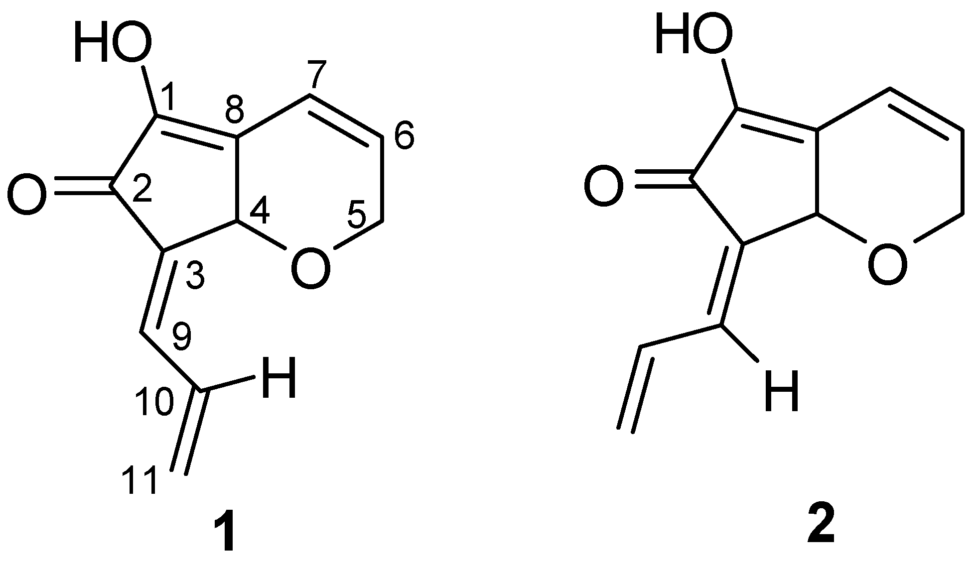
Figure 1.
Structures of compounds 1 and 2.

Compound 1 was identified by performing comprehensive spectral analyses (Figure 1 and Table 1) and by comparing resulting spectral data with those in the literature [15].
Analysis of the13C-NMR and HRFABMS data [m/z 191.0691 (M + H)+, ∆ -1.7 mmu] for compound 2 provided a molecular formula of C11H10O3, which suggested seven degrees of unsaturation. The IR absorption bands at 1680 and 3250 cm-1 indicated the presence of carbonyl and hydroxyl groups. The spectral data of compound 2 showed close similarities to those of 1. The 1H- and 13C-NMR data analysis indicated the presence of a carbonyl carbon (δC 187.8), a cis double bond [δC 134.2, 118.4; δH 6.17 (1H, ddd), 6.77 (1H, ddd)], a tetrasubstituted double bond (δC 128.3, 147.5), a conjugated diene [δC 131.6 (s), 136.6 (d); δH 6.65 (1H, br d) and δC 132.1 (d), 127.3 (t); δH 7.76 (1H, ddd), 5.57 (1H, dd), 5.61 (1H, dd)], an oxygenated methine [δC 71.9; δH 4.79 (1H, s)] and an oxygenated methylene [δC 67.2; δH 4.48 (1H, ddd), 4.57 (1H, ddd)].

| 1 | 2 | ||||||
|---|---|---|---|---|---|---|---|
| C no. | δC | δH (mult, J in Hz) | δC | δH (mult, J in Hz) | |||
| 1 | 147.2 | 147.5 | |||||
| 2 | 187.9 | 187.8 | |||||
| 3 | 132.6 | 131.6 | |||||
| 4 | 71.3 | 5.01 (s) | 71.9 | 4.79 (s) | |||
| 5α | 67.1 | 4.50 (ddd, 18.5, 4.2, 2.0) | 67.2 | 4.48 (ddd, 18.3, 4.2, 1.7) | |||
| 5β | 4.61 (ddd, 18.5, 2.4, 2.4) | 4.57 (ddd, 18.3, 2.4, 2.4) | |||||
| 6 | 134.0 | 6.17 (ddd, 10.0, 4.2, 2.4) | 134.2 | 6.17 (ddd, 10.0, 4.2, 2.4) | |||
| 7 | 118.4 | 6.78 (br d, 10.0) | 118.4 | 6.77 (ddd, 10.0, 2.4, 1.7) | |||
| 8 | 129.1 | 128.3 | |||||
| 9 | 133.6 | 7.04 (br d, 11.7) | 136.6 | 6.65 (br d, 11.5) | |||
| 10 | 132.4 | 6.89 (ddd, 16.8, 11.7, 10.0) | 132.1 | 7.76 (ddd, 17.1, 11.5, 10.0) | |||
| 11α | 127.9 | 5.64 (br d, 10.0) | 127.3 | 5.57 (dd, 10.0, 1.7) | |||
| 11β | 5.71 (br d, 16.8) | 5.61 (dd, 17.1, 1.7) | |||||
| OH | 6.08 (br s) | 6.12 (br s) | |||||
a 1H-NMR (400 MHz) and 13C-NMR (100 MHz) were recorded in CDCl3.
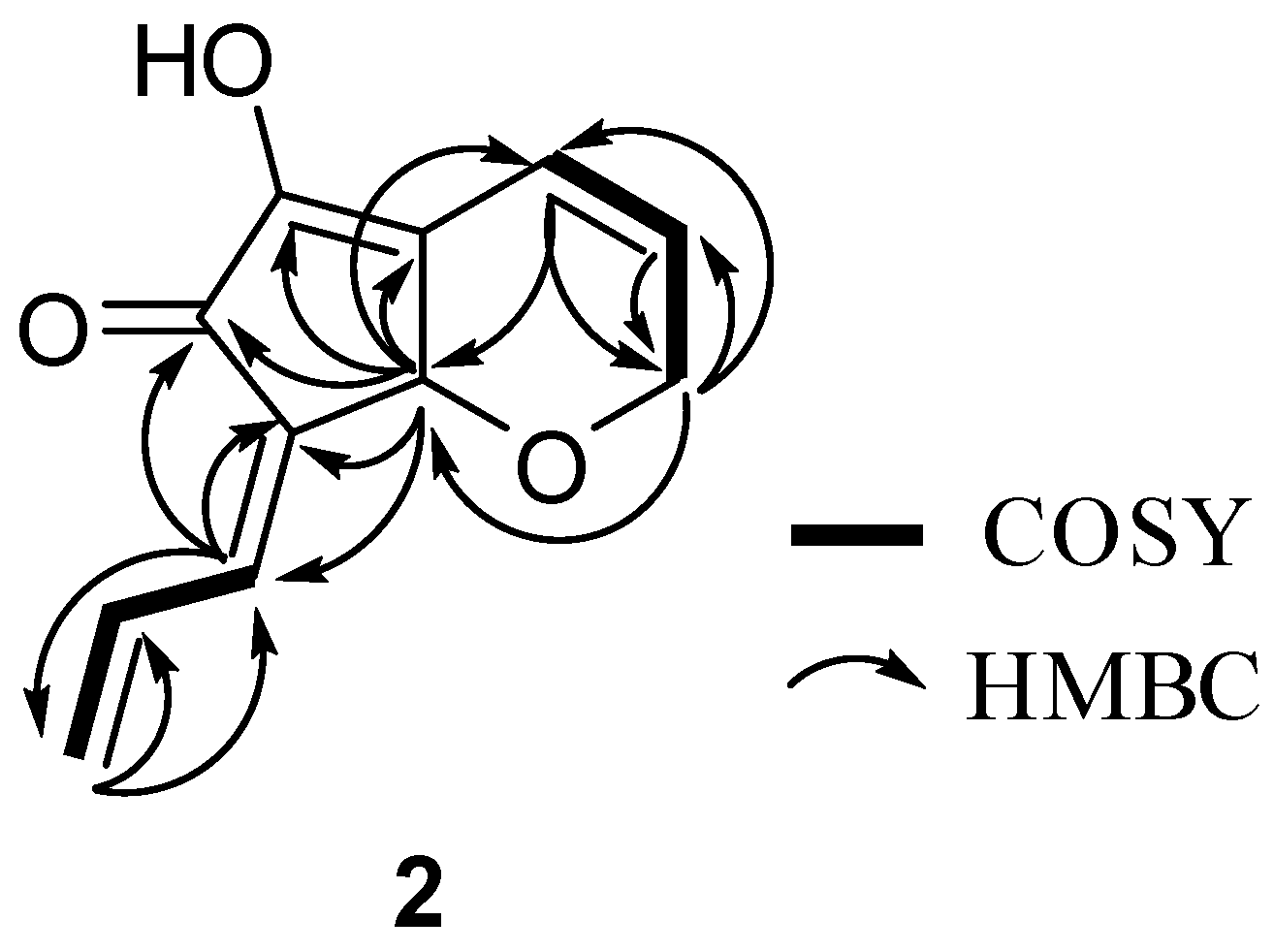
Extensive analysis of 1H-1H COSY demonstrated two isolated spin systems, C5-C7 and C9-C11 (Figure 2). The connectivity of the aforementioned partial structures was established from the HMBC correlations of H-4/C-1, H-4/C-2, H-9/C-2, H-4/C-3, H-9/C-3, H2-5/C-4, H-7/C-4, H-6/C-5, H-7/C-5, H-5/C-6, H-5/C-7, H-4/C-7, H-4/C-8, H-4/C-9, H-11/C-9, H-11/C-10 and H-9/C-11 as shown in Figure 2, to describe the entire carbon framework of 2.
Figure 2.
Planar structure of 2 based on COSY and HMBC correlations.

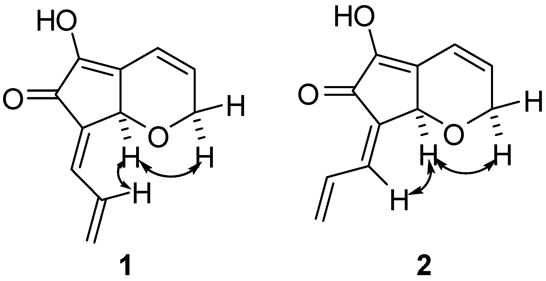
The chemical shift of H-10 in 2 (δH 7.76) was at lower field than in 1 (δH 6.89) owing to the magnetic anisotropy effect of the carbonyl group and the chemical shift of H-9 in 2 (δH 6.65) was at higher field than in 1 (δH 7.04), suggesting a Z configuration for the C-3, 9 double bond of 2. This was confirmed by an NOEDS experiment, in which NOE was observed between H-9 and H-4 (Figure 3).
Figure 3.
Selected NOEs of compounds 1 and 2.

Biological Activities
Compounds 1 and 2 showed cytotoxity against HCT116 cells (human colorectal cancer cells) in a dose dependent manner (Figure 4a). Necrosis is a form of traumatic cell death that results from acute cellular injury. In contrast, apoptosis is a form of programmed cell death involving a biochemical cascade that includes caspases and cysteine proteases. Caspase 3/7 exists downstream in the caspase cascade.
To examine the type of cell death induced by these compounds at 20 ppm, caspase 3/7 activity was measured in HCT116 cells in the presence of compounds 1 and 2. Caspase 3/7 activity in cells treated with compounds 1 and 2 was expressed as percent induced, compared to control cells not treated with the compound (Figure 4b). The caspase 3/7 induction associated with compounds 1 and 2 were 53.6% and 73.6%, respectively, indicating that these compounds induce apoptotic cell death by activating caspases through the mitochondrial/cytochrome C stress pathway that begins with the release of cytochrome C from mitochondria [16,17,18].
Figure 4.
Cytotoxicity and caspase 3/7 activity of compounds 1 and 2 against HCT116 cells. (a) Cytotoxicity of compounds 1 and 2; (b) caspase 3/7 induction due to compounds 1 and 2.
Figure 4.
Cytotoxicity and caspase 3/7 activity of compounds 1 and 2 against HCT116 cells. (a) Cytotoxicity of compounds 1 and 2; (b) caspase 3/7 induction due to compounds 1 and 2.

Conclusions
In this study we isolated compounds 1 and 2, constituents of the sponge Uloma sp. [15], as major components of the ascidian Diplosoma virens, and we confirmed the structure of 2 by interpreting its 2D-NMR and MS data, thus providing full spectral data for 2 and 1. Compounds 1 and 2 showed weak cytotoxity against HCT116 cells (human colorectal cancer cells) by triggering apoptotic cell death. C11 cyclopentenones (didemnenones) [6] and the related compounds (nakienone and terpiodiene) [19,20] have been isolated from ascidians, cyanobacteria and a sponge, respectively. Isolation of a series of the C11 compounds, including compounds 1 and 2, from unrelated marine organisms supports the potential microbial origin of these compounds. Studies on the origin of compounds 1 and 2 and their bioactivities are under way in our laboratory.
Experimental
General
Optical rotations were measured on a JASCO P-1020 polarimeter. UV spectra of the methanol solutions were measured on a JASCO V-550 spectrophotometer. IR spectra were recorded as dry films on a JASCO FT/IR-300 spectrometer. The 1H-, 13C-, and 2D-NMR spectra were recorded on a JEOL lambda 400 or a JEOL α-500 spectrometer, and 1H- and 13C- chemical shifts were referenced to the solvent peaks [δH 7.24 and δC 77.0 in CDCl3]. Mass spectra were measured on a Waters Quattro micro API triple quadruple mass analyzer. Open column chromatography was performed on Kieselgel 60 (70-230 mesh, Merck). Preparative HPLC was run on a Waters 600 multi solvent system using a reversed-phase column (YMC-Pack C8, 20 x 250 mm I. D., YMC). All solvents used were reagent grade.
Animal Collection
The small yellowish green ascidian was collected at low tide from the coast of Hateruma Island, Okinawa (Japan) in April 2006, and identified as Diplosoma virens by Professor Euichi Hirose, University of the Ryukyus, Japan. The identified ascidian was kept frozen at -80˚C until used. A voucher specimen was deposited at the University of the Ryukyus (Specimen no. 070222).
Extraction and Purification
The ascidian (72 g, wet weight) was exhaustively extracted with acetone (100 mL, 3 times) and filtered. The filtrates were combined and concentrated in vacuo to remove acetone. The resulting residue was first partitioned between MeOH/H2O (9:1, 100 mL) and hexane (100 mL). The aqueous MeOH phase was concentrated in vacuo, and then the resulting mixture was partitioned between CH2Cl2 (100 mL) and water (100 mL). The CH2Cl2 extract was column chromatographed on Si gel eluting with CH2Cl2 (100 mL), CH2Cl2: EtOAc (1:1 v/v, 100 mL), EtOAc (200 mL) and MeOH (200 mL). One twentieth of the CH2Cl2 eluate which contained compounds 1 and 2 was concentrated, and then the residue was separated by reversed-phase HPLC (YMC-Pack C8) using a linear gradient of water (eluent A) and acetonitrile (eluent B), (0 min, 30% B; 1 min, 30% B; 10 min, 40 % B and 25 min, 60 % B; flow rate, 15 mL min-1) to furnish compounds 1 (tR = 12.8 min, 4.8 mg) and 2 (tR = 11.7 min, 3.1 mg).
5-Hydroxy-7-prop-2-en-(E)-ylidene-7,7a-dihydro-2H-cyclopenta[b]pyran-6-one (1). Pale yellow powder; [α]21D +4.1 (c 0.15, MeOH); UV (MeOH) λmax (log ε) 249 (4.4) and 347 (4.2) nm; FT/IR (film) νmax 3250, 1680, 1620, 1420, 1360, 1220, 1150, 1080 and 770 cm-1; 1H- and 13C- NMR (CDCl3) data, see Table 1; for UV, IR and 1H-NMR data refer to [15]; LRESIMS m/z 191 (M+H)+, m/z 225 (M+Na)+ and m/z 189 (M-H)-; HRFABMS m/z (M+H)+ 191.0752 (calcd for C11H11O3, 191.0708).
5-Hydroxy-7-prop-2-en-(Z)-ylidene-7,7a-dihydro-2H-cyclopenta[b]pyran-6-one (2). Pale yellow powder; [α]21D +11 (c 0.32, MeOH); UV (MeOH) λmax 249 (log ε) (4.0), 346 (3.9) nm; FT/IR (film) νmax 3250, 1680, 1620, 1420, 1350, 1220, 1080 and 780 cm-1; 1H- and 13C-NMR (CDCl3) data, see Table 1; LRESIMS m/z 191 (M+H)+, m/z 225 (M+Na)+ and m/z 189 (M-H)-; HRFABMS m/z (M+H)+ 191.0691 (calcd for C11H11O3, 191.0708).
Biological Assay
Cells: HCT-116 cells (human colorectal cancer cells) were cultured in DMEM medium (including 10% FBS, 100 U/mL penicillin, and 100 ng/mL streptomycin) at 37˚C in a 5% CO2 atmosphere.
Cell viability: The MTT assay was used to examine the cell viability. Briefly, HCT116 cells were seeded at a density of 2.5 X 10⁴ cells/mL on 96-well plates and cultured for 17 hrs with or without the test compound. After incubation, 3-(4,5-dimethyl-2-thiazolyl)-2,5-diphenyl-2H-tetrazolium bromide (MTT, 0.5 mg/mL) was added to each well, the samples were again incubated for 3 hrs, and then they were removed from suspension. Extraction with DMSO (100 μL), measured at 570 nm, provided the reference for readings at 630 nm with a microplate reader (BIO-RAD Model 550, BIO-RAD, USA).
Caspase activity: HCT116 cells were plated at a density of 2.5 X 10 4 cells on 96-well plates, which were incubated for 17 hrs with or without the test compound. Caspase activation in cultured cells was measured using the commercially available caspase-3/7 assay kit (Promega, USA), following the protocol supplied by the manufacturer. Each cultured cell in the well was incubated at room temperature for 2 hrs 30 min with 100 μL of proluminescent substrate containing Z-DEVD (Caspase-GloTM 3/7), provided with the kit. Following caspase cleavage, a substrate reacts with luciferase and releases light in the presence of ATP and oxygen. The luminescence of the reaction products was measured with a CL-detector (CLD-110, Tohoku Electronic Co.).
Acknowledgements
We would like to thank Professor Euichi Hirose, University of the Ryukyus, for identifying the ascidian.
References
- Davidson, B.S. Ascidians: producers of amino acid-derived metabolites. Chem. Rev. 1993, 93, 1771–1791. [Google Scholar] [CrossRef]
- Faulkner, D.J. Marine Natural Products. Nat. Prod. Rep 2002, 19, 1–48, and previous reports in this series. [Google Scholar] Blunt, J.W.; Copp, B.R.; Munro, M.H.G.; Northcote, P.T.; Prinsep, M.R.J. Marine Natural Products. Nat. Prod. Rep 2006, 23, 26–78, and previous reports in this series. [Google Scholar]
- Lewin, R.A. Prochlorophyta as a proposed new division of algae. Nature 1976, 261, 697–698. [Google Scholar] [CrossRef]
- Withers, N.; Vidaver, W.; Lewin, R.A. Pigment Composition, Photosynthesis and Fine Structure of a Non-Blue-Green Prokaryotic Algal Symbiont (Prochloron Sp.) in a Didemnid Ascidian from Hawaiian Waters. Phycologia 1978, 17, 167–171. [Google Scholar] [CrossRef]
- Lewin, R.A.; Cheng, L. Prochrolon: A microbial Enigma; Chapman & Hall: New York, 1989; p. 129. [Google Scholar]
- Lindquist, N.; Fenical, W.; Sesin, D.F.; Ireland, C.M.; Duyne, G.D.V.; Forsyth, C.J.; Clardy, J. Isolation and structure determination of the didemnenones, novel cytotoxic metabolites from tunicates. J. Am. Chem. Soc. 1988, 110, 1308–1309. [Google Scholar] [CrossRef]
- Zabriskie, T.M.; Mayne, C.L.; Ireland, C.M. Patellazole C: a novel cytotoxic macrolide from Lissoclinum patella. J. Am. Chem. Soc. 1988, 110, 7919–7920. [Google Scholar] [CrossRef]
- Coreley, D.G.; Moore, R.E.; Paul, V.J. Patellazole B: a novel cytotoxic thiazole-containing macrolide from the marine tunicate Lissoclinum patella. J. Am. Chem. Soc. 1988, 110, 7920–7922. [Google Scholar] [CrossRef]
- Davidson, B.S.; Molinski, T.F.; Barrows, L.R.; Ireland, C.M. Varacin: a novel benzopentathiepin from Lissoclinum vareau that is cytotoxic toward a human colon tumor. J. Am. Chem. Soc. 1991, 113, 4709–4710. [Google Scholar] [CrossRef]
- Carroll, A.R.; Feng, Y.; Bowden, B.F.; Coll, J.C. Studies of Australian Ascidians. 5. Virenamides A-C, New Cytotoxic Linear Peptides from the Colonial Didemnid Ascidian Diplosoma virens. J. Org. Chem. 1996, 12, 4059–4061. [Google Scholar]
- Ueda, K.; Hu, Y. Haterumalide B: A new cytotoxic macrolide from an Okinawan ascidian Lissoclinum sp. Tetrahedron Lett. 1999, 40, 6305–6308. [Google Scholar] [CrossRef]
- Takada, N.; Sato, H.; Suenaga, K.; Arimoto, H.; Yamada, K.; Ueda, K.; Uemura, D. Isolation and structures of haterumalides NA, NB, NC, ND, and NE, novel macrolides from an Okinawan Sponge Ircinia sp. Tetrahedron Lett. 1999, 40, 6309–6312. [Google Scholar] [CrossRef]
- Kokubo, S.; Yogi, K.; Uddin, M.J.; Inuzuka, T.; Suenaga, K.; Ueda, K.; Uemura, D. Kohamaic Acids A and B, Novel Cytotoxic Sesterterpenic Acids, from the Marine Sponge Ircinia sp. Chem. Lett. 2001, 2, 176–177. [Google Scholar]
- Uddin, M.J.; Kokubo, S.; Ueda, K.; Suenaga, K.; Uemura, D. Haterumaimides F-I, Four New Cytotoxic Diterpene Alkaloids from an Ascidian Lissoclinum Species. J. Nat. Prod. 2001, 64, 1169–1173. [Google Scholar] [CrossRef]
- Wratten, S.J.; Faulkner, D.J. Antimicrobial metabolites from the marine sponge uloma sp. . Tetrahedron Lett. 1978, 19, 961–964. [Google Scholar] [CrossRef]
- Li, P.; Nijhawan, D.; Budihardjo, I.; Srinivasula, S.M.; Ahmad, M.; Alnemri, E.S.; Wang, X. Cytochrome c and dATP-Dependent Formation of Apaf-1/Caspase-9 Complex Initiates an Apoptotic Protease Cascade. Cell 1997, 91, 479–489. [Google Scholar] [CrossRef]
- Scaffidi, C.; Fulda, S.; Srinivasan, A.; Friesen, C.; Li, F.; Tomaselli, K.J.; Debatin, K.M.; Krammer, P.H.; Peter, M.E. Two CD95 (APO-1/Fas) signaling pathways. EMBO J. 1998, 17, 1675–1687. [Google Scholar] [CrossRef]
- Kuwana, T.; Smith, J.J.; Muzio, M.; Dixit, V.; Newmeyer, D.D.; Kornbluth, S. Apoptosis Induction by Caspase-8 Is Amplified through the Mitochondrial Release of Cytochrome c. J. Biol. Chem. 1998, 273, 16589–16594. [Google Scholar]
- Nagle, D.G.; Gerwick, W.H. Nakienones A-C and nakitriol, new cytotoxic cyclic C11 metabolites from an okinawan cyanobacterial (Synechocystis sp.) overgrowth of coral. Tetrahedron. Lett. 1995, 36, 849–852. [Google Scholar] [CrossRef]
- Teruya, T.; Nakagawa, S.; Koyama, T.; Suenaga, K.; Uemura, D. Terpiodiene: A Novel Tricyclic Alcohol from the Okinawan Sponge Terpios hoshinota. Chem. Lett. 2002, 38–39. [Google Scholar]
- Sample Availability: Samples of the compounds are available from authors.
© 2008 MDPI (http://www.mdpi.org). Reproduction is permitted for noncommercial purposes.
Share and Cite
MDPI and ACS Style
Ogi, T.; Taira, J.; Margiastuti, P.; Ueda, K. Cytotoxic Metabolites from the Okinawan Ascidian Diplosoma virens. Molecules 2008, 13, 595-602. https://doi.org/10.3390/molecules13030595
AMA Style
Ogi T, Taira J, Margiastuti P, Ueda K. Cytotoxic Metabolites from the Okinawan Ascidian Diplosoma virens. Molecules. 2008; 13(3):595-602. https://doi.org/10.3390/molecules13030595
Chicago/Turabian StyleOgi, Takayuki, Junsei Taira, Palupi Margiastuti, and Katsuhiro Ueda. 2008. "Cytotoxic Metabolites from the Okinawan Ascidian Diplosoma virens" Molecules 13, no. 3: 595-602. https://doi.org/10.3390/molecules13030595