Comparative Studies on Conventional and Microwave Synthesis of Some Benzimidazole, Benzothiazole and Indole Derivatives and Testing on Inhibition of Hyaluronidase

Abstract
:Introduction

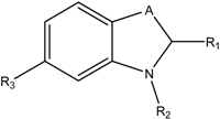
| Compound | A | R1 | R2 | R3 | Sag Hyal IC50 [µM], pH 5 |
|---|---|---|---|---|---|
| I |  | =S | -COCH3 | -H | 160 |
| II | -CH2- | -Ph (OSO2NH2) | -C10H21 | -OSO2NH2 | 11 |
| III | -O- | =S | -COCH2Ph | -H | 260 |
| IV | -O- | =S | -CO(CH2)2Ph | -H | 15 |
| V | -O- | =S | -CO(CH2)3Ph | -H | 110 |
Results and Discussion

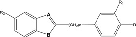
| Microwave heating | Conventional heating | |||||||||||||
|---|---|---|---|---|---|---|---|---|---|---|---|---|---|---|
| Comp No | A | B | n | R | R1 | R2 | Method* | Time (min) | Power (watt) | Yield (%) | Temp. (˚C) | Time (h) | Yield (%) | Mp. (˚C) |
| 1 | N | NH | 0 | H | H | H | A, D | 8 | 100 | 88 | 180 | 5 | 83 | 293 (17) |
| 2 | N | NH | 0 | F | H | H | A, E | 10 | 100 | 90 | 200 | 5 | 87 | 250 (18) |
| 3 | N | NH | 0 | OCH3 | H | H | A, E | 8 | 100 | 74 | 180 | 5 | 70 | 225 (17-19) |
| 4 | N | S | 0 | H | H | H | B, F | 5 | 50 | 90 | 30 | 2 | 70 | 115 (20) |
| 5 | N | S | 0 | OCH3 | H | H | B, F | 6 | 50 | 94 | 30 | 2 | 80 | 123 (21) |
| 6 | N | NH | 1 | H | H | H | A, G | 5 | 150 | 85 | Reflux | 7 | 50 | 182 (22) |
| 7 | N | NH | 1 | OCH3 | H | H | A, G | 5 | 150 | 80 | Reflux | 7 | 40 | 165 (23) |
| 8 | N | NH | 1 | H | OCH3 | H | C, H | 7 | 150 | 75 | Reflux | 8 | 35 | 152-155 |
| 9 | C-CH3 | NH | 0 | H | H | H | C, H | 3 | 50 | 80 | 100 | 2 | 70 | 88 (24) |
| 10 | C-CH3 | NH | 0 | H | H | CH3 | C, H | 3 | 50 | 78 | 100 | 2 | 70 | 105-108 |
| 11 | C-CH3 | N-CH3 | 0 | H | H | H | C, H | 4 | 50 | 70 | 100 | 3 | 50 | 69 (25) |
| 12 | C-Ph | NH | 0 | OH | H | H | C, H | 5 | 50 | 75 | 100 | 4 | 60 | 147 |
| Compound No | Inhibition [%] at 100 μM | IC50 [μM] | ||
|---|---|---|---|---|
| pH7 | 3.5 | pH7 | 3.5 | |
| 1 | 6 | 0 | n.d | n.d |
| 2 | 2 | 0 | n.d | n.d |
| 3 | 3 | 0 | n.d | n.d |
| 4 | 2 | 2 | n.d | n.d |
| 5 | 10 | 0 | n.d | n.d |
| 6 | 13 | 5 | n.d | n.d |
| 7 | 15 | 5 | n.d | n.d |
| 8 | 16 | 11 | n.d | n.d |
| 9 | 17 | 14 | n.d | n.d |
| 10 | 16 | 8 | n.d | n.d |
| 11 | 16 | 9 | n.d | n.d |
| 12 | 48 | 42 | 107 | 107 |
| Ascorbic acid palmitate | 99% | 99% | 18 | 8 |
Experimental
General
Chemistry
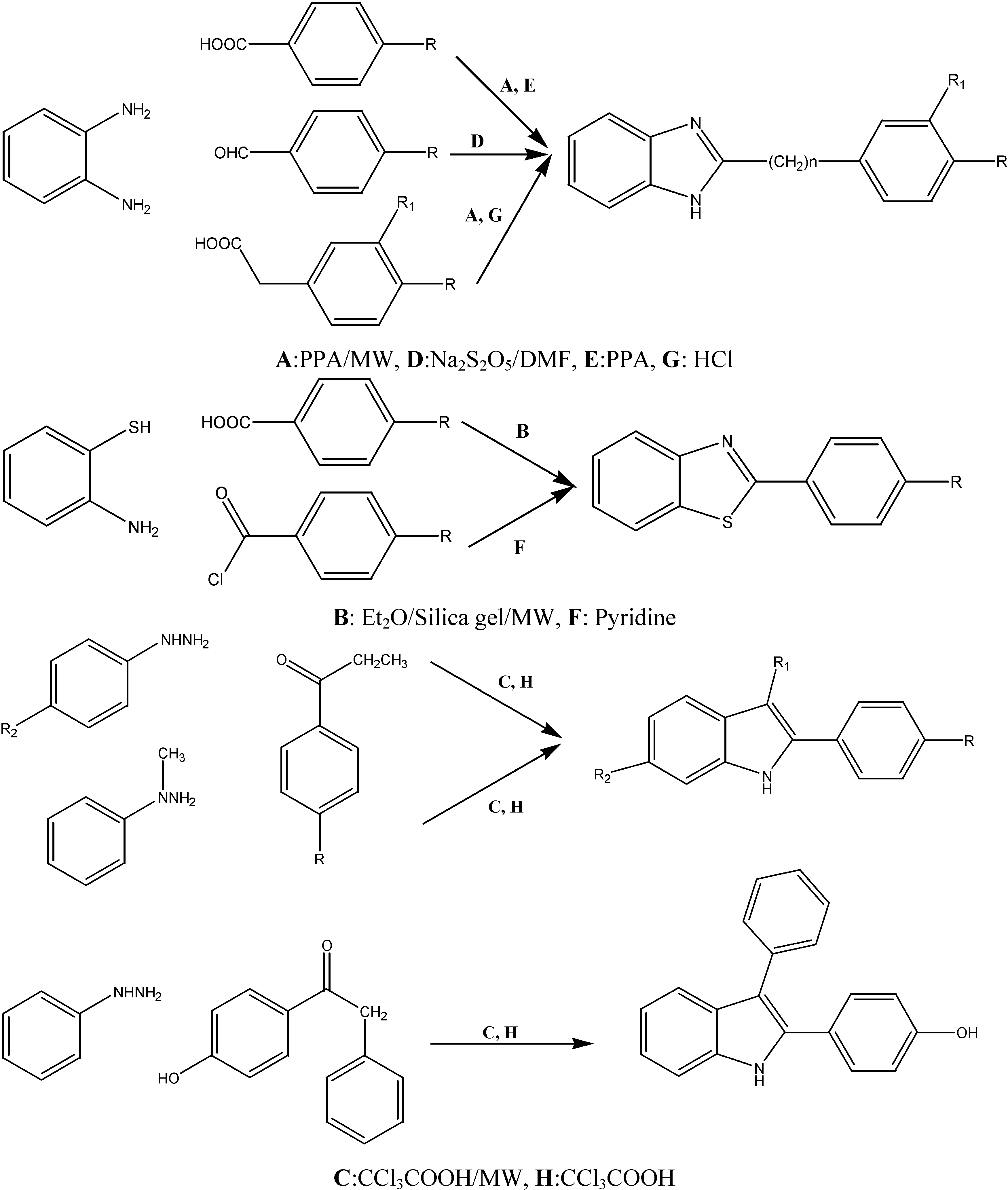
General method for the synthesis of 2-aryl-1H-benzimidazoles, 2-arylbenzoxazole and 2-arylindole derivatives
Microwave irradiation conditions:
Classical conditions:
Characterization data
Assays for the measurement of hyaluronidase activity
Stains-all-assay

Morgan-Elson Assay
Acknowledgements
References and Notes
- West, D.C.; Kumar, S. The Effect of Hyaluronate and its oligosaccharides on endothelial cell proliferation and monolayer integrity. Exp. Cell Res. 1989, 183, 179–196. [Google Scholar] [CrossRef]
- Auvinen, P.; Tammi, R.; Parkkinen, J.; Tammi, M.; Ågren, U.; Johansson, R.; Hirvikoski, P.; Ekselinen, M.; Kosma, V.M. Hyaluronan in peritumoral stroma and malignant cells associates with breast cancer spreading and predicts survival. Am. J. Pathol. 2000, 156, 342–347. [Google Scholar]
- Girish, S.; Kemparaju, K. The magic glue hyaluronan and its eraser hyaluronidase: A biological overview. Life Sci. 2007, 80, 1921–1943. [Google Scholar] [CrossRef]
- Lokeshwar, V.B.; Rubinowicz, D.; Schroeder, G.L.; Forgacs, E.; Minna, J.D.; Block, N.L.; Nadji, M.; Lokeshwar, B.L. Stromal and epithelial expression of tumor markers hyaluronic acid and HYAL1 hyaluronidase in prostate cancer. J. Biol. Chem. 2001, 275, 11922–11932. [Google Scholar]
- Liu, D.; Pearlman, E.; Diaconu, E.; Guo, K.; Mori, H.; Haqqi, T.; Markowitz, S.; Wilson, J.; Sy, M.-S. Expression of hyaluronidase by tumor cells induces angiogenesis in vivo. Proc. Natl. Acad. Sci. USA 1996, 93, 7832–7837. [Google Scholar] [CrossRef]
- Trochon, V.; Blot, E.; Cymbalista, F.; Engelmann, C.; Tang, R.-P.; Thomdis, A.; Vasse, M.; Soria, J.; Lu, H.; Soria, C. Apigenin inhibits endothelial-cell proliferation in g2/m phase whereas it stimulates smooth-muscle cells by inhibiting p21 and p27 expression. Int. J. Cancer 2000, 85, 691–696. [Google Scholar] [CrossRef]
- Herbst, R.S.; Madden, T.L.; Tran, H.T.; Blumenschein, G.R.; Meyers, C.A., Jr.; Seabrooke, L.F.; Khuri, F.R.; Puduvalli, V.K.; Allgood, V.; Fritsche, H.A.; Hinton, L., Jr.; Newman, R.A.; Crane, E.A.; Fossella, F.A.; Dordal, M.; Goodin, T.; Hong, W.K. Safety and pharmacokinetic effects of TNP-470, an angiogenesis inhibitor, combined with paclitaxel in patients with solid tumors: evidence for activity in non–small-cell lung cancer. J. Clin. Oncol. 2002, 20, 4440–4447. [Google Scholar] [CrossRef]
- Sridhar, S.S.; Shepherd, F.A. Targeting angiogenesis: a review of angiogenesis inhibitors in the treatment of lung cancer. Lung Cancer 2003, 42, 581–591. [Google Scholar]
- Jeong, S.-J.; Higuchi, R.; Ono, M.; Kuwano, M.; Kim, Y.-C.; Miyamoto, T. cis-Hinokresinol, a norlignan from anmemarrhena asphodeloides, inhibits angiogenic responce in vitro and in vivo. Biol. Pharm. Bull. 2003, 26, 1721–1724. [Google Scholar] [CrossRef]
- Laird, A.D.; Vajkoczy, P.; Shawver, L.K.; Thurnher, A.; Liang, C.; Mohammadi, M.; Schlessinger, J.; Ullrich, A.; Hubbard, S.R.; Blake, R.A.; Fong, A.T.; Strawn, L.M.; Sun, L.; Tang, C.; Hawtin, R.; Tang, F.; Shenoy, N.; Hirth, K.P.; McMahon, G.; Cherrington, J.M. SU6668 is a potent antiangiogenic and antitumor agent that induces regression of established tumors. Cancer Res. 2000, 60, 4152–4160. [Google Scholar]
- Jeong, S.J.; Ahn, N.H.; Kim, Y.C.; Inagaki, M.; Miyamoto, T.; Higuchi, R. Norlignans with hyaluronidase inhibitory activity from Anemarrhena asphodeloides. Planta Medica 1999, 65, 367–368. [Google Scholar] [CrossRef]
- Salmen, S. Inhibitors of Bacterial and Mammalian Hyaluronidases: Synthesis and Structure-Activity Relationships. Ph.D. Thesis, University of Regensburg Press, Regensburg, Germany, 2003. [Google Scholar]
- Botzki, A.; Salmen, S.; Bernhardt, G.; Buschauer, A.; Dove, S. Structure-based design of bacterial hyaluronan lyase inhibitors. QSAR Comb. Sci. 2005, 24, 458–469. [Google Scholar] [CrossRef]
- Rigden, D.J.; Botzki, A.; Nukui, M.; Mewbourne, R.B.; Lamani, E.; Braun, S.; von Angerer, E.; Bernhardt, G.; Dove, S.; Buschauer, A.; Jedrzejas, M.J. Design of new benzoxazole-2-thione-derived inhibitors of Streptococcus pneumoniae hyaluronan lyase: structure of a complex with a 2-phenylindole. Glycobiology 2006, 16, 757–765. [Google Scholar] [CrossRef]
- Olgen, S.; Kaeßler, A.; Nebioglu, D.; Jose, J. New potent indole derivatives as hyaluronidase inhibitors. Chem. Biol. Drug. Des. 2007, 70, 547–551. [Google Scholar] [CrossRef]
- Kaessler, A.; Algul, O.; Jose, J. A microplate based screening of benzimidazole derivatives on hyaluronidase inhibition at pH 7 and 3.5. Lett. Drug Des. Discov. 2007, 4, 562–569. [Google Scholar] [CrossRef]
- Du, L.-H.; Wang, Y.-G. A rapid and efficient synthesis of benzimidazoles using hypervalent iodine as oxidant. Synthesis 2007, 5, 675–678. [Google Scholar]
- Getvoldsen, G.; Fredriksson, A.; Elander, N.; Stone-Elander, S. Microwave-assisted cyclocondersation of 1,2-diaminobenzene with [4-18F]fluorobenzoic acid:microwave synthesis of 2-([4-18F]fluorophenyl)benzimidazole. J. Label. Compd. Radiopharm. 2004, 47, 139–145. [Google Scholar] [CrossRef]
- Arslan, H.; Algul, O. Vibrational spectrum and assignments of 2-(4-methoxyphenyl)-1H-benzo[d]imidazole by ab initio Hartree–Fock and density functional methods. Spectrochim. Acta A: Mol. Biomol. Spectrosc. [CrossRef]
- Kodomari, M.; Tamaru, Y.; Aoyama, T. Solvent-free synthesis of 2-aryl and 2-alkylbenzothiazoles on silica gel under microwave irradiation. Synth. Commun. 2004, 3029–3036. [Google Scholar] [CrossRef]
- Arslan, H.; Algul, O. Synthesis and Ab initio/DFT studies on 2-(4-methoxyphenyl)benzo- [d]thiazole. Int. J. Mol. Sci. 2007, 8, 1–18. [Google Scholar] [CrossRef]
- Charton, J.; Girault-Mizzi, S.; Debreu-Fontaine, M.-A.; Foufelle, F.; Hainault, I.; Bizot-Espiard, J.-G.; Caignard, D.-H.; Sergheraert, C. Conversion of sterically hindered diacylated 1,2-phenylenediamines into 2-substituted benzimidazoles. Chem. Pharm. Bull. 2005, 53, 492–497. [Google Scholar] [CrossRef]
- Gumus, F.; Demirci, A.B.; Özden, T.; Eroglu, H.; Diril, N. Synthesis, characterization and mutagenicity of new cis-[Pt(2-substituted-benzimidazole)2Cl2]complexes. Pharmazie 2003, 58, 303–307. [Google Scholar]
- Matsumoto, K.; Tanaka, A.; Yukio, I.; Hayashi, N.; Toda, M.; Bulman, R.A. Fischeer Indole Synthesis in the Absence of a Solvent. Heterocycl. Commun. 2003, 9, 9–12. [Google Scholar]
- Katritzky, A.R.; Fan, W.-Q.; Akutagawa, K.; Wang, J. Carbon dioxide: A reagemt for the simultaneous protection of nucleophilic and the activation of alternative locations to electrophilic attack. 16. A novel synthetic method for the side-chain functionalization of N-methyl-o-toluidine and for the preparation of 2-substituted N-methylindoles. Heterocycles 1990, 30, 407–414. [Google Scholar] [CrossRef]
- Benchetrit, L.C.; Pahuja, S.L.; Gray, E.D.; Edstrom, R.D. A sensitive method for the assay of hyaluronidase activity. Anal. Biochem. 1977, 79, 431–37. [Google Scholar] [CrossRef]
- Stern, R.; Jedrzejas, M. Hyaluronidases: their genomics, structures, and mechanisms of action. J. Chem. Rev. 2006, 106, 818–839. [Google Scholar] [CrossRef]
- Botzki, A.; Rigden, D.J.; Braun, S.; Nukui, M.; Salmen, S.; Hoechstetter, J.; Bernhardt, G.; Dove, S.; Jedrzejas, M.J.; Buschauer, A. L-Ascorbic acid 6-hexadecanoate, a potent hyaluronidase inhibitor: X-Ray structure and molecular modeling of enzyme-inhibitor complexes. J. Biol. Chem. 2004, 279, 45990–45997. [Google Scholar] [CrossRef] [Green Version]
- Sample Availability: Contact the authors.
© 2008 by MDPI (http://www.mdpi.org). Reproduction is permitted for noncommercial purposes.
Share and Cite
Algul, O.; Kaessler, A.; Apcin, Y.; Yilmaz, A.; Jose, J. Comparative Studies on Conventional and Microwave Synthesis of Some Benzimidazole, Benzothiazole and Indole Derivatives and Testing on Inhibition of Hyaluronidase. Molecules 2008, 13, 736-748. https://doi.org/10.3390/molecules13040736
Algul O, Kaessler A, Apcin Y, Yilmaz A, Jose J. Comparative Studies on Conventional and Microwave Synthesis of Some Benzimidazole, Benzothiazole and Indole Derivatives and Testing on Inhibition of Hyaluronidase. Molecules. 2008; 13(4):736-748. https://doi.org/10.3390/molecules13040736
Chicago/Turabian StyleAlgul, Oztekin, Andre Kaessler, Yagmur Apcin, Akin Yilmaz, and Joachim Jose. 2008. "Comparative Studies on Conventional and Microwave Synthesis of Some Benzimidazole, Benzothiazole and Indole Derivatives and Testing on Inhibition of Hyaluronidase" Molecules 13, no. 4: 736-748. https://doi.org/10.3390/molecules13040736