Article

Structure Determination of β-Glucans from Ganoderma lucidum with Matrix-assisted Laser Desorption/ionization (MALDI) Mass Spectrometry

by
Wei-Ting Hung
1,
Shwu-Huey Wang
2,
Chung-Hsuan Chen
1 and
Wen-Bin Yang
1,*
1
Genomics Research Center, Academia Sinica, No. 128, Academia Road Section 2, Nan-Kang, Taipei 11529, Taiwan
2
Instrument Center, Taipei Medical University, Taipei 11031, Taiwan
*
Author to whom correspondence should be addressed.
Molecules 2008, 13(8), 1538-1550; https://doi.org/10.3390/molecules13081538
Submission received: 26 May 2008 / Revised: 21 June 2008 / Accepted: 25 July 2008 / Published: 3 August 2008

Abstract

:
A novel method that uses matrix-assisted laser desorption/ionization (MALDI) mass spectrometry to analyze molecular weight and sequencing of glucan in Ganoderma lucidum is presented. Thus, β-glucan, which was isolated from fruiting bodies of G. lucidum, was measured in a direct and fast way using MALDI mass spectrometry. In addition, tandem mass spectrometry of permethylated glucans of G. lucidum, dextran, curdlan and maltohexaose were also pursued and different fragment patterns were obtained. The G. lucidum glucan structure was determined and this method for linkage analysis of permethylated glucan has been proven feasible.

Introduction

Ganoderma species (a group of medicinal fungi), including Ganoderma lucidum (Reishi in Japan or Ling-Zhi in China), has been used for a long time in China to prevent and treat various human diseases. Matrix-assisted laser desorption/ionization (MALDI) has been developed as a successful tool for the analysis of biopolymers and macromolecules [1,2,3]. The ions produced by MALDI are predominantly singly charged molecular ions so that simple mass spectra can be used for distribution analysis of neutral polysaccharides. In recent years, several mass spectrometric studies have been focused on the analysis of polysaccharide materials or oligosaccharides [4,5,6]. Direct detection of saccharides in sulfuric acid/phenol has been commonly used before, but it can not derive information on molecular weights of different saccharides and the differences among pentoses, hexoses and unusual sugars. Gel permeation chromatography (GPC) has been used for measurements of the size of polysaccharides by comparison of results with appropriate polysaccharide standards [7]. Nevertheless, the resolution of GPC is generally poor and the process is very time consuming. Since most polysaccharides are polydispersable to molecular weight, GPC is not a direct method for accurate mass measurements. On the other hand, MALDI-MS is practical for more qualitative and mass determination. In our previous study [8,9], we found that non-derivatized and derivatized polysaccharides were successfully analyzed by a straightforward MALDI mass spectrometry when 2’,4’,6’-trihydroxyacetophenone (THAP) or 2,5-dihydroxybenzoic acid (2,5-DHB) was used as matrix. In this work, we measured both non-derivatized and derivatized glucans from G. lucidum polysaccharides. In addition, comparisons of molecular weight and structural information are made between the G. lucidum polysaccharides and glucan standards from dextran, curdlan and starch using MALDI-TOF (time-of-flight) mass spectrometry. The linkage information of the permethylated oligomer ions from G. lucidum was established through the use of on-line MS/MS experiments.
Polysaccharides extracted from G. lucidum (also known as Ling-Zhi) have been widely used and studied for their immunomodulating and anti-cancer potential [10,11,12,13,14]. The main components of G. lucidum polysaccharides are (1→3) and (1→6)-β-d-glucan. β-d-Glucan is a carbohydrate polymer with chains of glucose molecules linked together by β-glycosidic linkages [15,16,17]. Up to now, β-glucan isolated from G. lucidum has not been studied using the MALDI-MS method. In this work, we have analyzed not only non-derivatized G. lucidum polysaccharides, but also the permethylated derivatized β-glucan.

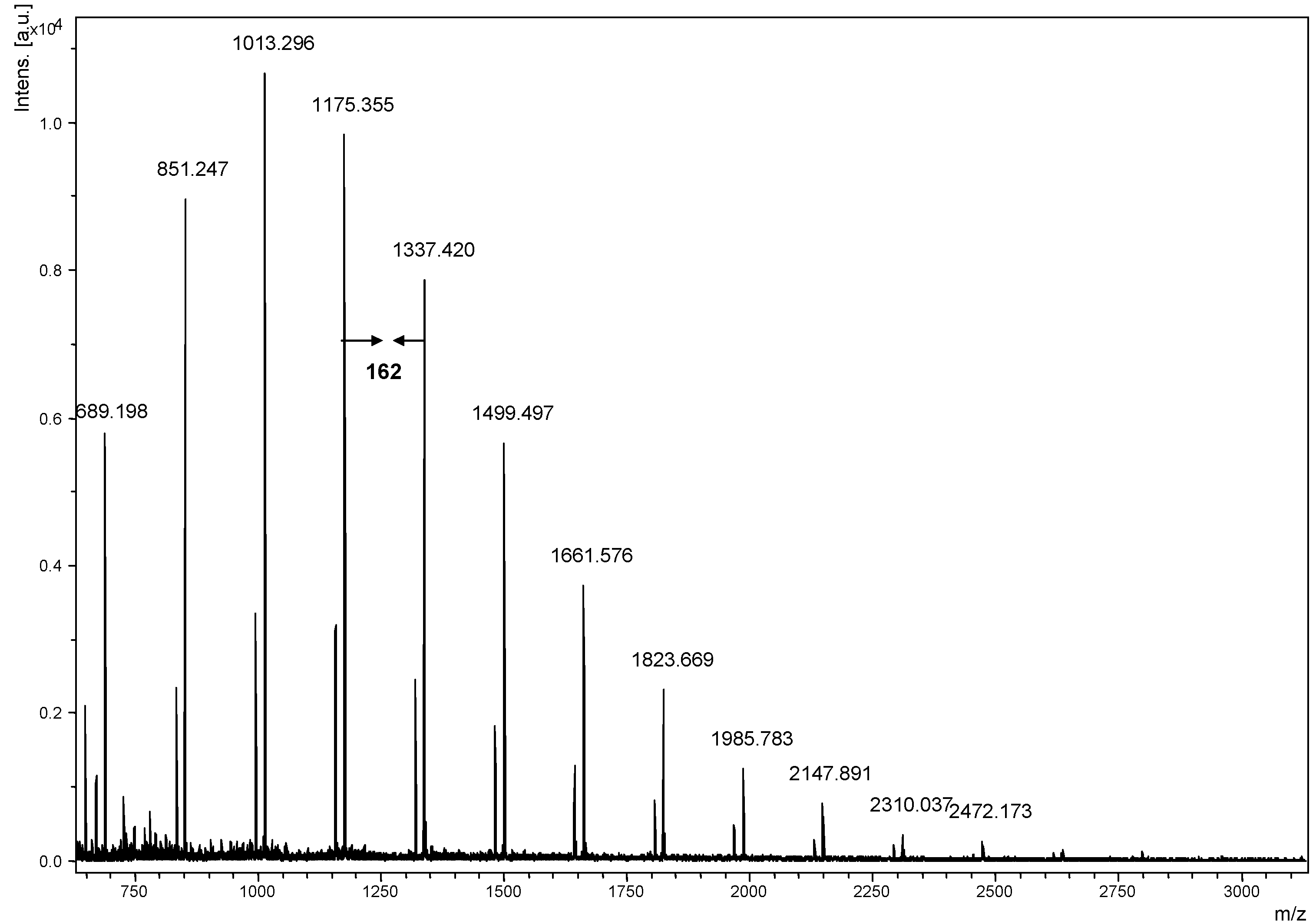
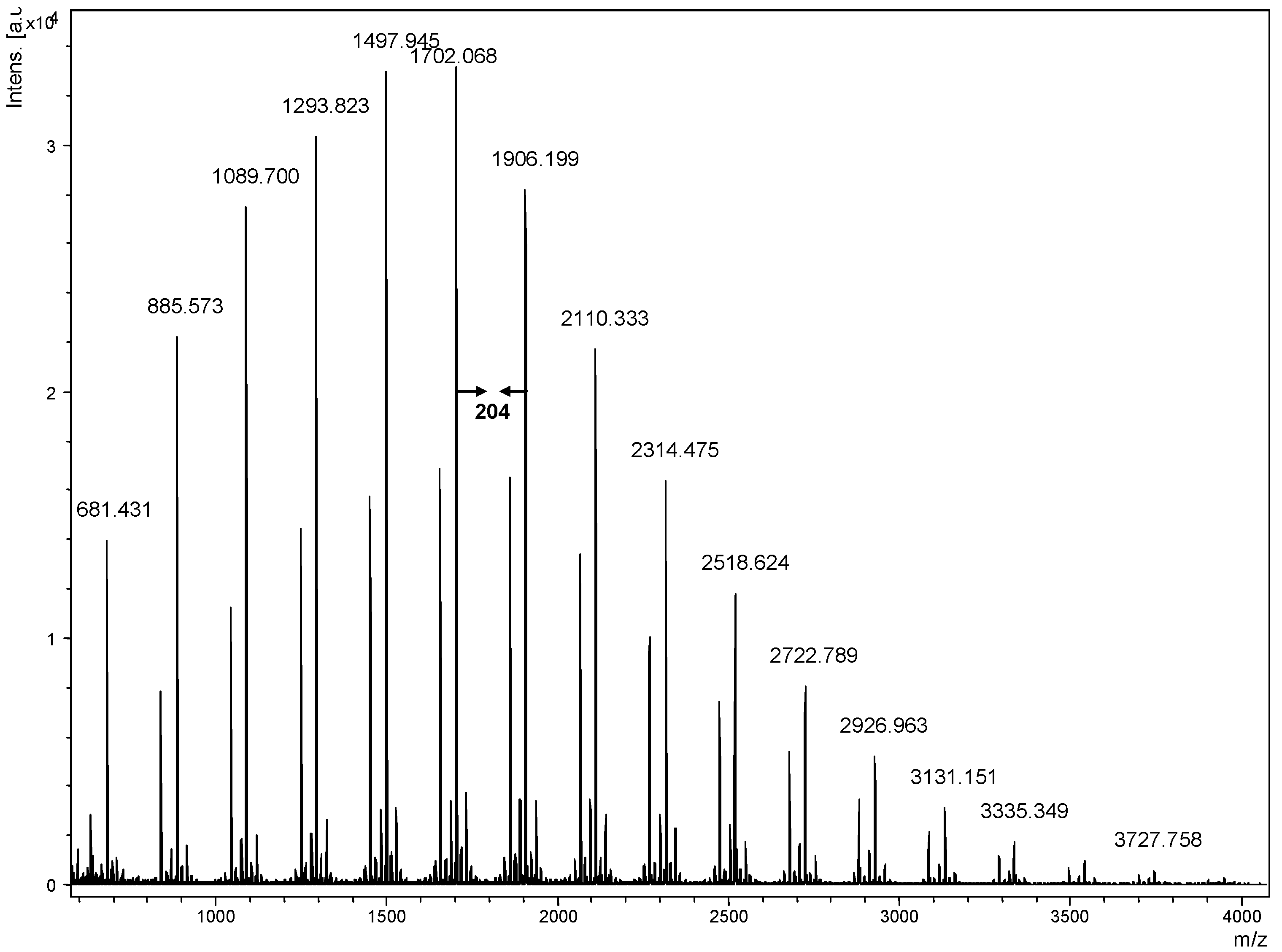
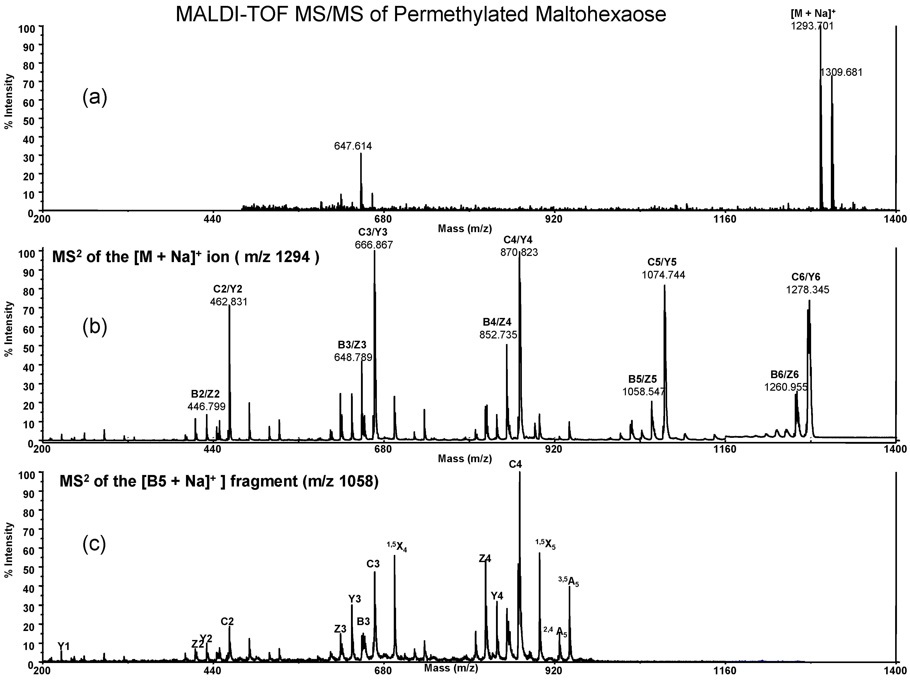
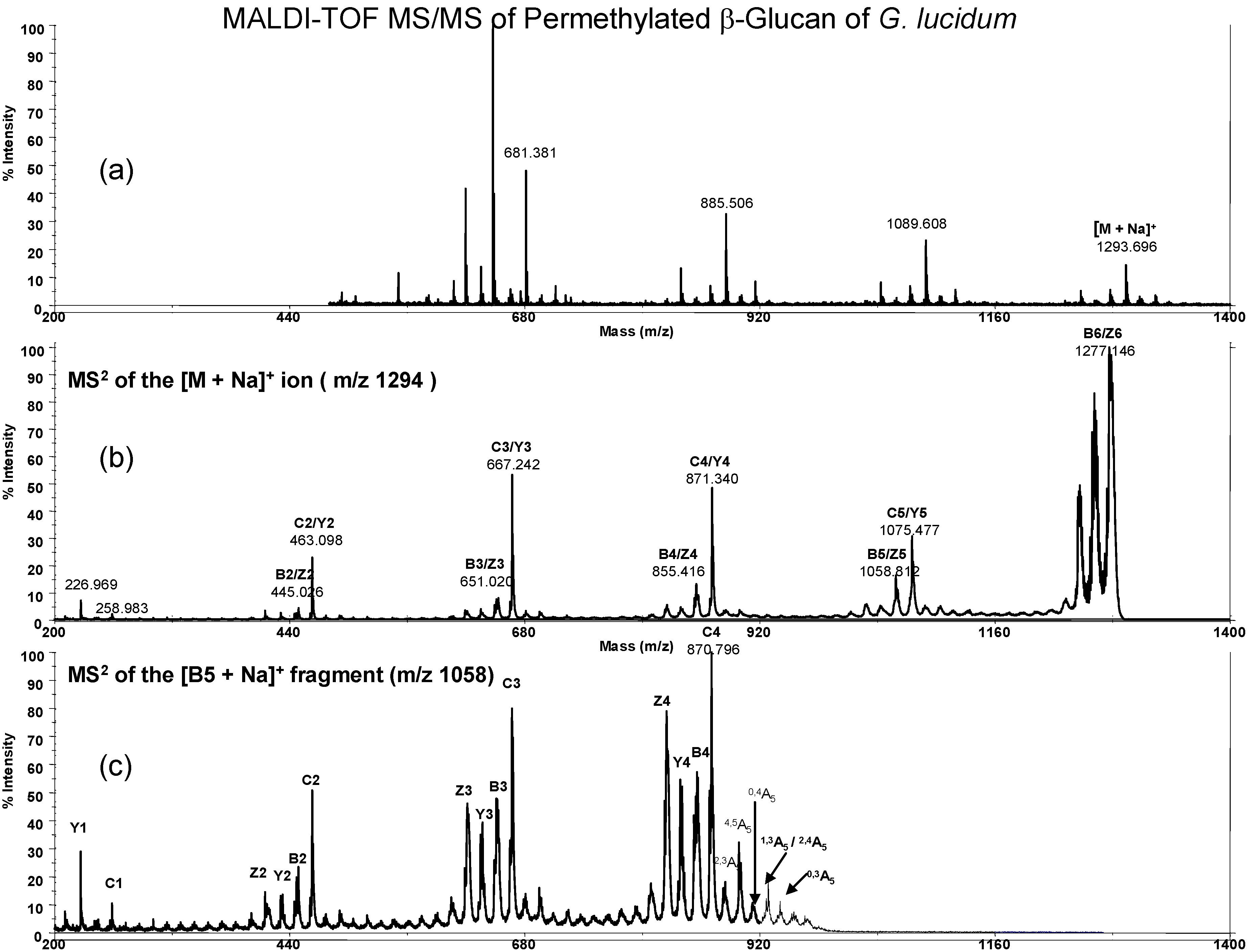
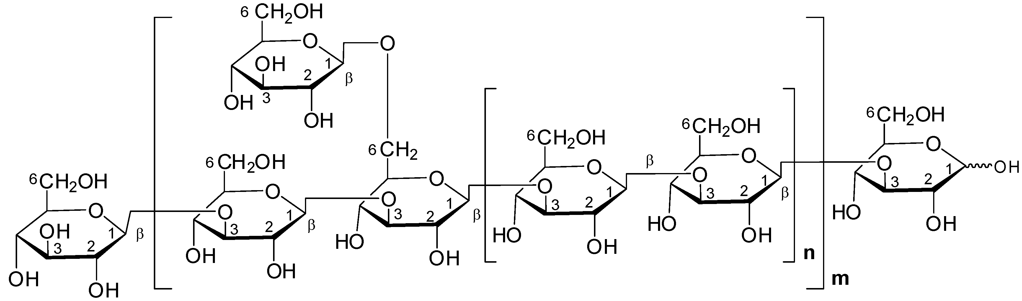
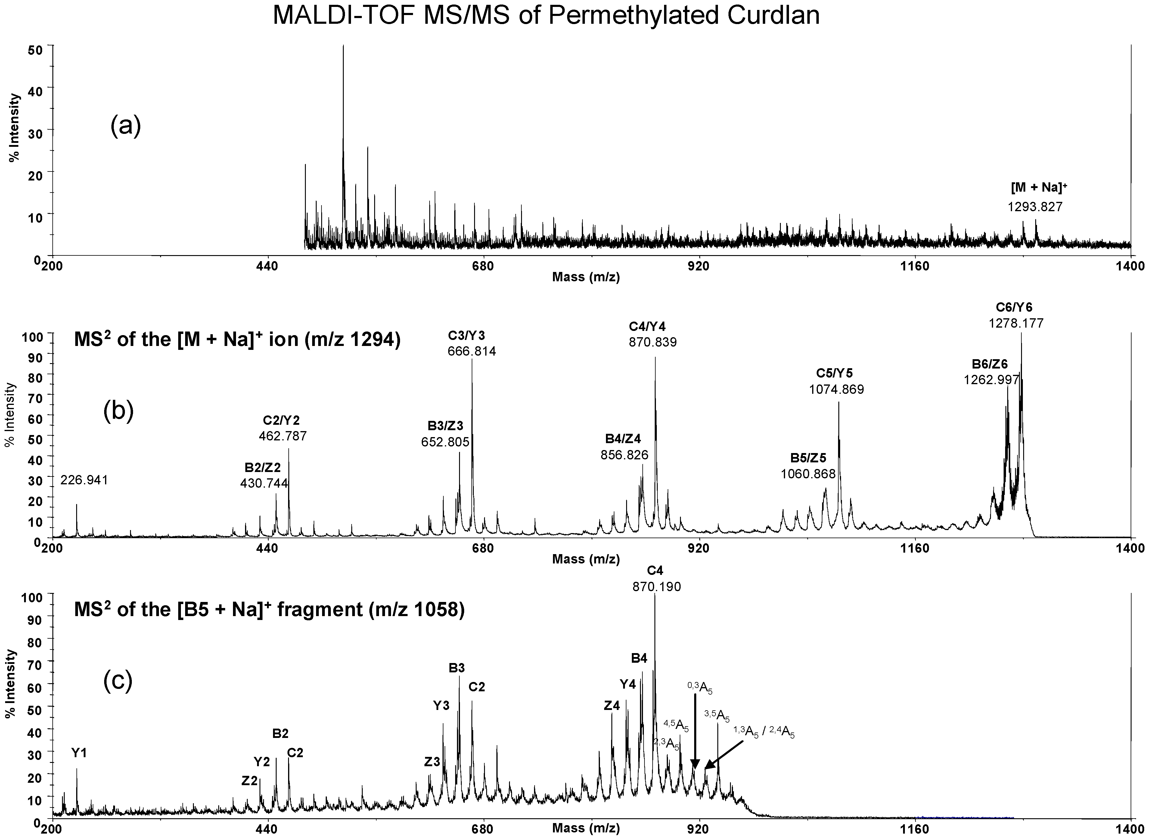
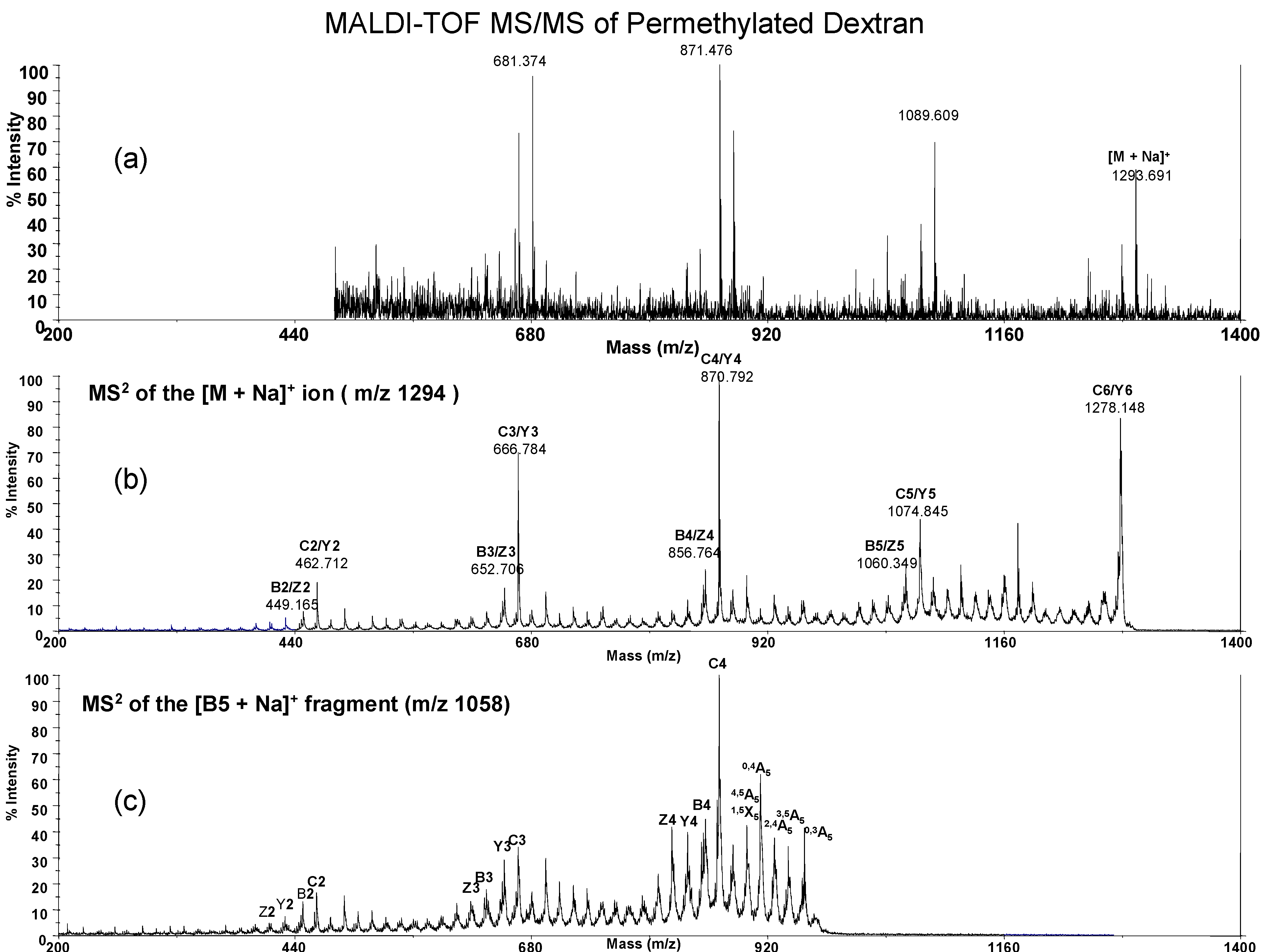
Results and Discussion

Our goal in this study was to examine the structure of glucans from G. lucidum. The ground fruiting body of G. lucidum was extracted by hot water to release polysaccharides and the crude polysaccharides were further hydrolyzed by a mild acid degradation (2M TFA, 50 oC, 2 hrs) to obtain a low molecular weight and water soluble polysaccharide fraction. In order to investigate the sugar composition of G. lucidum polysaccharide, this polysaccharide fraction was hydrolyzed into monosaccharides by acidic hydrolysis (4M TFA, 121 oC, 6 hrs) and high performance anion exchange chromatography with pulsed amperometric detection (HPAEC-PAD) was used to measure its sugar composition. We found only glucose present in this fraction of G. lucidum. By NMR analysis, we found that these novel glucans have a β-(1→3)/β-(1→6)-configuration at the glycosidic bonds (data not shown). This result is in agreement with the previous research in which polysaccharides are isolated from G. lucidum [18,19]. However, here we used MALDI-TOF mass spectrometry to determine G. lucidum glucan. For the analysis of native polysaccharides in our previous work [8,9] we observed THAP and 2,5-DHB are good matrices that substantially enhanced signals for native pullulans standard in MALDI mass spectrometry. In this study we also use both matrices for the mass spectrometry analysis. Both positive and negative ions of polysaccharides were obtained. In this study, neutral carbohydrates were found to prefer the positive ion mode, due to the attachment of alkali metal ions. The G. lucidum glucans were observed as sodiated ions ([Glcn + Na]+) and the corresponding mass spectrum is given in Figure 1. The mass of the singly charged molecular ion were calculated as 162.14n + 22.990 Da and 162.14n + 22.990 + 18.015 (mass of reducing end residue) Da respectively, where n is the number of glucose units. For example, if n = 10, the mass of the corresponding glucan should be 1662.4 Da respectively. The mass region of ion peaks was observed with a peak-to-peak mass difference of 162.1 Da, consistent with the repeating unit of the β-(1→3)-glucan. Similar observations have been reported previously [7,20,21] and were attributed to the fragmentation of the high mass polysaccharides. The MALDI-TOF mass spectrum in Figure 1 showed that G. lucidum glucans are composed of a mixture of glucose polymers with molecular weights around 689.2 ~ 2634.2 Da. This is the first report on measuring the glucan having DP = 4 ~ 16 in G. lucidum with mass spectrometry.
Figure 1. MALDI-TOF mass spectrum of Ganoderma lucidum glucans using 2,5-DHB as matrix. The mass difference of 162.1 Da between neighboring peaks is observed.
Molecules 13 01538 g001
The known glucans of different molecular weights such as dextran (1→6), amylose (1→4) and curdlan (1→3) have been analyzed by MALDI for molecular weight determination [20,21,22,23]. However, up to now the chemical linkages between these glucans still have not been determined by mass spectrometry. In this work, we determined the sequences of oligosaccharides and polysaccharides. We prepared the glucan samples by water extraction and a size exclusion chromatographic method to obtain low molecular weight oligosaccharides as well as polysaccharides and these glucans were derivatized by permethylation. Permethylation produced stronger sodium attached ions than non-methylated glucans for maltohexose, dextran, curdlan and G. lucidum. In Figure 2, we measured permethylated G. lucidum glucans using 2,5-DHB as a matrix. The G. lucidum glucans were observed as sodium attached ions and molecular masses were calculated as 219.13 (a terminal sugar) + 204.13n + 22.99 Da and 219.13 + 204.13n + 31.02 (mass of reducing end residue) + 22.99 Da, respectively, where n is the number of 2,4,6-trimethyl glucosyl unit. For example, if a decasaccharide, the mass of the corresponding glucan should be 2079.3 Da ([2,3,4,6-tetramethylglucosyl-(2,4,6-trimethyl glucosyl)9 + Na]+) and 2110.3 Da ([2,3,4,6-tetramethylglucosyl-(2,4,6-trimethyl glucosyl)9 + 31.02 + Na]+) respectively. The mass region of ion peaks was observed with a peak-to-peak mass difference of 204.1 Da consistent with the repeating unit of the permethylated β-glucan from 681.4 ~ 3727.8 Da.
Figure 2. MALDI-TOF mass spectrum of permethylated G. lucidum glucans with 2,5-DHB as matrix. A peak-to-peak mass difference of 204.1 Da is observed.
Molecules 13 01538 g002
In order to obtain sequencing information about G. lucidum glucans we used the tandem mass (MS/MS) method to get linkage information. MS2 spectra for various oligosaccharides such as dextran (1→6), oligomaltose (1→4), curdlan (1→3) and G. lucidum polysaccharides (1→3/1→6) were obtained to show different fragmented ions for linkage assignment. In the past, Creaser et al. [24] published the structural analysis of non-derivative maltoheptaose using the MALDI-TOF MSn method. They obtained a strong sodium attached singly charged ion peak at m/z 1175.3, which was subsequently analyzed by a tandem mass spectrometry to yield a number of fragment ions. Here, we investigated MALDI of permethylated maltohexaose and the peak showed at m/z 1293.7 as [M + Na]+ ion (Figure 3a). The MS/MS spectra of the [M + Na]+ ion of permethylated maltohexaose at m/z 1293.7 are shown in Figure 3b. The spectra were dominated by peaks resulting from cleavage at glycosidic bonds, giving the C/Y ion series (m/z 1278.3, 1074.7, 870.8, 666.9, 462.8) and a less intense series of B/Z ions with 18 Da lower (m/z 1260.9, 1058.8, 852.7, 648.8, 446.8), respectively. The nomenclatures for the fragment ions were introduced by Domon and Costello [25]. Although reducing terminal ions (C ions) are often more abundant, it is the B ions that are more informative when probed by MS/MS analysis. Pyranose unsaturation (B ions) coupled with metal ion chelation appears to lower energy barriers to ring rupture via a classical retro Diels-Alder decomposition reaction to give cross-ring cleavages (A-type or X-type fragments) [26,27]. Figure 3c showed the MS2 of the B5 ion, with m/z at 1058.8 and the major cross-ring fragment ions at m/z 1058.8 ~ 870.8 (B5~C4 region) are 941.0 (3,5A5), 927.0 (2,4A5) and 898.9 (1,5X5), respectively. We found a high intensity of 3,5A5 ion to indicate a 1→4 linkage while 1→3 linkage is absent. The 2,4A5 ion is present of only for the 1→4 or 1→3 linkage but not in 1→6 linkage and 1,5X5 fragment ions are X type ion with the similar ions also found for 1,5X4, 1,5X3 which also appear in MS/MS spectrum of permethylated maltohexaose at m/z 1293.7 (figure 3b). This method got a similar mode of fragment ions when probed B ions at B6, B4, B3 and B2 by MS2 analysis. Although the fragment ions from B5 ion still have not enough information for identification of linkage analysis between this region of ring fragments, its spectrum is quite different and can be used for the comparison of various glucans.
Figure 3. MALDI-TOF MS/MS of permethylated maltohexaose in 2,5-DHB as matrix. The ion of maltohexaose (m/z = 1293.8, [M + Na]+) shown in figure 3a. MS2 at the permethylated maltohexaose ion ([M + Na]+, m/z = 1293.8) shown in 3b and MS2 at the B5 sodiated fragment ion (m/z = 1058.8) shown in figure 3c. The major cross-ring fragment ions at B5~C4 region (m/z = 1058.8 ~ 870.8) are 941.0 (3,5A5), 927.0 (2,4A5) and 898.9 (1,5X5).
Molecules 13 01538 g003
Here we also measured permethylated G. lucidum glucan, which is derived from acidic degradation (Figure 4a). And MS2 at m/z = 1293.7 of this permethylated G. lucidum hexasaccharide (Figure 4b) was dominated by peaks resulting from cleavage at glycosidic bonds, giving the C/Y ion series and a less intense series of B/Z ions, respectively, which are same as the one mentioned in the above (Figure 3b). The MS2 spectrum of permethylated G. lucidum glucan B5 ion at m/z 1058.8 is shown in Figure 4c. The major fragment ions are the Y and C ions (1277.1, 1075.5, 871.3, 667.2, 463.1 m/z), as usual. However, the B5 ions differ between the permethylated G. lucidum hexasaccharide (Figure 4c) and permethylated maltohexaose (Figure 3c). The fragmented ions from m/z at 1058.8 ~ 871.3 are 940.7 (0,3A5), 928.7 (1,3A5/2,4A5), 912.3 (0,4A5), 898.7 (1,5X5) and 883.1 (2,3A5), respectively. For the ions with m/z at 912.3 (0,4A5) and 898.7 (4,5A5), it indicates that G. lucidum glucan has 1→6 linkage and 883.1 (2,3A5) indicates out the presence of 1→3 linkage. From Figure 4, we confirm that this methylated glucan has 1→3 and 1→6 linkage between glycosidic bonds.
Figure 4. MALDI-TOF MS/MS of permethylated G. lucidum glucans. The ion of permethylated G. lucidum hexasaccharide (m/z = 1293.7, [M + Na]+) shown in figure 4a. MS2 at the hexasaccharide ion ([M + Na]+, m/z = 1293.8) shown in 4b and MS2 at the B5 sodiated fragment ion (m/z = 1058.8) shown in figure 4c. The major cross-ring fragment ions at B5~C4 region are 940.7 (0,3A5), 928.7 (1,3A5/2,4A5), 912.3 (0,4A5), 898.7 (1,5X5/4,5A5) and 883.1 (2,3A5), respectively.
Molecules 13 01538 g004
Figure 5. Possible repeating units of G. lucidum glucans.
Molecules 13 01538 g005
Figure 6. MALDI-TOF MS/MS of permethylated curdlan in 2,5-DHB as matrix. Shown are the hexasaccharide ion (m/z = 1293.8, [M + Na]+) (6a), MS2 at the hexasaccharide ion ([M + Na]+, m/z = 1293.8) (6b) and MS2 at the B5 sodiated fragment ion (m/z = 1058.8) (6c). The major cross-ring fragment ions at B5~C4 region are 941.0 (0,3A5), 927.9 (1,3A5/2,4A5), 911.0 (0,4A5), 898.9 (1,5X5) and 883.1 (2,3A5).
Molecules 13 01538 g006
For comparison we also prepared permethylated low molecular weight glucan from curdlan (Figure 6) and dextran (Figure 7). The MS/MS spectrum as its B5 ion (m/z 1058.8) also gave the cross-ring fragment ions for glucan structural determination. In brief, permethylated G. lucidum glucan displayed similar cross-ring fragment ions with curdlan at 940.7 (0,3A5), 928.7 (1,3A5/2,4A5), 912.3 (0,4A5), 898.7 (1,5X5/4,5A5) and 883.1 (2,3A5) and G. lucidum glucan also has a weaker signal at 898.7 m/z (4,5A5) that is the same as dextran 4,5A5 ion. In this study we did not obtain the ratio of 1→3/1→6 linkage of G. lucidum glucan. However, the real glucan structure in G. lucidum contains trace amount of 1→6 side chains, which were previously identified by NMR and GC/MS analysis. The mass spectrometric study for quantification of branched glucan still faces challenges.
Here we focus on B type ions for linkage analysis, since the sugar ring cleavage is easier so that fragment ions are strong enough for the analysis. The ions from permethylated hexasaccharide (m/z = 1293.7) to disaccharide (m/z = 446.7) are examined for the study of different fragmented ions on these glucans. Based on the MALDI-TOF mass analysis we determined not only the molecular weights of glucans, but also the sugar sequences. The results shown that G. lucidum glucan has a branched (1→3/1→6)-glucan linkage structure and the proposed chemical structure of G. lucidum glucan is as shown in Figure 5.
Figure 7. MALDI-TOF MS/MS of permethylated dextran glucans. The hexasaccharide ion (m/z = 1293.8, [M + Na]+) is shown in 7a. MS2 at the hexasaccharide ion ([M + Na]+, m/z = 1293.8) is shown in 7b and MS2 at the B5 sodiated fragment ion (m/z = 1058.8) is shown in 7c. The major cross-ring fragment ions at B5~C4 region are 957.3 (0,3A5), 940.8 (3,5A5), 925.9 (1,4A5), 912.9 (0,4A5) and 898.9 (4,5A5/1,5X5) respectively.
Molecules 13 01538 g007

Conclusions

The use of MALDI-TOF mass spectrometry for analyzing polysaccharides of G. lucidum is successfully demonstrated. The results showed that G. lucidum has an abundance of glucan and the chemical structure involves (1→3/1→6) linkages at the glycosidic bonds. This is the first time a detailed analysis of the molecular weight and glycosidic bond linkages G. lucidum polysaccharides. The method can be applied to sequencing of polysaccharides from other natural products or synthetic organic chemicals in the future.

Experimental Section

Materials

Matrices of 2’,4’,6’-trihydroxyacetophenone (THAP) and 2,5-dihydroxybenzoic acid (2,5-DHB) were purchased from Sigma Chem. Co. and methanol, dimethyl sulfoxide, acetonitrile, methyl iodide, sodium hydride and trifluoroacetic acid were purchased from Merck & Co., Inc. Glucan from dextran, curdlan, oligomaltose were obtained from Sigma Chem. Co. All chemicals were analytical grade and used without further purification and double distilled water was used throughout polysaccharide extraction and purification. Glucan from ground fruiting body of G. lucidum was extracted by hot water and TFA degradation.

Instrumentation

MS spectra of the sodium adducts of oligosaccharides [M + Na]+ were acquired using an UltraflexTM TOF/TOF mass spectrometer (Bruker Daltonik GmbH, Bremen, Germany) with gridless ion optics under control of Flexcontrol 2.0. The instrument, equipped with the SCOUTTM high-resolution optics with X-Y multisample probe and gridless reflector, was used at a maximum accelerating potential of 25 kV and was operated in reflector mode for MS analysis. Ionization was accomplished with a 337-nm beam from a nitrogen laser with a repetition rate of 20 Hz. The output signal from the detector was digitized at a sampling rate of 2 GHz. The high-performance anion-exchange chromatography with a pulsed amperometric detection (HPAEC-PAD) for sugar composition analysis was performed on a Dionex BioLC system (Dionex, Sunnyvale, CA, USA) using a CarboPac PA10 analytical column (2 mm × 250 mm) and a CarboPac PA10 guard column (2 mm × 50 mm). GC-MS system (Thermo Fisher Scientific, Inc, Waltham, MA 024253, USA) with PolarisQ ion trap and BPX-70 column were used for alditol acetates and partially methylated alditol acetates analysis. The NMR spectra were recorded on a Bruker 500 MHz NMR spectrometer (Bruker BioSpin GmbH, Rheinstetten, Germany) with 5 mm Cryoprobe DCI 1H/13C.

Mass spectral analysis of glucans

For MALDI-MS analysis, the samples were prepared by standard dried-droplet preparation on stainless steel MALDI targets using 2,5-dihydroxybenzoic acid or 2’,4’,6’-trihydroxyacetophenone as matrix. The non-derivatized and derivatized (permethylation) oligo-, polysaccharides were mixed with 10 mM of 2,5-DHB or THAP in the ratio of 1:1. In MS mode, the spectra were accumulated in average of 2000–3000 shuts.

Preparation of glucans

The dried fruiting bodies of G. lucidum were ground and extracted with dd-water (double-distilled water) at 100 oC for 12 hr. The suspension was centrifuged (4000 rpm, 1 hr) to remove the insoluble materials, and the water-soluble crude polysaccharide was obtained (11%). The crude polysaccharide (0.5 g) was degraded into a low molecular weight glucan using a chemical method (2M TFA 50 oC, 2h) and then purified through a gel-filtration chromatography using a Sephadex G-15 column with dd-water as the eluent to obtain a glucan fraction (15~20%). Other low molecular weight glucans from dextran and curdlan were obtained by the similar method for G. lucidum glucan.

Composition analysis of G. lucidum glucan

The G. lucidum glucan (50 mg) was hydrolyzed with 4 M trifluoroacetic acid (TFA) at 121 ºC for 4 h. The mixture was cooled, and TFA was removed under reduced pressure. The acid hydrolysate was washed with 50% aqueous methanol, and then dried under vacuum. Reduction of monosaccharides in the hydrolysate was carried out by using NaBH4 (80 mg) in MeOH at room temperature for 30 min. This procedure was repeated for 3–5 times to assure complete reduction of monosaccharides to alditols. The mixture was washed with concentrated HCl and MeOH, and then dried under vacuum. The alditols reduction products were subjected to acetylation using acetic anhydride in pyridine (1:2) at 80 ºC for 2 h, followed by incubation at 25 ºC for 16 h. The alditol peracetates were extracted by chloroform/2 M HCl (2:1), and washed carefully with saturated NaHCO3. After removal of chloroform, the composition of G. lucidum glucan of alditol peracetates was determined by GC–MS analysis. In GC–MS the alditol acetates were separated by BPX-70 column (SGE Analytical Science Pty Ltd., Victoria 3134, Australia). Oven temperature was increased from 38 ºC to 150 ºC at 5 ºC per min, then to 230 ºC at 3 ºC per min and further increased to 260 ºC and kept there for 5 min with the carrier gas (He) kept at a flow rate of 1 mL/min. In HPAEC-PAD analysis, The G. lucidum glucan (50 mg) was hydrolyzed by 4 M TFA at 121 ºC for 4 h. TFA was removed under reduced pressure and the acid hydrolysate was determined by Dionex BioLC system. A CarboPac PA10 analytical column (2 × 250 nm) and a CarboPac PA10 guard column were used. Samples were eluted using a linear gradient of 16 mM NaOH over 20 min and flow rate was set as 0.25 mL/min. Determinations were performed by comparison of peak areas with monosaccharide standards. Analyses were performed twice for each sample.

Linkage analysis of G. lucidum glucan

The G. lucidum glucan (30 mg) was dissolved in DMSO (2 mL), treated with NaH (30 mg, 60% in mineral oil) for 10 min with continuous stirring, and ice cold MeI (1 mL) was added. After stirring at room temperature for 12 h, the methylated glucan in suspension were separated by addition of chloroform/water (2:1). The organic phase was washed with 10% Na2S2O3 and water, and then concentrated under reduced pressure to give the crude product of methylated polysaccharides. According to the above-described procedure, the product of methylated glucan was similarly hydrolyzed with TFA, reduced with NaBH4, and acetylated with Ac2O/pyridine to give methylated alditol peracetates. The composition of methylated alditol peracetates were determined by GC–MS analysis.

Acknowledgements

We thank Genomics Research Center at Academia Sinica for financial support and Mr. Sylvain Kuo-Shiang Liao for his helps on chemical operation. The fruiting bodies of Ganoderma lucidum samples were provided from Wyntek Biotech Co., Inc for this study.

References

  1. Karas, M.; Bachmann, D.; Bahr, U.; Hillenkamp, F. Matrix-assisted ultraviolet laser desorption of non-volatile compounds. Int. J. Mass Spectrom. Ion Process. 1987, 78, 53–68. [Google Scholar] [CrossRef]
  2. Bahr, U.; Karas, M.; Hillenkamp, F. Analysis of biopolymers by matrix-assisted laser desorption/ionization (MALDI) mass spectrmetry. Fresenius J. Anal. Chem. 1994, 348, 783–791. [Google Scholar] [CrossRef]
  3. Fitzgerald, M. C.; Smith, L. M. Mass spectrometry of nucleic acids: The promise of matrix-assisted laser desorption-ionization (MALDI) mass spectrometry. Annu. Rev. Biophys. Biomol. Struct. 1995, 24, 117–140. [Google Scholar] [CrossRef]
  4. Harvey, D. J. Analysis of carbohydrates and glycoconjugates by matrix-assisted laser desorption/ionization mass spectrometry: An update covering the period 2001-2002. Mass Spectrom. Rev. 2008, 27, 125–201. [Google Scholar] [CrossRef]
  5. Harvey, D. J. Analysis of carbohydrates and glycoconjugates by matrix-assisted laser desorption/ionization mass spectrometry: An update covering the period 1999-2000. Mass Spectrom. Rev. 2006, 25, 595–262. [Google Scholar] [CrossRef]
  6. Ojima, N.; Masuda, K.; Tanaka, K.; Nishimura, O. Analysis of neutral oligosaccharides for structural characterization by matrix-assisted laser desorption-ionization quadrupole ion trap time-off-flight mass spectrometry. J. Mass Spectrom. 2005, 40, 380–388. [Google Scholar] [CrossRef]
  7. Deery, M. J.; Stimson, E.; Chappell, C. G. Size exclusion chromatography/mass spectrometry applied to the analysis of polysaccharides. Rapid Commun. Mass Spectrom. 2001, 15, 2273–2283. [Google Scholar] [CrossRef]
  8. Hsu, N. Y.; Yang, W. B.; Wong, C. H.; Lee, Y. C.; Lee, R. T.; Wang, Y. S.; Chen, C. H. Matrix-assisted laser desorption-ionization mass spectrometry of polysaccharides with 2’,4’,6’-trihydroxyacetophenone as matrix. Rapid Commun. Mass Spectrom. 2007, 21, 2137–2146. [Google Scholar] [CrossRef]
  9. Chang, W. C.; Huang, L. C. L.; Wang, Y. S.; Peng, W. P.; Chang, H. C.; Hsu, N. Y.; Yang, W. B.; Chen, C. H. Matrix-assisted laser desorption-ionization (MALDI) mechanism revisited. Anal. Chim. Acta 2007, 582, 1–9. [Google Scholar] [CrossRef]
  10. Cheng, K. C.; Huang, H. C.; Chen, J. H.; Hsu, J. W.; Cheng, H. C.; Ou, C. H.; Yang, W. B.; Wong, C. H.; Juan, H. F. Ganoderma lucidum polysaccharides in human monocytic leukemia cells: from gene expression to network construction. BMC Genomics 2007, 8:411, 1–17. [Google Scholar]
  11. Hua, K. F.; Hsu, H. Y.; Chao, L. K.; Chen, S. T.; Yang, W. B.; Hsu, J.; Wong, C. H. Ganoderma lucidum polysaccharides enhance CD14 endocytosis of LPS and promote TLR4 signal transduction of cytokine expression. J. Cell. Physiol. 2007, 212, 537–550. [Google Scholar] [CrossRef] [Green Version]
  12. Lin, K. I.; Kao, Y. Y.; Kuo, H. K.; Yang, W. B.; Chou, A.; Lin, H. H.; Yu, A. L.; Wong, C. H. Reishi polysaccharides induce immunoglobulin production through the TLR4/TLR2-mediated induction of transcription factor Blimp-1. J. Biol. Chem. 2006, 281, 24111–24123. [Google Scholar]
  13. Ji, Z.; Tang, Q.; Zhang, J.; Yang, Y.; Jia, W.; Pan, Y. Immunomodulation of RAW264.7 macrophages by GLIS, a proteopolysaccharide from Ganoderma lucidum. J. Ethnopharmacol. 2007, 112, 445–450. [Google Scholar]
  14. Zhu, X. L.; Chen, A. F.; Lin, Z. B. Ganoderma lucidum polysaccharides inhance the function of immunological effector cells in immunosuppressed mice. J. Ethnopharmacol. 2007, 111, 219–226. [Google Scholar]
  15. Wang, Y. Y.; Khoo, K. H.; Chen, S. T.; Lin, C. C.; Wong, C. H.; Lin, C. H. Studies on the immuno-modulating and antitumor activities of Ganoderma lucidum (Reishi) polysaccharides: functional and proteomic analyses of a fucose-containing glycoprotein fraction responsible for the activities. Bioorg. Med. Chem. 2002, 10, 1057–1062. [Google Scholar]
  16. Usui, T.; Iwasaki, Y.; Mizuno, T. Isolation and characterization of antitumor active β-D-glucans from the fruit bodies of Ganoderma applanatum. Carbohydr. Res. 1983, 115, 273–280. [Google Scholar]
  17. Sone, Y.; Okuda, R.; Wada, N.; Kishida, E.; Misaki, A. Structures and antitumor activities of the polysaccharides isolated from fruiting body and the growing culture of mycelium of Ganoderma lucidum. Agric. Biol. Chem. 1985, 49, 2641–2653. [Google Scholar]
  18. Bao, X. F.; Zhen, Y.; Ruan, L.; Fang, J. N. Purification, characterization, and modification of T lymphocyte stimulating polysaccharide from spores of Ganoderma lucidum. Chem. Pharm. Bull. 2002, 50, 623–629. [Google Scholar] [CrossRef]
  19. Kim, Y. T.; Kim, E. H.; Cheong, C.; Williams, D. L.; Kim, C. W.; Lim, S. T. Structural characterization of β-D-(1→3, 1→6)-linked glucans using NMR spectroscopy. Carbohydr. Res. 2000, 328, 331–341. [Google Scholar] [CrossRef]
  20. Chan, T. W. D.; Tang, K. Y. Analysis of a bioactive β-(1→3) polysaccharide (Curdlan) using matrix-assisted laser desorption/ionization time-of-flight mass spectrometry. Rapid Commun. Mass Spectrom. 2003, 17, 887–896. [Google Scholar] [CrossRef]
  21. Wang, J.; Jiang, G.; Vasanthan, T. MALDI-MS characterization of maltooligo/polysaccharides from debranched starch amylopectin of corn and barley. Starch/Stärke 1999, 51, 243–248. [Google Scholar] [CrossRef]
  22. Chan, P. K.; Chan, T. W. D. Effect of sample preparation methods on the analysis of dispersed polysaccharides by matrix-assisted laser desorption/ionization time-of-flight mass spectrometry. Rapid Commun. Mass Spectrom. 2000, 14, 1841–1847. [Google Scholar] [CrossRef]
  23. Snovida, S. I.; Perreault, H. A 2,5-dihydroxybenzoic acid/N,N-dimethylaniline matrix for the analysis of oligosaccharides by matrix-assisted laser desorption/ionization mass spectrometry. Rapid Commun. Mass Spectrom. 2007, 21, 3711–3715. [Google Scholar] [CrossRef]
  24. Creaser, C. S.; Reynolds, J. C.; Harvey, D. J. Structural analysis of oligosaccharides by atmospheric pressure matrix-assisted laser desorption/ionization quadrupole ion trap mass spectrometry. Rapid Commun. Mass Spectrom. 2002, 16, 176–184. [Google Scholar] [CrossRef]
  25. Domon, B.; Costello, C. E. A systematic nomenclature for carbohydrate fragmentations in FAB-MS/MS spectra of glycoconjugates. Glycoconj. J. 1988, 5, 397–409. [Google Scholar] [CrossRef]
  26. Ashline, D.; Singh, S.; Hanneman, A.; Reinhold, V. Congruent strategies for carbohydrate sequencing mining structural detail by MSn. Anal. Chem. 2005, 77, 6250–6262. [Google Scholar] [CrossRef]
  27. Biemann, K. The application of mass spectrometry in organic chemistry: determination of the structure of natural products. Angew. Chem. Int. Ed. Engl. 1962, 1, 98–111. [Google Scholar] [CrossRef]

