Cytotoxic Components of Pereskia bleo (Kunth) DC. (Cactaceae) Leaves

Abstract
:Introduction
Results and Discussion
Extraction and isolation of pure compounds and the sterol mixture

In vitro Neutral Red cytotoxicity assay

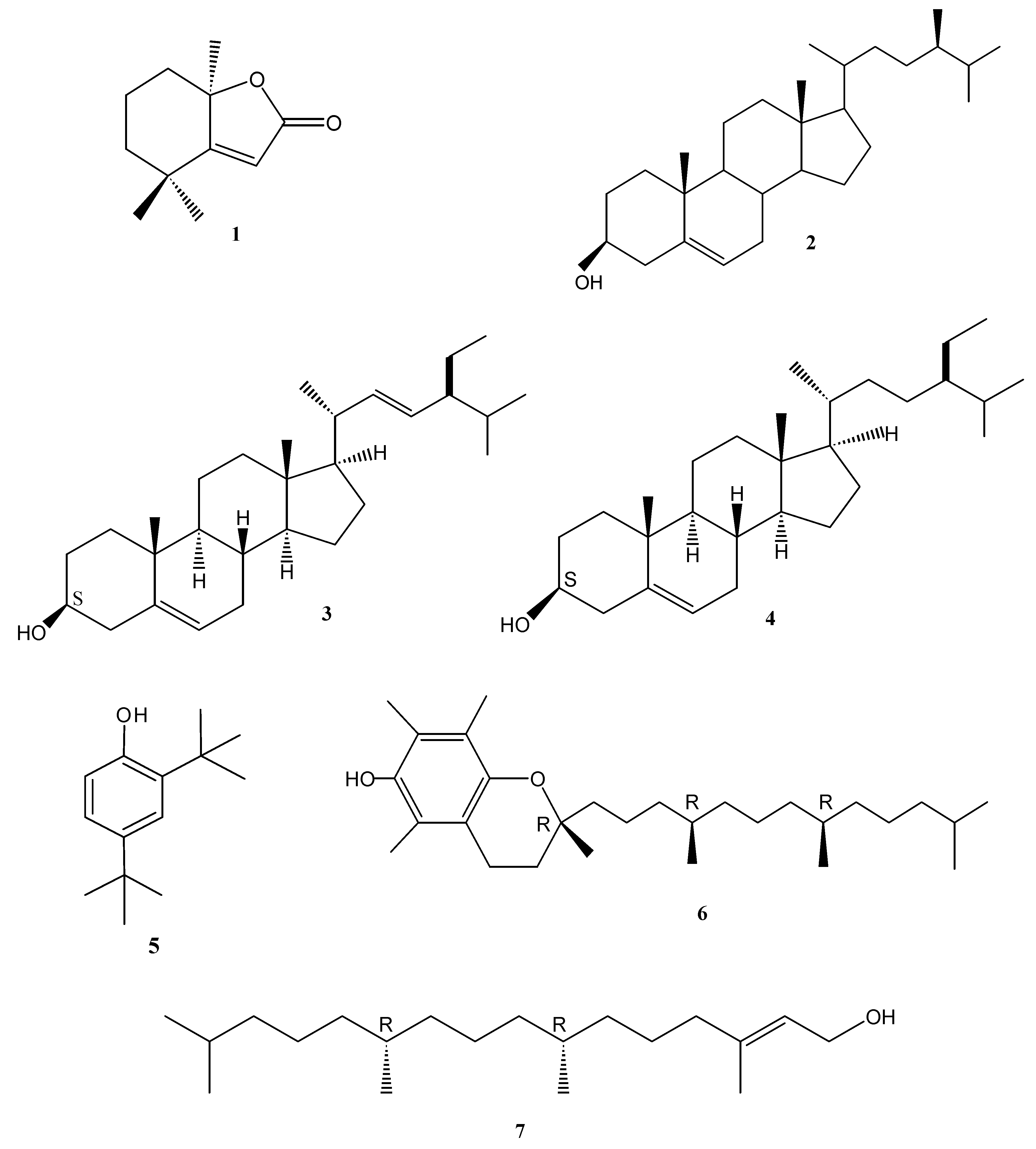
| Compound | Cytotoxicity (IC50) in µg/mL (µM) | |||||
|---|---|---|---|---|---|---|
| KB | MCF7 | CasKi | HCT 116 | A549 | MRC-5 | |
| Dihydroactinidiolide (1) | 6.7 | 30 | 40 | 5 | 97 | 91.3 |
| (37.22) | (166.67) | (222.22) | (27.78) | (538.89) | (507.22) | |
| β -sitosterol (4) | >100 | 72 | 62 | >100 | 78 | >100 |
| (>241.55) | (173.91) | (149.76) | (>241.55) | (188.41) | (>241.55) | |
| 2,4-di tert butylphenol (5) | 0.81 | 5.75 | 4.5 | 29 | 6 | 20 |
| (3.93) | (27.91) | (21.84) | (140.78) | (29.13) | (97.09) | |
| α-tocopherol (6) | 8 | 7.5 | 6 | 31 | 6 | 30.5 |
| (18.6) | (17.44) | (13.95) | (72.09) | (13.95) | (70.93) | |
| Phytol (7) | 7.1 | 34 | 18 | 100 | 31 | 74.3 |
| (23.99) | (114.86) | (60.81) | (337.84) | (104.73) | (251.01) | |
| Mixture | >100 | >100 | >100 | >100 | >100 | >100 |
| Doxorubicina | 1.3x10⁻² | 7.6x10⁻² | 6.0x10⁻³ | 3.6x10⁻¹ | 2.2x10⁻¹ | 5.5x10⁻¹ |
| (0.023) | (0.139) | (0.011) | (0.663) | (0.401) | (1.01) | |
Experimental
General
Plant sample collection and identification
Extraction and isolation of pure compound and mixture
Cell lines and culture medium
In vitro Neutral Red cytotoxicity assay

Conclusions
Acknowledgements
- Sample Availability: Samples are available from the authors.
References
- Goh, K.L. Malaysian herbaceous plants (in Chinese); Advanco Press: Malaysia, 2000; p. 142. [Google Scholar]
- Tan, M.L.; Sulaiman, S.F.; Najimuddin, N.; Samian, M.R.; Tengku Muhammad, T.S. Methanolic extract of Pereskia bleo (Kunth) DC. (Cactaceae) induces apoptosis in breast carcinoma, T47-D cell line. J. Ethnopharmacol. 2005, 96, 287–294. [Google Scholar] [CrossRef]
- Er, H.M.; Cheng, E.; Radhakrishnan, A.K. Anti-proliferative and mutagenic activities of aqueous and methanol extracts of leaves from Pereskia bleo (Kunth) DC leaves. J. Ethnopharmacol. 2007, 113, 448–456. [Google Scholar] [CrossRef]
- Doetsch, P.W.; Cassady, J.M.; McLaughlin, J.L. Cactus alkaloids: XL. Identification of mescaline and other β-phenethylamines in Pereskia, Pereskiopsis and Islaya by use of fluorescamine conjugates. J. Chromatogr. A 1980, 189, 79–85. [Google Scholar] [CrossRef]
- Sri Nurestri, A.B.; Norhanom, A.W.; Hashim, Y.; Sim, K.S.; Hong, S.L.; Lee, G.S.; Syarifah, N.S.A.R. Cytotoxic activity of Pereskia bleo (Cactaceae) against selected human cell lines. Int. J. Cancer Res. 2008, 4, 20–27. [Google Scholar] [CrossRef]
- Eidman, K.F.; MacDougall, B.S. Synthesis of loliolide, actinidiolide, dihydroactinidiolide, and aeginetolide via cerium enolate chemistry. J. Org. Chem. 2006, 71, 9513–9516. [Google Scholar] [CrossRef]
- Borse, B.B.; Rao, L.J.M.; Nagalakshmi, S.; Krishnamurthy, N. Fingerprint of black teas from India: identification of the regio-specific characteristics. Food Chem. 2002, 79, 419–424. [Google Scholar] [CrossRef]
- Huang, L.F.; Zhong, K.J.; Sun, X.J.; Wu, M.J.; Huang, K.L.; Liang, Y.Z.; Guo, F.Q.; Li, Y.W. Comparative analysis of the volatile components in cut tobacco from different locations with gas chromatography-mass spectrometry (GC-MS) and combined chemometric methods. Analytica Chimica Acta 2006, 575, 236–245. [Google Scholar] [CrossRef]
- Gutierrez, R.M.P. Identification of triterpenoids in chloroform extract of Agarista Mexicana by MS and NMR. Nat. Prod. Res. 2006, 20, 181–185. [Google Scholar] [CrossRef]
- Nes, W.; David, N.; Robert, A.; Benson, M. Carbon-13 NMR studies on sitosterol biosynthesized from [13C] mevalonates. Phytochemistry 1992, 31, 805–811. [Google Scholar]
- Borenfreund, E.; Puerner, J.A. A simple quantitative procedure using monolayer culture for toxicity assays. J. Tissue Cult. Meth. 1984, 9, 7–9. [Google Scholar] [CrossRef]
- Geran, R.I.; Greenberg, N.H.; McDonald, M.M.; Schumacher, A.M.; Abbott, B.J. Protocols for screening chemical agents and natural products against animal tumor and other biological systems. Cancer Chemother. Rep. 1972, 3, 17–19. [Google Scholar]
- Swanson, S.M.; Pezzuto, J.M. Bioscreening technique for cytotoxicity potential and ability to inhibit macromolecule biosynthesis. In Drug bioscreening: drug evaluation techniques in pharmacology; Thompson, E.B., Ed.; VCH Publishers: New York, NY, USA, 1990; pp. 273–297. [Google Scholar]
- Russo, A.; Cardile, V.; Ioannes, A.D.; Garbarino. Effect of litreol on the viability of human cancer cells. Chem. Biol. Interact. 2009, 179, 178–184. [Google Scholar] [CrossRef]
- Kozubek, A.; Tyman, J.H. Resorcinolic lipids, the natural non-isoprenoid phenolic amphiphiles and their biological activity. Chem. Rev. 1999, 99, 1–26. [Google Scholar] [CrossRef]
- Awad, A.B.; Downie, A.C.; Fink, C.S. Inhibition of growth and stimulation of apoptosis by β-sitosterol treatment of MDA-MB-231 human breast cancer cells in culture. Int. J. Mol. Med. 2000, 5, 541–545. [Google Scholar]
- Block, S.; Baccelli, C.; Tinant, B.; Meervelt, L.V.; Rozenberg, R.; Jiwan, J.L.; Habib, L.; Gabriel, P.G.; De, M.C.; Joelle, Q.L. Diterpenes from the leaves of Croton zambesicus. Phytochemistry 2004, 65, 1165–1171. [Google Scholar] [CrossRef]
- Bennani, H.; Drissi, A.; Giton, F.; Kheuang, L.; Fiet, J.; Adlouni, A. Antiproliferative effect of polyphenols and sterols of virgin argan oil on human prostate cancer cell lines. Cancer Det. Prev. 2007, 31, 64–69. [Google Scholar] [CrossRef]
- Jackson, S.J.; Houghton, P.J.; Retsas, S.; Photiou, A. In vitro cytotoxicty of norviburtinal and isopinnatal from Kigelia pinnata against cancer cell lines. Planta Med. 2000, 66, 758. [Google Scholar] [CrossRef]
- Moghadasian, M.H. Pharmacological properties of plant sterols: in vivo and in vitro observations. Life Sci. 2000, 67, 605–615. [Google Scholar] [CrossRef]
- NIST 05 MS Library; Agilent Technologies: Palo Alto, CA, USA, 2002.
- Lee, C.C.; Houghton, P. Cytotoxicity of plants from Malaysia and Thailand used traditionally to treat cancer. J. Ethnopharmacol. 2005, 100, 237–243. [Google Scholar] [CrossRef]
- Boik, J. Natural compounds in cancer therapy; Oregon Medical Press: Princeton, MN, USA, 2001; p. 25. [Google Scholar]
- Gille, L.; Kleiter, M.; Willmann, M.; Nohl, H. Paramagnetic species in the plasma of dogs with lymphoma prior to and after treatment with doxorubicin An ESR study. Biochem. Pharmacol. 2002, 64, 1737–1744. [Google Scholar] [CrossRef]
- Carter, S.K. Adriamycin: a review. J. Natl. Cancer Inst. 1975, 55, 1256–1274. [Google Scholar]
- Khanna, C.; Lund, E.M.; Redic, K.A.; Hayden, D.W.; Bell, F.W.; Goulland, E.L.; Klausner, J.S. Randomized controlled trial of doxorubicin versus dactinomycin in multi agent protocol for treatment of dogs with malignant lymphoma. J. Am. Vet. Med. Assoc. 1998, 213, 985–990. [Google Scholar]
- Houghton, P.J.; Raman, A. Laboratory Handbook for the Fractionation of Natural Extracts; Chapman & Hall: London, UK, 1998; pp. 1–13. [Google Scholar]
- Masatoshi, S.; Toshiko, A.; Kazue, S.; Mariko, I.; Ikuko, I.; Hiroshi, S.; Ichiro, Y.; Seiichiro, F. Radical production and cytotoxic activity of tert-butyl-substituted phenols. Vitro Mol. Toxicol. 2001, 14, 53–63. [Google Scholar] [CrossRef]
- Yoon, M.-A.; Jeong, T.-S.; Park, D.-S.; Xu, M.-Z.; Oh, H.-W.; Song, K.-B.; Lee, W.S.; Park, H.-Y. Antioxidant effects of quinoline alkaloids and 2,4-di-tert-butylphenol isolated from Scolopendra subspinipes. Biol. Pharm. Bull. 2006, 29, 735–739. [Google Scholar] [CrossRef]
- Nogueira, P.C.; de, L.; Bittrich, V.; Shepherd, G.J.; Lopes, A.V.; Marsaiolia, A.J. The ecological and taxonomic importance of flower volatiles of Clusia species (Guttiferae)s. Phytochemistry 2001, 56, 443–452. [Google Scholar] [CrossRef]
- Rana, V.S.; Blazquez, M. A. Chemical constituents of Gynura cusimbua aerial parts. J. Essent. Oil Res. 2007, 19, 21–22. [Google Scholar] [CrossRef]
- Moghadasian, M.H.; McManus, B.M.; Pritchard, P.H.; Frohlich, J.J. "Tall oil”- derived phytosterols reduce atherosclerosis in ApoE-deficient mice. Arterioscler. Thromb. Vasc. Biol. 1997, 17, 119–126. [Google Scholar] [CrossRef]
- Moghadasian, M.H.; McManus, B.M.; Godin, D.V.; Rodrigues, B.; Frohlich, J.J. Proatherogenic and antiatherogenic effects of probucol and phytosterols in apolipoprotein E-deficient mice: possible mechanisms of action. Circulation 1999, 99, 11733–1739. [Google Scholar]
- Bouic, P.J. The role of phytosterols and phytosterolins in immune modulation: a review of the past 10 years. Curr. Opin. Clin. Nutr. Metab. Care 2001, 4, 471–475. [Google Scholar] [CrossRef]
- Rensburg, S.J.; Daniels, W.M.; Zyl, J.M.; Taljaard, J.J. A comparative study of the effects of cholesterol, beta-sitosterol, beta- sitosterol glucoside, dehydroepiandrosterone sulphate and melatonin on in vitro lipid peroxidation. Metab. Brain Dis. 2000, 15, 257–265. [Google Scholar] [CrossRef]
- Yasukawa, K.; Takido, M.; Matsumoto, T.; Takeuchi, M.; Nakagawa, S. sterol and triterpene derivatives from plants inhibit the effects of tumour promoter and sitosterol and betulinic acid inhibit tumour formation in mouse skin two-stage carcinogenesis. Oncology 1991, 41, 72–76. [Google Scholar]
- Hao, J.; Zhang, B.; Liu, B.; Lee, M.; Hao, X.; Reuhl, K.R.; Chen, X.; Yang, C.S. Effect of α-tocopherol, N-acetylcysteine and omeprazole on esophageal adenocarcinoma formation in a rat surgical model. Int. J. Cancer 2009, 124, 1270–1275. [Google Scholar] [CrossRef]
- Bermudez, Y.; Ahmadi, S.; Lowell, N.E.; Kruk, P.A. Vitamin E suppreses telomerase activity in ovarian cancer cells. Cancer Det. Prev. 2007, 31, 119–128. [Google Scholar] [CrossRef]
- Lamson, D.W.; Brignall, M.S. Antioxidants in cancer therapy: their actions and interactions with oncologic therapies. Altern. Med. Rev. 1999, 4, 30–329. [Google Scholar]
- Drisko, J.A.; Chapman, J.; Hunter, V.J. The use of antioxidant therapies during chemotherapy. Gynecol. Oncol. 2003, 88, 434–439. [Google Scholar] [CrossRef]
- Amir, D.; Sunil, K.; Gautam, S.; Bharat, B. A. Back to basics: how natural products can provide the basis for new therapeutics. Expert Opin. Investig. Drugs 2007, 16, 1753–1773. [Google Scholar] [CrossRef]
- Ju, Y.H.; Clausen, L.M.; Allred, K.F.; Almada, A.L.; Helferich, W.G. ß-Sitosterol, ß-sitosterol glucoside, and a mixture of ß-sitosterol and ß-sitosterol glucoside modulate the growth of estrogen-responsive breast cancer cells in vitro and in ovariectomized athymic mice. J. Nutr. 2004, 134, 1145–1151. [Google Scholar]
- Osborne, C.K.; Hobbs, K.; Trent, J.M. Biological differences among MCF-7 human breast cancer cell lines from different laboratories. Breast Cancer Res. Treat. 1987, 9, 111–121. [Google Scholar] [CrossRef]
- Bolt, M.W.; Racz, W.J.; Brien, J.F.; Massey, T.E. Effects of vitamin E on cytotoxicity of amiodarone and N-desethylamiodarone in isolated hamster lung cells. Toxicology 2001, 166, 109–118. [Google Scholar] [CrossRef]
- Przybyszewski, W.M.; Kopec-Szlezak, J.; Malec, J. Protection of L5178Y cells by vitamin E against acute hydroxyurea-induced cytotoxic events. Cancer Lett. 1987, 34, 337–344. [Google Scholar] [CrossRef]
- Xie, R.-N.; Shen, X.-N.; Zhong, W.-J.; Zhou, X.-F.; Ying, X.-P. Effect of lycopene alone or with Vitamin E on BALB/c-3T3 cell transformation induced by benzo (a) pyrene. Huanjing Yu Zhiye Yixue (in Chinese) 2004, 21, 124–126. [Google Scholar]
- Al-Sherbini, E-S.A.M.; El Noury, A.H.; El Rouby, M.N. Vitamin E (α-tocopherol) enhances the PDT action of hematoporphyrin derivatives on cervical cancer cells. Med. Laser Appl. 2009, 24, 65–73. [Google Scholar] [CrossRef]
- Kogure, K.; Manabe, S.; Suzuki, I.; Tokumura, A.; Fukuzawa, K. Cytotoxicity of α-tocopheryl succinate, malonate and oxalate in normal and cancer cells in vitro and their anti-cancer effects on mouse melanoma in vivo. J. Nutr. Sci. Vitaminol. 2005, 51, 392–397. [Google Scholar] [CrossRef]
- Campbell, S.E.; Stone, W.L.; Lee, S.; Whaley, S.; Yang, H.; Qiu, M.; Goforth, P.; Sherman, D.; McHaffie, D.; Krishnan, K. Comparative effects of RRR-alpha- and RRR-gamma-tocopherol on proliferation and apoptosis in human colon cancer cell lines. BMC Cancer 2006, 6, 13. [Google Scholar] [CrossRef]
- Wu, X.-X.; Kakehi, Y.; Jin, X.-H.; Inui, M.; Sugimoto, M. Induction of apoptosis in human renal cell carcinoma cells by vitamin E succinate in caspase-independent manner. Urology 2009, 73, 193–199. [Google Scholar] [CrossRef]
© 2009 by the authors; licensee Molecular Diversity Preservation International, Basel, Switzerland. This article is an open access article distributed under the terms and conditions of the Creative Commons Attribution license ( http://creativecommons.org/licenses/by/3.0/).
Share and Cite
Malek, S.N.A.; Shin, S.K.; Wahab, N.A.; Yaacob, H. Cytotoxic Components of Pereskia bleo (Kunth) DC. (Cactaceae) Leaves. Molecules 2009, 14, 1713-1724. https://doi.org/10.3390/molecules14051713
Malek SNA, Shin SK, Wahab NA, Yaacob H. Cytotoxic Components of Pereskia bleo (Kunth) DC. (Cactaceae) Leaves. Molecules. 2009; 14(5):1713-1724. https://doi.org/10.3390/molecules14051713
Chicago/Turabian StyleMalek, Sri Nurestri Abdul, Sim Kae Shin, Norhanom Abdul Wahab, and Hashim Yaacob. 2009. "Cytotoxic Components of Pereskia bleo (Kunth) DC. (Cactaceae) Leaves" Molecules 14, no. 5: 1713-1724. https://doi.org/10.3390/molecules14051713