Article

LYRM1, a Gene that Promotes Proliferation and Inhibits Apoptosis during Heart Development

1
Department of Pediatrics, Nanjing Maternal and Child Health Hospital of Nanjing Medical University, No.123 Tianfei Road, Nanjing 210004, China
2
Department of Cardiology, The First Affiliated Hospital of Nanjing Medical University, No.300 Guangzhou Road, Nanjing 210029, China
*
Author to whom correspondence should be addressed.
These authors contributed equally to this work.
Molecules 2010, 15(10), 6974-6982; https://doi.org/10.3390/molecules15106974
Submission received: 18 September 2010 / Revised: 28 September 2010 / Accepted: 2 October 2010 / Published: 11 October 2010

Abstract

:
Congenital heart disease (CHD) is the most common type of birth defect, but its underlying molecular mechanisms remain unidentified. Previous studies determined that Homo sapiens LYR motif containing 1 (LYRM1) is a novel nucleoprotein expressed at the highest level in adipose tissue and in high levels in heart tissue. The LYRM1 gene may play an important role in the development of the human heart. This study was designed to identify the biological characteristics of the LYRM1 gene in heart development. On the basis of expression-specific differentiation markers identified with quantitative real-time RT-PCR and the morphology of LYRM1-overexpressing cells during differentiation, ectopic expression was not found to significantly affect differentiation of P19 cells into cardiomyocytes. MTT assays and cell cycle analysis showed that LYRM1 dramatically increases the proliferation of P19 cells. Furthermore, data from annexin V-FITC binding and caspase-3 activity revealed that LYRM1 can inhibit the apoptosis of P19 cells. Our data suggest that LYRM1 might have the potential to modulate cell growth, apoptosis, and heart development.

1. Introduction

Congenital heart disease (CHD) is the most common type of human birth defect; its incidence is 6–8 per 1,000 live births [1]. In China, there are more than 4 million untreated CHD patients [2]. Although the mortality of children with CHD has decreased because of improved surgical treatments, not all CHD patients survive to adulthood. CHD is a multifactorial disease, with environmental and genetic factors playing important roles [3]. According to existing epidemiologic studies, many genes are involved in the regulation of heart development, yet its molecular etiology is poorly understood.
There are at least eight LYR motif (LYRM) proteins in Homo sapiens. LYRM1, a novel gene, could play a role in cell growth and apoptosis. Previous studies have shown that besides showing the highest expression in adipose tissue, LYRM1 is also abundantly expressed in human heart tissue [4]. Therefore, this gene was selected for further analysis of heart development. In this study, we initially examined the effect of LYRM1 on cell differentiation. We found that overexpression of LYRM1 did not affect the differentiation of P19 cells into cardiomyocytes. Furthermore, we examined the effect of LYRM1 on cell proliferation and apoptosis and found that it dramatically increased the growth rate of the P19 cells and protected P19 cells from apoptosis.

2. Results and Discussion

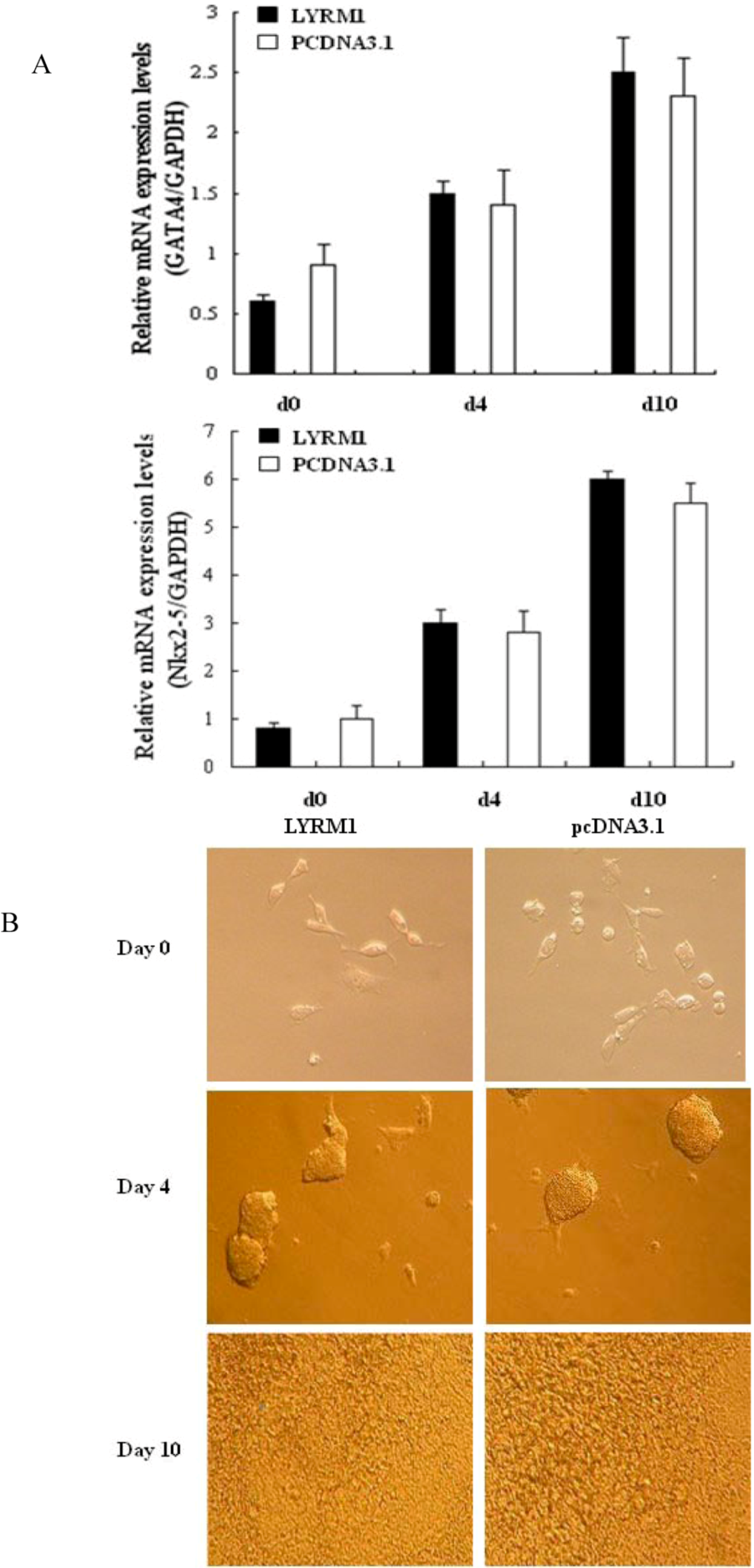
2.1. Effects of LYRM1 on cell differentiation

In order to assess whether LYRM1 affects the differentiation of P19 cells into cardiomyocytes, we established P19 cells that stably overexpressed LYRM1 and assayed the ability of these cells to differentiate into cardiomyocytes. The control was P19 cells transfected with pcDNA 3.1/HisB. No significant difference in morphology between P19 cell lines overexpressing the LYRM1 gene on days 0, 4, and 10 of differentiation and the control cells was found (Figure 1A. In a subsequent study, the expression of cardiomyogenesis-specific markers was detected during the differentiation of P19 cells. The molecular markers included GATA4 and Nkx2-5. Results from real-time qRT-PCR showed that there was no difference in the mRNA expression of these marker genes between LYRM1-overexpressing cells and controls at the same time points (days 0, 4, and 10) (Figure 1B).

2.2. Effect of LYRM1 on cell proliferation

Cell proliferation rate was analyzed with MTT assays every day for 7 days. P19 cells stably transfected with the pcDNA3.1Myc/HisB vector served as controls. Overexpression of LYRM1 resulted in a much higher growth rate compared with control cells ( 2A, *P = 0.001, **P < 0.001). Cell cycle analysis of stably transfected cells was used to further evaluate the effect of LYRM1 on cell proliferation. Data from flow cytometry assays showed that the percentage of P19 cells overexpressing LYRM1 in the S-phase compartment increased from 12 h after serum stimulation compared with control cells (Figure 2B, **P < 0.001).
Figure 1. Effect of LYRM1 on cell differentiation. (A) Expression of specific differentiation markers as determined by quantitative real-time RT-PCR at various time points during the stimulation of differentiation (days 0, 4, and 10). No difference in the expression levels of these markers was found between cells with or without exogenous expression of LYRM1 (n = 6; P < 0.05). (B) Morphology of P19 cells during differentiation (days 0, 4, and 10). P19 cells were transfected with pcDNA3.1Myc/HisB-LYRM1 or an empty vector and stimulated to differentiate over 10 days. This experiment is representative of three independent assays.
Molecules 15 06974 g001
Figure 2. Effect of LYRM1 on cell proliferation. The growth of cells was detected by MTT assays (A) and cell cycle analysis (B). Ectopic expression of LYRM1 increases P19 cells proliferation. The figure represents one of three independent experiments (*P = 0.001, **P < 0.001).
Molecules 15 06974 g002

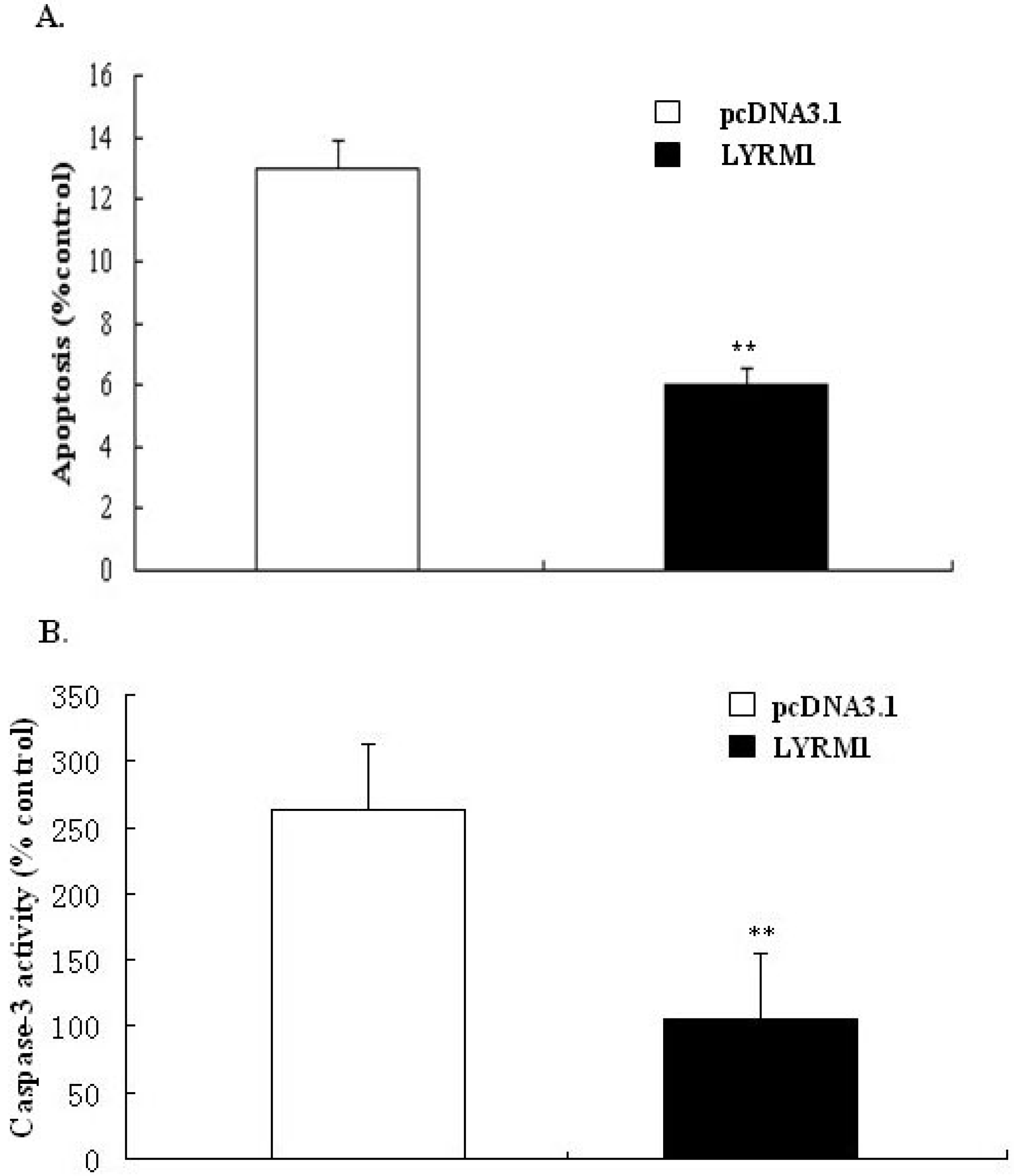
2.3. Effect of LYRM1 on cell apoptosis

To test the effect of LYRM1 on cell apoptosis, we cultured P19 cells in the absence of FBS for 24 h. The number of phosphatidylserine-positive cells was quantified using annexin V staining and by measuring caspase-3 activity; these assays were used to measure apoptosis. Our results suggest that LYRM1 protects against serum deprivation-induced apoptosis (Figure 3).
Figure 3. Effect of LYRM1 on cell apoptosis. Apoptosis was assayed by detecting binding of annexin V-FITC (A) and caspase-3 activity (B). Data from both assays showed that LYRM1 inhibits serum deprivation-induced apoptosis (n = 6; **P < 0.001)
Molecules 15 06974 g003

2.4. Discussion

Chromosomal and Mendelian syndromes account for approximately 20% of human CHD [5]. Although many genes have been found to be important for cardiac development in animal studies, only a few genes related to CHD in humans have been identified [6]. The results of multi-tissue expression analysis support a role for LYRM1 in human heart development. However, its function in heart development is currently unknown [4].
The heart is the first organ to form in the embryo, and a complex series of morphogenetic interactions involving cells from several embryonic origins is involved in its development [7]. Embryonic-fetal heart growth depends on the balance between proliferation and apoptosis of myocytes [8]. Previous reports have shown that P19 cells are a powerful model for studying the regulation of myocardial differentiation at the molecular and functional levels [9,10]. Therefore, in the present study, we used the P19 cell line as a research model. By establishing a stably transfected P19 line that overexpressed LYRM1, we found that LYRM1 does not affect the differentiation of P19 cells into cardiomyocytes, as shown by the morphology and expression of cardiomyogenesis-specific molecular markers. GATA4 is a cardiac-specific member of the GATA family of zinc finger transcription factors, and it is essential for cardiac differentiation of embryonal carcinoma cell lines [11,12,13]. Nkx2.5, a member of the NK2 class homeodomain protein, is required for early heart development [14,15,16]. Moreover, early studies found that GATA4 and Nkx2.5 interact physically and synergistically to activate cardiac transcription, suggesting that they play vital roles in mesoderm formation and specification of cardiac progenitors [17,18,19]. The MTT assays revealed that P19 cells overexpressing LYRM1 grew faster than the controls, and cell cycle analysis indicated a dramatic increase in the percentage of LYRM1-overexpressing P19 cells in the S phase. These data showed that LYRM1 increases the proliferation of P19 cells. Furthermore, the results from the analysis of annexin V-FITC staining and caspase-3 activity indicate that LYRM1 can prevent apoptosis induced by serum removal.

3. Experimental

3.1. Cell culture and differentiation of P19 cells

P19 cells were obtained from the American Type Culture Collection (ATCC, Manassas, VA, USA). The cells were cultured in α-modified Eagle’s medium (α-MEM; Gibco BRL, Grand Island, NY, USA) containing 10% fetal bovine serum (FBS, Gibco BRL), 100 U/mL penicillin, and 100 μg/mL streptomycin at 37 °C in 5% CO2. In order to induce cardiac differentiation, embryoid body (EB) formation was induced by plating 1 × 10⁶ P19 cells on 10-cm bacterial dishes in 15 mL of α-MEM supplemented with 1% dimethyl sulfoxide (DMSO, Sigma, St. Louis, MO, USA), 10% FBS, 100 U/mL penicillin, and 100 μg/mL streptomycin for 96 h. The formed EBs were transferred to 6-cm bacterial dishes and cultured in α-MEM with 10% FBS for an additional 4 or 6 days. The morphologic changes in the P19 cells were examined and photographed using an inverted microscope (Nikon, Japan).

3.2. Quantitative real-time RT-PCR

Total RNA was extracted from the P19 cells with Trizol reagent (Invitrogen, Carlsbad, CA, USA). Then, cDNA was synthesized from 1 μg of total RNA using an AMV Reverse Transcriptase Kit (Promega A3500; Promega, Madison, WI, USA). Real-time PCR was performed using the SYBR Green method in an Applied Biosystems 7300 Sequence Detection System (ABI 7300 SDS; Foster City, CA, USA) following the manufacturer’s protocols. The PCR conditions included a denaturation step (95 °C for 10 min), followed by amplification and quantification repeated 40 times (95 °C for 15 s and 60 °C for 1 min). Gene expression was measured in triplicate. The relative gene expression levels were quantified based on the Ct and normalized to a reference gene, GAPDH. The sequences of the primers used are shown in Table 1.
Table 1. Sequences of the primers used in this study.
Gene name Product sizeReverse and forward primers (5'-3')
GATA4136bpF:5'-CCTGCGGCCTCTACATGA-3'
R:5'-AGGGTCTCACCAGCAGGA-3'
Nkx2-5222bpF:5'-CCTGCGGCCTCTACATGA-3'
R:5'-AGGGTCTCACCAGCAGGA-3'
GAPDH237bpF:5'-TTCACCACCATGGAGAAGGC-3'
R:5'-GGCATGGACTGTGGTCATGA-3'

3.3. Establishment of a stable cell line overexpressing LYRM1

The coding sequence of LYRM1 was subcloned into the BamHI and the XhoI sites of the pcDNA3.1Myc/HisB vector to generate a plasmid expressing the LYRM1-6×His fusion protein. Expression vectors carrying the LYRM1 coding sequence, or empty vectors, were transfected into P19 cells. Two days after transfection, neomycin (G418, Roche, Basel, Switzerland) was added to the medium (600 μg/mL) to select for transfected P19 cells. After 2 weeks, individual colonies were isolated, propagated, and the LYRM1-6×His fusion protein was confirmed by western blotting. The anti-6×His antibody was obtained from Clontech. Colonies expressing the highest levels of LYRM1 were selected for further studies.

3.4. MTT assay

Cells were seeded in 96-well plates at a density of 2 × 10² cells/well and maintained in media supplemented with 10% FBS and G418 250 μg/mL. The growth of the cells was analyzed for 7 consecutive days by 3-(4,5-dimethylthiazol-2-yl)-2,5-diphenyltetrazolium bromide (MTT) assays, using a Cell Proliferation MTT Kit (Roche Diagnostics). Absorbance was measured at 560 nm and 660 nm, as recommended by the manufacturer.

3.5. Cell cycle assay

Before analysis, the cells were starved in serum-free α-MEM for 24 h to synchronize the cell cycle. Cells were incubated with α-MEM containing 10% (v/v) FBS for various times (0, 6, 12, 18, and 24 h) after serum deprivation. Cells were harvested using trypsin/EDTA, fixed in 70% ethanol, and cellular DNA was stained with 500 μL propidium iodide (PI). Cultured cells were analyzed by flow cytometry, and data were analyzed using CellQuest software.

3.6. Apoptosis assay

Cells were cultured in serum-free α-MEM for 24 h to induce apoptosis, and then harvested using trypsin/EDTA and washed with PBS. The cells were centrifuged and resuspended in 1 mL binding buffer and then stained with 10 μL annexin V-FITC and 10 μL PI at room for 5 min (Biovision, CA, USA). The stained cells were immediately analyzed using flow cytometry as previously described [20].

3.7. Measurement of caspase-3 activity

Caspase-3 activity was measured using a commercially available kit (KeyGen, Nanjing, China). The cells (5 × 10⁶) were lysed with 50 μL of chilled cell lysis buffer on ice for 20 min. After microcentrifugation (10,000 rpm, 1 min, 4 °C), the supernatant was used for a caspase-3 colorimetric protease assay. Next, 150 μg of protein was diluted to 50 μL with cell lysis buffer and then incubated with 5 μL substrate at 37 °C for 4 h. The samples were read at 405 nm in a microtiter plate reader.

3.8. Statistical analysis

The data are presented as the mean ± SD. All values were analyzed using the Student’s t test with the SPSS 15.0 statistical package. P values < 0.05 were considered statistically significant.

4. Conclusions

In summary, we have described several useful features of the novel gene LYRM1 in cardiac development. However, the precise molecular function of this gene remains unknown. For example, the in vivo function of LYRM1 remains to be clarified. Further studies are also required to elucidate the biological function of LYRM1 in the differentiation of human cardiac cells.

Acknowledgements

This work was supported by a grant from the Natural Science Foundation of Jiangsu Province, China (No BK2010582) and National Natural Science Foundation of China (No 81070138).
  • Sample Availability: Contact the authors.

References

  1. Sadowski, S.L. Congenital cardiac disease in the newborn infant: Past, present, and future. Crit. Care. Nurs. Clin. North Am. 2009, 21, 37–48. [Google Scholar] [CrossRef]
  2. Gorenflo, M.; Gu, H.; Xu, Z. Peri-operative pulmonary hypertension in paediatric patients: Current strategies in children with congenital heart disease. Cardiology 2010, 116, 10–17. [Google Scholar] [CrossRef]
  3. Huang, J.B.; Liu, Y.L.; Sun, P.W.; Lv, X.D.; Du, M.; Fan, X.M. Molecular mechanisms of congenital heart disease. Cardiovasc Pathol. 2010, 19, e183–e193. [Google Scholar] [CrossRef]
  4. Qiu, J.; Gao, C.L.; Zhang, M.; Chen, R.H.; Chi, X.; Liu, F.; Zhang, C.M.; Ji, C.B.; Chen, X.H.; Zhao, Y.P.; Li, X.N.; Tong, M.L.; Ni, Y.H.; Guo, X.R. LYRM1, a novel gene promotes proliferation and inhibits apoptosis of preadipocytes. Eur. J. Endocrinol. 2009, 160, 177–184. [Google Scholar]
  5. Bentham, J.; Bhattacharya, S. Genetic mechanisms controlling cardiovascular development. Ann. N Y Acad. Sci. 2008, 1123, 10–19. [Google Scholar]
  6. Joziasse, I.C.; van de Smagt, J.J.; Smith, K.; Bakkers, J.; Sieswerda, G.J.; Mulder, B.J.; Doevendans, P.A. Genes in congenital heart disease: atrioventricular valve formation. Basic Res. Cardiol. 2008, 103, 216–227. [Google Scholar] [CrossRef]
  7. Srivastava, D. Genetic regulation of cardiogenesis and congenital heart disease. Annu. Rev. Pathol. 2006, 1, 199–213. [Google Scholar] [CrossRef]
  8. Fiorina, P.; Corradi, D.; Pinelli, S.; Maestri, R.; Lagrasta, C.; Buscaglia, M.; Davalli, A.; Folli, F.; Astorri, E. Apoptotic/mytogenic pathways during human heart development. Int. J. Cardiol. 2004, 96, 409–417. [Google Scholar] [CrossRef]
  9. Heyden, M.A.; Kempen, M.J.; Tsuji, Y.; Rook, M.B.; Jongsma, H.J.; Opthof, T. P19 embryonal carcinoma cells: A suitable model system for cardiac electrophysiological differentiation at the molecular and functional level. Cardiovasc. Res. 2003, 58, 410–422. [Google Scholar] [CrossRef]
  10. Heyden, M.A.; Defize, L.H. Twenty one years of P19 cells: what an embryonal carcinoma cell line taught us about cardiomyocyte differentiation. Cardiovasc. Res. 2003, 58, 292–302. [Google Scholar] [CrossRef]
  11. Grepin, C.; Nemer, G.; Nemer, M. Enhanced cardiogenesis in embryonic stem cells overexpressing the GATA-4 transcription factor. Development 1997, 124, 2387–2395. [Google Scholar]
  12. Peterkin, T.; Gibson, A.; Loose, M.; Patient, R. The roles of GATA-4, -5 and -6 in vertebrate heart development. Semin. Cell Dev. Biol. 2005, 16, 83–94. [Google Scholar]
  13. Pikkarainen, S.; Tokola, H.; Kerkela, R.; Ruskoaho, H. GATA transcription factors in the developing and adult heart. Cardiovasc. Res. 2004, 63, 196–207. [Google Scholar] [CrossRef]
  14. Lints, T.J.; Parsons, L.M.; Hartley, L.; Lyons, I.; Harvey, R.P. 5: A novel murine homeobox gene expressed in early heart progenitor cells and their myogenic descendants. Development 1993, 119, 419–431. [Google Scholar]
  15. Komuro, I.; Izumo, S. Csx: A murine homeobox-containing gene specifically expressed in the developing heart. Proc. Natl. Acad. Sci. USA 1993, 90, 8145–8149. [Google Scholar] [CrossRef]
  16. Ueyama, T.; Kasahara, H.; Ishiwata, T.; Nie, Q.; Izumo, S. Myocardin expression is regulated by Nkx2.5, and its function is required for cardiomyogenesis. Mol. Cell Biol. 2003, 23, 9222–9232. [Google Scholar]
  17. Durocher, D.; Charron, F.; Warren, R.; Schwartz, R.T.; Nemer, M. The cardiac transcription factors Nkx2-5 and GATA-4 are mutual cofactors. EMBO J. 1997, 16, 5687–5696. [Google Scholar] [CrossRef]
  18. Brown, CO., 3rd.; Chi, X.; Garcia-Gras, E.; Shirai, M.; Feng, X.H.; Schwartz, R.T. The cardiac determination factor, Nkx2-5, is activated by mutual cofactors GATA-4 and Smad1/4 via a novel upstream enhancer. J. Biol. Chem. 2004, 279, 10659–10669. [Google Scholar]
  19. Jiang, Y.; Drysdale, T.A.; Evans, T. A role for GATA-4/5/6 in the regulation of Nkx2.5 expression with implications for patterning of the precardiac field. Dev. Biol. 1999, 216, 57–71. [Google Scholar] [CrossRef]
  20. Vermes, I.; Haanen, C.; Steffens-Nakken, H.; Reutelingsperger, C. A novel assay for apoptosis: Flow cytometric detection of phosphatidylserine expression on early apoptotic cells using fluorescein labeled annexin-V. J. Immunol. Methods 1995, 180, 39–52. [Google Scholar]

