Synthesis and Biological Evaluation of Novel 99mTc-Labelled Bisphosphonates as Superior Bone Imaging Agents

Key Laboratory of Nuclear Medicine, Ministry of Health, Jiangsu Key Laboratory of Molecular Nuclear Medicine, Jiangsu Institute of Nuclear Medicine, Wuxi 214063, China
*
Author to whom correspondence should be addressed.
Molecules 2011, 16(8), 6165-6178; https://doi.org/10.3390/molecules16086165
Submission received: 10 June 2011
/
Revised: 15 July 2011
/
Accepted: 19 July 2011
/
Published: 25 July 2011
(This article belongs to the Special Issue Radical Chemistry)
Abstract
:A series of novel zoledronic acid (ZL) derivatives 1-hydroxy-3-(2-methyl-1H-imidazol-1-yl)propane-1,1-diyldiphosphonic acid (MIPrDP), 1-hydroxy-4-(2-methyl-1H-imidazol-1-yl)butane-1,1-diyldiphosphonic acid (MIBDP), and 1-hydroxy-5-(2-methyl-1H-imidazol-1-yl)pentane-1,1-diyldiphosphonic acid (MIPeDP) were prepared and successfully labeled with 99mTc in high labeling yields. The in vitro stability and in vivo biodistribution of 99mTc-MIPrDP, 99mTc-MIBDP and 99mTc-MIPeDP were investigated and compared. The biodistribution studies indicate that the radiotracer 99mTc-MIPrDP has highly selective uptake in the skeletal system and rapid clearance from soft tissues. The present findings indicate that 99mTc-MIPrDP holds great potential for use in bone imaging.
1. Introduction
Technetium-99m, with its excellent physical characteristics and easy availability from a generator, has become the most important nuclide for organ imaging in nuclear medicine. Since Fleisch et al. described that bisphosphonates had high affinity for bone mineral in 1968, several 99mTc-labeled phosphate compounds have been developed for skeletal imaging, including pyrophosphate [1], polyphosphates [2,3], and diphosphonates [4,5].
Complexes of technetium-99m labeled with methylenediphosphonate (99mTc-MDP) and hydroxymethylenediphosphonate (99mTc-HMDP) have been widely used, both experimentally and clinically, for detection of skeletal metastases and other focal bone lesions [4,6,7]. However, an interval of 2–6 h is needed between injecting these agents into the patient and bone scanning [8]. Shorting this interval would lessen the burden on patients in terms of the total length of the examination. To enable imaging at an earlier time after injection, a radiopharmaceutical with higher affinity for bone, larger ratio of bone-to-soft tissue uptake and more rapid clearance from blood is required, accordingly [9]. Consequently, the nature of ligands (diphosphonic acid) is a key factor to determine the advantages of the radiopharmaceuticals.
Diphosphonates have a fundamental P–C–P backbone structure, and have high affinity for bone mineral. ZL [1-hydroxy-2-(imidazol-1-yl)-ethylidene-1,1-bisphosphonic acid], one kind of the typical third-generation DPs, is currently the most potent bisphosphonate. In preclinical models of bone resorption, for example, ZL is at least 100 times more potent than clodronate and pamidronate, and it is at least 1,000 times more potent than etidronate [10].
In the previous work of our group, a series of technetium-99m labeled DPs with the alkyl substituent in the imidazole ring of ZL have been prepared and their in vivo biological properties were systematically investigated [11,12,13,14]. However, to the best of our knowledge, extension and optimization of the linker chain between the imidazolyl and geminal bisphosphonate group to develop novel bone imaging agent has been largely unexplored.
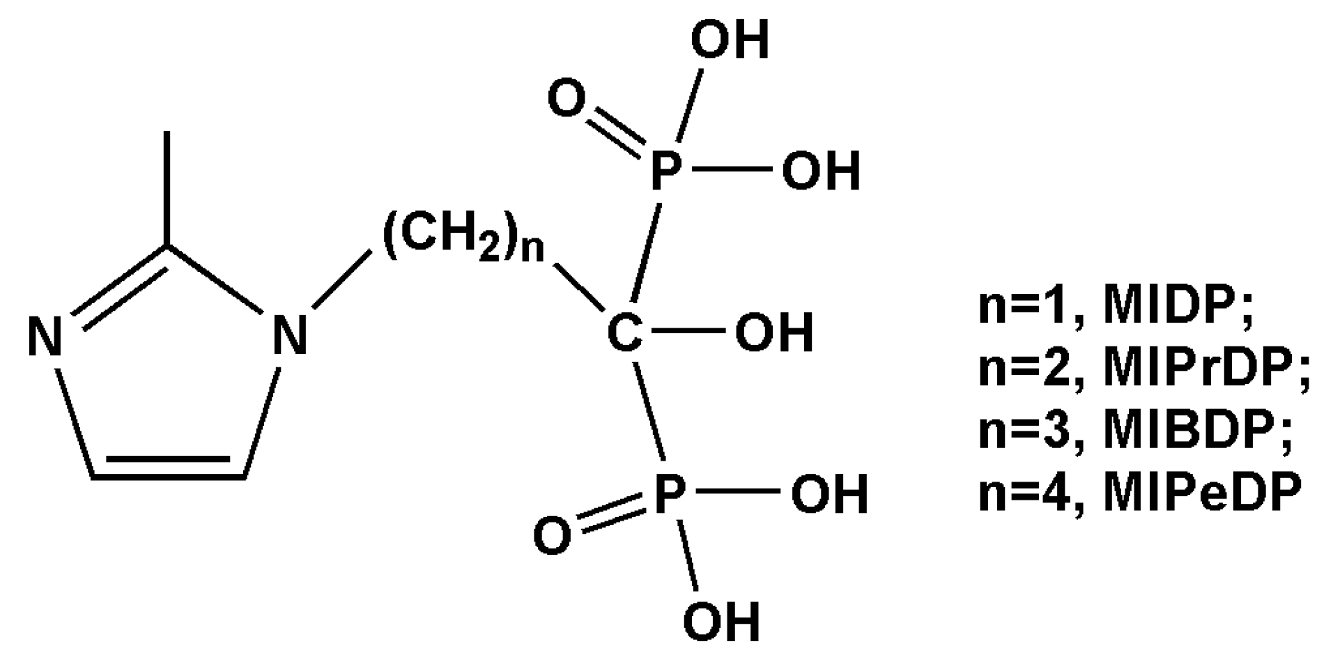
Based on our primary studies, we found that optimization of imidazolyl ring or extension of the linker chain between the imidazolyl and geminal bisphosphonate group in the ZL to be labeled with technetium-99m can bring significant influence on the biological properties of the bone resorption and the clearance from blood and soft tissues [15,16]. For the purpose of developing novel bone imaging agent with excellent biological properties, we continue to extend the number of the methylene chain between the methylimidazolyl and geminal bisphosphonate group in the diphosphonate acid (see Figure 1). In this work, a series of novel 99mTc-labeled bisphosphonates were prepared and reported, i.e., 99mTc-MIPrDP, 99mTc-MIBDP and 99mTc-MIPeDP. Their in vitro stability and in vivo biodistribution were also studied.
2. Results and Discussion
2.1. Chemistry and Radiolabeling
MIPrDP, MIBDP and MIPeDP were synthesized by three step reactions from the starting material 2-methylimidazole. The target compounds were identified by elemental analysis, MS and 1H-NMR, and the results agree well with the expected chemical structures. 99mTc-MIPrDP, 99mTc-MIBDP and 99mTc-MIPeDP were labeled with 99mTcO4− by reduction with stannous chloride. For TLC analysis, with the H2O system, the Rf values of Na99mTcO4 and 5a-5c were about 0.9-1, while 99mTc-colloidal impurities remain at 0-0.1. With the acetone system, the 99mTc-colloidal impurities and 5a-5c remain at the origin and Na99mTcO4 moves with the solvent front.
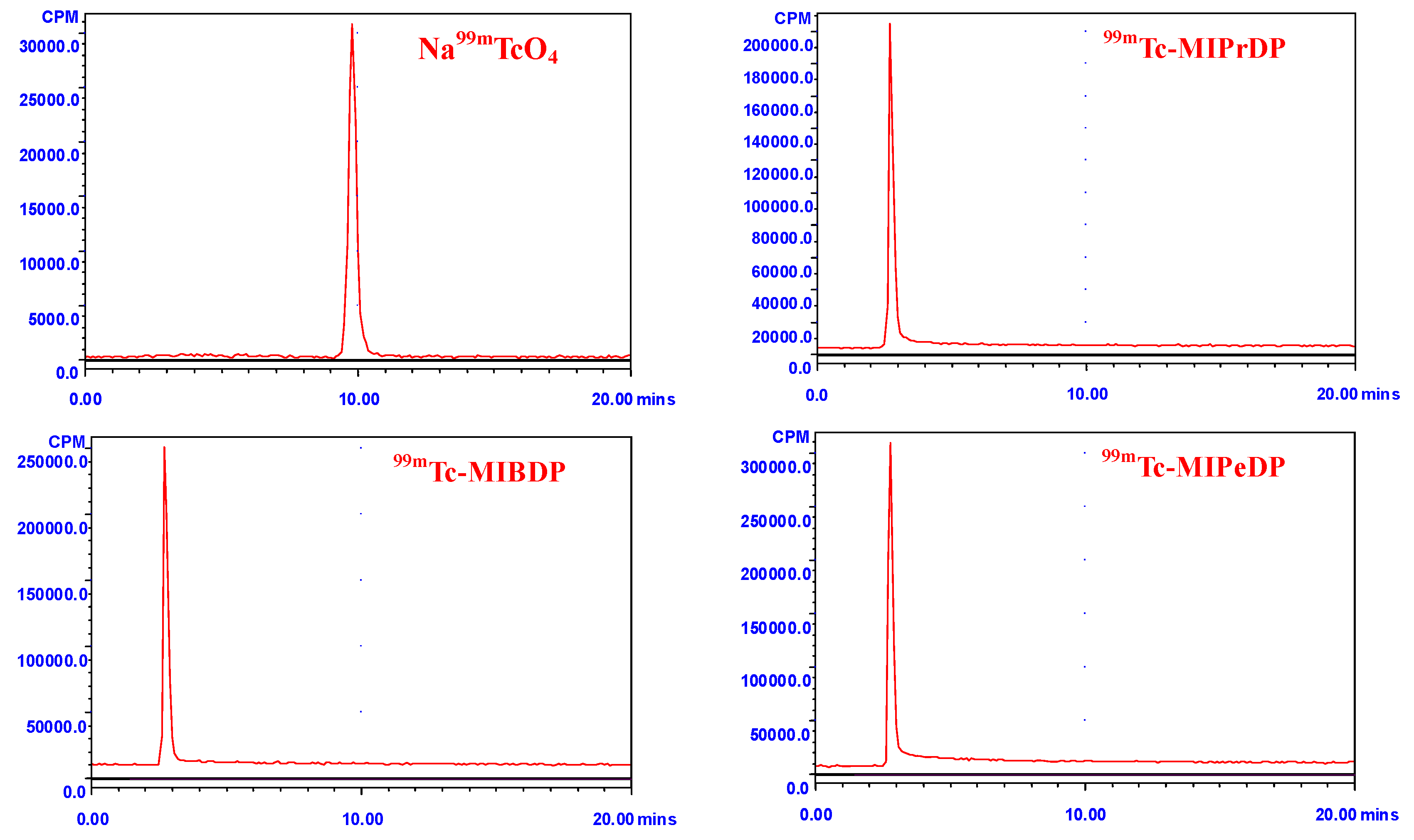
HPLC analysis revealed the formation of free technetium (Na99mTcO4) that was eluted at a retention time of 9.8 min, whereas 5a-5c eluted at a retention time of 2.8 ± 0.1 min (see Figure 2). The identical retention times revealed the structural analogy of these 99mTc-DPs [9]. For each radiolabeled complex, the single peak in the HPLC-chromatogram clearly shows the formation of only one complex and excludes the possibility of residual Na99mTcO4 or other components [17]. That is, the chelation of technetium with the bisphosphonates moiety is unique and complete.
According the TLC and HPLC analysis, the radiochemical purities of these 99mTc-DPs were all greater than 97%. This labeling method also meets the clinical requirement of other 99mTc-labeled bisphosphonates such as 99mTc-MDP. The radiolabeled compounds were used immediately after the formulation for both in vitro and in vivo studies.
2.2. In Vitro Stability and Octanol-Water Partition Coefficient
The in vitro stabilities of 99mTc-MIPrDP, 99mTc-MIBDP and 99mTc-MIPeDP were studied in PBS (pH = 7.4) for different time intervals (1, 2, 3, 4, 5, 6 h) at 37 °C. The stability was presented as RCP on the basis of the HPLC analysis. After 6 h of incubation, more than 97% of 99mTc-MIPrDP, 99mTc-MIBDP and 99mTc-MIPeDP remained intact in the PBS. The results indicated that the labeling efficiency of these complexes was high and their stability duration was long enough to allow further biodistribution and imaging studies.
The octanol-water partition coefficients (logP) for 99mTc-MIPrDP, 99mTc-MIBDP and 99mTc-MIPeDP were −1.89, −1.93, −2.10 and −1.71, −1.77, −1.89 in PBS at two different pH values of 7.0 and 7.4 respectively (see Table 1), which demonstrated that logP decreased from 99mTc-MIPrDP to 99mTc-MIPeDP and the liposolubility at pH = 7.4 was higher than that at pH = 7.0. From 99mTc-MIPrDP to 99mTc-MIPeDP, the longer the carbon chain, the smaller the log P. As well known, the log P value is a very useful parameter that can be used to understand the behavior of a drug and predict its distribution in the organism in combination with other parameters [18]. The different lipotropy among three technetium-99m labeled zoledronic acid derivatives may be attributed to the change in the linker chain between the methylimidazolyl and the geminal bisphosphonate group.
2.3. Plasma Protein Binding
The percentage of protein binding was 11.2%, 12.4%, and 14.7% for 99mTc-MIPrDP, 99mTc-MIBDP and 99mTc-MIPeDP, respectively. As known, different protein binding rate has significant influence on the bone uptake of the tracer agent [19]. Here, with the extension of carbon chain, the plasma protein binding increases and the maximum bone uptake decreases gradually. The structural difference further affects the blood clearance of the radiolabeled complexes [20], which will be discussed in detail in the next blood kinetics studies.
2.4. Blood Kinetics Studies
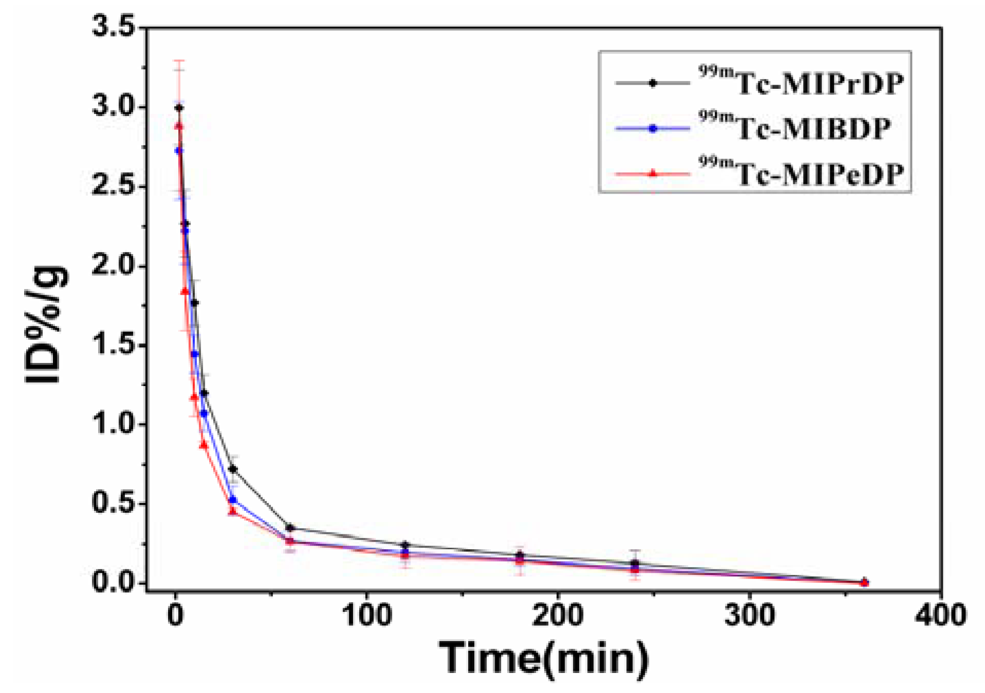
Pharmacokinetic parameters were listed in Table 2. Figure 3 shows the blood clearance of 5a-5c in the mice 6 h post injection. Pharmacokinetics of 5a-5c comply with the two-compartment model with the pharmacokinetic equations of C = 2.66e−0.16t + 0.90e−0.01t, C = 2.53e−0.056t + 0.282e−0.0034t and C = 2.03e−0.061t + 0.736e−0.0067t, respectively. The values of CL were 2.58, 2.90 and 3.84 and the AUC were 142.9, 127.4, and 96.2 for 99mTc-MIPrDP, 99mTc-MIBDP and 99mTc-MIPeDP, respectively.
In the early phase, the blood clearance of 99mTc-MIPrDP was slower than 99mTc-MIBDP and 99mTc-MIPeDP. After 2 h, the radioactivity concentration of three tracer agents in blood reaches an equilibrium which coincides with the pharmacokinetic parameters CL, AUC and the pharmacokinetic curves. Ranging from 5 to 10 min, the bone uptake increases with the increased carbon chain. That is, the uptake of 99mTc-MIPeDP in bone increases faster than 99mTc-MIPrDP and 99mTc-MIBDP, which consists with the variation of K12. It was concluded that 99mTc-MIPeDP can be absorbed quickly and eliminated from the blood rapidly. In this study, the blood clearance (see Figure 3) of 99mTc-MIPrDP, 99mTc-MIBDP and 99mTc-MIPeDP were very fast, due to low protein binding.
2.5. Biodistribution Studies
Biodistributions of 5a-5c were determined in ICR mice, and the data is shown in Table 3 as the percentage administered activity (injected dose) per gram of tissue (%ID/g). Although ZL has been known to possess low toxicity and can be used therapeutically at a high dose [21,22], the toxicity profile of its derivatives are uncertain. Therefore, after the complexes 99mTc-DPs (1.85 MBq) in a volume of 0.2 mL were given to the normal mice via intravenous injection respectively, the mice were watched carefully for any sign of adverse reaction. As expected, the mice showed no signs of toxicity through the overall study period.
Inspecting Table 3, one can observe that 5a-5c are mainly accumulated in the bone, kidneys, and liver. The uptake of 5a-5c in bone increased steadily from 5 to 60 min post injection. At 2 h post injection, the bone uptake was 19.6 ± 0.87, 11.2 ± 0.13 and 17.6 ± 0.42 %ID/g for 99mTc-MIPrDP, 99mTc-MIBDP and 99mTc-MIPeDP, respectively. From previous work of our group [16], it was found that the bone uptake increases with the increasing carbon chain between the imidazolyl and geminal bisphosphonate group in ZL (i.e., from 99mTc-ZL to 99mTc-IPrDP). However, in the present work the bone uptake of 99mTc-MIPeDP was smaller than those of 99mTc-MIPrDP and 99mTc-MIBDP (see Table 3). This indicates that limitless extension of the carbon chain is not always beneficial to improve the bone uptake. Furthermore, the bone uptake efficiency of these radiotracers was compared. For example, after injection of 99mTc-MIPrDP, the bone uptake was up to 6.43 %ID/g at 5 min and it increased continuously to a maximum of 19.6 %ID/g at 120 min, which was larger than the corresponding values of 99mTc-IPrDP (5.37 and 11.14 %ID/g respectively) [16]. This implied that the bone uptake efficiency of the former with a methyl in the imidazole ring is larger than the latter with no substituent in the imidazole ring. Considering that 99mTc-IPrDP is better than 99mTc-MDP and 99mTc-ZL in the bone imaging, it is therefore concluded that 99mTc-MIPrDP is the best one among these bone scanning agents.
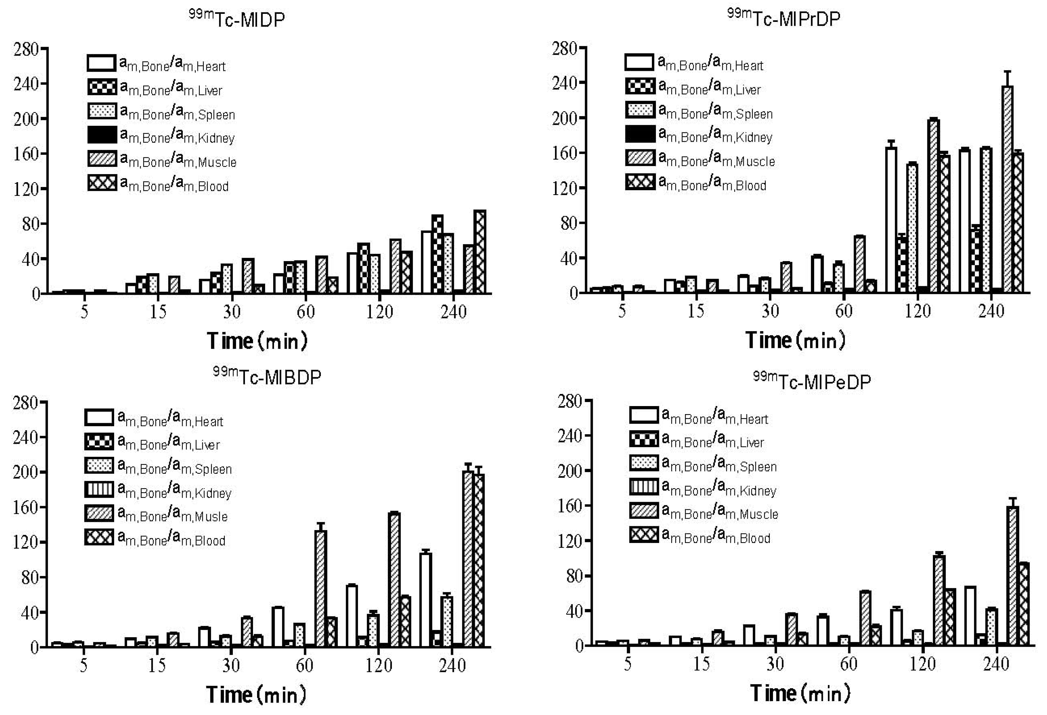
Compounds 5a-5c also showed predominant kidney and liver uptake, indicating that they were not only excreted through the kidneys, but also absorbed and eliminated by the liver. It was noted that the uptake of 99mTc-MIBDP and 99mTc-MIPeDP in liver, spleen and kidneys were larger than those of 99mTc-MIPrDP. For instance, the kidneys and liver uptake of 5a-5c at 2 h post injection were 3.21 ± 0.50, 4.45 ± 0.22 and 4.80 ± 0.14 %ID/g for kidneys and 0.32 ± 0.05, 1.14 ± 0.04 and 2.25 ± 0.24 %ID/g for liver, respectively. The uptake ratios of bone to liver and kidneys for 99mTc-MIPrDP were 61.25 ± 1.89 and 6.10 ± 0.78 respectively, while those of 99mTc-MIBDP and 99mTc-MIPeDP were 9.81 ± 1.23, 2.51 ± 0.69 and 7.81 ± 0.74, 3.6 ± 1.21 (see Figure 4). Thus, we can see that from 99mTc-MIPrDP to 99mTc-MIPeDP with the extension of the linker chain between the methylimidazolyl and geminal bisphosphonates group, the former exhibits a significant advantage in the clearance from soft tissues and it can better satisfy the clinical requirements of a good bone imaging agent.
Radioactivity levels of 5a-5c in the blood were 5.00 ± 0.02, 3.79 ± 0.07 and 3.40 ± 0.14 %ID/g at 5 min post injection, respectively, which followed by a rapid clearance. At 4 h post injection, the radioactivity in blood was only 0.08, 0.09 and 0.12 %ID/g for 5a-5c respectively. This indicates that all these radiotracers can eliminate quickly from the blood, which agrees well with the pharmacokinetics studies.
For better understanding the biodistributions and metabolism of 5a-5c in vivo, we further compared them with the radiotracer 99mTc-MIDP (1-hydroxy-2-(2-methyl-1H-imidazolyl)ethane-1,1-diyl diphosphonic acid) [11]. The biodistribution data also show that 99mTc-MIDP has high kidney uptake, indicating that it is excreted quickly through the kidneys. Comparison of the uptake ratios of bone to soft tissue (including heart, liver, spleen, muscle and kidneys) among these homologues shows that these uptake ratios of 99mTc-MIDP are all very small while those of 99mTc-MIPrDP are all larger than others after 120 min post injection (see Figure 4). This showed 99mTc-MIPrDP was not only superior to its parent complex 99mTc-MIDP but also superior to 99mTc-IPrDP judged from the biodistribution studies. In summary, the radiotracer 99mTc-MIPrDP better meets the clinical requirement of bone imaging agent with higher bone uptake and lower background among this kind of homologues.
3. Experimental
3.1. General
All analytical chemical reagents employed were purchased from commercial sources and used without further purification. Na99mTcO4 was supplied by the Affiliated Jiangyuan Hospital of the Jiangsu Institute of Nuclear Medicine. All melting points were measured on a Yanaco MP-500 melting point apparatus (Shimadzu, Japan). Elemental analysis was carried out using an Elementar Vario EL III analyzer. Electron spray ion (ESI) mass spectra were determined using a Waters Platform ZMD4000 LC/MS (Waters, U.S.A.). Nuclear magnetic resonance (NMR) spectra were obtained on a Bruker DRX-500 spectrometer (Bruker, Germany), and the chemical shift value was given relative to the internal tetramethylsilane (TMS). Xinhua chromatography paper (Shanghai, China) was used for thin layer chromatography (TLC). A Packard-multi-prias γ Counter (Perkins Elmer, U.S.A.) was used. High performance liquid chromatography (HPLC) analysis was performed on a Waters 600-type high-performance liquid chromatography (Waters, U.S.A.) equipped with a dural k absorbance detector (Waters 2487), binary HPLC pump (Waters 1525) and Cd (Te) detector equipped with a flow scintillation analyzer (Perkin Elmer). Normal Institute of Cancer Research (ICR) mice (weighting 18~20 g) were supplied by the Shanghai SLAC Laboratory Animal CO., Ltd. (Shanghai, China). The animal experiment was approved by the Animal Care and Ethics Committee of the Jiangsu Institute of Nuclear Medicine.
3.2. Syntheses of Diphosphonic Acids
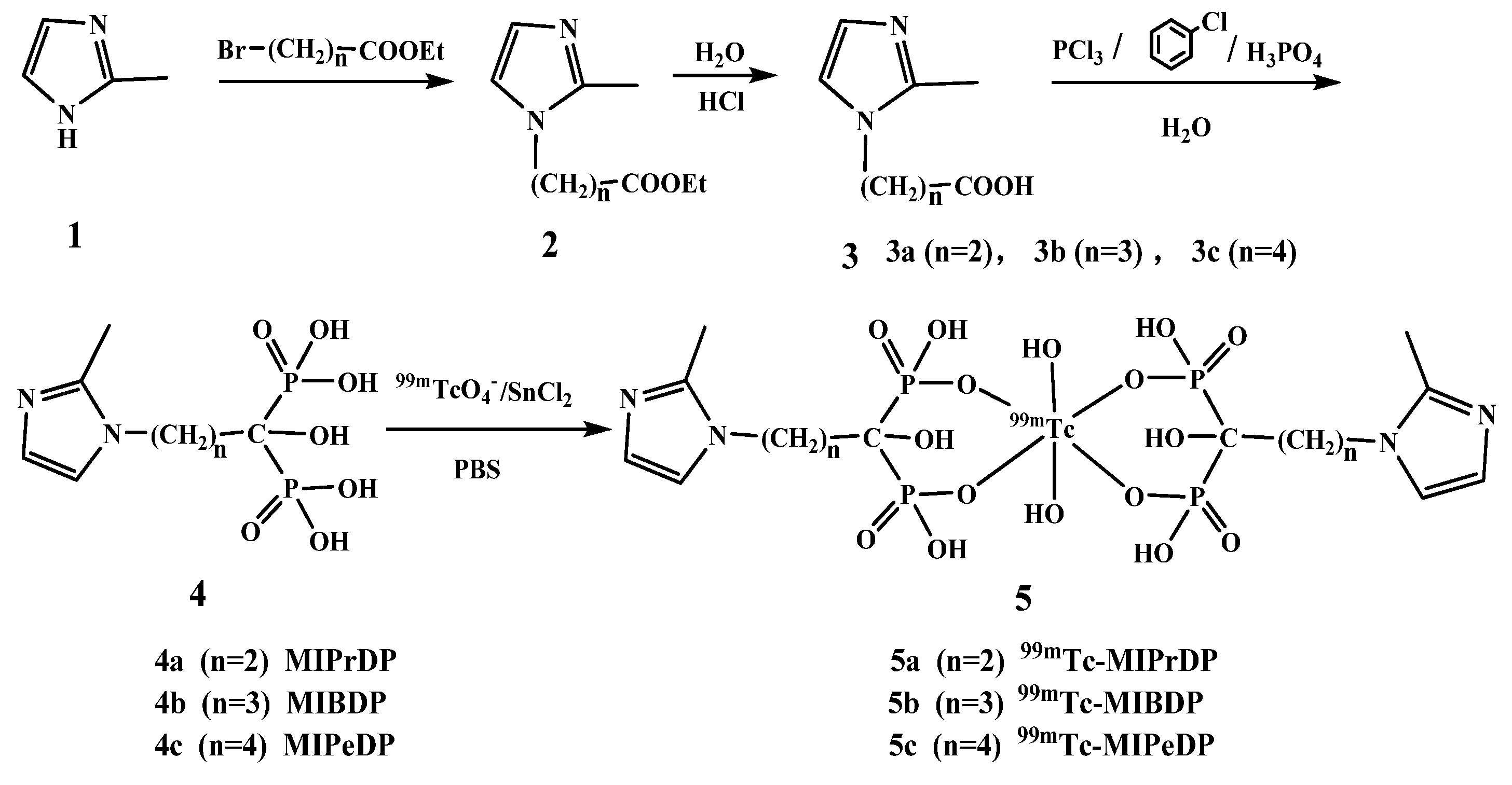
Three 99mTc-diphosphonates were synthesized according to the procedure outlined in Scheme 1 as described previously [16,23].
3.2.1. General Procedure for the Preparation of Compounds 3a-3c
2-Methylimidazole (1, 8.2 g, 0.1 mol) was dissolved in CH2Cl2 (75 mL). Then KOH (8.4 g, 0.15 mol), K2CO3 (13.8 g, 0.0835 mol) and tetrabutylammonium bromide (0.7 g, 0.002 mol) were added and stirred at room temperature for 30 min. The solution was treated dropwise with the corresponding ethyl bromoacetate homolog (0.1 mol) at room temperature. The mixture was heated to reflux for 7 h. The inorganic salt was removed by filtration and the filtrate was washed with saturated NaCl solution (30 mL × 4). Finally, the organic layer was evaporated under vacuum to give crude compound 2 as a brown gum, which was used without any further purification for the next step. Water (90 mL) and HCl (12 mol/L, 10 mL) were successively added to compound 2, and the mixture was refluxed 8 h, the solution was concentrated and recrystallized from isopropanol to give the white crystalline product 3.
3-(2-Methyl-1H-imidazol-1-yl)propanoic acid (3a): Yield: 34%. mp 125-128 °C; ESI-MS, m/z (%): 153 (100) = M-H+.
4-(2-Methyl-1H-imidazol-1-yl)butanoic acid (3b): Yield: 42%. mp 120-122 °C; ESI-MS, m/z (%): 167 (100) = M-H+.
5-(2-Methyl-1H-imidazol-1-yl)pentanoic acid (3c): Yield: 30%. mp 123-125 °C; ESI-MS, m/z (%): 181 (100) = M-H+.
3.2.2. General Procedure for the Preparation of Compound 4
Compound 3 (20 mmol) was dissolved in chlorobenzene (25 mL) and heated to 120 °C for 30 min, then phosphoric acid (85%, 4.2 mL) was added. The solution was treated dropwise with phosphorus trichloride (7.6 mL), and kept at 120 °C for 4 h. The chlorobenzene was decanted. Then, the yellow residue was redissolved in HCl (9 mol/L, 20 mL) and heated to reflux for 5 h. The mixture was treated with charcoal before filtration and concentration. Finally, the crude product was recrystallized from ethanol to give the white crystalline product 4.
1-Hydroxy-3-(2-methyl-1H-imidazol-1-yl)propane-1,1-diyldiphosphonic acid (4a): Yield: 65%. mp 183-186 °C; 1H-NMR (400 MHz, D2O): δ7.332 (d, 1H, CH-ring), 7.208 (d,1H, CH-ring), 4.378 (t, 2H, N-CH2), 2. 557 (s, 1H, ring-CH3), 2.419 (q, 2H, OH-C-CH2); 13C-NMR (125 MHz, D2O) δ: 144.216, 121.349, 117.808 (3C, C-ring), 71.896 (C-OH), 43.167 (N-CH2), 33.599 (CH2), 9.943 (-CH3); ESI-MS, m/z (%): 299 (100) = M-H+. Anal. calcd for C7H14N2O7P2 (%): C, 28.01; H, 4.70; N, 9.33; Found (%): C, 28.22; H, 4.82; N, 9.51.
1-Hydroxy-4-(2-methyl-1H-imidazol-1-yl)butane-1,1-diyldiphosphonic acid (4b): Yield: 58%. mp 142-145 °C; 1H-NMR (400 MHz, D2O): δ7.635 (d, 1H, CH-ring), 7.547 (d, 1H, CH-ring), 4.115 (t, 2H, N-CH2), 2.592 (s, 1H, ring-CH3), 2.320 (q, 2H, OH-C-CH2), 2.004 (m, 2H, CH2-CH2-CH2); 13C-NMR (125 MHz, D2O) δ: 146.160, 124.981, 120.525 (3C, C-ring), 74.692 (C-OH), 46.669 (N-CH2), 31.901, 25.431 (2C, CH2), 11.813 (-CH3); ESI-MS, m/z (%): 231 (100) = M+-83. Anal. calcd for C8H16N2O7P2 (%): C, 30.61; H, 5.09; N, 8.92; Found (%): C, 30.72; H, 5.24; N, 9.01.
1-Hydroxy-5-(2-methyl-1H-imidazol-1-yl)pentane-1,1-diyldiphosphonic acid (4c): Yield: 70%. mp 212-215 °C; 1H-NMR (400 MHz, D2O): δ7.295 (d, 1H, CH-ring), 7.208 (d, 1H, CH-ring), 4.072 (t, 2H, N-CH2), 2.533 (s, 1H, ring-CH3), 1.943 (q, 2H, OH-C-CH2), 1.824 (m, 2H, CH2-CH2-COH), 1.606 (m, 2H, ring-CH2CH2); 13C-NMR (125 MHz, D2O) δ: 144.388, 121.271, 119.475 (3C, C-ring), 74.209 (C-OH), 46.965 (N-CH2), 33.678, 30.169, 21.143(3C, CH2), 10.396 (-CH3); ESI-MS, m/z (%): 245 (100) = M+-83. Anal. calcd for C9H18N2O7P2 (%): C, 32.93; H, 5.49; N, 8.54; Found (%): C,33.04; H, 5.52; N, 8.39.
3.3. Radiochemical Syntheses of 5a-5c
To a 10 mL vial, aqueous solution of 4a-4c (100 μL, 0.25 g DPs dissolved in 5.0 mL 0.2 mol/L sodium hydroxide solution), freshly prepared solution of stannous chloride dehydrate (100 μL, 10 mg SnCl2·2H2O dissolved in 10.0 mL 0.5 mol/L HCl), and 74.0 MBq freshly eluated Na99mTcO4 were added. The reaction solution was adjusted to pH = 6.0 by adding 0.2 mol/L phosphate buffered solution (PBS) and diluted to 2 mL. It was vortexed adequately and reacted at 70 °C for 30 min.
3.4. Quality Control of 5a-5c
The radiochemical purity (RCP) and radiolabeling yield (RLY) of 5a-5c was determined by TLC and HPLC.
3.4.1. TLC
About 3 μL 5a-5c solutions were spotted with a microcap at 1.5 cm from the bottom of paper strips. The paper strips were placed in two developing agents, acetone and distilled water. With the distilled water, 99mTc-colloidal impurities remain at the bottom on the paper strip, while Na99mTcO4 and 5a-5c both migrate with the solvent front. With the acetone, the 99mTc-colloidal impurities and 5a-5c remain at the origin and Na99mTcO4 moves with the solvent front. The strips were cut into pieces of 1 cm and the activity of these pieces was counted to determine the RCP value by a well-type γ counter.
3.4.2. HPLC
The RCP of 5a-5c were determined a Waters 600-type high-performance liquid chromatography. The sample was passed through a millipore filter carefully and injected into the HPLC column (SinoChrom ODS-BP, PN: E2117215-080108, 4.6 mm × 250 mm × 10 μm, DaLian, China). The absorbance was measured on the UV detector at 210 nm. Radioanalysis of the labeled compound was conducted using a Cd (Te) detector. The flow rate was adjusted to 0.9 mL·min−1 and the isocratic mobile phase was 70% H2O and 30% CH3CN.
3.5. In Vitro Stability of 5a-5c
The in vitro stabilities of 99mTc-MIPrDP, 99mTc-MIBDP and 99mTc-MIPeDP were studied in PBS (pH = 7.4) after different interval (1, 2, 3, 4, 5 and 6 h) at physiological temperature of 37 °C. The RCP values were evaluated by HPLC at different time points to determine if they were stable in vitro.
3.6. Octanol-Water Partition Coefficients of 5a-5c
Octanol–water partition coefficients were determined for 5a-5c at two different pH values of 7.0 and 7.4 by measuring the distribution of radiolabeled compounds in n-octanol and PBS, respectively. A sample of radiolabeled compound 5a-5c (100 μL) was diluted with PBS (900 μL). Then, it was mixed with n-octanol (1 mL) and vortexed at room temperature for 2 min. The mixture was further centrifuged at 4,000 rpm for 5 min to ensure complete separation of layers. Both layers were collected and the radioactivity counts from 100 μL aliquots of both the organic and aqueous phases were measured with a γ counter. Log P values were calculated using the formula of log P = log (CPM of octanol/CPM of water).
3.7. Plasma protein Binding Assay
The radiolabled compounds (100 μL, 37 KBq) were mixed with the human plasma (100 μL) in the centrifuge tube. After the mixture was incubated at 37 °C for 2 h, the plasma protein was precipitated by adding trichloroacetic acid (1 mL, 250 g/L) to the mixture. The supernatant and precipitate were separated by centrifugation at 2,000 rpm for 5 min. The radioactivities of both phases were measured separately. The above experimental procedure was repeated three times. The percentage of protein binding was determined by the following equation: plasma binding % = [plasma (CPM) − supernatant (CPM)/plasma (CPM)] × 100 = [precipitate (CPM)/plasma (CPM)] × 100%
3.8. In Vivo Distribution of 5a-5c
Thirty mice were randomly divided into six groups and injected via the tail vein with the test agent (99mTc-MIPrDP, 99mTc-MIBDP and 99mTc-MIPeDP) in the volume of 0.2 mL and activity of approximately 7.4 MBq. Groups of mice were sacrificed by decapitation at 5, 15, 30, 60, 120 and 240 min after injection. Tissue samples of interest were removed and weighed, as well as 200 μL blood were taken from carotid artery. The activity for each sample was determined by a γ counter. Distribution of the radioactivity in different tissues and organs was calculated and expressed as the %ID/g, where:
3.9. Blood Kinetics Studies
For pharmacokinetics study, 5a-5c (7.4 MBq, 0.2 mL) was administered to the mice via intravenous injection in the tail vein respectively. A series of blood samples (20 μL) were collected in the microcap tube by nicking the tail with a needle at 2, 5, 10, 15, 30, 60, 120, 180, and 240 min after injecting 5a-5c. The radioactivity of each blood sample was counted and expressed as %ID/g. Pharmacokinetic parameters were analyzed by the program 3P97, and the radioactivity can be expressed as a function of time with the following equation C = Ae−αt + Be−βt.
4. Conclusions
A series of novel ZL derivatives MIPrDP, MIBDP and MIPeDP have been prepared and successfully labeled with 99mTc in a high labeling yield and good in vitro stability. Compared with 99mTc-MIDP and 99mTc-IPrDP, the radiolabeled complex 99mTc-MIPrDP shows higher selective uptake in the skeletal system and rapider clearance from soft tissues. The results indicate that 99mTc-MIPrDP is a better bone-imaging agent among our designed homologous radiotracers, and it is worthy of further preclinical investigation (such as SPECT imaging studies) to determine if it is better for bone scintigraphy. This work will be continued later.
Acknowledgment
The authors are very grateful to the National Natural Science Foundation of China (20801024 and 21001015), Natural Science Foundation of Jiangsu Province (BK2009077) and Science Foundation of Health Department of Jiangsu Province (H200963) for their financial support.
References
- King, M.A.; Weber, D.A.; Casarett, G.W.; Burgener, F.A.; Corriveau, O. A study of irradiated bone. Part II: Changes in Tc-99m pyrophosphate bone imaging. J. Nucl. Med. 1980, 21, 22–30. [Google Scholar] [PubMed]
- Valdez, V.A.; Jacobstein, J.G. Decreased bone uptake of technetium-99m polyphosphate inthalassemia major. J. Nucl. Med. 1980, 21, 47–49. [Google Scholar] [PubMed]
- Davis, M.A.; Jones, A.G. Comparison of 99mTc-labeled phosphate and phosphonate agents for skeletal imaging. J. Nucl. Med. 1976, 6, 19–31. [Google Scholar] [CrossRef]
- Subramanian, G.; McAfee, J.G.; Blair, R.J. Technetium-99m-methylene diphosphonate-a superior agent for skeletal imaging: Comparison with other technetium complexes. J. Nucl. Med. 1975, 16, 744–755. [Google Scholar] [PubMed]
- Fogelman, I.; Pearson, D.W.; Bessent, R.G.; Tofe, A.J.; Francis, M.D. A comparison of skeletal uptakes of three diphosphonates by whole-body. J. Nucl. Med. 1981, 22, 880–883. [Google Scholar] [PubMed]
- Cole, T.J.; Balseiro, J.; Lippman, H.R. Technetium-99m-methylene diphosphonate (MDP) uptake in a sympathetic effusion: An index of malignancy and a review of the literature. J. Nucl. Med. 1991, 32, 325–327. [Google Scholar] [PubMed]
- Shalaby-Rana, E.; Majd, M. 99mTc-MDP scintigraphic findings in children with leukemia: Value of early and delayed whole-body imaging. J. Nucl. Med. 2001, 42, 878–883. [Google Scholar] [PubMed]
- Love, C.; Din, A.S.; Tomas, M.B.; Kalapparambath, T.P.; Palestro, C.J. Radionuclide bone imaging: An illustrative review. Radiographics 2003, 23, 341–358. [Google Scholar] [CrossRef] [PubMed]
- Ogawa, K.; Mukai, T.; Inoue, Y.; Ono, M.; Saji, H. Development of a novel 99mTc-chelate conjugated bisphosphonate with high affinity for bone as a bone scintigraphic agent. J. Nucl. Med. 2006, 47, 2042–2047. [Google Scholar] [PubMed]
- Smith, M.R. Osteoclast targeted therapy for prostate cancer: Bisphosphonates and beyond. Urol. Oncol.: Semin. Orig. Investig. 2008, 26, 420–425. [Google Scholar] [CrossRef] [PubMed]
- Wang, H.Y.; Luo, S.N.; Xie, M.H.; Liu, X.Y.; Feng, Y.Y.; Chen, Z.M. The new bone imaging agent: Preparation and biodistribution of 99Tcm-ZL. Nucl. Tech. (in Chinese) 2006, 29, 438–441. [Google Scholar]
- Niu, G.S.; Luo, S.N.; Yan, X.H.; Yang, M.; Ye, W.Z.; Wang, H.Y. The preparation and biodistribution of 99Tcm-EIDP. Nucl. Tech. (in Chinese) 2008, 31, 698–701. [Google Scholar]
- Chen, C.Q.; Luo, S.N.; Lin, J.G.; Yang, M.; Ye, W.Z.; Qiu, L. Preparation and biodistribution of 99Tcm-PIDP as bone imaging agent. Nucl. Sci. Tech. 2009, 20, 302–306. [Google Scholar]
- Lin, J.G.; Luo, S.N.; Chen, C.Q.; Qiu, L.; Wang, Y.; Cheng, W. Preparation and preclinical pharmacological study on a novel bone imaging agent 99mTc-EMIDP. Appl. Radiat. Isot. 2010, 9, 1616–1622. [Google Scholar] [CrossRef] [PubMed]
- Wang, Y.; Luo, S.N.; Lin, J.G.; Qiu, L.; Cheng, W.; Zhai, H.Z.; Nan, B.B.; Ye, W.Z.; Xia, Y.Y. Animal studies of 99mTc-i-PIDP: A new bone imaging agent. Appl. Radiat. Isot. 2011, 69, 1169–1175. [Google Scholar] [CrossRef] [PubMed]
- Lin, J.G.; Qiu, L.; Cheng, W.; Luo, S.N.; Ye, W.Z. Preparation and in vivo biological investigations on a novel radioligand for bone scanning: Technetium-99m-labeled zoledronic acid derivative. Nuel. Med. Biol. 2011, 38, 619–629. [Google Scholar] [CrossRef] [PubMed]
- Verbeke, K.; Rozenski, J.; Cleynhens, B.; Vanbilloen, H.; Groot, T.; Weyns, N.; Bormans, G.; Verbruggen, A. Development of a conjugate of 99mTc-EC with aminomethylene diphosphonate in the search for a bone tracer with fast clearance from soft Tissue. Bioconjug. Chem. 2002, 13, 16–22. [Google Scholar] [CrossRef] [PubMed]
- Valko, K. Application of high-performance liquid chromatography based measurements of lipophilicity to model biological distribution. J. Chromatogr. A 2004, 1037, 299–310. [Google Scholar] [CrossRef] [PubMed]
- Kroesbergen, J.; Roozen, A.M.P.; Wortelboer, M.R.; Gelsema, W.J.; DeLigny, C.L. 99mTc bone scanning agents-VI. Gel chromatographic analysis of the plasma protein binding of 99mTc(Sn)pyrophosphate, 99mTc(Sn)MDP and 99mTc(Sn)HMDP. Nucl. Med. Biol. 1988, 5, 479–487. [Google Scholar] [CrossRef]
- Vallner, J.J. Binding of drugs by albumin and plasma protein. J. Pharm. Sci. 1977, 66, 447–465. [Google Scholar] [CrossRef] [PubMed]
- Major, P.; Lortholary, A.; Hon, J.; Abdi, E.; Mills, G.; Menssen, H.D.; Yunus, F.; Bell, R.; Body, J.; Quebe-Fehling, E.; Seaman, J. Zoledronic acid is superior to pamidronate in the treatment of hypercalcemia of malignancy: A pooled analysis of two randomized, controlled clinical trials. J. Clin. Oncol. 2001, 19, 558–567. [Google Scholar] [CrossRef] [PubMed]
- Berenson, J.R. Recommendations for zoledronic acid treatment of patients with bone metastases. Oncologist 2005, 10, 52–62. [Google Scholar] [CrossRef] [PubMed]
- Widler, L.; Jaeggi, K.A.; Glatt, M.; Müller, K.; Bachmann, R.; Bisping, M.; Born, A.R.; Cortesi, R.; Guiglia, G.; Jeker, H.; et al. Highly potent geminal bisphosphonates. From pamidronate disodium (aredia) to zoledronic acid (zometa). J. Med. Chem. 2002, 45, 3721–3738. [Google Scholar] [CrossRef] [PubMed]
Sample Availability: Samples of the compounds are available from the authors. |
Figure 1.
Structures of MIDP and its derivatives.

Figure 2.
HPLC chromatograms (5a rt = 2.8 min, 5b rt = 2.9 min, 5c rt = 2.9 min and 99mTcO4− rt = 9.8 min).
Figure 2.
HPLC chromatograms (5a rt = 2.8 min, 5b rt = 2.9 min, 5c rt = 2.9 min and 99mTcO4− rt = 9.8 min).

Figure 3.
Pharmacokinetic curves in the mice for 99mTc-MIPrDP, 99mTc-MIBDP and 99mTc-MIPeDP (n = 5, mean ± SD).
Figure 3.
Pharmacokinetic curves in the mice for 99mTc-MIPrDP, 99mTc-MIBDP and 99mTc-MIPeDP (n = 5, mean ± SD).

Figure 4.
Bone to tissue uptake ratio in mice at different time post injection of 99mTc-MIDP and 5a-5c.
Figure 4.
Bone to tissue uptake ratio in mice at different time post injection of 99mTc-MIDP and 5a-5c.
Scheme 1.
Syntheses of 99mTc-MIPrDP, 99mTc-MIBDP and 99mTc-MIPeDP.


Table 1.
Octanol-water partition coefficient.
| Constituent | pH = 7.0 | pH = 7.4 |
|---|---|---|
| 99mTc-MIPrDP | −1.89 ± 0.05 | −1.71 ± 0.12 |
| 99mTc-MIBDP | −1.93 ± 0.09 | −1.77 ± 0.05 |
| 99mTc-MIPeDP | −2.10 ± 0.07 | −1.89 ± 0.07 |
Table 2.
Pharmacokinetic parameters of the 5a-5c in mice (mean ± SD, n = 5, %ID/g).
| Parameters | 99mTc-MIPrDP | 99mTc-MIBDP | 99mTc-MIPeDP |
|---|---|---|---|
| K12 (min−1) | 0.0266 | 0.0286 | 0.0873 |
| K21 (min−1) | 0.0209 | 0.0087 | 0.0493 |
| Ke (min−1) | 0.0193 | 0.0221 | 0.0369 |
| CL (%ID/g/min) | 2.5887 | 2.9035 | 3.8432 |
| T1/2α (min) | 11.519 | 12.369 | 4.269 |
| T1/2β (min) | 102.78 | 202.12 | 61.64 |
| AUC (%ID/g * min) | 142.92 | 127.43 | 96.21 |
Table 3.
Biodistribution of 99mTc-MIDP and 5a-5c in mice (mean ± SD, n = 5, %ID/g).
| Tissue | Time after injection | |||||
|---|---|---|---|---|---|---|
| 5 min | 15 min | 30 min | 60 min | 120 min | 240 min | |
| 99mTc-MIDP [11] | ||||||
| Heart | 3.03 ± 0.18 | 1.23 ± 0.05 | 0.84 ± 0.01 | 0.54 ± 0.02 | 0.31 ± 0.02 | 0.20 ± 0.01 |
| Liver | 1.52 ± 0.03 | 0.68 ± 0.02 | 0.56 ± 0.03 | 0.33 ± 0.03 | 0.25 ± 0.01 | 0.16 ± 0.01 |
| Spleen | 1.73 ± 0.01 | 0.60 ± 0.01 | 0.40 ± 0.02 | 0.32 ± 0.04 | 0.32 ± 0.01 | 0.21 ± 0.02 |
| Kidney | 19.7 ± 1.22 | 12.1 ± 2.11 | 8.42 ± 0.22 | 7.82 ± 0.88 | 4.54 ± 0.52 | 4.14 ± 0.67 |
| Muscle | 1.65 ± 0.34 | 0.66 ± 0.02 | 0.34 ± 0.01 | 0.28 ± 0.01 | 0.23 ± 0.02 | 0.26 ± 0.03 |
| Blood | 8.54 ± 0.92 | 3.90 ± 0.11 | 1.35 ± 0.22 | 0.63 ± 0.02 | 0.30 ± 0.01 | 0.15 ± 0.01 |
| All bone | 11.1 ± 0.08 | 12.8 ± 0.23 | 13.2 ± 1.11 | 11.6 ± 1.22 | 14.2 ± 0.45 | 14.2 ± 0.23 |
| 99mTc-MIPrDP | ||||||
| Heart | 1.41 ± 0.03 | 0.71 ± 0.07 | 0.53 ± 0.02 | 0.30 ± 0.01 | 0.12 ± 0.01 | 0.07 ± 0.00 |
| Liver | 1.17 ± 0.03 | 0.87 ± 0.06 | 1.35 ± 0.09 | 1.15 ± 0.02 | 0.32 ± 0.05 | 0.16 ± 0.00 |
| Spleen | 0.86 ± 0.03 | 0.59 ± 0.03 | 0.64 ± 0.01 | 0.38 ± 0.02 | 0.14 ± 0.00 | 0.07 ± 0.00 |
| Kidney | 8.35 ± 0.11 | 4.58 ± 0.19 | 3.44 ± 0.23 | 2.54 ± 0.05 | 3.21 ± 0.50 | 2.78 ± 0.09 |
| Muscle | 0.86 ± 0.02 | 0.58 ± 0.11 | 0.34 ± 0.03 | 0.19 ± 0.00 | 0.10 ± 0.01 | 0.05 ± 0.00 |
| Blood | 5.00 ± 0.02 | 2.69 ± 0.19 | 1.54 ± 0.12 | 0.74 ± 0.05 | 0.13 ± 0.01 | 0.08 ± 0.00 |
| All bone | 6.43 ± 0.40 | 10.5 ± 0.12 | 10.2 ± 0.56 | 12.2 ± 0.05 | 19.6 ± 0.87 | 11.5 ± 0.27 |
| 99mTc-MIBDP | ||||||
| Heart | 1.43 ± 0.04 | 1.02 ± 0.08 | 0.63 ± 0.03 | 0.36 ± 0.02 | 0.16 ± 0.00 | 0.11 ± 0.01 |
| Liver | 1.85 ± 0.08 | 2.96 ± 0.07 | 3.59 ± 0.25 | 1.79 ± 0.12 | 1.14 ± 0.04 | 0.81 ± 0.01 |
| Spleen | 1.12 ± 0.03 | 1.05 ± 0.04 | 0.95 ± 0.03 | 0.52 ± 0.03 | 0.41 ± 0.03 | 0.29 ± 0.01 |
| Kidney | 8.36 ± 0.28 | 6.53 ± 0.33 | 5.62 ± 0.46 | 5.21 ± 0.40 | 4.45 ± 0.22 | 5.78 ± 0.20 |
| Muscle | 0.87 ± 0.08 | 0.55 ± 0.11 | 0.41 ± 0.03 | 0.15 ± 0.00 | 0.08 ± 0.00 | 0.05 ± 0.00 |
| Blood | 3.79 ± 0.07 | 1.54 ± 0.06 | 0.81 ± 0.00 | 0.35 ± 0.02 | 0.19 ± 0.00 | 0.09 ± 0.00 |
| All bone | 6.56 ± 0.20 | 11.1 ± 1.45 | 12.9 ± 0.42 | 17.1 ± 1.08 | 11.2 ± 0.13 | 16.8 ± 0.88 |
| 99mTc-MIPeDP | ||||||
| Heart | 1.83 ± 0.05 | 0.99 ± 0.03 | 0.52 ± 0.02 | 0.39 ± 0.02 | 0.32 ± 0.03 | 0.16 ± 0.00 |
| Liver | 1.89 ± 0.04 | 5.31 ± 0.33 | 4.58 ± 0.16 | 4.20 ± 0.13 | 2.25 ± 0.24 | 0.90 ± 0.01 |
| Spleen | 1.07 ± 0.04 | 1.80 ± 0.14 | 1.45 ± 0.04 | 1.27 ± 0.03 | 0.67 ± 0.02 | 0.28 ± 0.02 |
| Kidney | 8.15 ± 0.26 | 7.05 ± 0.24 | 6.16 ± 0.16 | 5.31 ± 0.08 | 4.80 ± 0.14 | 4.06 ± 0.03 |
| Muscle | 1.08 ± 0.06 | 0.53 ± 0.05 | 0.37 ± 0.04 | 0.17 ± 0.01 | 0.11 ± 0.01 | 0.08 ± 0.01 |
| Blood | 3.40 ± 0.14 | 1.18 ± 0.08 | 0.91 ± 0.01 | 0.53 ± 0.01 | 0.18 ± 0.00 | 0.12 ± 0.01 |
| All bone | 7.22 ± 0.33 | 11.3 ± 1.76 | 11.4 ± 0.05 | 12.6 ± 0.13 | 17.6 ± 0.42 | 11.4 ± 0.31 |
© 2011 by the authors; licensee MDPI, Basel, Switzerland. This article is an open access article distributed under the terms and conditions of the Creative Commons Attribution license (http://creativecommons.org/licenses/by/3.0/).
Share and Cite
MDPI and ACS Style
Qiu, L.; Cheng, W.; Lin, J.; Luo, S.; Xue, L.; Pan, J. Synthesis and Biological Evaluation of Novel 99mTc-Labelled Bisphosphonates as Superior Bone Imaging Agents. Molecules 2011, 16, 6165-6178. https://doi.org/10.3390/molecules16086165
AMA Style
Qiu L, Cheng W, Lin J, Luo S, Xue L, Pan J. Synthesis and Biological Evaluation of Novel 99mTc-Labelled Bisphosphonates as Superior Bone Imaging Agents. Molecules. 2011; 16(8):6165-6178. https://doi.org/10.3390/molecules16086165
Chicago/Turabian StyleQiu, Ling, Wen Cheng, Jianguo Lin, Shineng Luo, Li Xue, and Jing Pan. 2011. "Synthesis and Biological Evaluation of Novel 99mTc-Labelled Bisphosphonates as Superior Bone Imaging Agents" Molecules 16, no. 8: 6165-6178. https://doi.org/10.3390/molecules16086165