Article

Furocoumarin Derivatives from Radix Angelicae Dahuricae and Their Effects on RXRα Transcriptional Regulation

1
School of Pharmaceutical Sciences, Xiamen University, Xiamen 361005, China
2
Sanford-Burnham Medical Research Institute, La Jolla, CA 92037, USA
*
Authors to whom correspondence should be addressed.
These authors contributed equally to this work.
Molecules 2011, 16(8), 6339-6348; https://doi.org/10.3390/molecules16086339
Submission received: 12 May 2011 / Revised: 5 July 2011 / Accepted: 14 July 2011 / Published: 26 July 2011
(This article belongs to the Section Natural Products Chemistry)

Abstract

:
A novel furocoumarin derivative named oxyalloimperatorin (1), together with seventeen furocoumarins 2–18 were isolated from the radix of Angelica dahurica. The chemical structure of new metabolite was characterized by analysis of IR, NMR, and HR-ESI-MS spectroscopic data. Among the isolated compounds, 13, 16, and 18 (each at 20 μM) could significantly promote the gene transcriptional function of nuclear receptor RXRα. While 79, 13, 14, and the new structure 1 (each at 20 μM) showed significant reduction in RXRα gene transcriptional activities induced by 9-cis-retinoid acid. The findings indicated that these furocoumarin skeleton derivatives might hold beneficial effects on many intractable diseases, such as cancer and metabolic diseases, due to their potential activities on regulating the transcriptional activation function of RXRα.

1. Introduction

Radix Angelicae Dahuricae, the root of Angelica dahurica (Fisch. ex Hoffm) Benth, et Hook. f. var.formosana (Boiss.) Shan et Yuan, has been widely used as a traditional medicine in China for the treatment of toothache, headache, cough, asthma, coryza, etc. [1]. Previous phytochemical investigation revealed that the root of A. dahurica possesses various chemical composition including volatile oil, coumarins and glycosides. In them, coumarins were the most important major components with many remarkable activities, such as anti-inflammation [2], anti-bacteria [3], and lipogenetic inhibition [4]. In addition, many studies have been concerned about the anticancer effect of coumarins from A. dahurica [5,6,7,8].
The retinoid X receptor-α (RXRα) is a member of the nuclear receptor superfamily of ligand-activated transcription factors and an obligatory heterodimer partner for many nuclear receptors such as the peroxisome proliferator-actived receptor (PPAR), the retinoic acid receptor (RAR), and the liver X receptor (LXR) [9]. It plays key roles in various biological processes including cancer, diabetes, obesity, and atherosclerosis, and both agonist and antagonist of RXRα have been revealed to exert beneficial effects in such diseases [10,11,12]. In recent years, more and more studies have been focused on screening small molecules with regulatory function to RXRα from nature [13,14,15]. The specific aim of our present study was to identify novel naturally occurring metabolites with regulatory effects on RXRα gene transcriptional activation. Several furocoumarins were isolated from the radix of A. dahuricae and their transcriptional activities were examined by reporter gene assay.

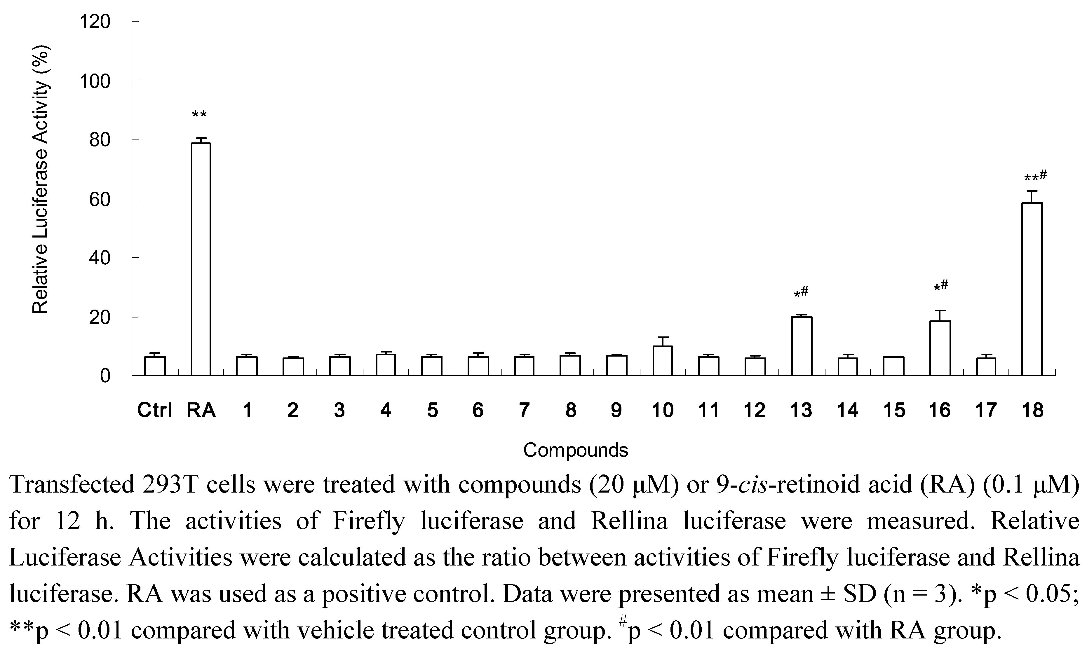
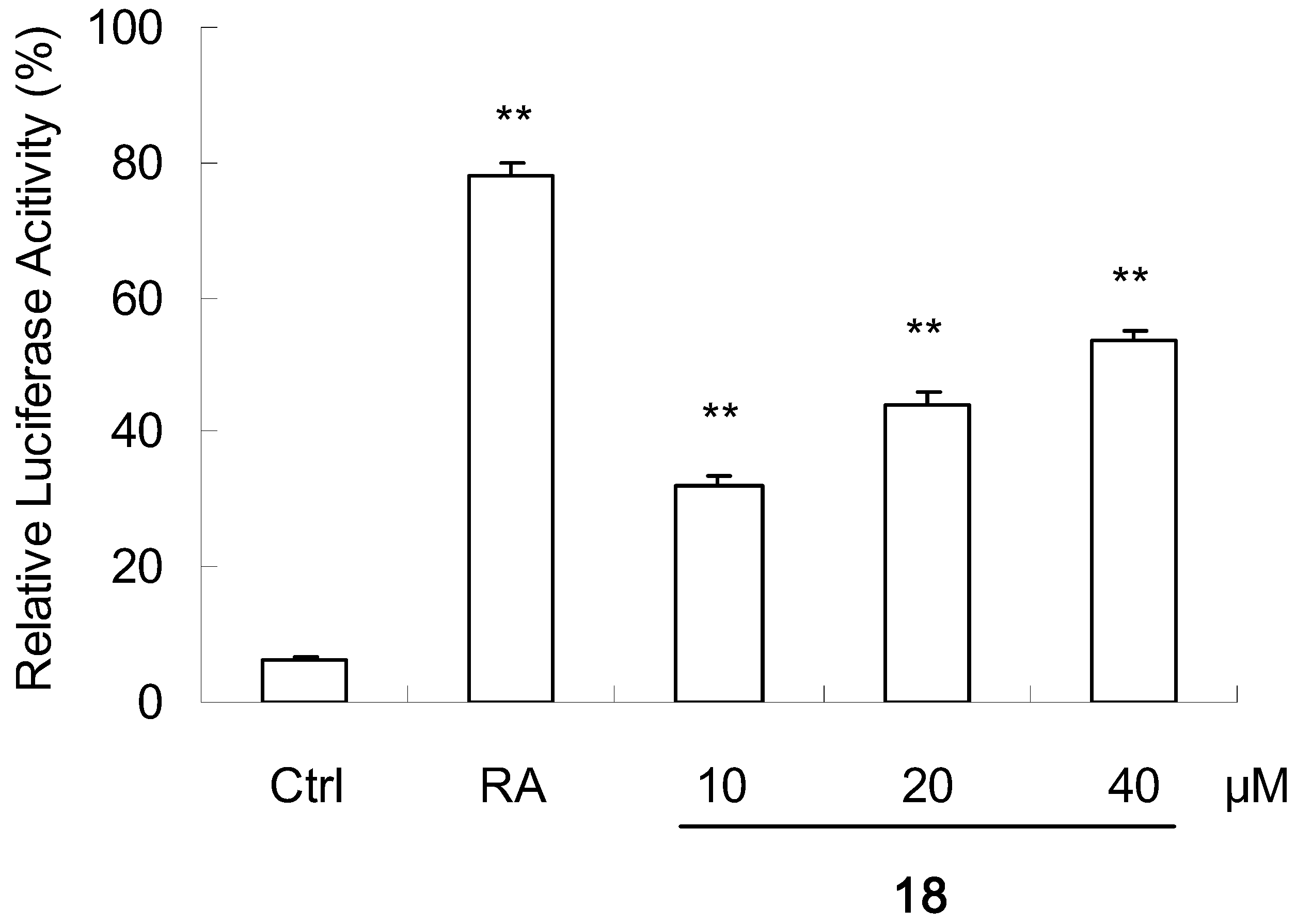
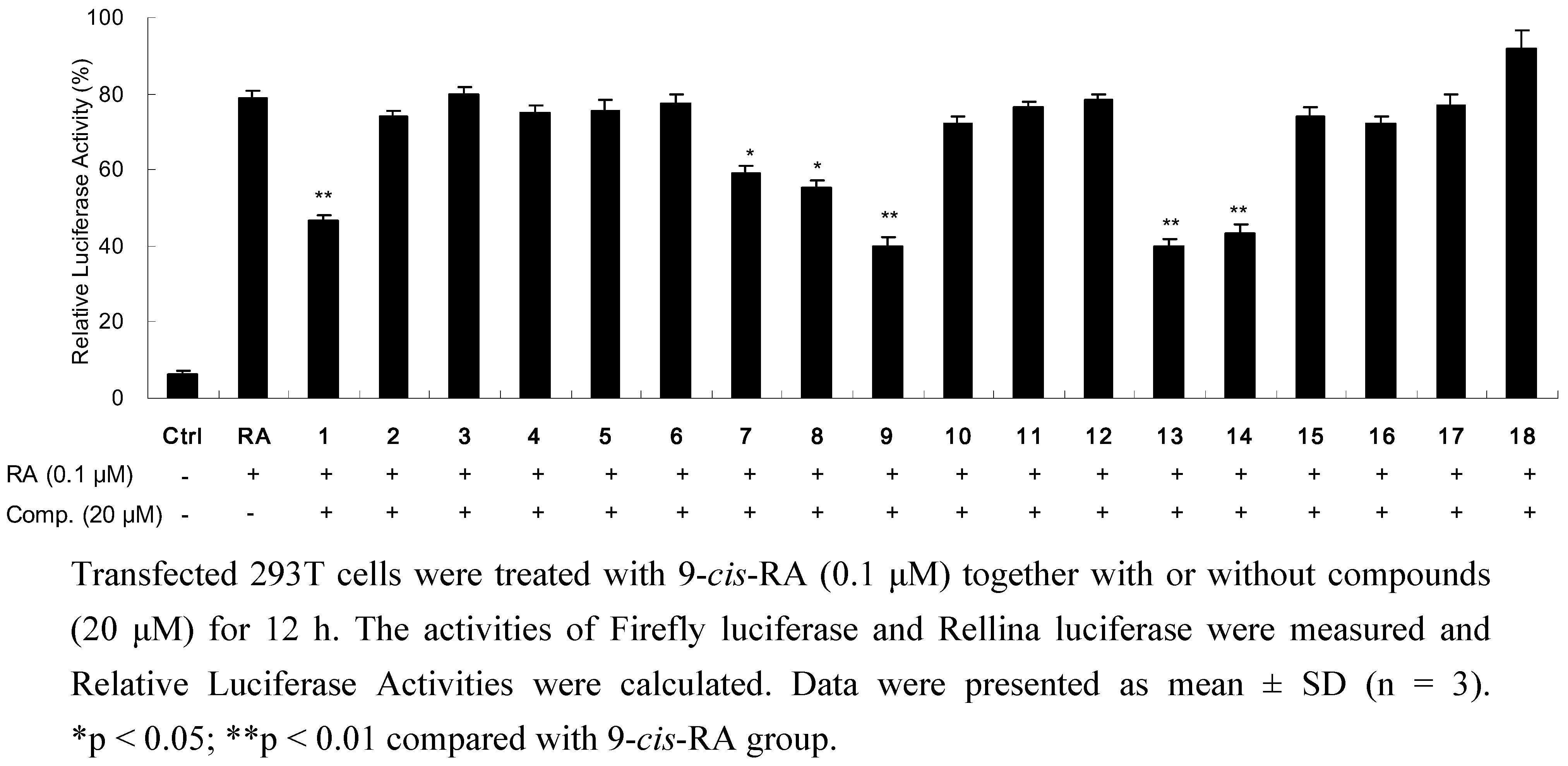
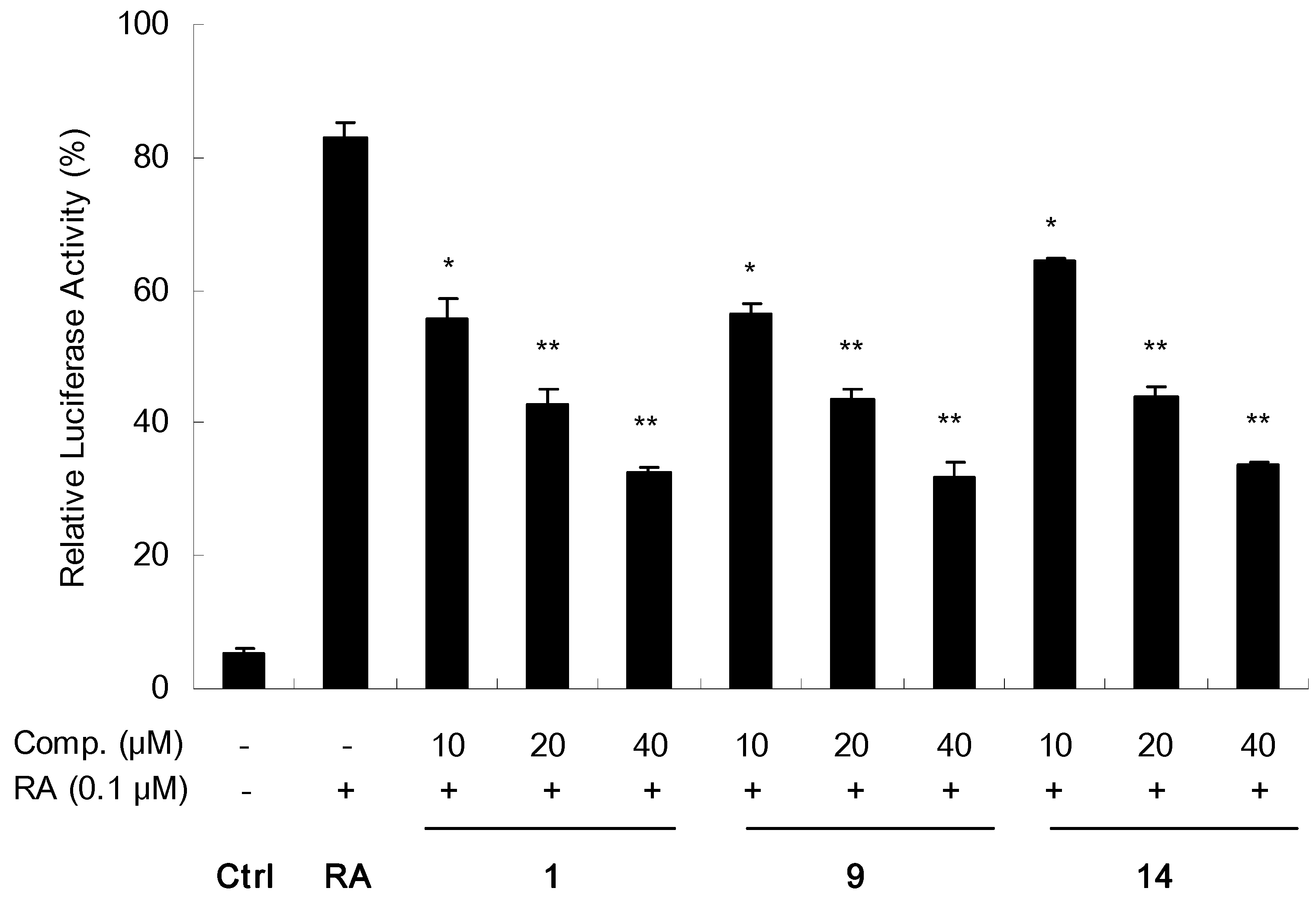
2. Results and Discussion
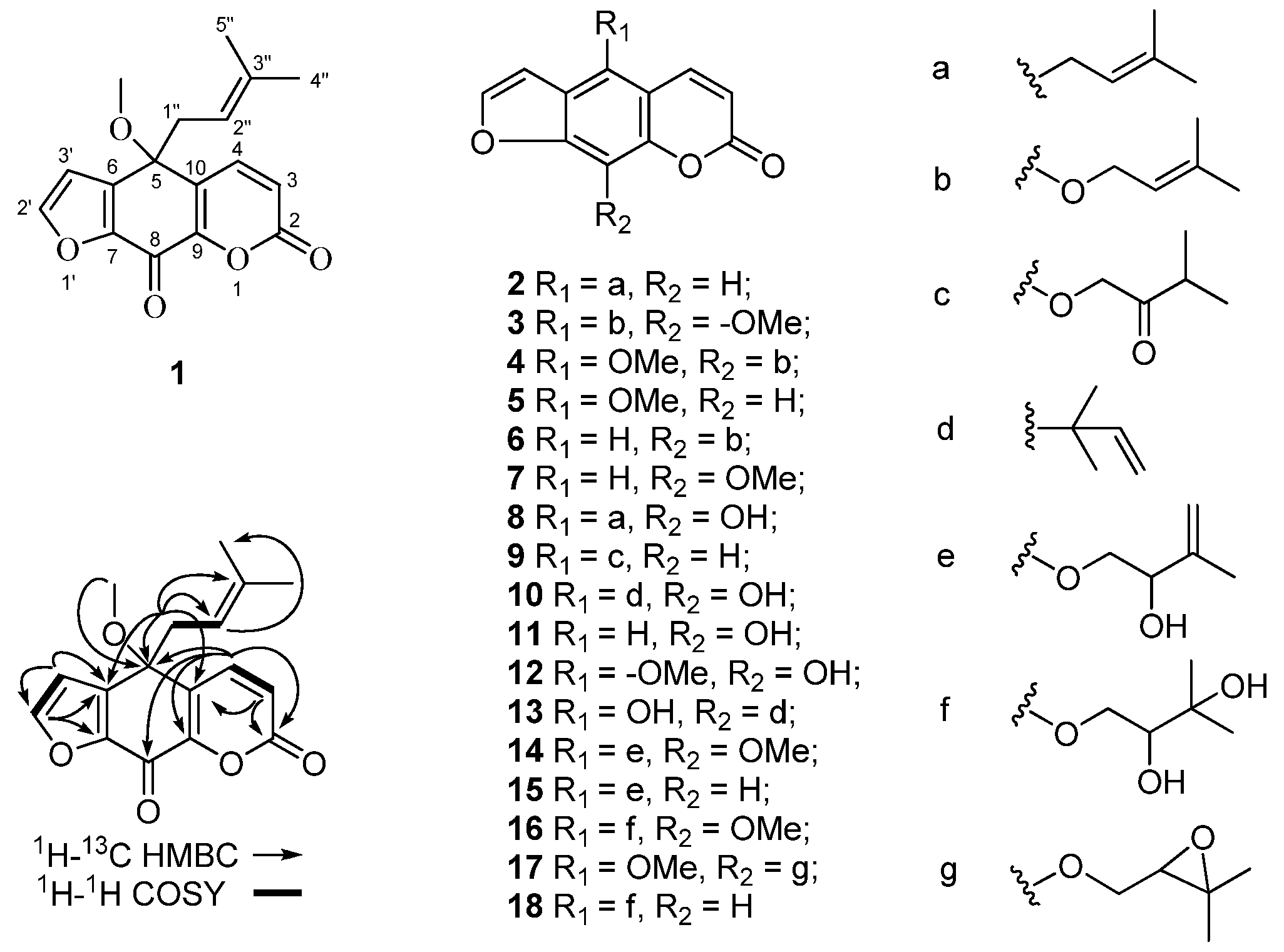
Chromatography of the EtOAc-soluble extract of the Radix Angelicae Dahuricae produced a novel furocoumarin derivative 1, together with seventeen furocoumarins 2-18. The known compounds were identified as isoimperatorin (2) [16], cnidilin (3) [17], phellopterin (4) [18], bergapten (5) [18], imperatorin (6) [19], xanthotoxin (7) [5], alloimperatorin (8) [16], isooxypeucedanin (9) [20], isodemethylfuropinarine (10) [21], xanthotoxol (11) [16], 5-methoxy-8-hydroxypsoralen (12) [22], demethylfuropinarine (13) [23], apaensin (14) [24], pabulenol (15) [25], isobyakangelicin (16) [26], byakangelicol (17) [18] and oxypeucedanin hydrate (18) [17], respectively. Among them, 10 and 13 were isolated from A. dahurica for the first time.
Compound 1 was obtained as a white amorphous powder, [α]22D +5 (c 0.4, MeOH). Its molecular formula, C17H16O5, was established by HR-ESI-MS with a mass of [M + Na]+ (m/z 323.0894, calcd. 323.0890). The IR spectrum of 1 showed characteristic absorptions of α,β-unsaturated lactone (νmax 1738 cm–1), and α,β-unsaturated carbonyl (νmax 1687 cm–1) groups. The 1H-NMR spectrum of 1 showed two methyl, one methoxyl, five olefinic, and one methylene proton. The 13C-NMR, and DEPT spectra of 1 displayed 18 carbons, including two methyls, one methoxyl, five olefinic methines, two ketone carbonyl, and five quarternary carbons (Table 1). Proton signals were all allocated by observation of HMQC correlations from δH 6.69 (1H, d, J = 9.6 Hz) to δC 120.2, δH 7.90 (1H, d, J = 9.6 Hz) to δC 140.7, δH 8.09 (1H, d, J = 2.0 Hz) to δC 149.6, δH 6.92 (1H, d, J = 2.0 Hz) to δC 110.0, δH 2.84 (2H, br d, J = 7.6 Hz) to δC 39.1, δH 4.78 (1H, tq, J = 7.6, 1.2 Hz) to δC 115.9, δH 1.53 (3H, d, J = 1.2 Hz) to δC 24.7, δH 1.42 (3H, d, J = 1.2 Hz) to δC 17.1, and δH 3.03 (3H, s) to δC 51.8. The above 1H- and 13C-NMR data were partly similar to those of alloimperatorin (8), which is a furocoumarin conjugated a prenyl unit [16]. Firstly, the proton signal of olefinic methine at δH 4.78 (tq, J = 7.6, 1.2 Hz) showed the same couple constants as two methyl protons (δH 1.53, d, J = 1.2 Hz and 1.42, d, J = 1.2 Hz) and methylene protons at δH 2.84 (br d, J = 7.6 Hz). HMBC spectra showed correlations from the olefinic triplet (δH 4.78) to the methyl resonances δC 24.7, from the methylene proton (δH 2.84) to δC 115.9 and 136.3, from the two methyl proton resonances (δH 1.53 and 1.42) to δC 115.9 and 136.3. The above data together with the 1H-1H COSY correlations between δH 4.78 and the methylene proton at δH 2.84 indicated the presence of a prenyl unit in 1. Secondly, a pyrone ring residue of coumarin including resonances of δC 158.7, 120.2, 140.7, 150.9, 126.6 and δH 6.69, 7.90 could be identified by combining analysis of 1H-, 13C-NMR and HMBC spectra. In which, two coupling proton signals at δH 6.69 (1H, d, J = 9.6 Hz, H-3) and δH 7.90 (1H, d, J = 9.6 Hz, H-4) assigned to the double bond protons of pyrone ring presented HMBC correlations with δC 158.7 (C-2), 150.9 (C-9), and 126.6 (C-10). In addition, HMBC correlations from δH 8.09 (1H, d, J = 2.0 Hz) and 6.92 (1H, d, J = 2.0 Hz) to δC 138.4 and 147.3 suggested the presence of a furan ring which included resonances of δH 8.09 (H-2′), 6.92 (H-3′), δC 149.6 (C-2′), 110.0 (C-3′), 138.4 (C-6), and 147.3 (C-7). Interestingly, the methylene proton (δH 2.84) of prenyl was largely upfield shifted comparing with that of 8, with chemical shift at δH 3.72 (2H, d, J = 7.2 Hz), indicating that the prenyl was no longer attached to an unsaturated carbon of the furocoumarin moiety. The observation of HMBC correlations from δH 2.84 to carbon signals at δC 76.4, 126.6, and 138.4 illustrated that the prenyl located at the quaternary carbon at δC 76.4 which was adjacent to the pyrone and furan ring. In addition, the resonance δC 164.7 at downfield shift in 13C-NMR spectrum could be assigned as a conjugated ketone carbon according with the presence of α, β-unsaturated carbonyl stretching vibration (νmax 1687 cm–1) in IR spectrum. Long-rang HMBC correlations from H-4 (δH 7.90) to δC 164.7 suggested the adjacency relation between the pyrone ring and the ketone carbon. The location of the methoxyl substituent was confirmed by observing the long-range correlation from methoxyl proton signal at δH3.03 (3H, s) to the quaternary carbon at δC 76.4. Thus, the chemical structure of 1 was finally elucidated as illustrated in Figure 1, and was named oxyalloimperatorin. The stereochemistry of C-5 remains to be determined.
Figure 1. Chemical structures of compounds 118 from Radix Angelicae Dahuricae and the Key HMBC and 1H-1H COSY correlations of compound 1.
Molecules 16 06339 g001
Table 1. 1H-NMR and 13C-NMR data of compound 1 (Acetone-d4, 400 MHz).
PositionδC, mult.δH (J in Hz)
2158.7, C
3120.2, CH6.69 d (9.6)
4140.7, CH7.90 d (9.6)
576.4, C
6138.4, C
7147.3, C
8164.7, C
9150.9, C
10126.6, C
2'149.6, CH8.09 d (2.0)
3'110.0, CH6.92 d (2.0)
1"39.1, CH22.84 br d (7.6)
2"115.9, CH4.78 tq (7.6, 1.2)
3"136.3, C
4"24.7, CH31.53 d (1.2)
5"17.1, CH31.42 d (1.2)
5-OCH351.8, CH33.03 s
All isolated compounds were furocoumarin skeleton derivatives. Their effects on gene transactivity of RXRα were evaluated by a Dual-Luciferase reporter assay system. It is well known that 9-cis-retinoid acid (9-cis-RA) could largely promote the reporter transcription [27]. As shown in Figure 2, consistent with previous results, treatment of cells with 9-cis-RA (0.1 μM) significantly (P < 0.01) induced the reporter transcription with a relative luciferase activity of 79%.
Figure 2. Promoting effects of isolated compounds on reporter transcription activities of RXRα.
Molecules 16 06339 g002
Among the isolated compounds, 18 (20 μM) significantly (P < 0.01) increased the transcriptional activation of RXRα, while 13 and 16 (each at 20 μM) showed weak effects (P < 0.05) on increasing the reporter transcription (Figure 2). Furthermore, 18 with concentration-dependent effect in the range of 10~40 μM is shown in Figure 3.
Figure 3. Promoting effect of compound 18 on reporter transcription activities of RXRα with concentration-dependence.
Molecules 16 06339 g003
All the isolated compounds were also examined for their possibility to inhibit the transactivity of RXRα using a similar assay as previous report, in which 9-cis-RA was simultaneously added to strongly induce RXRα gene transactivity [14]. Among tested compounds, 79, 13, 14, and the new structure 1 (each at 20 μM) showed significant reduction in the relative luciferase activity induced by 9-cis-RA (Figure 4). In addition, 1, 9 and 14 were further measured in three different concentrations at 10, 20 and 40 μM. As shown in Figure 5, all three metabolites exhibited good concentration-dependent inhibitory effects.
The above findings indicate that these furocoumarin skeleton derivatives might have useful impact on many intractable diseases, such as cancer and metabolic diseases, due to their potential effects on regulating the transcriptional activation function of RXRα. Interestingly, compound 13 showed not only weakly increased the reporter transcriptional activation of RXRα but it also reduced the transactivity of RXRα induced by 9-cis-RA. One of the possible reasons could be that 13 and 9-cis-RA bind competitively with RXRα when they were simultaneously added to cells. Because of the transactive effect of 13 was rather weaker than that of 9-cis-RA (Figure 2), the transactivity of RXRα induced by 9-cis-RA was therefore decreased. However, this speculation and whether all the candidate compounds could bind to RXRα to regulate its transcriptional expression or not needs to be confirmed by further study.
Figure 4. Inhibitory effects of isolated compounds on reporter transcription activities of RXRα.
Molecules 16 06339 g004
Figure 5. Inhibitory effects of compounds 1, 9 and 14 on reporter transcription activities of RXRα in concentration-dependent manner.
Molecules 16 06339 g005

3. Experimental

3.1. General

High-performance liquid chromatography (HPLC)-grade solvents were purchased from Merck KGaA (Darmstadt, Germany). Analytical reagents were obtained from Sinopharm Chemical Reagent co., Ltd (Shanghai, China). Silica gel (200–300 mesh) used in column chromatography and TLC plates were bought from Qingdao Haiyang Chemical Co., Ltd (Qingdao, China). YMC gel ODS-A was purchased from YMC co., Ltd (Allentown, USA). NMR spectra were recorded on a Bruker Avance 400 spectrometer using tetramethylsilane as the internal reference. HR-ESI-MS were recorded on Bruker FT-MS. IR spectrum was carried on a Nicolet IR200 (Thermo Electron Corporation, U.S.A.). Rotation data was obtained from a 341 polarimeter (PerkinElmer Co. Ltd. U.S.A). The values of luciferases were measured on a 1420 VICTOR3TM V (PerkinElmer, Boston, MA, USA).

3.2. Materials

Radix Angelicae Dahuricae, the dry root of A. dahurica (Fisch. ex Hoffm) Benth, et Hook. f, was purchased from a store of Tongrentang Pharmacy (Hangzhou City, Zhejiang Province, China) and identified by Mrs. Xiuhong Zhou (Senior Engineer, Forestry Bureau of Yongchun, Quanzhou City, China). A voucher specimen was deposited at the School of Pharmaceutical Sciences in Xiamen University, Xiamen, China. Plasmids (pBind RXRα LBD and pG5 luc) were provided by Dr. Xiao-kun Zhang from the Burnham Institute for Medical Research, Cancer Center, La Jolla, CA, USA. Dual-Luciferase Reporter Assay System Kit was purchased from Promega Corporation. Lipofectamine 2000 reagent was bought from Invitrogen Co., Ltd.

3.3. Extraction and Isolation

The dried root of A. dahurica (3 kg) was boiled and refluxed for 2 h with 60% of aqueous ethanol solution (5 L × 3 times). After filtration, the extracted solution was concentrated in vacuo. The condensate was then suspended in H2O (5 L) and partitioned with EtOAc (5 L × 3 times). The EtOAc extracts were combined and evaporated under vacuum to afford an EtOAc-soluble extract (48.3 g). The EtOAc-soluble extract (45.0 g) was chromatographed on silica gel column using stepwise gradient elution with CHCl3-MeOH (100:0~0:100) to obtain 11 fractions (Fr. 1-11). Fr.2 (12.1 g) was subjected to silica gel column chromatography eluting with n-hexane-EtOAc (98:2~1:1) to get 10 subfractions (Fr.2-1 ~ Fr.2-10). Fr.2-5 (1.9 g) was applied to YMC ODS column chromatography and eluted with aqueous acetonitrile (55~100%) to give compound 2 (257.0 mg). Fr.2-6 (1.3 g) was subjected to YMC gel ODS column chromatography and eluted with aqueous methanol (40~100%) to give 3 (14.0 mg). Fr.2-7 (2.0 g) was chromatographed on YMC gel ODS-A column and eluted with aqueous methanol (50~100%) to give 4 (300 mg) and a subfraction (Fr.2-7-2) which was further purified through preparative HPLC (Restek Prinnacle DB C18, 5 μm, 250×10 mm) eluting with 60% of aqueous methanol solution to afford 5 (40.0 mg, Rt 14.0 min) and 6 (61.0 mg, Rt 17.5 min). Fr.2-8 (782.0 mg) was subjected to YMC gel column chromatography using the elution of aqueous methanol (30~100%) to yield 7 (13.0 mg). Fr.2-9 (738.0 mg) was applied to YMC gel column chromatography and eluted with aqueous methanol solution (40~100%) to produce 8 (58.0 mg) and a subfraction (Fr.2-9-2) which was further purified through preparative HPLC eluting with MeOH-H2O (60:40) to afford 9 (39.0 mg, Rt 6.5 min) and 10 (23.0 mg, Rt 10.5 min). Fr.2-10 (883.0 mg) was subjected to YMC gel column and eluted with aqueous methanol solution (40~100%) to get 11 (44.0 mg), 12 (21.0 mg), 1 (12.0 mg), and subfraction Fr.2-10-4 (100 mg) was chromatographed on YMC gel to get 13 (14.0 mg) and Fr.2-10-4-1 which was further purified through preparative HPLC eluting with MeOH-H2O (70:30) to afford 14 (29.0 mg, Rt 6.5 min) and 15 (38.0 mg, Rt 7.5 min). Fr.4 (2.5 g) was applied to YMC gel column chromatography with aqueous methanol solution (40~100%) as elution to afford 16 (256.0 mg). Fr.5 (2.0 g) was subjected to silica gel column eluted with EtOAc-MeOH (96:4~0:100) to afford Fr.5-7(155 mg) which was crystallized in chloroform to obtain 17 (50.0 mg). Fr.7 (5.18 g) was subjected to silica gel column chromatography eluted with CHCl3-MeOH (99:1~0:100) to afford Fr.7-3. The subfraction (0.8 g) was applied to silica gel column and eluted with CHCl3-Acetone (95:5~0:100) to afford 18 (131.0 mg).
Oxyalloimperatorin (1): white, amorphous powder, [α]22D +5 (c 0.4, MeOH). UV (MeOH) λmax 361 (log ε 3.50) nm; IR (MeOH) νmax 2923, 1738, 1687, 1625, 1438, 1082 cm–1; 1H-NMR (400 MHz, acetone-d4) and 13C-NMR (100 MHz, acetone-d4), see Table 1; HR-ESI-MS m/z 323.0894 [M + Na]+ (calcd for C17H16O5, 323.0890), m/z 339.0643 [M + K]+ (calcd 339.0629).

3.4. Cell Culture and Dual-Luciferase Reporter Gene Assay

Human embryonic kidney 293T cells were cultured in DMEM medium containing 10% fetal bovine serum (FBS). The previous dual-luciferase reporter gene assay with some modification was used in the present study [14,27]. In brief, approximately 4 × 10⁴ cells/well were seeded in 24-well plates. Cells were transfected with two plasmids, 30 ng pBind RXRα LBD and 60 ng pG5 luc using Lipofectamine 2000 (Invitrogen). After 24 h, cells were exposed to tested compounds for 12 h. Then, the cells were washed with PBS and lysed with passive lysis buffer (1 × PLB) on rocking platform for 15 min. The activities of Firefly luciferase and Rellina luciferase were examined according to the introduction of Dual-Luciferase Reporter Assay System Kit. Relative luciferase activities were obtained as the ratio between activities of Firefly luciferase and Rellina luciferase.

3.5. Statistical Analysis

The results were expressed as mean ± standard diviation (S.D.) from at least three independent experiments. Statistical significances were compared between two groups. Statistical analysis was performed with the Student’s t-test. The value of P < 0.05 was considered statistically significant.

4. Conclusions

One novel furocoumarin derivative 1, together with seventeen furocoumarins 2–18 were isolated from the radix of Angelica dahurica. The chemical structure of new metabolite was characterized by analysis of IR, NMR, and HR-ESI-MS spectroscopic data. Among these furocoumarin skeleton derivatives, the new compound 1, and known compounds 7–9, 13, 14, 16 and 18 showed the potential activities in regulating transcriptional activation function of RXRα. These metabolites might show beneficial effects against intractable diseases with relation to RXRα, for example anti-cancer and anti-diabetes. The various bioactivities of these metabolites and their molecular mechanism of action relating to nuclear receptor RXRα could be examined in the future study.

Acknowledgments

This work was financially supported by the Xiamen Science and Technology Key program grant (No. 3502Z20100006), Fujian major program grant (No. 2009Y3004), the Fundamental Research Funds for the Central Universities (No. 20101211000) and the National Natural Science Foundation of China (NSFC) (No. 30873146)

References

  1. Liu, R.M.; Li, A.F.; Sun, A.L. Preparative isolation and purification of coumarins from Angelica dahurica (Fisch. ex Hoffm) Benth, et Hook. f (Chinese traditional medicinal herb) by high-speed counter-current chromatography. J. Chromatogr. A 2004, 1052, 223–227. [Google Scholar]
  2. Kimura, Y.; Okuda, H.; Baba, K. Histamine-release effectors from Angelica dahurica var. dahurica root. J. Nat. Prod. 1997, 60, 249–251. [Google Scholar] [CrossRef]
  3. Kwon, Y.S.; Kobayashi, A.; Kajiyama, S.I.; Kawazu, K.; Kanzaki, H.; Kim, C.M. Antimicrobial constituents of Angelica dahurica roots. Phytochemistry 1997, 44, 887–889. [Google Scholar]
  4. Kimura, Y.; Ohminami, H.; Arichi, H.; Okuda, H.; Baba, K.; Kozawa, M.; Arichi, S. Effects of various coumarins from roots of Angelica dahurica on actions of adrenaline, ACTH and insulin in fat cells. Planta Med. 1982, 45, 183,187. [Google Scholar]
  5. Thanh, P.N.; Jin, W.Y.; Song, G.Y.; Bae, K.H.; Kang, S.S. Cytotoxic Coumarins from the root of Angelica dahurica. Arch. Pharm. Res. 2004, 27, 1211–1215. [Google Scholar] [CrossRef]
  6. Pae, H.O.; Oh, H.; Yun, Y.G.; Oh, G.S.; Il Jang, S.; Hwang, K.M.; Kwon, T.O.; Lee, H.S.; Chung, H.T. Imperatorin, a furanocoumarin from Angelica dahurica (Umbelliferae), induces cytochrome c-dependent apoptosis in human promyelocytic leukaemia, HL-60 cells. Pharmacol. Toxicol. 2002, 91, 40–48. [Google Scholar] [CrossRef]
  7. Kim, E.K.; Kwon, K.B.; Shin, B.C.; Seo, E.A.; Lee, Y.R.; Kim, J.S.; Park, J.W.; Park, B.H.; Ryu, D.G. Scopoletin induces apoptosis in human promyeloleukemic cells, accompanied by activations of nuclear factor kappa B and caspase-3. Life Sci. 2005, 77, 824–836. [Google Scholar] [CrossRef]
  8. Yang, J.Y.; Della-Fera, M.A.; Baile, C.A. Esculetin induces mitochondria-mediated apoptosis in 3T3-L1 adipocytes. Apoptosis 2006, 11, 1371–1378. [Google Scholar] [CrossRef]
  9. Szanto, A.; Narkar, V.; Shen, Q.; Uray, I.P.; Davies, P.J.A.; Nagy, L. Retinoid X receptors: X-ploring their (patho)physiological functions. Cell Death Differ. 2004, 11, S126–S143. [Google Scholar]
  10. Zhou, H.; Liu, W.; Su, Y.; Wei, Z.; Liu, J.; Kolluri, S.K.; Wu, H.; Cao, Y.; Chen, J.; Wu, Y.; et al. NSAID sulindac and its analog bind RXRα and inhibit RXRα-dependent AKT signaling. Cancer Cell 2010, 17, 560–573. [Google Scholar] [CrossRef]
  11. Mukherjee, R.; Davies, P.J.A.; Crombie, D.L.; Bischoff, E.D.; Cesario, R.M.; Jow, L.; Hamann, L.G.; Boehm, M.F.; Mondon, C.E.; Nadzan, A.M.; et al. Sensitization of diabetic and obese mice to insulin by retinoid X receptor agonists. Nature 1997, 386, 407–410. [Google Scholar]
  12. Lenhard, J.M.; Lancaster, M.E.; Paulik, M.A.; Weiel, J.E.; Binz, J.G.; Sundseth, S.S.; Gaskill, B.A.; Lightfoot, R.M.; Brown, H.R. The RXR agonist LG100268 causes hepatomegaly, improves glycaemic control and decreases cardiovascular risk and cachexia in diabetic mice suffering from pancreatic beta-cell dysfunction. Diabetologia 1999, 42, 545–554. [Google Scholar] [CrossRef]
  13. Duan, Y.H.; Dai, Y.; Wang, G.H.; Chen, H.F.; Gao, H.; Chen, J.B.; Yao, X.S.; Zhang, X.K. Xanthone and benzophenone glycosides from the stems of Cratoxylum formosum ssp. pruniflorum. Chem. Pharm. Bull. 2011, 59, 231–234. [Google Scholar]
  14. Duan, Y.H.; Dai, Y.; Wang, G.H.; Zhang, X.; Chen, H.F.; Chen, J.B.; Yao, X.S.; Zhang, X.K. Bioactive Xanthones from the stems of Cratoxylum formosum ssp. pruniflorum. J. Nat. Prod. 2010, 73, 1283–1287. [Google Scholar] [CrossRef]
  15. Kotani, H.; Tanabe, H.; Mizukami, H.; Makishima, M.; Inoue, M. Identification of a naturally occurring rexinoid, honokiol, that activates the retinoid X receptor. J. Nat. Prod. 2010, 73, 1332–1336. [Google Scholar]
  16. Kang, J.; Zhou, L.; Sun, J.H.; Han, J.; Guo, D.A. Chromatographic fingerprint analysis and characterization of furocoumarins in the roots of Angelica dahurica by HPLC/DAD/ESI-MSn technique. J. Pharm. Biomed. Anal. 2008, 47, 778–785. [Google Scholar]
  17. Baek, N.I.; Ahn, E.M.; Kim, H.Y.; Pauk, Y.D. Furanocoumarins from the root of Angelica dahurica. Arch. Pharm. Res. 2000, 23, 467–470. [Google Scholar] [CrossRef]
  18. Bergendorff, O.; Dekermendjian, K.; Nielsen, M.; Shan, R.; Witt, R.; Ai, J.; Sterner, O. Furanocoumarins with affinity to brain benzodiazepine receptors in vitro. Phytochemistry 1997, 44, 1121–1124. [Google Scholar]
  19. Wei, Y.; Ito, Y. Preparative isolation of imperatorin, oxypeucedanin and isoimperatorin from traditional Chinese herb "bai zhi" Angelica dahurica (Fisch ex Hoffm) Benth. et Hook using multidimensional high-speed counter-current chromatography. J. Chromatogr. A 2006, 1115, 112–117. [Google Scholar]
  20. Shin, K.H.; Moon, K.H.; Woo, W.S. Two minor furanocoumarins of Angelica dahurica. Arch. Pharm. Res. 1991, 14, 165–166. [Google Scholar] [CrossRef]
  21. Murray, R.D.H.; Jorge, Z.D.; Boag, D.M. Claisen rearrangements - XIV. Synthesis of the coumarin, benahorin and revision of the structure of marmelide. Tetrahedron 1984, 40, 5225–5227. [Google Scholar]
  22. Avramenko, L.G.; Sklyar, Y.E.; Pimenov, M.G. Coumarins of Peucedanum baicalense. Khim. Prir. Soedin. 1975, 11, 421–422. [Google Scholar]
  23. Kozawa, M.; Fukumoto, M.; Matsuyama, Y.; Baba, K. Chemical studies on the constituents of the Chinese crude drug "Quiang Huo". Chem. Pharm. Bull. 1983, 31, 2712–2717. [Google Scholar] [CrossRef]
  24. Snu, H.; Lin, Z.; Niu, F.; Ding, J. Studies on the Chinese drugs of Umbelliferae. IV. Structure of apaensin. Yunnan Zhiwu Yanjiu 1981, 3, 279–281. [Google Scholar]
  25. Basa, S.C.; Chatterjee, J.; Chatterjee, A. Pabulenol, a biological transformation product of oxypeucedanin. Tetrahedron Lett. 1971, 12, 1977–1978. [Google Scholar] [CrossRef]
  26. Thastrup, O.; Lemmich, J. Furocoumarin glucosides of Angelica archangelica subspecies litoralis. Phytochemistry 1983, 22, 2035–2037. [Google Scholar]
  27. Zhang, X.-K.; Lehmann, J.; Hoffmann, B.; Dawson, M.I.; Cameron, J.; Graupner, G.; Hermann, T.; Tran, P.; Pfahl, M. Homodimer formation of retinoid X receptor induced by 9-cis retinoic acid. Nature 1992, 358, 587–591. [Google Scholar]

