Article

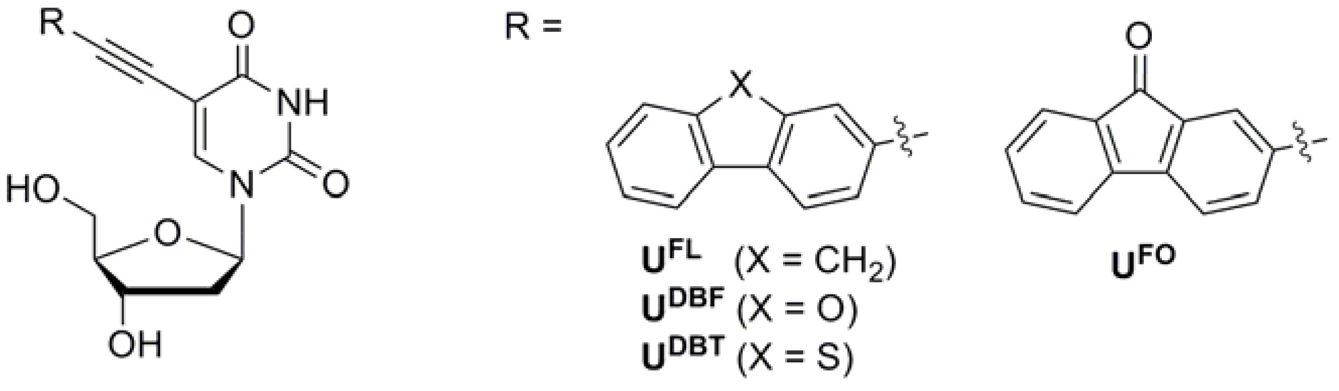
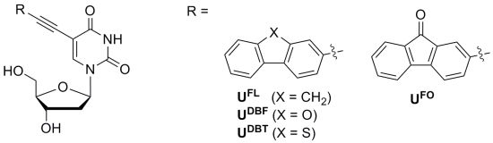
Synthesis and Photophysical Study of 2′-Deoxyuridines Labeled with Fluorene Derivatives

1
Department of Chemistry and Green-Nano Materials Research Center, Kyungpook National University, Daegu 702-701, Korea
2
Molecular Imaging Research Center, Korea Institute of Radiological and Medical Sciences, 75 Nowon-gil, Seoul 139-706, Korea
*
Authors to whom correspondence should be addressed.
Molecules 2012, 17(10), 12061-12071; https://doi.org/10.3390/molecules171012061
Submission received: 14 September 2012 / Revised: 3 October 2012 / Accepted: 10 October 2012 / Published: 15 October 2012
(This article belongs to the Special Issue Nucleic Acid Analogs)

Abstract

:
We examined microenvironment-sensitive fluorescent 2′-deoxyuridines labeled with fluorene derivatives that exhibited solvent-dependent photophysical properties. The high sensitivity of the fluorescence shift and the nucleoside intensity dependence on solvent polarity provided information useful for estimating the polarity of the environment surrounding the fluorescent nucleoside.

Graphical Abstract

1. Introduction

Fluorescent nucleosides which are structurally noninvasive, forming stable Watson-Crick base pairs, and sensitive to their physical conditions and molecular species in solution, exhibiting environmental-specific changes in their fluorescent properties, have become powerful tools for the investigation of nucleic acid structure, recognition of single nucleotide polymorphisms (SNPs), and studies on enzymatic processes involving DNA [1,2,3,4,5,6,7,8].
In order to design fluorescent nucleosides, we utilized an ethynyl linker at the 5 position of uracil to maintain the hybridization properties of the parent nucleoside. This substitution is expected to have very little influence on the stability of the resulting duplex DNA [9,10,11,12,13,14,15,16,17,18,19,20]. Among fluorophores, fluorene derivatives have moderate quantum yields and are less bulky than other commonly used fluorophores, e.g., pyrene, fluorescein, rhodamine, and cyanine dyes [21]. Previously, we reported fluorene (FL)- and 9-fluorenone (FO)-labeled deoxyuridine (UFL and UFO), which we incorporated at the central positions of oligodeoxynucleotides in an attempt to examine the effect of electronic modification of the fluorophore scaffold on the potential of the molecular beacon (MB) for SNP typing (Figure 1) [9,10,11]. When such a quencher-free MB hybridizes with its perfectly matched target DNA, it exhibits strong fluorescence. In contrast, when it forms duplexes with single-base-mismatched target DNAs, the UFL and UFO units display quenched fluorescence as a result of photoinduced charge transfer originating from interactions with neighboring nucleobases. These changes in fluorescence are extremely dependent on the electronic and conformational microenvironments of the flanking bases. Therefore, we sought to synthesize other fluorescent uridines labeled with new FL derivatives, dibenzofuran (DBF) and dibenzothiophene (DBT), in order to examine changes in their photophysical properties through modifications of the fluorene unit and to develop these nucleosides as microenvironment-sensitive fluorescent nucleosides [17,22,23]. Although FL, FO, DBF, and DBT are structural analogs that differ only in the type of atoms bridging the two aromatic rings, they have dramatically different photophysical properties [24,25,26]. Here, we report the synthesis and photophysical properties of fluorescent FL derivative-conjugated 2′-deoxyuridine analogs.
Figure 1. Fluorescent nucleosides used in this study.
Molecules 17 12061 g001

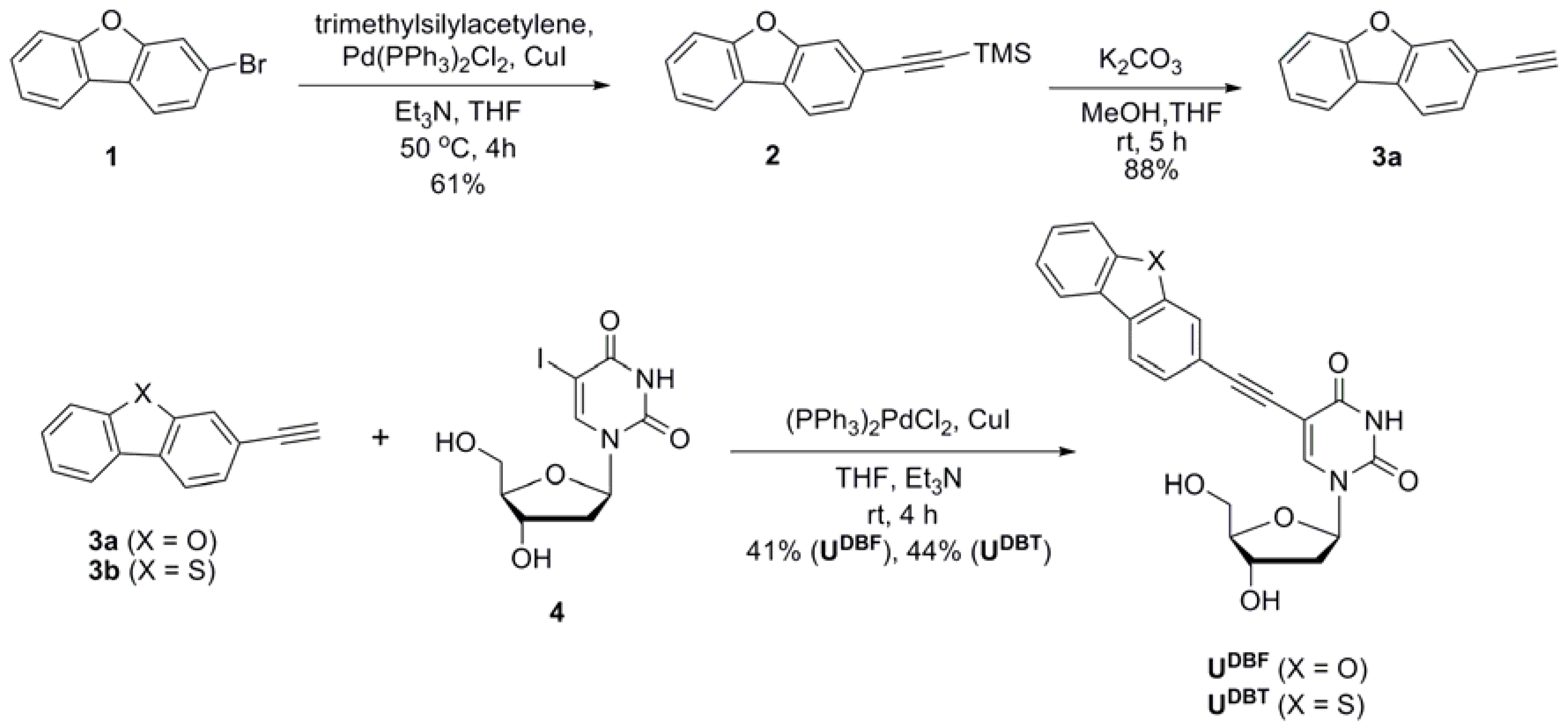
2. Results and Discussion
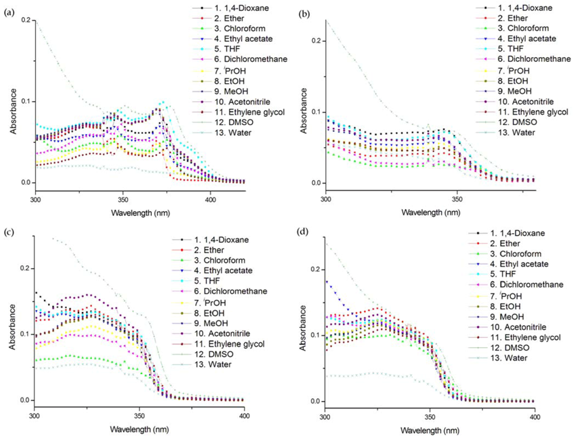
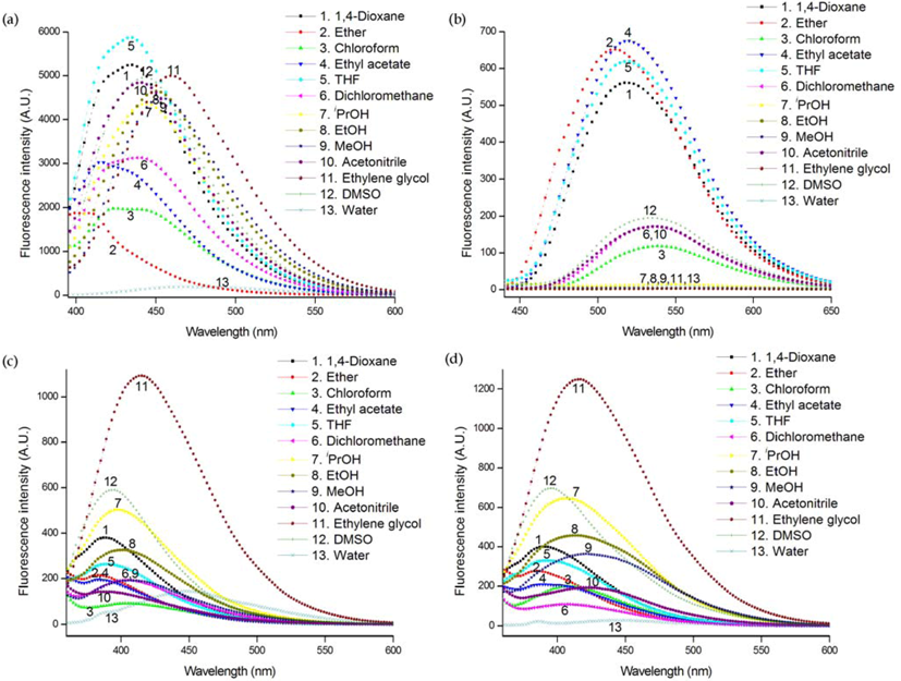
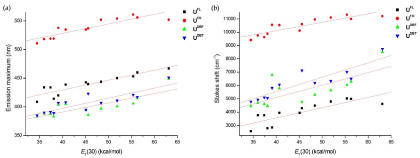
The synthetic route of the DBF- and DBT-labeled 2′-deoxyuridine derivatives UDBF and UDBT is outlined in Scheme 1. 3-Ethynyldibenzofuran (3a) was prepared by Pd/Cu-catalyzed Sonogashira coupling [27,28] of 3-bromodibenzofuran (1) with trimethylsilylacetylene followed by desilylation. 3-Ethynyldibenzothiophene (3b) was also synthesized according to the reported protocol [29]. We synthesized UDBF and UDBT from the corresponding 2′-deoxy-5-iodouridine (4) through a palladium catalyzed cross-coupling reaction with 3-ethynyldibenzofuran (3a) or 3-ethynyldibenzothiophene (3b). The syntheses of UFL and UFO were conducted as reported [9,10,11].
Generally, solvent polarity is of primary interest when considering environmental effects [30]. Therefore, we first measured the absorption and emission spectra of nucleosides in thirteen solvents of different polarities. Solvent marginally affected the absorption, probably due to the weak interaction between the nucleosides and solvent in the ground state (Figure 2). However, solvent polarity had a significant influence on both the emission maximum and intensity (Figure 3). All nucleosides exhibited different emission intensities and maxima depending on the solvent they were in, indicating that they are all environmentally sensitive.
Scheme 1. Route for the synthesis of UDBF and UDBT.
Molecules 17 12061 g002
Figure 2. Absorption spectra of (a) UFL (3 μM), (b) UFO (3 μM), (c) UDBF (5 μM), and (d) UDBT (5 μM) in different solvents at 25°C. All samples contain 0.5% THF/MeOH (1:1 v/v) to ensure solubility.
Molecules 17 12061 g003
Figure 3. Emission spectra of (a) UFL, (b) UFO, (c) UDBF, and (d) UDBT in different solvent at 25°C (all at 3 μM concentration). The excitation wavelengths were 370 nm for UFL and 340 nm for the others. All samples contain 0.5% THF/MeOH (1:1 v/v) to ensure solubility.
Molecules 17 12061 g004
Table 1 summarizes the photophysical properties of nucleosides in thirteen different solvents. The fluorescence quantum yields (ΦF) of the nucleosides were determined using a 0.1 N aqueous H2SO4 solution of quinine sulfate (λex = 350 nm) as a standard [31]. There are some noteworthy features: (a) generally, the presence of a heteroatom in the fluorene unit of nucleoside UFO, UDBF, and UDBT diminishes its fluorescence yield and fluorescence brightness (i.e., the product of its molar extinction coefficient and quantum yield) drastically when compared with UFL. (2) UDBF and UDBT showed very similar photophysical properties in various solvents. (3) The quantum yield and fluorescence brightness of nucleosides is highest in iPrOH for UFL, ethyl acetate for UFO, and ethylene glycol for UDBF and UDBT. The lowest fluorescence brightness, however, was observed in ethylene glycol for UFO and water for UFL, UDBF, and UDBT. These results indicate that the nucleosides exhibit highly solvent-dependent photophysical properties despite their structural similarities. UFO, interestingly, exhibited a strong solvent dependency–namely, higher fluorescence brightness in aprotic solvents relative to those in protic solvents such as iPrOH, EtOH, MeOH, ethylene glycol, and water which was attributable to the hydrogen bonding between the carbonyl group of UFO and solvent.
Table 1. Photophysical characteristics of nucleosides in different solvents at 25°C.
SolventCompoundET(30)11λmax (nm) aε (M−1 cm−1)λem (nm) bΦF cBrightness d
1,4-DioxaneUFL3637325,0004340.338,250
Ether 34.537027,2004090.0551,500
Chloroform 39.137520,1004200.183,620
Ethyl acetate 38.137119,9004140.142,790
THF 37.437329,7004340.319,200
Dichloromethane 40.737422,2004390.235,100
iPrOH 48.437020,9004440.5010,500
EtOH 51.937024,4004500.256,100
MeOH 55.436925,1004530.266,530
Acetonitrile 45.637026,2004400.287,340
Ethylene glycol 56.337412,9004600.273,480
DMSO 45.137727,1004430.184,880
Water 63.13844,9304670.062305
1,4-DioxaneUFO3634420,8005180.0561,160
Ether 34.534521,2005110.0801,700
Chloroform 39.134314,3005380.029415
Ethyl acetate 38.134319,4005190.0901,750
THF 37.434621,9005190.0641,400
Dichloromethane 40.734215,8005350.040632
iPrOH 48.434416,5005520.003456.1
EtOH 51.934316,3005540.0009715.8
MeOH 55.434217,9005580.001425.1
Acetonitrile 45.634218,4005370.0219403
Ethylene glycol 56.334514,0005560.0007610.6
DMSO 45.134721,5005350.018387
Water 63.134113,2005520.003850.2
1,4-DioxaneUDBF3632823,3003880.0491,140
Ether 34.532727,3003830.029792
Chloroform 39.131717,1004040.0861,470
Ethyl acetate 38.132724,5003830.027662
THF 37.432924,7003880.035865
Dichloromethane 40.732821,4004050.044942
iPrOH 48.432822,5003970.0861,940
EtOH 51.932724,3004010.0741,800
MeOH 55.432623,0004060.0471,080
Acetonitrile 45.632624,0003860.026624
Ethylene glycol 56.332921,3004150.234,900
DMSO 45.1nd end e3940.084nd e
Water 63.132511,7004490.047550
1,4-DioxaneUDBT3632726,5003900.0471,250
Ether 34.532528,9003580.020578
Chloroform 39.132920,5004070.0501,030
1,4-DioxaneUDBT3632726,5003900.0471,250
Ethyl acetate 38.132524,2003890.024581
THF 37.432624,9003910.033822
Dichloromethane 40.732724,6004080.029713
iPrOH 48.432524,3004070.0641,560
EtOH 51.932622,9004110.0611,400
MeOH 55.432524,4004210.0451,100
Acetonitrile 45.632524,8004230.020496
Ethylene glycol 56.332821,9004170.112,410
DMSO 45.1nd end e3950.082nd e
Water 63.13248,6004510.007967.9
a Only the largest absorption maxima are listed; b Wavelength of emission maximum when excited at the absorption maximum; c Quantum efficiencies using 0.1 N aqueous H2SO4 solution of quinine sulfate as a standard, λex = 350 nm. Data shown are the mean values of three independent experiments; d The fluorescence brightness = ε × ΦF; e Not detectable due to overlapping absorption bands of a nucleoside and DMSO.
In polar solvents such as iPrOH, EtOH, MeOH, acetonitrile, ethylene glycol, DMSO, and water substantially larger red-shifts in emission maxima of nucleosides were observed. Because it is instructive to calculate the magnitude of the expected spectral shifts due to solvent polarity effects, we plotted the fluorescence emission maxima and Stokes shifts (νabsνem) of nucleosides in thirteen different solvents against Reichardt’s microscopic solvent parameter, ET(30) (Figure 4) [32]. It is interesting to note that there is a linear correlation between emission maxima and ET(30) regardless of the aproticity of the solvent. The red-shift of the fluorescence could be due to the significant difference between the excited‐state charge distribution in the solute and the ground‐state charge distribution, resulting in stronger interactions with polar solvents in the excited state.
Figure 4. Effect of ET(30) on (a) the fluorescence emission maxima and (b) the Stokes shifts of nucleosides.
Molecules 17 12061 g005
Emission maxima of UFO were red-shifted relative to those of other nucleosides. This higher Stokes shift of UFO is probably because the carbonyl group allows for hydrogen bonding and charge separation better than do the other nucleosides [30]. Interestingly, the Stokes shifts of UDBF and UDBT exhibited a more gradual shift to longer wavelengths with increasing solvent polarity compared to the slopes of other nucleosides, as shown in Figure 4b. In order to compare the sensitivity of our molecules of interest to environmental polarity with that of reported polarity-sensitive nucleosides [12,33], we examined the photophysical properties of fluorescent nucleosides in binary water/1,4-dioxane mixtures (Table S1, Figure 5), which is an established method for estimating the microenvironment polarity of fluorophores [34]. The Stokes shifts plotted against the ET(30) values of the samples is shown in Figure 5. The slopes obtained from the linear plots indicated that UFO, UDBF, and UDBT are highly sensitive to environmental polarity and are comparable to the slopes of reported nucleosides such as pyridine- and furan-labeled uridines. UDBF and UDBT revealed a seemingly exponential trend, leading us to conclude that a more appropriate expression for the interactions between these nucleosides and solvents should be explored.
Figure 5. Dependence of the Stokes shift of nucleosides in water/1,4-dioxane binary solvent mixture on the empirical solvent polarity parameter, ET(30).
Molecules 17 12061 g006

3. Experimental

3.1. General

All reactions were performed in dry glassware under Ar atmospheres. Analytical thin layer chromatography (TLC) was performed using Merck 60 F254 silica gel plates; column chromatography was performed using Merck 60 silica gel (230–400 mesh). Melting points were determined using an Electrothermal IA 9000 series melting point apparatus and are uncorrected. Infrared (IR) spectra were recorded using a JASCO FT/IR-4100 spectrometer. 1H- and 13C-NMR spectra were recorded using a Bruker NMR spectrometer (AVANCE digital 400 MHz). High-resolution electron impact (EI) mass spectra were recorded using a JEOL JMS-700 mass spectrometer at the Daegu center of KBSI, Korea.

3.2. Materials

All commercially available chemicals were used without further purification; solvents were carefully dried and distilled prior to use. 3-Bromobenzofuran (1) [35] and 3-ethynyldibenzothiophene (3b) [29] have been reported previously. UFL and UFO were synthesized according to the reported protocol [9,10,11].

3.3. Preparation of 3-[2-(Trimethylsilyl)ethynyl]dibenzofuran (2)

A solution of 1 [35] (580 mg, 2.35 mmol), (PPh3)2PdCl2 (165 mg, 0.235 mmol), and CuI (44.8 mg, 0.235 mmol) in THF (12 mL) and Et3N (3.9 mL) was degassed with nitrogen. Trimethylsilylacetylene (500 μL, 3.52 mmol) was added at 50 °C and the mixture stirred for 4 h. After evaporation of solvent in vacuo, the residue was subjected to chromatography on a silica gel column with hexane as eluent to give 2 (380 mg, 61%): M.p. 110–113 °C; IR (film): ν 3063, 2954, 2896, 2144, 1453, 1416, 1340, 1315, 1248, 1201, 1133, 940, 831, 743, 629 cm–1; 1H-NMR (CDCl3): δ 7.93 (dq, J = 8.0, 0.67 Hz, 1H; H-6), 7.86 (dd, J = 8.0, 0.40 Hz, 1H; H-1), 7.67 (q, J = 0.80 Hz, 1H; H-4), 7.57 (dt, J = 8.0, 0.80 Hz, 1H; H-2), 7.49–7.45 (m, 2H; H-7 and H-9), 7.35 (td, J = 7.4, 0.80 Hz, 1H; H-8), 0.29 (s, 9H; SiCH3); 13C-NMR (CDCl3): δ 156.9, 155.7 127.8, 127.1, 124.8, 123.9, 123.1, 121.8, 121.0, 120.5, 115.3, 111.9, 105.3, 95.0, 0.1; HRMS–EI (m/z): [M]+ calcd for C17H16OSi 264.0970; found, 264.0968.

3.4. Preparation of 3-Ethynyldibenzofuran (3a)

A solution of 2 (600 mg, 2.27 mmol) and K2CO3 (345 mg, 2.25 mmol) in MeOH (6.7 mL) and THF (6.7 mL) was stirred at rt for 5 h. After evaporation of the solvent in vacuo, dichloromethane and water were added and the product was extracted into the organic phase which was then concentrated. The residue was purified by chromatography (SiO2; hexane/EtOAc, 10:1) to give 3a (385 mg, 88%): M.p. 83–86 °C; IR (film): ν 3264, 2920, 2854, 2098, 1641, 1595, 1446, 1364, 1193, 1107, 926, 880, 821, 742, 666, 606 cm–1; 1H-NMR (CDCl3): δ 7.94 (dq, J = 7.4, 0.8 Hz, 1H; H-6), 7.89 (dd, J = 8.0, 0.8 Hz, 1H; H-1), 7.70 (q, J = 0.53 Hz, 1H; H-4), 7.58 (dt, J = 8.0, 0.8 Hz, 1H; H-2), 7.50–7.46 (m, 2H: H-7 and H-9), 7.36 (td, J = 7.4, 0.80 Hz, 1H; H-8), 3.17 (s, 1H; CCH); 13C-NMR (CDCl3): δ 156.9, 155.7, 127.9, 127.1, 125.1, 123.8, 123.2, 121.1, 120.7, 120.6, 115.5, 111.6, 83.9, 77.8; HRMS–EI (m/z): [M]+ calcd for C14H8O 192.0575; found, 192.0573.

3.5. General Procedure for Nucleoside Synthesis

(PPh3)2PdCl2 (36.5 mg, 0.0520 mmol) and CuI (9.9 mg, 0.0520 mmol) were added to a solution of 2′-deoxy-5-iodouridine 2 (184 mg, 0.520 mmol) and 2-ethynylfluorene derivative 3 (0.520 mmol) in Et3N (2.6 mL) and THF (7.8 mL). Argon was bubbled through the mixture for 2 min before the mixture was subjected 10 times to a pump/purge cycle, and then it was stirred at rt for 4 h. After evaporation of solvent in vacuo, the residue was subjected to chromatography (SiO2; CH2Cl2/MeOH, 40:1) to yield UDBF (41%) or UDBF (44%).
2′-Deoxy-5-(3-dibenzofuranylethynyl)uridine (UDBF). M.p. >164 °C dec.; IR (film): ν 3383, 3162, 3049, 2922, 2855, 1664, 1455, 1275, 1195, 1099, 987, 860, 740, 633 cm–1; 1H-NMR (DMSO-d6): δ 11.74 (s, 1H; NH), 8.46 (s, 1H; H-6), 8.18 (dd, J = 7.8, 0.60 Hz, 2H; DBF-H), 7.81 (q, J = 0.67 Hz, 1H; DBF-H), 7.74–7.72 (m, 1H; DBF-H), 7.58–7.54 (m, 1H; DBF-H), 7.51 (dd, J = 7.8, 1.4 Hz, 2 H; DBF-H), 7.43 (td, J = 7.3, 0.6 Hz, 1H; DBF-H), 6.15 (t, J = 6.4 Hz, 1H; H-1′), 5.30 (d, J = 4.4 Hz, 1H; OH-3′), 5.23 (t, J = 4.8 Hz, 1H; OH-5′), 4.30–4.26 (m, 1H; H-3′), 3.83 (q, J = 3.3 Hz, 1H; H-4′), 3.71–3.59 (m, 2H; H-5′), 2.20–2.17 (m, 2H; H-2′); 13C-NMR (DMSO-d6): δ 161.5, 156.1, 155.1, 149.5, 144.2, 131.6, 128.3, 126.5, 124.0, 123.5, 123.1, 121.6, 121.3, 114.3, 111.8, 98.1, 92.0, 87.6, 84.9, 83.3, 69.9, 60.8; HRMS–EI (m/z): [M]+ calcd for C23H18N2O6, 418.1165; found, 418.1167.
2'-Deoxy-5-(3-dibenzothiophenylethynyl)uridine (UDBT). M.p. >165°C dec.; IR (film): ν 3377, 3155, 3053, 2923, 2852, 1660, 1455, 1272, 1228, 1195, 1094, 987, 919, 825, 747, 635 cm–1; 1H-NMR (DMSO-d6): δ 11.72 (s, 1H; NH), 8.46 (s, 1H; H-6), 8.40–8.38 (m, 2H; DBT-H), 8.178 (dd, J = 1.4, 0.60 Hz, 1H; DBT-H), 8.07–8.04 (m, 1H; DBT-H), 7.58 (dd, J = 8.2, 1.4 Hz, 1H; DBT-H), 7.55–7.53 (m, 2H; DBT-H), 6.144 (t, J = 6.402, 1H; H-1′), 5.30 (d, J = 4.4 Hz, 1H; OH-3′), 5.23 (t, J = 4.6 Hz, 1H; OH-5′), 4.30–4.26 (m, 1H; H-3′), 3.83 (q, J = 3.3 Hz, 1H; H-4′), 3.71–3.59 (m, 2H; H-5′), 2.20–2.16 (m, 2H; H-2′); 13C-NMR (DMSO-d6): δ 161.6, 149.6, 144.2, 139.3, 138.9, 135.0, 134.5, 127.6, 125.7, 125.0, 123.2, 122.4, 122.2, 120.9, 98.1, 91.9, 87.6, 84.9, 83.5, 79.2, 69.9, 60.8, 55.0; HRMS-EI (m/z): [M]+ calcd for C23H18N2O5S, 434.0936; found, 434.0935.

3.6. UV and Fluorescence Measurements

Ultraviolet (UV) spectra were recorded using a Cary 100 UV-Vis spectrophotometer and 10-mm-path quartz cell, with respect to a pure-solvent reference. Fluorescence spectra were recorded using a Hitachi F4500 spectrofluorometer. All samples were prepared from a stock solution in THF/MeOH (1:1 v/v) to ensure solubility, and hence, all samples contain 0.5% THF/MeOH (1:1 v/v). The excitation and emission bandwidth was 1 nm. The fluorescence quantum yields (ΦF) were determined using 0.1 N aqueous H2SO4 solution of quinine sulfate as a reference [31].

4. Conclusions

We designed structurally similar fluorescent 2′-deoxyuridine derivatives that exhibit solvent-dependent photophysical properties via drastic changes in emission intensity as well as emission wavelength. These microenvironment-sensitive nucleosides may be used as probes for investigating nucleic acid dynamics and the recognition process. A deeper understanding of how the photophysical properties relate to chemical structures may allow for the design of ideal environmentally sensitive fluorescent nucleosides towards the development of DNA probes. Efforts in these directions are currently in progress.

Supplementary Materials

Supplementary materials can be accessed at: https://www.mdpi.com/1420-3049/17/10/12061/s1.

Acknowledgments

This research was supported by Basic Science Research Program through the National Research Foundation of Korea (NRF) funded by the Ministry of Education, Science and Technology (2010-0007605 and 2012-0002831).
  • Sample Availability: Samples of the compounds used in this study are available from the authors.

References

  1. Ranasinghe, R.T.; Brown, T. Fluorescence based strategies for genetic analysis. Chem. Commun. 2005, 5487–5502. [Google Scholar] [CrossRef]
  2. Wilson, J.N.; Kool, E.T. Fluorescent DNA base replacements: Reporters and sensors for biological systems. Org. Biomol. Chem. 2006, 4, 4265–4274. [Google Scholar] [CrossRef]
  3. Venkatesan, N.; Seo, Y.J.; Bang, E.K.; Park, S.M.; Lee, Y.S.; Kim, B.H. Chemical modification of nucleic acids toward functional nucleic acid systems. Bull. Korean Chem. Soc. 2006, 28, 613–630. [Google Scholar]
  4. Venkatesan, N.; Seo, Y.J.; Kim, B.H. Quencher-free molecular beacons: A new strategy in fluorescence based nucleic acid analysis. Chem. Soc. Rev. 2008, 37, 648–663. [Google Scholar]
  5. Dodd, D.W.; Hudson, R.H.E. Intrinsically fluorescent base-discriminating nucleoside analogs. Mini-Rev. Org. Chem. 2009, 6, 378–391. [Google Scholar]
  6. Sinkeldam, R.W.; Greco, N.J.; Tor, Y. Fluorescent analogs of biomolecular building blocks: Design, properties, and application. Chem. Rev. 2010, 110, 2579–2619. [Google Scholar]
  7. Dai, N.; Kool, E.T. Fluorescent DNA-based enzyme sensors. Chem. Soc. Rev. 2011, 40, 5756–5770. [Google Scholar] [CrossRef]
  8. Østergaard, M.E.; Hrdlicka, P.J. Pyrene-functionalized oligonucleotides and locked nucleic acids (LNAs): Tools for fundamental research, diagnostics, and nanotechnolog. Chem. Soc. Rev. 2011, 40, 5771–5788. [Google Scholar] [CrossRef]
  9. Hwang, G.T.; Seo, Y.J.; Kim, B.H. A highly discriminating quencher-free molecular beacon for probing DNA. J. Am. Chem. Soc. 2004, 126, 6528–6529. [Google Scholar] [CrossRef]
  10. Ryu, J.H.; Seo, Y.J.; Hwang, G.T.; Lee, J.Y.; Kim, B.H. Triad base pairs containing fluorene unit for quencher-free SNP typing. Tetrahedron 2007, 63, 3538–3547. [Google Scholar]
  11. Ryu, J.H.; Heo, J.Y.; Bang, E.-K.; Hwang, G.T.; Kim, B.H. Quencher-free linear beacon systems containing 2-ethynylfluorenone-labeled 2′-deoxyuridine units. Tetrahedron 2012, 68, 72–78. [Google Scholar] [CrossRef]
  12. Sinkeldam, R.W.; Greco, N.J.; Tor, Y. Polarity of major grooves explored by using an isosteric emissive nucleoside. ChemBioChem 2008, 9, 706–709. [Google Scholar] [CrossRef]
  13. Østergaard, M.E.; Guenther, D.C.; Kumar, P.; Baral, B.; Deobald, L.; Paszczynski, A.J.; Sharma, P.K.; Hrdlicka, P.J. Pyrene-functionalized triazole-linked 2′-deoxyuridines-probes for discrimination of single nucleotide polymorphisms (SNPs). Chem. Commun. 2010, 46, 4929–4931. [Google Scholar]
  14. Varghese, R.; Wagenknecht, H.-A. Non-covalent versus covalent control of self-assembly and chirality of Nile red-modified nucleoside and DNA. Chem. Eur. J. 2010, 16, 9040–9046. [Google Scholar] [CrossRef]
  15. Bag, S.S.; Kundu, R.; Matsumoto, K.; Saito, Y.; Saito, I. Singly and doubly labeled base-discriminating fluorescent oligonucleotide probes containing oxo-pyrene chromophore. Bioorg. Med. Chem. Lett. 2010, 20, 3227–3230. [Google Scholar]
  16. Pawar, M.G.; Srivatsan, S.G. Synthesis, photophysical characterization, and enzymatic incorporation of a microenvironment-sensitive fluorescent uridine analog. Org. Lett. 2011, 13, 1114–1117. [Google Scholar]
  17. Saito, Y.; Miyamoto, S.; Suzuki, A.; Matsumoto, K.; Ishihara, T.; Saito, I. Fluorescent nucleosides with “on-off”switching function, pH-responsive fluorescent uridine derivatives. Bioorg. Med. Chem. Lett. 2012, 22, 2753–2756. [Google Scholar]
  18. Tanaka, M.; Oguma, K.; Saito, Y.; Saito, I. Enhancement of fluorescence quenching and exciplex formation in DNA major groove by double incorporation of modified fluorescent deoxyuridines. Bioorg. Med. Chem. Lett. 2012, 22, 4103–4106. [Google Scholar] [CrossRef]
  19. Barrois, S.; Beyer, C.; Wagenknecht, H.-A. Covalent modification of 2′-deoxyuridine with two different molecular switches. Synlett 2012, 23, 711–716. [Google Scholar] [CrossRef]
  20. Tanaka, M.; Oguma, K.; Saito, Y.; Saito, I. Drastic enhancement of excess electron-transfer efficiency through DNA by inserting consecutive 5-phenylethynyl-2'-deoxyuridines as a modulator. Chem. Commun. 2012, 48, 9394–9396. [Google Scholar]
  21. Parker, C.A. Photoluminescence of Solutions; Elsevier: Amsterdam, The Netherlands, 1968. [Google Scholar]
  22. Saito, Y.; Koda, M.; Shinohara, Y.; Saito, I. Synthesis and photophysical properties of 8-arylbutadienyl 2′-deoxyguanosines. Tetrahedron Lett. 2011, 52, 491–494. [Google Scholar] [CrossRef]
  23. Saito, Y.; Shinohara, Y.; Ishioroshi, S.; Suzuki, A.; Tanaka, M.; Saito, I. Synthesis of environmentally sensitive 2′-deoxyguanosine containing solvatochromic pyrene dluorophore. Tetrahedron Lett. 2011, 52, 2359–2361. [Google Scholar]
  24. Panozzo, S.; Vial, J.-C.; Kervella, Y.; Stéphan, O. Fluorene-fluorenone copolymer: Stable and efficient yellow-emitting material for electroluminescent devices. J. Appl. Phys. 2002, 92, 3495–3502. [Google Scholar] [CrossRef]
  25. Józefowicz, M. Determination of reorganization energy of fluorenone and 4-hydroxyfluorenone in neat and binary solvent mixtures. Spectrochim. Acta A 2007, 67, 444–449. [Google Scholar] [CrossRef]
  26. Nguyen, D.D.; Trunk, J.; Nakhimovsky, L.; Spanget-Larsen, J. Electronic transitions of fluorene, dibenzofuran, carbazole, and dibenzothiophene: From the onset of absorption to the ionization threshold. J. Mol. Spectrosc. 2010, 264, 19–25. [Google Scholar] [CrossRef]
  27. Sonogashira, K.; Tohda, Y.; Hagihara, N. A convenient synthesis of acetylenes: Catalytic substitutions of acetylenic hydrogen with bromoalkenes, iodoarenes and bromopyridines. Tetrahedron Lett. 1975, 16, 4467–4470. [Google Scholar] [CrossRef]
  28. Hwang, G.T.; Son, H.S.; Ku, J.K.; Kim, B.H. Synthesis and photophysical studies of bis-enediynes as tunable fluorophores. J. Am. Chem. Soc. 2003, 125, 11241–11248. [Google Scholar]
  29. Sekine, C.; Ishitobi, M.; Iwakura, K.; Minai, M.; Fujisawa, K. Novel high birefringence dibenzothiophenylacetylene liquid crystals. Liq. Cryst. 2002, 29, 355–367. [Google Scholar] [CrossRef]
  30. Lakowicz, J.R. Principles of Fluorescence Spectroscopy, 3rd ed; Springer: New York, NY, USA, 2006; pp. 205–235. [Google Scholar]
  31. Eastman, J.W. Quantitative spectrofluorimetry—The fluorescence quantum yield of quinine sulfate. Photochem. Photobiol. 1967, 6, 55–72. [Google Scholar] [CrossRef]
  32. Reichardt, C. Solvatochromic dyes as solvent polarity indicators. Chem. Rev. 1994, 94, 2319–2358. [Google Scholar] [CrossRef]
  33. Sinkeldam, R.W.; Marcus, P.; Uchenik, D.; Tor, Y. Multisensing emissive pyrimidine. Chem. Phys. Chem. 2011, 12, 2260–2265. [Google Scholar] [CrossRef]
  34. Sinkeldam, R.W.; Tor, Y. To D or not to D? On estimating the microenvironment polarity of biomolecular cavities. Org. Biomol. Chem. 2007, 5, 2523–2528. [Google Scholar] [CrossRef]
  35. Cullinane, N.M.; Padfield, H.J.H. Investigations in the diphenylene oxide series. Part V. J. Chem. Soc. 1935, 1131–1134. [Google Scholar]

