Communication

Growth Inhibition of Streptococcus from the Oral Cavity by α-Amyrin Esters

by
Gloria Díaz-Ruiz
1,
Liliana Hernández-Vázquez
2,
Héctor Luna
2,
María Del Carmen Wacher-Rodarte
1 and
Arturo Navarro-Ocaña
1,*
1
Departamento de Alimentos y Biotecnología, Facultad de Química, UNAM. Circuito Exterior, Ciudad Universitaria 04510, Mexico
2
Departamento de Sistemas Biológicos, Universidad Autónoma Metropolitana-Xochimilco, Calz. del Hueso 1100, Col. Villa Quietud 04960, Mexico
*
Author to whom correspondence should be addressed.
Molecules 2012, 17(11), 12603-12611; https://doi.org/10.3390/molecules171112603
Submission received: 19 September 2012 / Revised: 19 October 2012 / Accepted: 22 October 2012 / Published: 25 October 2012
(This article belongs to the Section Natural Products Chemistry)

Abstract

:
Five terpenoids were tested by the macrodilution broth method to determine their inhibition activity on cariogenic bacterial growth. In general, α-, β-amyrin and α-amyrin phenylacetate proved to be active, reducing the bacterial viability to less than 20%.

1. Introduction

The terpenoids α-amyrin (3) and β-amyrin (5) are commonly found in medicinal plants and oleo-resins obtained by bark incision of several species of Bursera or Protium spp. of the Burseraceae family (Figure 1) [1]. They have shown various pharmacological activities in vitro and in vivo against several health-related conditions, including microbial, fungal, and viral infections, cancer and inflammation. In addition, there are abundant examples of α-amyrin-containing plants used in folk medicine. For example Strobilanthes callosus, which has anti-inflammatory and antimicrobial activities [2], and Decalepis hamiltonii, which is used as an appetite stimulant, blood purifier and food preservative [3,4].
α/β-Amyrin modulate acute periodontal inflammation by reducing neutrophils infiltration, oxidative stress, and the production of pro-inflammatory cytokine TNF-α, which suggests they might be useful as therapeutic agents for the treatment of gingivitis and periodontitis [5]. Other studies have demonstrated that the α/β-amyrin mixture also has anti-inflammatory, gastroprotective, antiallergenic and antinociceptive activities [6].
The antifungal activity of amyrin and some 3-O-acyl derivatives has been evaluated against Candida species. Additionally, inhibition of the adhesion of C. albicans to human epithelial cells in vitro was also determined. Amyrin formate and acetate derivatives were the most active compounds, inhibiting all the opportunistic Candida species when tested in concentrations from 30 to 250 mg/mL. The formate also showed interesting inhibition of the adhesion ability of C. albicans [7]. β-Amyrin acetate isolated from Heliotropium marifolum showed potent activity against Penicillum chrysogenum, Escherichia coli and Klebsiella pneumonia [8].
Dental caries is a common bacterial pathology caused by a biofilm consisting of a multitude of microorganisms present on the tooth surface [9], being streptococci the bacteria most frequently associated with cariogenesis [10]. One method for caries prevention is the use of antimicrobial agents to limit the growth of microorganisms such as Streptococcus mutans. Extracts of miswak, tea tree oil, peppermint, green tea and manuka honey have all been recently incorporated into mouth rinses and toothpastes [11,12,13,14].
Some compounds with potential activity against oral microorganisms have been found in Nature, e.g., S. mutans was sensitive to totarol, a diterpenoid isolated from the bark of Podocarpus nagi with MICs of 0.78 μg/mL and MBC of 0.78 μg/mL [15]. The methanolic extract from the bark of B. crassifolia and the β-amyrin isolated therefrom inhibited the growth of bacteria, including S. mutans, at concentrations ranging from 64 to 1088 μg/mL; the minimum inhibitory concentration (MIC) for β-amyrin was 128 μg/mL [16].
The aim of this work was to isolate β-amyrin from nanche (Byrsonima crassifolia) and α-amyrin from copal (Bursera spp) and prepare some esters for the evaluation of their capacity to inhibit the growth of oral streptococci associated with dental caries.

2. Results and Discussion

2.1. Isolation of α- and β-Amyrin

The triterpenes α-amyrin (3) and β-amyrin (5) were isolated from copal and nanche bark in 7.0% and 2.0%, respectively [17]. Because in preliminary studies a greater activity was observed with α-amyrin than with β-amyrin, we decided to prepare some esters of α-amyrin to assay their activity against cariogenic microorganisms.

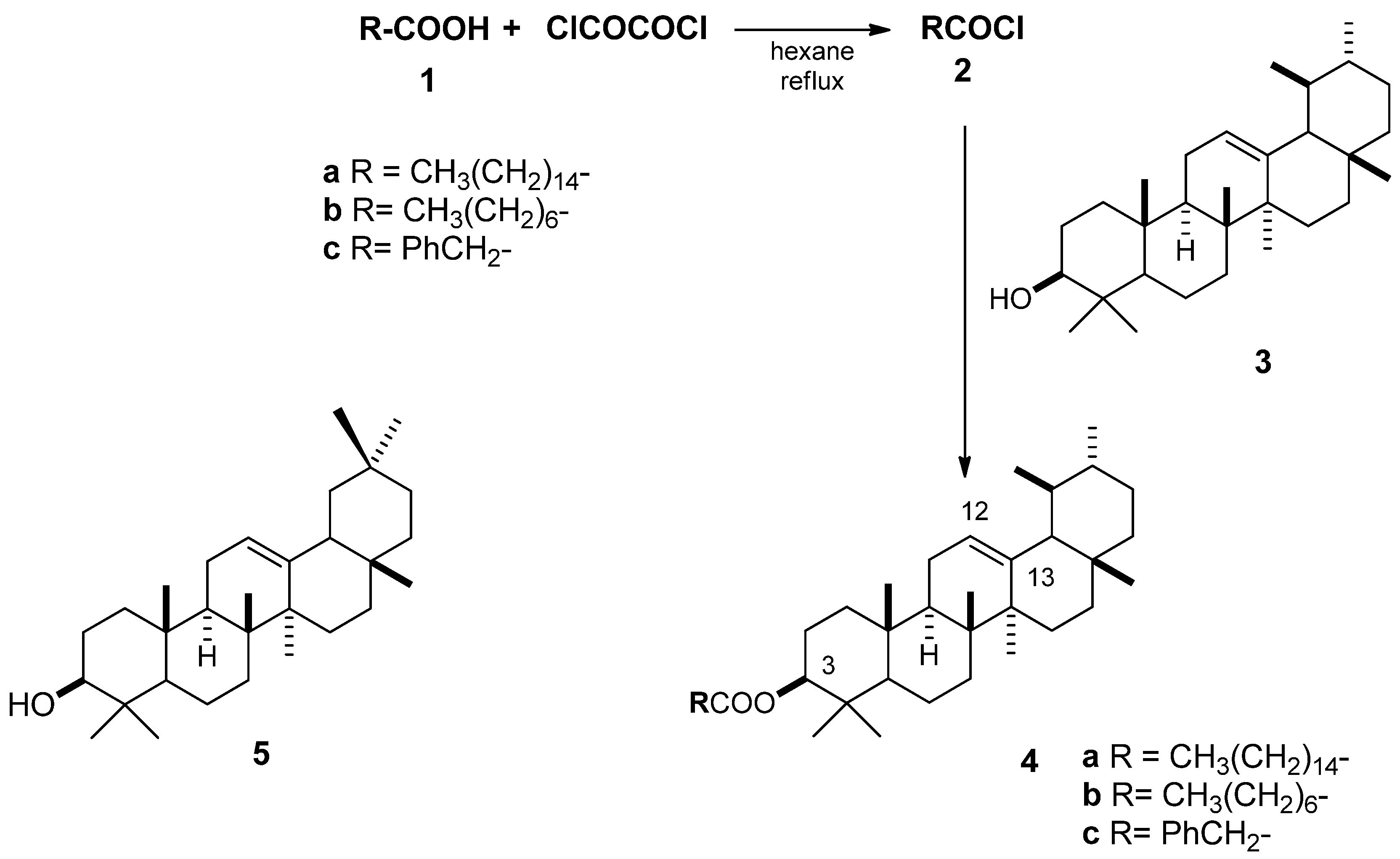
2.2. Esterification of α-Amyrin

Three esters of α-amyrin 4ac were synthesized in good to excellent yields (70–98%) (Scheme 1). The spectroscopic data of 4a and 4b are in agreement with those reported in the literature [6,18]. To the best of our knowledge, α-amyrin phenylacetate (4c) is reported and completely characterized for the first time. Its IR spectra showed an absorption band at 1724 cm−1 assigned to C=O of the ester group, and absorption bands between 1404–1583 of (C=C) for the aromatic ring. In the 1H-NMR spectrum of 4c, signals at 0.75–2.03 ppm showed the characteristic terpene profile. The protons alpha to the C=O group of the phenylacetate fragment appeared as a singlet at 3.72 ppm; H-3 appeared as a double-doublet at 4.48 ppm with a J = 9.2, 5.6 Hz; and H-12 as a triplet at 5.11 ppm, with a J = 3.6 Hz. The five protons corresponding to the aromatic fragment appeared as multiplets at 7.24–7.33 ppm. In the 13C-NMR the signal for C-5 was at 55.0 ppm, C-3 at 81.2 ppm, C-12 at 123.9 ppm, C-13 at 139.3 ppm, and the C=O appeared at 170.9 ppm.
Scheme 1. Structure of α and β-amyrin and esterification reactions of α-amyrin.
Molecules 17 12603 g002

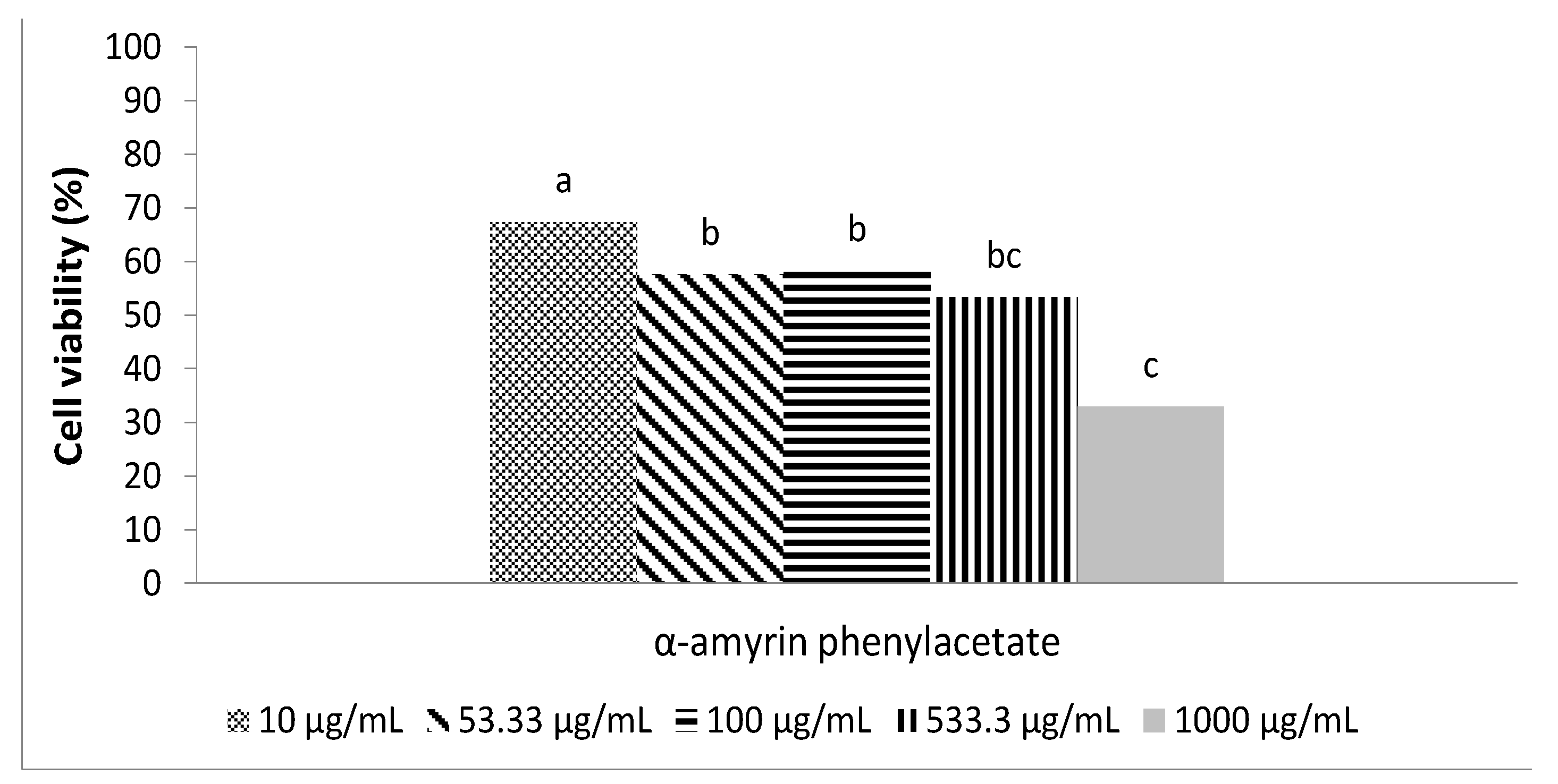
2.3. Antimicrobial Activity Using the Macrodilution Broth Method

In this work, five compounds (namely 3, 4ac, 5) were screened for antimicrobial activity using the macrodilution broth method, which is commonly use in the susceptibility assay for the antimicrobial evaluation of organic compounds [19]. Two concentrations of S. mutans were used, a lower (1 × 10³ cfu/mL) and a higher concentration (1 × 10⁶ cfu/mL), because a low concentration of S. mutans is associated with caries-free subjects, and the tooth surfaces susceptible to becoming carious are associated with a higher concentration [20].
During the experiments, chlorhexidine dihydrochloride (CHX) was used as a positive control, since produced 0% cell viability for all the assayed strains. The negative control consisted of a broth solution with 3.6 µL of Tween 80, which resulted in 100% cell viability for all strains tested.
To obtain information about the dosis-response relationship, S. mutans (at 106 cfu/mL) was exposed to the five compounds α-amyrin (3), β-amyrin (5), α-amyrin palmitate (4a), α-amyrin octanoate (4b) and α-amyrin phenylacetate (4c) (Scheme 1), at the following concentrations: 10, 53.33, 100, 533.33, 1,000 μg/mL. The results shown in Table 1 indicate that the palmitate (83–43% viability) and the octanoate esters were less active (89–39% viability), the phenylacetate (19–9% viability) being the most active, followed by α-amyrin (3) and β-amyrin (5). It is worth mentioning that the effect of the highest concentration of α-amyrin (16% viability) was comparable with the effect produced by the lowest concentration of α-amyrin phenylacetate (19% viability), which proved to be about 100 times more active than α-amyrin (3). The results showed statistically significant differences between concentrations according the Kruskal-Wallis test, at α = 0.05.
Table 1. Percentage of viability of Streptococcus mutans after 24 h exposure to the five terpenes at the concentrations used in this study. The viability was adjusted using BHI broth + Tween 80 as a control (100%).The initial cellular concentration was adjusted to 106 cfu/mL.
CompoundTerpene concentration (μg/mL)
10 53.33100 533.331000
α-amyrin (3)6246252516
β-amyrin (5)7261584637
α-amyrin palmitate (4a)8382684643
α-amyrin octanoate (4b)8962604039
α-amyrin phenylacetate (4c)191612129
Continuing the study, activity against S. salivarius, S. sanguinis, S. oralis (at 1 × 10⁶ cfu/mL) was tested, but only with the three most active compounds, using the same range of concentrations, and the results are shown in Table 2.
Table 2. Percentage of viability, related to terpene concentration, of Streptococcus salivarius, Streptococcus sanguinis and Streptococcus oralis after 24 h exposure to three selected derivatives at the concentrations used in this study 10–1,000 (µg/mL). The viability has been adjusted using BHI broth + Tween 80 as a control (100%).The initial cellular concentration was adjusted to 106 cfu/mL.
CompoundS. salivariusS. sanguinisS. oralis
α-amyrin (3)68–56%56–20%95–47%
β-amyrin (5)68–57%90–63%100–46%
α-amyrin phenylacetate (4c)86–12%83–48%3–1%
The range of cell development was related to the concentration of the three terpenes used for the experiments. In general, α-amyrin phenylacetate (4c) was the most active, especially at higher concentrations, except against S. sanguinis, for which α-amyrin was slightly more active at all the concentrations tested. For S. oralis, the inhibition activity of 4c was outstanding at all concentrations, showing a maximum of cell growth of 3% at the lowest concentration (10 µg/mL).
The high bacteriostatic effect displayed by α-amyrin phenylacetate (4c) against microorganisms responsible for dental caries at an initial bacteria concentration of 1 × 10⁶ cfu/mL prompted us to test its activity at a lower bacteria concentration (1 × 10³ cfu/mL) with the same concentrations of phenylacetate as before, and the results for Streptococcus mutans are shown in Figure 1. The cell viability was similar for all concentrations (around 60%), except for the 1,000 µg/mL, where the viability was 33%.
Figure 1. Viability of Streptococcus mutans after 24 h exposure to α-amyrin phenylacetate at the concentrations used in this study. The viability was adjusted using BHI broth + Tween 80 as a control (100%).The initial cellular concentration was adjusted to 103 cfu/mL. abc indicates if there is a statistically significant difference between concentrations by derivate (Kruskal-Wallis test, α = 0.05). A different letter indicates there is a statistically significant difference.
Molecules 17 12603 g001

3. Experimental

3.1. General

All reagents were from Aldrich and used without further purification; chlorhexidine gluconate (CHX) was from Sigma. 1H-NMR and 13C-NMR spectra were recorded on a 400 MHz Varian instrument in CDCl3, with tetramethylsilane (TMS) as the internal standard. FAB mass spectra were recorded on Thermo-Electron DFS. For preparative TLC, silica Gel 60, 20 × 20 cm plates of 0.25 mm thickness (Merck) were used. The triterpenoids were detected by spraying the plates with anisaldehyde-sulphuric acid (AS) reagent, and heating at 80–100 °C for 3 min.

3.2. Microorganisms

The following microorganisms were used: Streptococcus mutans (ATCC 10449), S. salivarius (field strain), S. sanguinis (field strain), S. oralis (field strain), which are all associated with the oral cavity. The field strains were collected from the Medicine School of Universidad Nacional Autónoma de México (UNAM). All microorganisms were stored at −65 °C in 20% glycerol.

3.2.1. Inoculum Preparation

Bacteria were refreshed in Brain Heart Infusion (BHI) broth (Difco) at 37 °C for 17 h, and later incubated at the same conditions for an additional 4 h. The optical density (OD600) of the Streptococcus culture was measured at 600 nm; an OD = 0.01 corresponds to approximately 106 cells/mL.

3.2.2. Statistical Analysis

The Kruskal-Wallis analysis of variance test was used for the determination of significant differences in the study [21].

3.3. Isolation of Triterpenes

Mexican copal resin was purchased in Tepoztlan, Morelos, and nanche bark was collected in the state of Veracruz, México. α-Amyrin (3) and β-amyrin (5) were isolated from Mexican copal resin and barks of nanche by a reported extraction procedure in 7.0% and 2.0% yield, respectively [14]. Briefly, the crude Mexican copal Tepoztlan resin (3 g) was dissolved in n-hexane (volume, time, temperature), and the insoluble fraction (200 mg) was used for the extraction of α-amyrin, being dissolved in dichloromethane (0.5 mL) and applied to the preparative TLC plates. After developing the plate twice in hexane-dichloromethane-methanol (10:3:0.5), the reference band was exposed to the developer (anisaldehyde-sulphuric acid), while the sample area was covered with a glass plate. Two zones (α-amyrin and a mixture of 3-epi-lupeol and another unidentified compound) were scraped off and each fraction was extracted three times with ethyl acetate (20 mL). The resulting solution was filtered and the solvent evaporated, yielding 160 mg (80%) of amyrin [17]. The air-dried and powdered stem barks of nance residues (1 kg) were extracted with n-hexane (2 L × 2) at room temperature for 48 h and 20.6 g (2.06% dry weight) of a solid white extract was obtained after filtration and evaporation of the solvent under reduced pressure. Crystallization of β-amyrin was performed by dissolving the solid white extract (4 g) in dichloromethane (20 mL) at room temperature. After dissolution, 15–20 mL of methanol was added, and the mixture was kept at room temperature for 12 h before isolation from the mother liquor by suction filtration [17].

3.4. Esterification of α-Amyrin

1.21 mmol of each acid 1ac were dissolved in n-hexane (20 mL), and oxalyl chloride (3.63 mmol) was added. The mixture was refluxed for 24 h, and then the excess of reagents was removed by evaporation under vacuum, yielding the corresponding acid chlorides 2ac, which were used without any purification. To a solution of the acid chloride in dry dichloromethane (20 mL) were added α-amyrin (0.5 mmol), pyridine (1.0 mmol) and DMAP (0.5 mmol). The resulting mixture was refluxed and monitored by TLC (Scheme 1). The reaction mixture was purified by column chromatography using different proportions of hexane-ethyl acetate, yielding the palmitate 4a, 71%, the octanoate 4b, 80%, and the phenylacetate 4c, 98% isolated yields. The spectroscopic data of 4ab are consistent with those reported in the literature [18,22].
α-Amyrin phenylacetate (4c). The purified product was obtained as a white solid (98%). Rf: 0.8 hexane-ethyl acetate (90:10). IR (film) ν 3024, 2945, 2906, 1724 cm−1. 1H-NMR: δ (ppm) 0.75 (3H, s), 0.79 (3H, s), 0.90 (3H, s), 0.95 (3H, s), 1.05 (3H, s), 3.72 (2H, s, -CH2-Ph), 4.48 (1H, dd, J = 9.2, 5.6, H-3), 5.11 (1H, t, J = 3.6, H-12), 7.24–7.33 (5H, m). 13C-NMR: δ (ppm) 38.2 (C-1), 23.4 (C-2), 81.2 (C-3), 37.6 (C-4), 55.0 (C-5), 18.1 (C-6), 32.7 (C-7), 39.8 (C-8), 47.4 (C-9), 36.6 (C-10), 23.3 (C-11), 123.9 (C-12), 139.3 (C-13), 41.9 (C-14), 26.5 (C-15), 28.0 8 (C-16), 33.7 (C-17), 58.9 (C-18), 39.5 (C-19), 39.9 (C-20). MS calculated for C38H57O2: 545.4353; Found: 545.4336.

3.5. Macrodilution Broth Method for Growth Inhibition Determination

Stock solutions (1.25 and 12.5 mg/mL) of each compound 3, 4ac, 5 were prepared in BHI broth supplemented with Tween 80 (3.6 µL/mL of broth) and sonicated before use. A series of dilutions was prepared from 10–1,000 µg/mL of α-amyrin (3), β-amyrin (5), α-amyrin phenylacetate (4c), α-amyrin octanoate (4b), and α-amyrin palmitate (4a). Each vial contained the Streptococcus strains with concentrations of 1 × 10³ or 1 × 10⁶ cfu/mL.
Control reactions were included consisting of inoculated growth medium without the terpene compounds; for the antimicrobial positive control, chlorhexidine gluconate (0.05%) was used. After 24 h of incubation at 37 °C, the surviving microorganisms were determined by the viable count cells.
Viable count cells: appropriate 1:10 dilutions in 0.85% sodium chloride were prepared. Each dilution (0.1 mL) was aseptically transferred to nutrient agar and the inoculum was spread with a sterile bacteriological loop. The inoculated plates were incubated for 24–48 h at 37 °C and the colonies were counted. The experiment for each dilution was done in triplicate.

4. Conclusions

α- and β-Amyrin were isolated from commercial crude copal resin and nanche bark, respectively. Three esters 4ac of α-amyrin (3) were synthesized in good to excellent yields (70–98%). To the best of our knowledge, α-amyrin phenylacetate (4c) is reported for the first time. Regarding biological activity, S. mutans was the most sensitive strain to the effect of the antimicrobial compounds evaluated in this work. α-amyrin phenylacetate showed the greatest bacteriostatic activity on an initial concentration of 103 and 106 cfu/mL of S. mutans. This property makes it a very promising compound for caries prevention.

Acknowledgments

The authors are grateful for the financial support of Instituto de Ciencia y Tecnología del D.F. Grant number: PICS08-40-ICYTDF.

References

  1. Lavoie, J.M.; Stevanovic, T. Variation of chemical composition of the lipophilic extracts from yellow birch (Betula alleghaniensis) foliage. J. Agric. Food Chem 2005, 53, 4747–4756. [Google Scholar] [CrossRef]
  2. Singh, B.; Sahu, P.M.; Sharma, M.K. Anti-inflamatory and antimicrobial activities of triterpenoids from Strobilantes callosus Ness. Phytomedicine 2002, 9, 355–359. [Google Scholar] [CrossRef]
  3. Nagarajan, S.; Mohan Rao, L.J. Triterpenoids from swallow roots a convenient HPLC method for separation. J. Chromatogr. Sci. 2007, 45, 189–194. [Google Scholar]
  4. Hernandez-Vazquez, L.; Palazon, J.; Navarro-Ocaña, A. The Pentacyclic Triterpenes α, β-amyrins: A review of sources and Biological Activities. In Phytochemicals, A Global Perspective of the Role in Nutrition and Health; Venketeshwer, R., Ed.; InTech: Rijeka, Croatia, 2012; pp. 487–501. [Google Scholar]
  5. Pinto, H.S.A.; Pinto, L.M.S.; Cunha, G.M.A.; Chaves, M.H.; Santos, F.A.; Rao, V.S. Anti-inflammatory effect of α, β-amyrin, a pentacyclic triterpene from Protium heptaphyllum in rat model of acute periodontitis. Inflammopharmacology 2008, 16, 48–52. [Google Scholar] [CrossRef]
  6. Barros, F.W.; Bandeira, P.N.; Lima, D.J.; Meira, A.S.; de Farias, S.S.; Albuquerque, M.R.; dos Santos, H.S.; Lemos, T.L.; de Morais, M.O.; Costa-Lotufo, L.V.; et al. Amyrin esters induce cell death by apoptosis in HL-60 leukemia cells. Bioorg. Med. Chem. 2011, 19, 1268–1276. [Google Scholar]
  7. Johann, S.; Soldi, C.; Lyon, J.P.; Pizzolatti, M.G.; Resende, M.A. Antifungal activity of the amyrin derivatives and In vitro inhibition of Candida albicans adhesion to human epithelial. Lett. Appl. Microbiol. 2007, 45, 148–153. [Google Scholar]
  8. Singh, B.; Dubey, M.M. Estimation of Triterpenoids from Heliotropium marifolium Koen ex Retz. in vivo and in vitro. I. Antimicrobial screening. Phytother. Res. 2001, 15, 231–234. [Google Scholar] [CrossRef]
  9. Porto, S.T.; Rangel, R.; Furtado, A.J.C.N.; de Carvalho, T.C.; Martins, G.H.C.; Veneziani, C.S.R.; da Costa, B.F.; Vinholis, H.C.A.; Cunha, R.W.; Heleno, C.G.V.; et al. Pimarane-type diterpernes: Antimicrobial activity against oral pathogens. Molecules 2009, 14, 191–199. [Google Scholar] [CrossRef]
  10. Castillo, A.; Rubiano, S.; Gutiérrez, J.; Hermoso, A.; Liébana, J. Post-pH effect in oral streptococci. Clin. Microbiol. Infect. 2000, 6, 142–146. [Google Scholar] [CrossRef]
  11. Allaker, R.P.; Douglas, C.W. Novel anti-microbial therapies for dental plaque-related diseases. Int. J. Antimicrob. Agents 2009, 33, 8–13. [Google Scholar] [CrossRef]
  12. Sasaki, H.; Matsumoto, M.; Maeda, M.; Hamada, S.; Ooshima, T. Antibacterial activity of polyphenol components in oolong tea extract against Streptococus mutans. Caries Res. 2004, 38, 2–8. [Google Scholar] [CrossRef]
  13. Ferrazzano, G.F.; Amato, I.; Ingenito, A.; Zarreli, A.; Pinto, G.; Pollio, A. Plant polyphenols and their anti-cariogenic properties: A review. Molecules 2011, 16, 1486–1507. [Google Scholar]
  14. Ferrazzano, G.F.; Roberto, L.; Amato, I.; Cantile, T.; Sangianantonio, G.; Ingenito, A. Antimicrobial propierties of Green tea extract against cariogenic microflora: An in vivo study. J. Med. Food 2011, 14, 907–911. [Google Scholar] [CrossRef]
  15. Kubo, I.; Muroi, H.; Himejima, M. Antibacterial activity of totarol and its potentiation. J. Nat. Prod. 1992, 55, 1436–1440. [Google Scholar] [CrossRef]
  16. Rivero-Cruz, J.F.; Sánchez-Nieto, S.; Benítez, G.; Casimiro, X.; Ibarra-Alvarado, C.; Rojas-Molina, A.; Rivero-Cruz, B. Antibacterial compounds isolated from Byrsonima crassifolia. Rev. Latinoam. Quím. 2009, 37, 155–162. [Google Scholar]
  17. Hernández-Vázquez, L.; Mangas, S.; Palazón, J.; Navarro-Ocaña, A. Valuable medicinal plants and resins: Commercial phytochemicals with bioactive properties. Ind. Crops Prod. 2010, 31, 476–480. [Google Scholar] [CrossRef]
  18. Serreqi, A.N.; Leone, R.; del Rio, L.F.; Mei, S.; Fernandez, M.; Breuil, C. Identification and quantificacion of important Steryl esteres in Aspen Wood. J. Am. Oil Chem. Soc. 2000, 77, 413–419. [Google Scholar] [CrossRef]
  19. Zayka, L.L. Spices and herbs: their antimicrobial activity and its determination. J. Food Saf. 1988, 9, 97–118. [Google Scholar] [CrossRef]
  20. Van Houte, J. Role of micro-organisms in caries etiology. J. Dent. Res. 1994, 73, 672–681. [Google Scholar]
  21. Soldi, C.; Pizzolatti, M.G.; Luiz, A.P.; Marcon, R.; Meoti, F.C.; Mioto, L.A.; Santos, R.S. Synthetic derivatives of the α- and β-amirina triterpenes and their antinociceptive properties. Bioorg. Med. Chem. 2008, 16, 3377–3386. [Google Scholar]
  22. Soldi, C.; Pizzolatti, M.G.; Luiz, A.P.; Marcon, R.; Meoti, F.C.; Mioto, L.A.; Santos, R.S. Synthetic derivatives of the α- and β-amirina triterpenes and their antinociceptive properties. Bioorg. Med. Chem. 2008, 16, 3377–3386. [Google Scholar] [CrossRef]
  • Sample Availability: Samples of the compounds α and β-amyrin are available from the authors.

