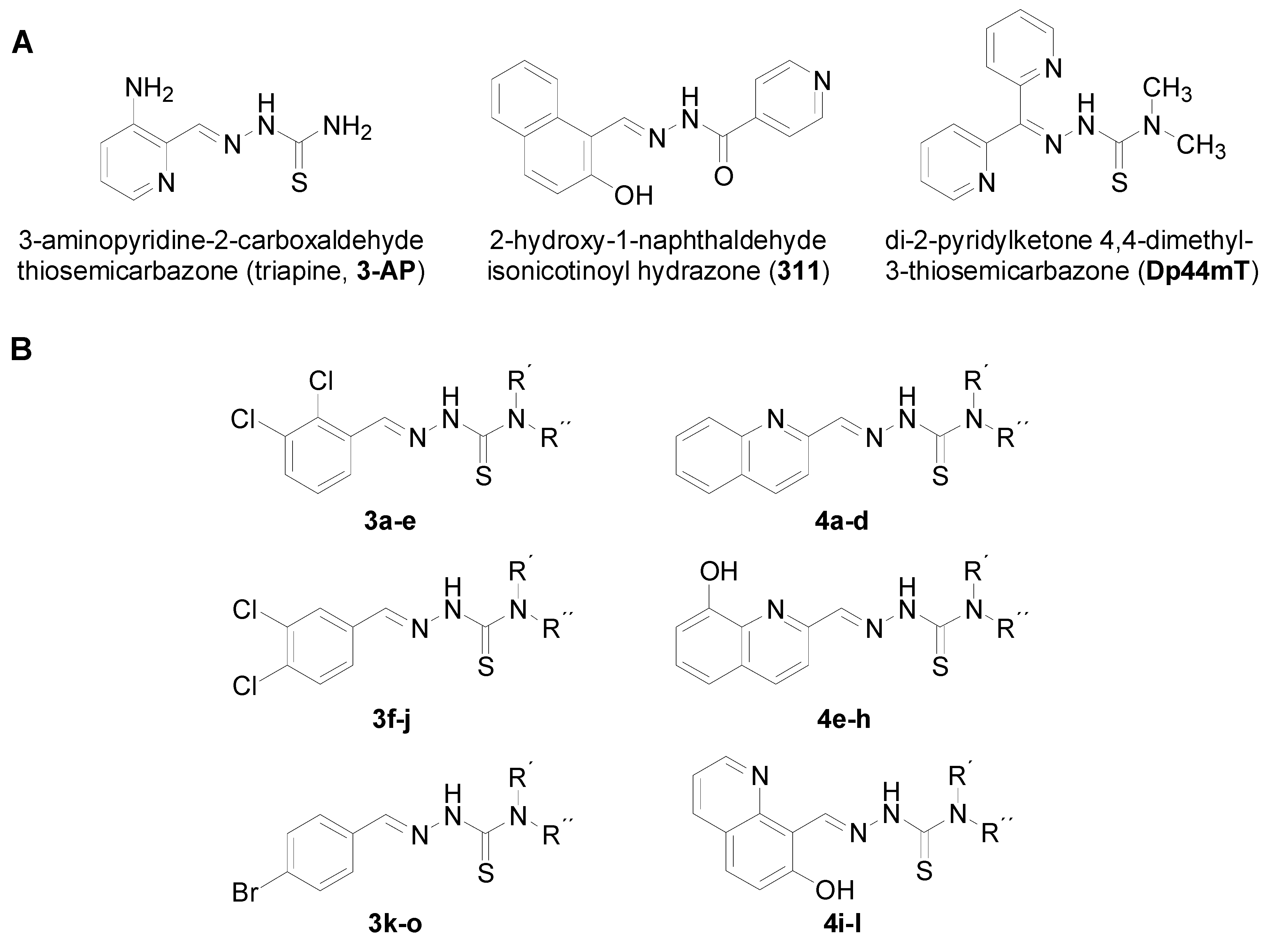
Investigation of the Biological Properties of (Hetero)Aromatic Thiosemicarbazones

 ,
,
Abstract
:1. Introduction

2. Results and Discussion
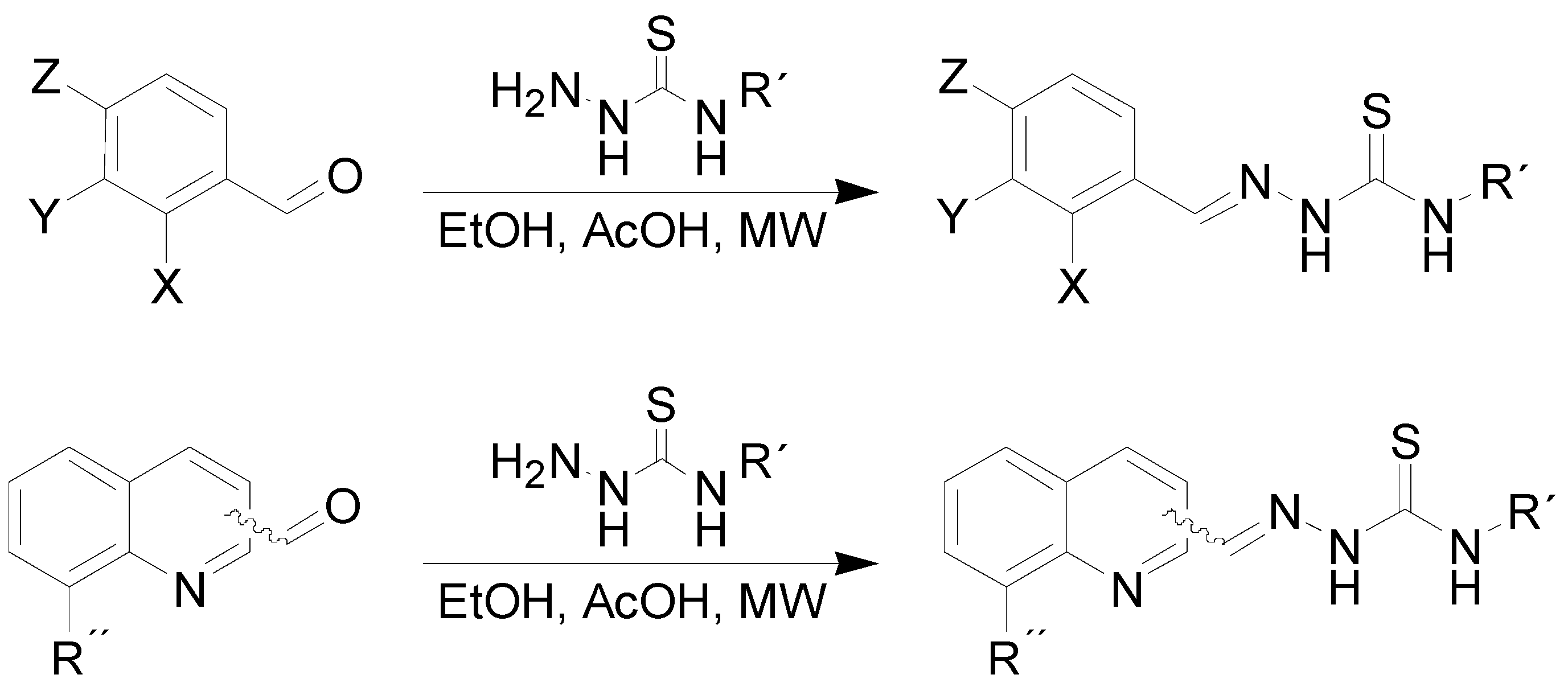
2.1. Chemistry

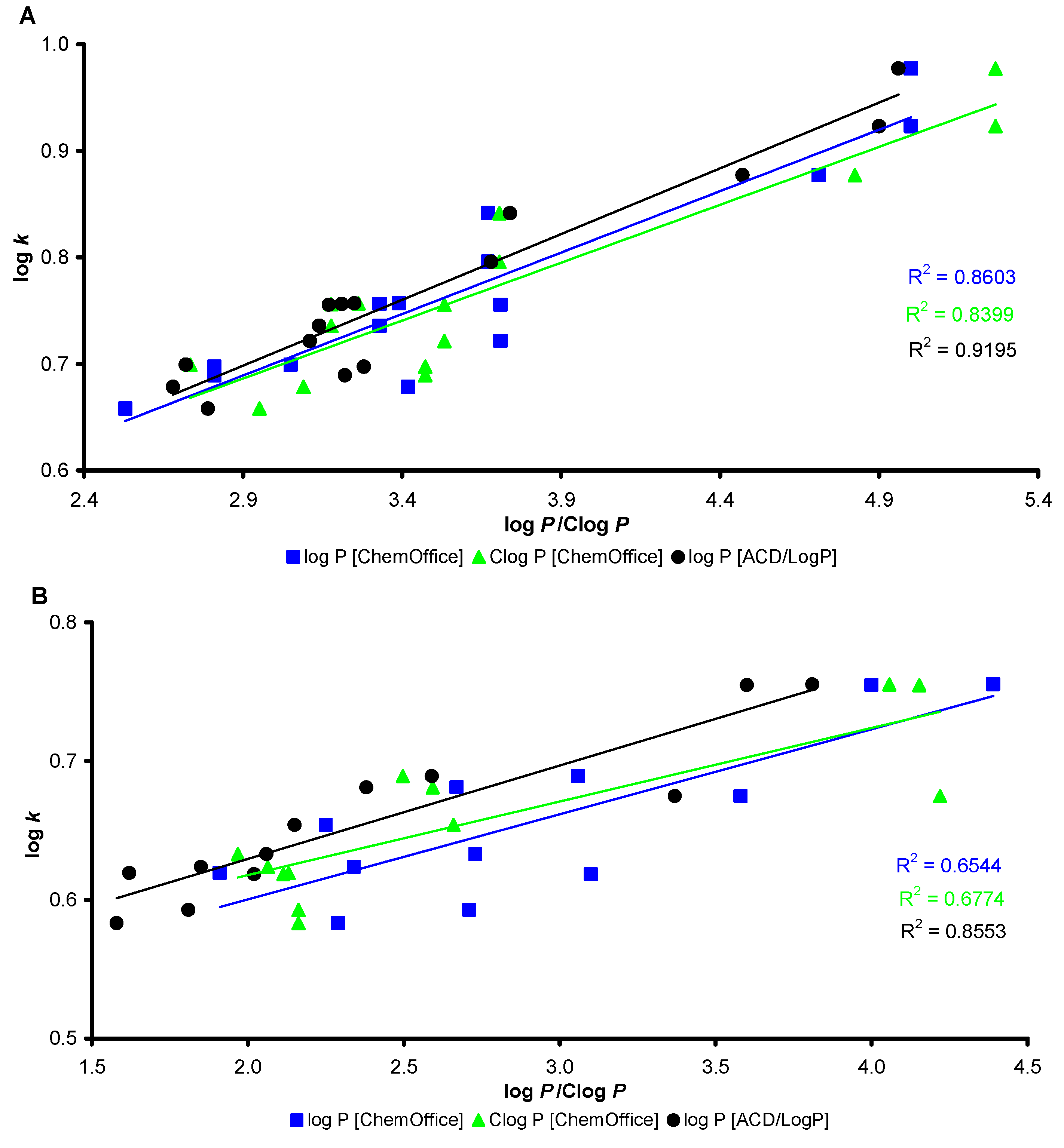
2.2. Lipophilicity

 | ||||||
|---|---|---|---|---|---|---|
| Comp. | R1 | R2 | R3 | log k | log P/Clog P ChemOffice | log P ACD/LogP |
| 3a | 2,3-Cl | H | H | 0.6892 | 2.81/3.473 | 3.22 ± 0.38 |
| 3b | 2,3-Cl | H | CH3 | 0.7357 | 3.33/3.177 | 3.14 ± 0.59 |
| 3c | 2,3-Cl | CH3 | CH3 | 0.7214 | 3.71/3.533 | 3.11 ± 0.60 |
| 3d | 2,3-Cl | H | C2H5 | 0.7958 | 3.67/3.706 | 3.68 ± 0.59 |
| 3e | 2,3-Cl | H | C6H5 | 0.9230 | 5.00/5.266 | 4.90 ± 0.59 |
| 3f | 3,4-Cl | H | H | 0.6974 | 2.81/3.473 | 3.28 ± 0.38 |
| 3g | 3,4-Cl | H | CH3 | 0.7563 | 3.33/3.177 | 3.21 ± 0.59 |
| 3h | 3,4-Cl | CH3 | CH3 | 0.7555 | 3.71/3.533 | 3.17 ± 0.60 |
| 3i | 3,4-Cl | H | C2H5 | 0.8415 | 3.67/3.706 | 3.74 ± 0.59 |
| 3j | 3,4-Cl | H | C6H5 | 0.9774 | 5.00/5.266 | 4.96 ± 0.59 |
| 3k | 4-Br | H | H | 0.6580 | 2.53/2.952 | 2.79 ± 0.39 |
| 3l | 4-Br | H | CH3 | 0.6993 | 3.05/2.734 | 2.72 ± 0.61 |
| 3m | 4-Br | CH3 | CH3 | 0.6784 | 3.42/3.090 | 2.68 ± 0.62 |
| 3n | 4-Br | H | C2H5 | 0.7568 | 3.39/3.263 | 3.25 ± 0.61 |
| 3o | 4-Br | H | C6H5 | 0.8772 | 4.71/4.823 | 4.47 ± 0.61 |
 | ||||||
|---|---|---|---|---|---|---|
| Comp. | R1 | R2 | R3 | log k | log P/Clog P ChemOffice | log P ACD/LogP |
| 4a |  | H | CH3 | 0.6329 | 2.73/1.968 | 2.06 ± 0.59 |
| 4b | | CH3 | CH3 | 0.6184 | 3.10/2.114 | 2.02 ± 0.60 |
| 4c | | H, | C2H5 | 0.6892 | 3.06/2.497 | 2.59 ± 0.59 |
| 4d | | H | C6H5 | 0.7553 | 4.39/4.057 | 3.81 ± 0.59 |
| 4e |  | H | CH3 | 0.6236 | 2.34/2.064 | 1.85 ± 0.84 |
| 4f |  | CH3 | C6H5 | 0.5927 | 2.71/2.163 | 1.81 ± 0.85 |
| 4g | | H | C2H5 | 0.6810 | 2.67/2.593 | 2.38 ± 0.84 |
| 4h | | H | C6H5 | 0.7548 | 4.00/4.153 | 3.60 ± 0.84 |
| 4i |  | H | CH3 | 0.6193 | 1.91/2.131 | 1.62 ± 1.09 |
| 4j | | CH3 | CH3 | 0.5831 | 2.29/2.163 | 1.58 ± 1.10 |
| 4k | | H | C2H5 | 0.6539 | 2.25/2.660 | 2.15 ± 1.09 |
| 4l | | H | C6H5 | 0.6748 | 3.58/4.220 | 3.37 ± 1.09 |

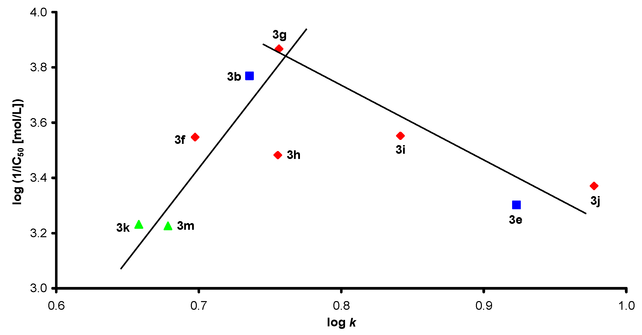
2.3. Biological Activities
2.3.1. Inhibition of Photosynthetic Electron Transport (PET) in Spinach Chloroplasts
| Comp. | PET IC50 | 1,2 MIC (a IC80 / b IC50) [µmol/L] | HCT-116 IC50 | |||||||
|---|---|---|---|---|---|---|---|---|---|---|
| CA a | CT a | CK a | CG a | TB a | AF b | AC b | TM b | |||
| 24 h | 24 h | 24 h | 24 h | 24 h | 24 h | 24 h | 72 h | |||
| 48 h | 48 h | 48 h | 48 h | 48 h | 48 h | 48 h | 120 h | |||
| 3a | ND | >125 | >125 | >125 | >125 | >125 | >125 | >125 | >125 | 41.5 ± 1.7 3 |
| 3b | 170.1 | >500 | >500 | >500 | >500 | >500 | >500 | >500 | >500 | >60 |
| 3c | ND | >125 | >125 | >125 | >125 | >125 | >125 | >125 | >125 | - |
| 3d | ND | >500 | >500 | >500 | >500 | >500 | >500 | >500 | >500 | 58.5 ± 0.2 3 |
| 3e | 499.3 | >125 | >125 | >125 | >125 | >125 | >125 | >125 | >125 | >60 |
| 3f | 283.3 | >500 | >500 | >500 | >500 | >500 | >500 | >500 | >500 | 46.0 ± 0.9 |
| 3g | 135.6 | >125 | >125 | >125 | >125 | >125 | >125 | >125 | >125 | 55.5 ± 6.2 |
| 3h | 329.3 | 15.62 | 500 | 500 | 500 | 125 | >500 | >500 | 31.25 | 47.0 ± 1.3 |
| 31.25 | 500 | 500 | 500 | 500 | >500 | >500 | 62.5 | |||
| 3i | 280.2 | >500 | >500 | >500 | >500 | >500 | >500 | >500 | >500 | 48.5 ± 2.8 |
| 3j | 425.9 | >125 | >125 | >125 | >125 | >125 | >125 | >125 | >125 | >60 |
| 3k | 586.4 | 500 | >500 | >500 | >500 | 500 | >500 | >500 | >500 | >60 |
| 500 | >500 | >500 | >500 | 500 | >500 | >500 | >500 | |||
| 3l | ND | >500 | >500 | >500 | >500 | >500 | >500 | >500 | >500 | >60 |
| 3m | 594.6 | 62.5 | >500 | >500 | >500 | 15.62 | >500 | >500 | 62.5 | 47.5 ± 2.4 |
| 250 | >500 | >500 | >500 | 62.5 | >500 | >500 | 62.5 | |||
| 3n | ND | >500 | >500 | >500 | >500 | >500 | >500 | >500 | >500 | >60 3 |
| 3o | ND | >500 | >500 | >500 | >500 | >500 | >500 | >500 | >500 | >60 |
| 4a | ND | >500 | >500 | >500 | >500 | >500 | >500 | >500 | >500 | >25 |
| 4b | 1368 | 1.95 | 1.95 | 3.9 | 1.95 | 1.95 | 3.9 | 3.9 | 3.9 | 4.86 ± 1.48 3 |
| 1.95 | 7.81 | 3.9 | 1.95 | 1.95 | 3.9 | 3.9 | 3.9 | |||
| 4e | 302 | >125 | >125 | >125 | >125 | >125 | >125 | >125 | >125 | 1.71 ± 0.34 3 |
| 4f | ND | >500 | >500 | >500 | >500 | >500 | >500 | >500 | >500 | - |
| 4h | ND | >125 | >125 | >125 | >125 | >125 | >125 | >125 | >125 | 24.97 ± 4.29 3 |
| >125 | >125 | >125 | >125 | >125 | >125 | >125 | >125 | |||
| 4i | ND | >500 | >500 | >500 | >500 | >500 | >500 | >500 | >500 | >25 3 |
| >500 | >500 | >500 | >500 | >500 | >500 | >500 | >500 | |||
| 4j | 520.4 | 125 | >250 | >250 | 125 | 125 | >250 | >250 | 125 | 20.75 ± 5.34 3 |
| >250 | >250 | >250 | 125 | >250 | >250 | >250 | 125 | |||
| 4k | ND | >500 | >500 | >500 | 62.5 | >500 | >500 | >500 | 62.5 | >25 3 |
| >500 | >500 | >500 | 500 | >500 | >500 | >500 | 62.5 | |||
| 4l | ND | 31.25 | 125 | 31.25 | 15.62 | 15.62 | 125 | 15.62 | 15.62 | 16.28 ± 1.69 3 |
| 62.5 | 125 | 125 | 31.25 | 125 | 125 | 62.5 | 15.62 | |||
| DCMU | 1.9 | - | - | - | - | - | - | - | - | |
| FLU | - | 0.06 | 0.12 | 3.91 | 0.98 | 0.24 | >125 | >125 | 1.95 | - |
| 0.12 | >125 | 15.62 | 3.91 | 0.48 | >125 | >125 | 3.91 | |||
| DXR | - | - | - | - | - | - | - | - | - | 10 ± 1.1 |
2.3.2. In Vitro Antifungal Susceptibility Testing

2.3.3. In Vitro Antiproliferative Activity
3. Experimental
3.1. General
3.2. Synthesis
3.2.1. General Procedure for Synthesis of Thiosemicarbazones 3a–o
3.2.2. General Procedure for the Synthesis of Thiosemicarbazones 4a–l
3.3. Lipophilicity Determination Using HPLC (Capacity Factor k/Calculated log k)
3.4. Lipophilicity Calculations
3.5. Study of the Inhibition Photosynthetic Electron Transport (PET) in Spinach Chloroplasts
3.6. In Vitro Antifungal Susceptibility Testing
3.7. Cell Culture and in Vitro Antiproliferative Activity
4. Conclusions
Supplementary Materials
Acknowledgements
- Sample Availability: Samples of the compounds are available from the authors.
References
- Wachtershauser, G. Before enzymes and templates: Theory of surface metabolism. Microbiol. Rev. 1988, 52, 452–484. [Google Scholar]
- Wachtershauser, G. Groundworks for an evolutionary biochemistry: The iron-sulphur world. Prog. Biophys. Mol. Biol. 1992, 58, 85–201. [Google Scholar] [CrossRef]
- Danielson, P.B. The cytochrome P450 superfamily: Biochemistry, evolution and drug metabolism in humans. Curr. Drug Metab. 2002, 3, 561–597. [Google Scholar] [CrossRef]
- Pietrangelo, A.; Schilsky, M. Metal storage disorders. Forward. Semin. Liver Dis. 2011, 31, 231–232. [Google Scholar] [CrossRef]
- Kalinowski, D.S.; Richardson, D.R. The evolution of iron chelators for the treatment of iron overload disease and cancer. Pharmacol. Rev. 2005, 57, 547–583. [Google Scholar] [CrossRef]
- Kalinowski, D.S.; Richardson, D.R. Future of toxicology-iron chelators and differing modes of action and toxicity: The changing face of iron chelation therapy. Chem. Res. Toxicol 2007, 20, 715–720. [Google Scholar] [CrossRef]
- Richardson, D.R.; Kalinowski, D.S.; Lau, S.; Jansson, P.J.; Lovejoy, D.B. Cancer cell iron metabolism and the development of potent iron chelators as anti-tumour agents. Biochim. Biophys. Acta 2009, 1790, 702–717. [Google Scholar] [CrossRef]
- Richardson, D.R.; Tran, E.H.; Ponka, P. The potential of iron chelators of the pyridoxal isonicotinoyl hydrazone class as effective antiproliferative agents. Blood 1995, 86, 4295–4306. [Google Scholar]
- Richardson, D.R. The therapeutic potential of iron chelators. Expert Opin. Investig. Drugs 1999, 8, 2141–2158. [Google Scholar] [CrossRef]
- Yu, Y.; Kalinowski, D.S.; Kovacevic, Z.; Siafakas, A.R.; Jansson, P.J.; Stefani, C.; Lovejoy, D.B.; Sharpe, P.C.; Bernhardt, P.V.; Richardson, D.R. Thiosemicarbazones from the old to new: Iron chelators that are more than just ribonucleotide reductase inhibitors. J. Med. Chem. 2009, 52, 5271–5294. [Google Scholar] [CrossRef]
- Richardson, D.R.; Baker, E. The uptake of iron and transferrin by the human malignant melanoma cell. Biochim. Biophys. Acta 1990, 1053, 1–12. [Google Scholar] [CrossRef]
- Richardson, D.R.; Baker, E. Two mechanisms of iron uptake from transferrin by melanoma cells. The effect of desferrioxamine and ferric ammonium citrate. J. Biol. Chem. 1992, 267, 13972–13979. [Google Scholar]
- Thelander, L.; Gräslund, A.; Thelander, M. Continual presence of oxygen and iron required for mammalian ribonucleotide reduction: Possible regulation mechanism. Biochem. Biophys. Res. Commun. 1983, 110, 859–865. [Google Scholar] [CrossRef]
- Yuan, J.; Lovejoy, D.B.; Richardson, D.R. Novel di-2-pyridyl-derived iron chelators with marked and selective antitumor activity: In vitro and in vivo assessment. Blood 2004, 104, 1450–1458. [Google Scholar] [CrossRef]
- Prasad, T.; Chandra, A.; Mukhopadhyay, C.K.; Prasad, R. Unexpected link between iron and drug resistance of Candida spp.: Iron depletion enhances membrane fluidity and drug diffusion, leading to drug-susceptible cells. Antimicrob. Agents Chemother. 2006, 50, 3597–3606. [Google Scholar] [CrossRef]
- Holbein, B.E.; Mira de Orduña, R. Effect of trace iron levels and iron withdrawal by chelation on the growth of Candida albicans and Candida vini. FEMS Microbiol. Lett. 2010, 307, 19–24. [Google Scholar] [CrossRef]
- Nevitt, T. War-Fe-re: Iron at the core of fungal virulence and host immunity. Biometals 2011, 24, 547–558. [Google Scholar] [CrossRef]
- Kuipers, M.E.; Beljaars, L.; van Beek, N.; de Vries, H.G.; Heegsma, J.; van Den Berg, J.J.M.; Meijer, D.K.F.; Swart, P.J. Conditions influencing the in vitro antifungal activity of lactoferrin combined with antimycotics against clinical isolates of Candida. Impact on the development of buccal preparations of lactoferrin. Acta Pathol. Microbiol. Immunol. Scand. 2002, 110, 290–298. [Google Scholar]
- Kuipers, M.E.; de Vries, H.G.; Eikelboom, M.C.; Meijer, D.K.; Swart, P.J. Synergistic fungistatic effects of lactoferrin in combination with antifungal drugs against clinical Candida isolates. Antimicrob. Agents Chemother. 1999, 43, 2635–2641. [Google Scholar]
- Spellberg, B.; Ibrahim, A.S.; Chin-Hong, P.V.; Kontoyiannis, D.P.; Morris, M.I.; Perfect, J.R.; Fredricks, D.; Brass, E.P. The deferasirox-ambisome therapy for mucormycosis (DEFEAT Mucor) study: A randomized, double-blinded, placebo-controlled trial. J. Antimicrob. Chemother. 2012, 67, 715–722. [Google Scholar] [CrossRef]
- Donnelly, J.P.; Lahav, M. Deferasirox as adjunctive therapy for mucormycosis. J. Antimicrob. Chemother. 2012, 67, 201–202. [Google Scholar]
- Konstantinovic, S.; Radovanovic, B.; Sovilj, S.; Stanojevic, S. Antimicrobial activity of some isatin-3-thiosemicarbazone complexes. J. Serb. Chem. Soc. 2008, 73, 7–13. [Google Scholar] [CrossRef]
- Kizilcikli, I.; Kurt, Y.D.; Akkurt, B.; Genel, A.Y.; Birteksoz, S.; Otuk, G.; Ulkuseven, B. Antimicrobial activity of a series of thiosemicarbazones and their Zn(II) and Pd(II) complexes. Folia Microbiol. 2007, 52, 15–25. [Google Scholar] [CrossRef]
- Kulandaivelu, U.; Padmini, V.G.; Suneetha, K.; Shireesha, B.; Vidyasagar, J.V.; Rao, T.R.KN.J.; Basu, A.; Jayaprakash, V. Synthesis, antimicrobial and anticancer activity of new thiosemicarbazone derivatives. Arch. Pharm. 2011, 344, 84–90. [Google Scholar] [CrossRef]
- Pervez, H.; Iqbal, M.S.; Tahir, M.Y.; Nasim, F.H.; Choudhary, M.I.; Khan, K.M. In vitro cytotoxic, antibacterial, antifungal and urease inhibitory activities of some N4-substituted isatin-3-thiosemicarbazones. J. Enzym. Inhib. Med. Chem. 2008, 23, 848–854. [Google Scholar] [CrossRef]
- Opletalova, V.; Kalinowski, D.S.; Vejsova, M.; Kunes, J.; Pour, M.; Jampilek, J.; Buchta, V.; Richardson, D.R. Identification and characterization of thiosemicarbazones with antifungal and antitumor effects: Cellular iron-chelation mediating cytotoxic activity. Chem. Res. Toxicol. 2008, 21, 1878–1889. [Google Scholar] [CrossRef]
- Li, Z.-C.; Chen, L.-H.; Yu, X.-J.; Hu, Y.-H.; Song, K.-K.; Zhou, X.-W.; Chen, Q.-X. Inhibition kinetics of chlorobenzaldehyde thiosemicarbazones on mushroom tyrosinase. J. Agric. Food Chem. 2010, 12537–12540. [Google Scholar]
- de Aquino, T.M.; Liesen, A.P.; da Silva, R.E.; Lima, V.T.; Carvalho, C.S.; de Faria, A.R.; de Araujo, J.M.; de Lima, J.G.; Alves, A.J.; de Melo, E.J.T.; et al. Synthesis, anti-Toxoplasma gondii and antimicrobial activities of benzaldehyde 4-phenyl-3-thiosemicarbazones and 2-[(phenylmethylene)hydrazono]-4-oxo-3-phenyl-5-thiazolidineacetic acids. Bioorg. Med. Chem. 2008, 16, 446–456. [Google Scholar]
- Jampilek, J.; Musiol, R.; Finster, J.; Pesko, M.; Carroll, J.; Kralova, K.; Vejsova, M.; O’Mahony, J.; Coffey, A.; Dohnal, J.; Polanski, J. Investigating biological activity spectrum for novel styrylquinazoline analogues. Molecules 2009, 14, 4246–4265. [Google Scholar] [CrossRef]
- Jampilek, J.; Musiol, R.; Pesko, M.; Kralova, K.; Vejsova, M.; Carroll, J.; Coffey, A.; Finster, J.; Tabak, D.; Niedbala, H.; et al. Ring-substituted 4-hydroxy-1H-quinolin-2-ones: Preparation and biological activity. Molecules 2009, 14, 1145–1159. [Google Scholar] [CrossRef]
- Musiol, R.; Serda, M.; Hensel-Bielowka, S.; Polanski, J. Quinoline-based antifungals. Curr. Med. Chem. 2010, 17, 1960–1973. [Google Scholar] [CrossRef]
- Musiol, R.; Jampilek, J.; Buchta, V.; Silva, L.; Niedbala, H.; Podeszwa, B.; Palka, A.; Majerz-Maniecka, K.; Oleksyn, B.; Polanski, J. Antifungal properties of new series of quinoline derivatives. Bioorg. Med. Chem. 2006, 14, 3592–3598. [Google Scholar] [CrossRef]
- Musiol, R.; Jampilek, J.; Nycz, J.E.; Pesko, M.; Carroll, J.; Kralova, K.; Vejsova, M.; O’Mahony, J.; Coffey, A.; Mrozek, A.; Polanski, J. Investigating the activity spectrum for ring-substituted 8-hydroxyquinolines. Molecules 2010, 15, 288–304. [Google Scholar] [CrossRef]
- Serda, M.; Kalinowski, D.S.; Mrozek-Wilczkiewicz, A.; Musiol, R.; Szurko, A.; Ratuszna, A.; Pantarat, N.; Kovacevic, Z.; Merlot, A.M.; Richardson, D.R.; Polanski, J. Synthesis and characterization of quinoline-based thiosemicarbazones and correlation of cellular iron-binding efficacy to anti-tumor efficacy. Bioorg. Med. Chem. Lett. 2012, 22, 5527–5531. [Google Scholar]
- Green, D.A.; Antholine, W.E.; Wong, S.J.; Richardson, D.R.; Chitambar, C.R. Inhibition of malignant cell growth by 311, a novel iron chelator of the pyridoxal isonicotinoyl hydrazone class: effect on the R2 subunit of ribonucleotide reductase. Clin. Cancer Res. 2001, 7, 3574–3579. [Google Scholar]
- Richardson, D.R.; Sharpe, P.C.; Lovejoy, D.B.; Senaratne, D.; Kalinowski, D.S.; Islam, M.; Bernhardt, P.V. Dipyridyl thiosemicarbazone chelators with potent and selective antitumor activity form iron complexes with redox activity. J. Med. Chem. 2006, 49, 6510–6521. [Google Scholar] [CrossRef]
- Kalinowski, D.S.; Yu, Y.; Sharpe, P.C.; Islam, M.; Liao, Y.T.; Lovejoy, D.B.; Kumar, N.; Bernhardt, P.V.; Richardson, D.R. Design, synthesis, and characterization of novel iron chelators: Structure−activity relationships of the 2-benzoylpyridine thiosemicarbazone series and their 3-nitrobenzoyl analogues as potent antitumor agents. J. Med. Chem. 2007, 50, 3716–3729. [Google Scholar] [CrossRef]
- Musiol, R.; Kowalczyk, W. Azole antimycotics-A highway to new drugs or a dead end? Curr. Med. Chem. 2012, 19, 1378–1388. [Google Scholar] [CrossRef]
- Roche, J.L.; Boyd, P.W.; McKay, R.M.L.; Geider, R.J. Flavodoxin as an in situ marker for iron stress in phytoplankton. Nature 1996, 382, 802–805. [Google Scholar] [CrossRef]
- Pospisil, P. Enzymatic function of cytochrome b559 in photosystem II. J. Photochem. Photobiol. B 2011, 104, 341–347. [Google Scholar] [CrossRef]
- Hansch, R.; Mendel, R.R. Physiological functions of mineral micronutrients (Cu, Zn, Mn, Fe, Ni, Mo, B, Cl). Curr. Opin. Plant Biol. 2009, 12, 259–266. [Google Scholar] [CrossRef]
- Nouet, C.; Motte, P.; Hanikenne, M. Chloroplastic and mitochondrial metal homeostasis. Trends Plant Sci. 2011, 16, 395–404. [Google Scholar] [CrossRef]
- Pilon, M.; Cohu, C.M.; Ravet, K.; Abdel-Ghany, S.E.; Gaymard, F. Essential transition metal homeostasis in plants. Curr. Opin. Plant Biol. 2009, 12, 347–357. [Google Scholar] [CrossRef]
- Serda, M.; Malecki, J.G.; Mrozek-Wilczkiewicz, A.; Musiol, R.; Polanski, J. Microwave assisted synthesis, X-ray crystallography and DFT calculations of selected aromatic thiosemicarbazones. J. Mol. Struct. 2012, in press. [Google Scholar]
- CS ChemOfficeUltra, version 12.0; The World’s Premier Desktop Chemistry Software, CambridgeSoft: Cambridge, MA, USA, 2008.
- ACD/LogP version 1.0; Software used for calculating logP values, Advanced Chemistry Development: Toronto, ON, Canada, 2006.
- Musiol, R.; Jampilek, J.; Podeszwa, B.; Finster, J.; Tabak, D.; Dohnal, J.; Polanski, J. RP-HPLC determination of lipophilicity in series of quinoline derivatives. Cent. Eur. J. Chem. 2009, 7, 586–597. [Google Scholar] [CrossRef]
- Taft, R.W. Steric Effects in Organic Chemistry; Newman, M.S., Ed.; John Willey & Sons: New York, NY, USA, 1956; pp. 556–675. [Google Scholar]
- Dimmock, J.R.; Puthucode, R.N.; Lo, M.S.; Quail, J.W.; Yang, J.; Stables, J.P. Structural modifications of the primary amino group of anticonvulsant aryl semicarbazones. Pharmazie 1996, 51, 83–88. [Google Scholar]
- Walter, W.; Rohloff, C. Oxidation-products of carbothioamides. 39. Configuration and hindered rotation of 4-monodisubstituted and 4,4-disubstituted thiosemicarbazone S,S,S-trioxides. Justus Liebigs Ann. Chem. 1977, 447-462. [Google Scholar]
- Masarovicova, E.; Kralova, K. Approaches to measuring plant photosynthesis activity. In Handbook of Photosynthesis, 2nd; Pessarakli, M., Ed.; Taylor & Francis Group: Boca Raton, FL, USA, 2005; pp. 617–656. [Google Scholar]
- Kralova, K.; Sersen, F.; Sidoova, E. Photosynthesis inhibition produced by 2-alkylthio-6-R-benzothiazoles. Chem. Pap. 1992, 46, 348–350. [Google Scholar]
- Sheehan, D.J.; Espinel-Ingroff, A.; Steele, M.; Webb, C.D. Antifungal susceptibility testing of yeasts: A brief overview. Clin. Infect. Dis. 1993, 17, 494–500. [Google Scholar] [CrossRef]
- Clinical Laboratory Standards Institute (CLSI), Reference Method for Broth Dilution Testing of Yeasts: Approved Standard M27-A2; CLSI: Wayne, PA, USA, 2002.
- Clinical Laboratory Standards Institute (CLSI), Method for Antifungal Disk Diffusion Susceptibility Testing of Yeasts: Approved Guideline M44-A; CLSI: Wayne, PA, USA, 2004.
© 2012 by the authors; licensee MDPI, Basel, Switzerland. This article is an open-access article distributed under the terms and conditions of the Creative Commons Attribution license (http://creativecommons.org/licenses/by/3.0/).
Share and Cite
Serda, M.; Mrozek-Wilczkiewicz, A.; Jampilek, J.; Pesko, M.; Kralova, K.; Vejsova, M.; Musiol, R.; Ratuszna, A.; Polanski, J. Investigation of the Biological Properties of (Hetero)Aromatic Thiosemicarbazones. Molecules 2012, 17, 13483-13502. https://doi.org/10.3390/molecules171113483
Serda M, Mrozek-Wilczkiewicz A, Jampilek J, Pesko M, Kralova K, Vejsova M, Musiol R, Ratuszna A, Polanski J. Investigation of the Biological Properties of (Hetero)Aromatic Thiosemicarbazones. Molecules. 2012; 17(11):13483-13502. https://doi.org/10.3390/molecules171113483
Chicago/Turabian StyleSerda, Maciej, Anna Mrozek-Wilczkiewicz, Josef Jampilek, Matus Pesko, Katarina Kralova, Marcela Vejsova, Robert Musiol, Alicja Ratuszna, and Jaroslaw Polanski. 2012. "Investigation of the Biological Properties of (Hetero)Aromatic Thiosemicarbazones" Molecules 17, no. 11: 13483-13502. https://doi.org/10.3390/molecules171113483