Article

The Incubation of 13a,17-Dihydroxystemodane with Cephalosporium aphidicola

by
Braulio M. Fraga
1,*,
Ricardo Guillermo
2,
Melchor G. Hernández
1,
María C. Chamy
3 and
Juan A. Garbarino
4
1
Instituto de Productos Naturales y Agrobiología, C.S.I.C., Avda. Astrofísico F. Sánchez 3, La Laguna, Tenerife, Canary Islands, 38206, Spain
2
Instituto Universitario de Bioorgánica “Antonio González”, Departamento de Química Orgánica, Universidad de La Laguna, Tenerife, 38206, Spain
3
Departamento de Química, Universidad Andrés Bello, Viña del Mar, Chile
4
Departamento de Química, Universidad Técnica Federico Santa María, Casilla-110V, Valparaiso, Chile
*
Author to whom correspondence should be addressed.
Molecules 2012, 17(2), 1744-1750; https://doi.org/10.3390/molecules17021744
Submission received: 30 November 2011 / Revised: 19 January 2012 / Accepted: 3 February 2012 / Published: 9 February 2012
(This article belongs to the Special Issue Terpenoids)

Abstract

:
The biotransformation of 13α,17-dihydroxystemodane (3) with the fungus Cephalosporium aphidicola afforded 13α,17,18-trihydroxystemodane (4), 3β,13α,17-tri-hydroxystemodane (5), 13α,17-dihydroxy-stemodan-18-oic acid (6), 3β,11β,13α,17-tetra-hydroxystemodane (7), 11β,13α,17,18-tetrahydroxystemodane (8) and 3β,13α,17,18-tetra-hydroxystemodane (9). The hydroxylation at C-18 of the substrate points to a biosynthetically-directed transformation, because aphidicolin (2) is hydroxylated at this carbon. However, the C-3(β) and C-11(β) hydroxylations seem to indicate a xenobiotic biotransformation.

Graphical Abstract

1. Introduction

Microbiological transformations can be divided into two groups: xenobiotic biotransformations, in which the substrate is strange to the transforming organism, and biosynthetically-directed transformations, also known as “analogue biosynthesis”, in which the substrate possesses a structure analogous to a natural biosynthetic intermediate found in the microorganism [1,2]. We have carried out both types of biotransformations using the fungi Mucor plumbeus [3] and Gibberella fujikuroi [4] respectively. Now, in this work we have used another fungus, Cephalosporium aphidicola, which occupies the borderline between the xenobiotic and biosynthetically-directed biotransformations, because it achieves both [5,6,7,8].
Diterpenes with a stemodane skeleton (i.e., 1) have a structural similarity with aphidicolin (2), an antiviral substance and a inhibitor of DNA polymerase, which was isolated from C. aphidicola [9], although the C/D ring junctions and the configuration at C-13 in stemodanes and aphidicolanes are different (Scheme 1). Thus, biotransformations of stemodane diterpenes, stemodin and stemodinone, with this fungus, have been carried out [10,11]. Some of us had isolated 13α,17-dihydroxystemodane (3), and analogous compounds of this type, from Stemonia chilensis, a plant that grows in the littoral zone of central Chile [12]. This compound had been incubated with M. plumbeus [13], a fungus used in xenobiotic biotransformations.
Scheme 1. Stemodane (1) and aphidicolin (2).
Molecules 17 01744 g001

2. Results and Discussion

The microbiological transformation of 13α,17-dihydroxystemodane (3) with the fungus C. aphidicola afforded 13α,17,18-trihydroxystemodane (4), 3β,13α,17-trihydroxystemodane (5), 13α,17-dihydroxystemodan-18-oic acid (6), 3β,11β,13α,17-tetrahydroxystemodane (7), 11β,13α,17,18-tetra-hydroxystemodane (8) and 3β,13α,17,18-tetrahydroxystemodane (9). Some of these metabolites were obtained as their acetates by acetylation of chromatographic fractions containing them.
The metabolite 4 showed in the HRMS spectrum the ion of higher mass at m/z 304.2407, formed from the molecular ion by loss of water, which indicated its molecular formula, C20H34O3. Thus, a new oxygen had been introduced in the molecule during the incubation. In the 1H-NMR spectrum the signal of a new AB system appears at δ 3.11 and 3.35 (1H each, d, J = 11 Hz). 13C-NMR spectrum showed a new signal at δ 72.6 (t). This last value is characteristic of an equatorial -CH2OH group at C-4 [14,15], confirmed in the HMBC experiment with correlations of H-18 with C-3, and H-19 with C-3, C-5 and C-18. Therefore, the structure of this compound was determined as 13α,17,18-trihydroxystemodane (4).
Compound 6 was obtained as its diacetate 6a by acetylation of the fractions containing it. The molecular formula of 6a was determined as C24H38O6 considering HRMS data. Therefore, the substrate had gained two oxygens and lost two hydrogens during the fermentation. The two oxygens must form a part of an acid, because in the 13C-NMR spectrum a new signal was detected at δ 182.0, typical of this group, while the disappearance of a methyl signal was noted. The presence of this new group was confirmed because 6a formed a methyl ester (compound 6am) by treatment with diazomethane. We considered that the C-18 acid must be formed by oxidation of the corresponding alcohol in 4 (Scheme 2), and consequently assigned the structure of 13α,17-dihydroxystemodan-18-oic acid (6) to the original metabolite formed in the biotransformation.
Table 1. 13C-NMR data of compounds 3, 4, 6a, 6am, 8a and 9a (CDCl3).
Position346a6am8a9a
136.235.835.735.936.0 a33.5
218.818.118.118.717.623.2
341.835.236.836.836.3 b74.6
433.237.6 a47.247.736.540.8
547.240.441.641.941.139.5
622.222.024.624.622.121.6
736.636.236.736.734.636.1
837.337.237.737.733.637.2
950.750.850.650.655.050.9
1038.538.3 a37.938.039.138.1
1127.227.326.026.171.727.1
1228.128.227.027.136.6b28.1
1374.374.284.484.574.074.6
1440.440.539.139.140.341.0
1537.437.435.235.235.5 a37.2
1629.729.829.829.928.429.4
1768.068.164.764.769.969.8
1834.572.6182.0179.673.365.9
1922.818.617.717.818.513.9
2018.819.619.019.020.619.4
a,b These values can be interchanged
Compound 8 was obtained as its triacetate 8a, the mass spectrum of which showed a peak at m/z 446.2687 formed from the molecular ion by loss of water. Thus, its molecular formula was determined as C26H40O7. Its NMR spectra showed two –CH2OAc groups, one corresponding to the acetylated C-17 alcohol of the substrate, and the other, formed by acetylation of an hydroxyl group introduced in the incubation, resonates at δH 3.61 and 3.98 (each 1H, d, J = 10.8 Hz) and at δC 73.3 (t). These signals are characteristic of an equatorial acetoxymethylene group at C-4 [14,15]. Other signals observed in the spectra of 8a were those of an oxymethine group at δH 5.36 (t, J = 7.6 Hz) and δC 71.7(d). These chemical shifts and couplings were analogous to those of an 11β-acetoxy derivative described in the biotransformation of the substrate 3 by M. plumbeus [13]. The HMBC experiment of 8a confirmed these assignments with the following crosspeaks: H-11 with C-8; H-18 with C-3, C-4 and C-5; H-19 with C-3, C-4, C-5 and C-18; H-20 with C-1, C-5, C-9 and C-10. Thus, the structure 11β,13α,17,18-tetrahydroxystemodane was assigned to the metabolite 8 (Scheme 2) obtained in this fermentation.
Acetylation and chromatography of the fractions containing 9 led to the triacetate 9a, which is an isomer of 8a. In addition to the signals of the 17-CH2OAc, in the 1H-NMR spectrum of the triacetate 8a another acetoxymethylene group was detected at δH 3.67 and 3.88 (each 1H, d, J = 11.6 Hz) and δC 65.9(t), which was assigned to C-4 with an α-equatorial configuration. Thus, in the HMBC experiment the main observed correlations were: H-3 with C-1, C-2, C-4, C-18 and C-19; H-18 with C-3, C-5 and C-19; H-19 with C-3, C-4, C-5 and C-18. These crosspeaks also showed that another acetoxy group was located at C-3, with resonances of this oxymethine at δC 74.6 and δH 4.78 (dd, J = 11.7 and 4.1 Hz). The coupling constant of this geminal proton to this acetoxy group indicated a β-equatorial configuration for this oxygenated function. Consequently, the structure of the original alcohol formed in the feeding was determined as 3β,13α,17,18-tetrahydroxystemodane (9).
Compounds 4 and 6–9 are described here for the first time, whilst 3β,13α,17-trihydroxystemodane (5) and 3β,11β,13α,17-tetrahydroxystemodane (7) were already isolated from the biotransformation of 3 with M. plumbeus [13].
Scheme 2. Biotransformation of 1 by Cephalosporium aphidicola.
Molecules 17 01744 g002

3. Experimental

3.1. General Procedures

1H- and 13C-NMR spectra were recorded at 500.13 and 125.03 MHz, respectively, in a Bruker AMX-500 spectrometer. Mass spectra were taken at 70 eV (probe) in a Micromass Autospec spectrometer. HPLC was performed using a Beckman System Gold 125P. Purification by HPLC was achieved using a silica gel column (Ultrasphere Si 5 μm, 10 × 250 mm). Dry column chromatography was carried out on silica gel Merck 0.040–0.063 mm.

3.2. Microorganism

The fungus strain Cephalosporium aphidicola IMI 68689 was a gift from Prof. J. R. Hanson, School of Chemistry, University of Sussex, UK.

3.3. Incubation of 3

C. aphidicola was grown in shake culture at 25 °C, in 20 conical flasks (250 mL), each containing 100 mL of a sterile medium comprising (per L) glucose (80 g), NH4NO3 (0.48 g), KH2PO4 (5 g), MgSO4 (1 g), and trace elements solution (2 mL). The trace elements solution contained (per 100 mL) Co(NO3)2 (0.01 g), CuSO4 (0.015 g), ZnSO4 (0.16 g), MnSO4 (0.01 g), (NH4)6Mo7O24 (0.01 g). 13α,17-Dihydroxystemodane (3, 230 mg) dissolved in EtOH (4.5 mL) was evenly distributed in 20 flasks after one day growth. After a further eight days, the fermentation was harvested. The mycelium was filtered and the culture filtrate was extracted with EtOAc. The extract was dried over Na2SO4 and the solvent evaporated to yield a residue (740 mg) that was chromatographed on a silica gel column in a petroleum ether-EtOAc gradient, to afford starting material 3 (30 mg), 13α,17,18-tri-hydroxystemodane (4, 4 mg), 3β,13α,17-trihydroxystemodane (5, 6 mg), 13α,17-dihydroxy-stemodane-18-oic acid (6) (1 mg), 3β,11β,13α,17-tetrahydroxystemodane (7, 2 mg), 11β, 13α,17,18-tetrahydroxystemodane (8, 1,5 mg) and 3β,13α,17,18-tetrahydroxystemodane (9, 3 mg).
13α,17,18-Trihydroxystemodane (4). 1H-NMR (CDCl3): δ 0.83 (3 H, s, H-19), 1.01 (3H, s, H-20), 1,60 (1H, dd, J = 11,4 and 2,4 Hz, H-5), 1.79 (2H, br s, H-16), 1.89 (1 H, ddt, J = 13.2, 7.4 and 3.1 Hz, H-7β), 2.15 (1H, br s, W1/2 = 16 Hz, H-14), 3.11 and 3.35 (each 1H, d, J = 11.0 Hz, H-18), 3.38 and 3.45 (each 1H, d, J = 10.9 Hz, H-17). EIMS m/z (rel. int.): 304 [M−H2O]+ (2), 291 (66), 286 (17), 273 (100), 255 (54), 230 (8), 215 (12), 206 (18), 203 (40), 173 (29), 159 (22). Found [M−H2O]+ at m/z 304.2407. C20H32O2 requires 304.2402.
13α,17-Dihydroxystemodane-18-oic acid (6). Obtained as its diacetate 5a by acetylation and chromatography of the fractions containing it, 1H-NMR (CDCl3): δ 1.00 (3H, s, H-20), 1.25 (3H, s, H-19), 1.87 (2H, m, H-1 and H-16), 1.97 (1H, dd, J = 13.7 and 5.6 Hz, H-11), 2.05 and 2.06 (each 3H, s), 2.16 (1H, dd, J = 12.1 and 2.0 Hz, H-5), 2.80 (1 H, br t, J = 7.0 Hz, H-14), 4.36 and 4.51 (each 1H, d, J = 12.2 Hz, H-17). EIMS m/z (rel. int.): 360 [M−C2H4O2]+ (5), 318 (33), 305 (16), 300 (24), 285 (12), 277 (19), 255 (12), 239 (7), 220 (22), 204 (17), 184 (35), 159 (20). Found [M-C2H4O2]+ at m/z 360.2291. C22H32O4 requires 360.2301. Acetate methyl ester (5am). 1H-NMR (CDCl3): δ 1.00 (3H, s, H-20), 1.25 (3H, s, H-19), 2.04 and 2.06 (each 3H, s), 2.14 (1H, dd, J = 12.1 and 2.2 Hz, H-5), 2.81 (1 H, br t, J = 7.0 Hz, H-14), 3.65 (3H, s, -OMe), 4.35 and 4.51 (each 1H, d, J = 12.1 Hz, H-17). EIMS m/z (rel. int.): 374 [M-C2H4O2]+ (9), 332 (82), 314 (52), 299 (24), 277 (38), 255 (75), 239 (27), 234 (44), 220 (17), 199 (15), 185 (27). Found [M−C2H4O2]+ at m/z 374.2456. C23H34O4 requires 374.2457.
11α,13α,17,18-Tetrahydroxystemodane (8). Obtained as its triacetate 8a from the fractions containing it, 1H-NMR (CDCl3): δ 0.89 (3H, s, H-19), 1.02 (3 H, s, H-20), 1.72 (2H, m, H-1 and H-15), 2.00, 2.06 and 2.08 (each 3H, s), 2.10 (1 H, m, H-14), 2.33 (1H, m, H-8) 3.61 and 3.98 (each 1H, d, J = 10.8 Hz, H-18), 3.96 and 3.99 (each 1 H, d, J 11.2 Hz, H-17), 5.36 (1H, t, J = 7.6 Hz, H-11). EIMS m/z (rel. int.): 446 [M−H2O]+ (1), 404 (2), 386 (33), 371 (6), 344 (23), 331 (21), 313 (10), 276 (14), 274 (13), 253 (17), 215 (76), 201 (14), 189 (47), 129 (100). Found [M−H2O]+ at m/z 446.2687. C26H38O6 requires 446.2668.
3β,13α,17,18-Tetrahydroxystemodane (9). Obtained as its triacetate 9a from the fractions containing it, 1H-NMR (CDCl3): δ 0.90 (3 H, s, H-19), 1.03 (3H, s, H-20), 1.30 (2H, m, H-6), 1.74 (1H, m, H-8), 1.91 (2H, m, H-7 and H-16), 2.02, 2.07 and 2.09 (each 3H, s), 2.13 (1H, br t, J = 7.0 Hz, H-14), 3.67 and 3.88 (each 1H, d, J 11.6 Hz, H-18), 3.91 and 4.00 (each 1H, d, J = 11.3 Hz, H-17), 4.78 (1H, dd, J 11.7 and 4.1 Hz, H-3). EIMS m/z (rel. int.): 404 [M−C2H4O2]+ (6), 391 (24), 331 (9), 326 (29), 311 (14), 284 (16), 271 (14), 269 (12), 266 (49), 251 (39), 223 (17), 197 (14), 186 (100). Found [M−C2H4O2]+ at m/z 404.2544. C23H35O5 requires 404.2563.

4. Conclusions

Several conclusions can be deduced from the microbiological transformation of 13α,17-dihydroxystemodane (3) with C. aphidicola:
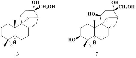
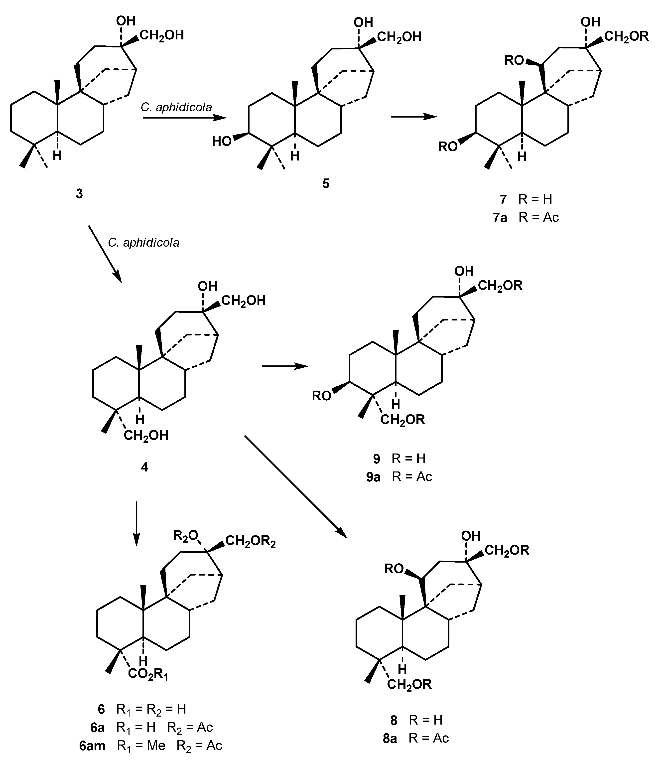
  • The hydroxylations produced in the substrate 3 by this fungus occurred at C-3(β), C-11(β) and C-18.
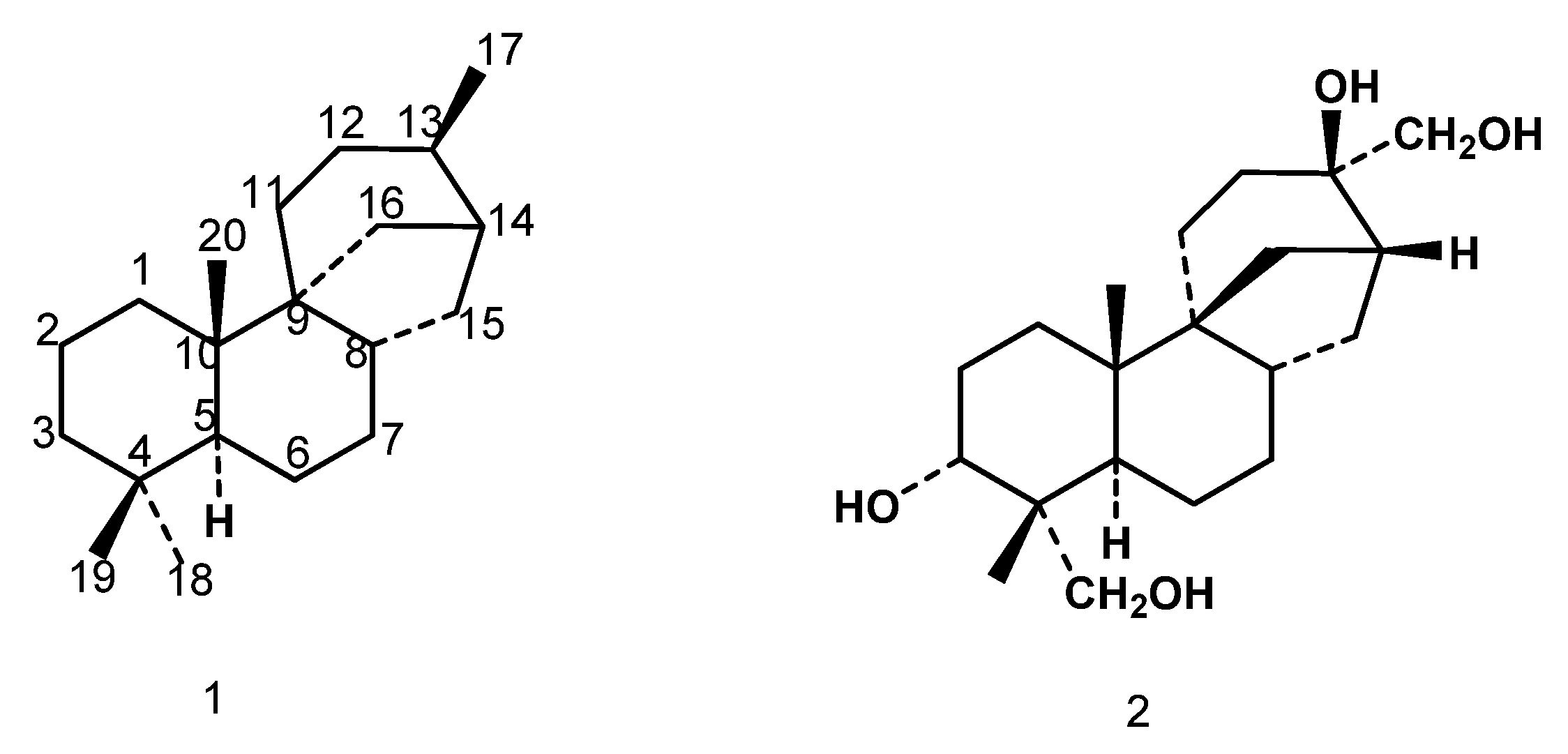
  • The hydroxylation at C-18 points to a biosynthetically-directed transformation, since aphidicolin (2) is also hydroxylated at this carbon. This position was also functionalized in the biotransformation of stemodine and stemodinone with C. aphidicola [10,11]. However, the C-3(β) and C-11(β) hydroxylations, also observed in the incubation of 3, seem to indicate a xenobiotic biotransformation. These hydroxylations were also observed in the feeding of 3 with M. plumbeus [13], a fungus used in the latter type.
  • The oxidation of C-18 to acid level, as occurs in the formation of 6 from 4, has now been observed for the first time in a biotransformation with C. aphidicola.
  • It is probable that the formation of 9 only occurs from 4, and not from 5. Thus the hydroxylation of the C-18 methyl in 5 to form 9 could be inhibited by the presence of the equatorial β-hydroxyl group at C-3 (Scheme 2). In aphidicolin biosynthesis has been noted that an axial α-hydroxyl at C-3 blocks the hydroxylation of C-18 [8], whilst in gibberellin biosynthesis has been observed that an equatorial 3α-OH inhibits hydroxylation of C-19 [16].

Acknowledgements

This work has been supported by grant CTQ2009-14629-C02-01, Ministerio de Ciencia e Innovación (MICINN), Spain.

References and Notes

  1. Thiericke, R.; Rohr, J. Biological variation of microbial metabolites by precursor-directed biosynthesis. Nat. Prod. Rep. 1993, 10, 265–289. [Google Scholar] [CrossRef]
  2. Hanson, J.R. The microbiological transformation of diterpenoids. Nat. Prod. Rep. 1992, 9, 139–151. [Google Scholar] [CrossRef]
  3. Fraga, B.M.; González-Vallejo, V.; Guillermo, R. On the biotransformation of ent-trachylobane to ent-kaur-11-ene diterpenes. J. Nat. Prod. 2011, 74, 1985–1989. [Google Scholar] [CrossRef]
  4. Fraga, B.M.; González-Vallejo, V.; Guillermo, R.; Díaz, L.N. Biotransformation of 7α-hydroxy- and 7-oxo-ent-atis-16-ene derivatives by the fungus Gibberella fujikuroi. Phytochemistry 2010, 71, 1313–1321. [Google Scholar]
  5. Hanson, J.R.; Nasir, H.; Parvez, A. The hydroxylation of testosterone and some relatives by Cephalosporium aphidicola. Phytochemistry 1996, 42, 411–415. [Google Scholar]
  6. Atta-ur-Rahman; Yaqoob, A.; Farooq, A.; Anjun, S.; Fahim, A.; Choudhary, M.I. Fngal transformation of (1R, 2S, 5R)-(−)-menthol by Cephalosporium aphidicola. J. Nat. Prod. 1998, 61, 1340–1342. [Google Scholar] [CrossRef]
  7. Hanson, J.R.; Jarvis, A.J.; Ratcliffe, A.H. Biotransformation of some aphidicolane derivatives by Cephalosporium aphidicola. Phytochemistry 1992, 31, 3851–3853. [Google Scholar]
  8. Hanson, J.R.; Jarvis, A.J.; Laboret, F.; Takahashi, J. The incubation of 3α,16β-dihydroxyaphidicolane with Cephalosporium aphidicola. Phytochemistry 1995, 38, 73–75. [Google Scholar]
  9. Dalziel, W.; Hesp, B.; Stevenson, K.M.; Jarvis, J.A.J. The structure and absolute configuration of the antibiotic aphidicolin: A tetracyclic diterpenoid containing a new ring system. J. Chem. Soc. Perkin Trans. I 1973, 2841–2851. [Google Scholar]
  10. Badria, F.A.; Hufford, C.D. Microbial transformations of stemodin, a Stemodia diterpene. Phytochemistry 1991, 30, 2265–2268. [Google Scholar]
  11. Hanson, J.R.; Reese, P.B.; Takahashi, J.A.; Wilson, M.R. Biotransformation of some stemodane diterpenoids by Cephalosporium aphidicola. Phytochemistry 1994, 36, 1391–1393. [Google Scholar]
  12. Chamy, M.C.; Piovano, M.; Garbarino, J.A.; Gambaro, V. Stemodane diterpenes from Stemodia chilensis. Phytochemistry 1991, 30, 1719–1721. [Google Scholar]
  13. Fraga, B.M.; Guillermo, R.; Hernández, M.G.; Chamy, M.C.; Garbarino, J.A. Biotransformation of two stemodane diterpenes by Mucor plumbeus. Tetrahedron 2004, 60, 7921–7932. [Google Scholar]
  14. González, A.G.; Arteaga, J.M.; Bretón, J.L.; Fraga, B.M. Five new labdane diterpene oxides from Eupatorium jhanii. Phytochemistry 1977, 16, 107–110. [Google Scholar]
  15. González, A.G.; Fraga, B.M.; Hernández, M.G.; Hanson, J.R. The 13C NMR spectra of some ent-18-hydroxy-kaur-16-enes. Phytochemistry 1981, 20, 846–847. [Google Scholar]
  16. Fraga, B.M.; González, A.G.; Hanson, J.R.; Hernández, M.G. The microbiological transformation of some ent-3β-hydroxykaur-16-enes by Gibberella fujikuroi. Phytochemistry 1981, 20, 57–61. [Google Scholar]
  • Sample Availability: Samples of the compounds 3 and 5 are available from the authors.

