Article

Regulated Expressions of MMP-2, -9 by Diterpenoids from Euphorbia formosana Hayata

1
Graduate Institute of Pharmacognosy, Taipei Medical University, 250 Wu Xin Street, Taipei 110, Taiwan
2
Taipei Medical University Hospital, Taipei Medical University, 252 Wu Xin Street, Taipei 110, Taiwan
3
Department of Pharmacology, Taipei Medical University, 250 Wu Xin Street, Taipei 110, Taiwan
4
School of Pharmacy, Taipei Medical University, 250 Wu Xin Street, Taipei 110, Taiwan
5
Taiwan Forestry Research Institute, No. 53, Nan-Hai Road, Taipei, 100, Taiwan
*
Authors to whom correspondence should be addressed.
These authors contributed equally to this work.
Molecules 2012, 17(2), 2082-2090; https://doi.org/10.3390/molecules17022082
Submission received: 29 November 2011 / Revised: 3 February 2012 / Accepted: 6 February 2012 / Published: 21 February 2012

Abstract

:
Two new abietane type diterpenoids, namely seco-helioscopinolide (1) and 3β,7β-dihydroxy-ent-abieta-8,13-diene-12,16-olide (2) were isolated from the aerial parts of Euphorbia formosana Hayata together with helioscopinolide A (3), helioscopinolide B (4), helioscopinolide C (5) and ent-(5β,8α,9β,10α,12α)-12-hydroxyatis-16-ene-3,14-dione (6). The structures of compounds 16 were elucidated by analyzing their spectroscopic data and comparison with the literature. Further biological tests by gelatin zymographic analysis revealed that 35 significantly up-regulated the expressions and activation of MMP-2 and -9 in human fibrosarcoma cell line HT1080.

1. Introduction

Increasing ultraviolet exposure of skin leads to acute and chronic detrimental cutaneous effects, which may result in the development of skin malignancies and photoaging [1]. Matrix metalloproteinases (MMPs) were known not only to exert functions in the resolution phase of wound healing but influence other wound-healing responses, such as inflammatory and re-epithelization [2]. Therefore, the screening of bioactive compounds from natural resources which can modulate the activity of MMPs, should prove worthwhile. Euphorbia formosana Hayata has long been used in folk medicine for the treatment of snakebite as well as many other dermatoses in Taiwan [3]. In this paper we describe the isolation and identification of a series of ent-abietane-type diterpenoids 16 (Figure 1) from E. formosana. During the past few years analogues of 16 were found to exhibit cytotoxic [4], antimicrobial [5], spasmolytic [6], antioxidant [7] and gastroprotective activities [8]. The bioactivities of 16 on MMP-2 and -9 in HT-1080 cells were evaluated in this study.
Figure 1. Chemical structures of compound 16.
Molecules 17 02082 g001

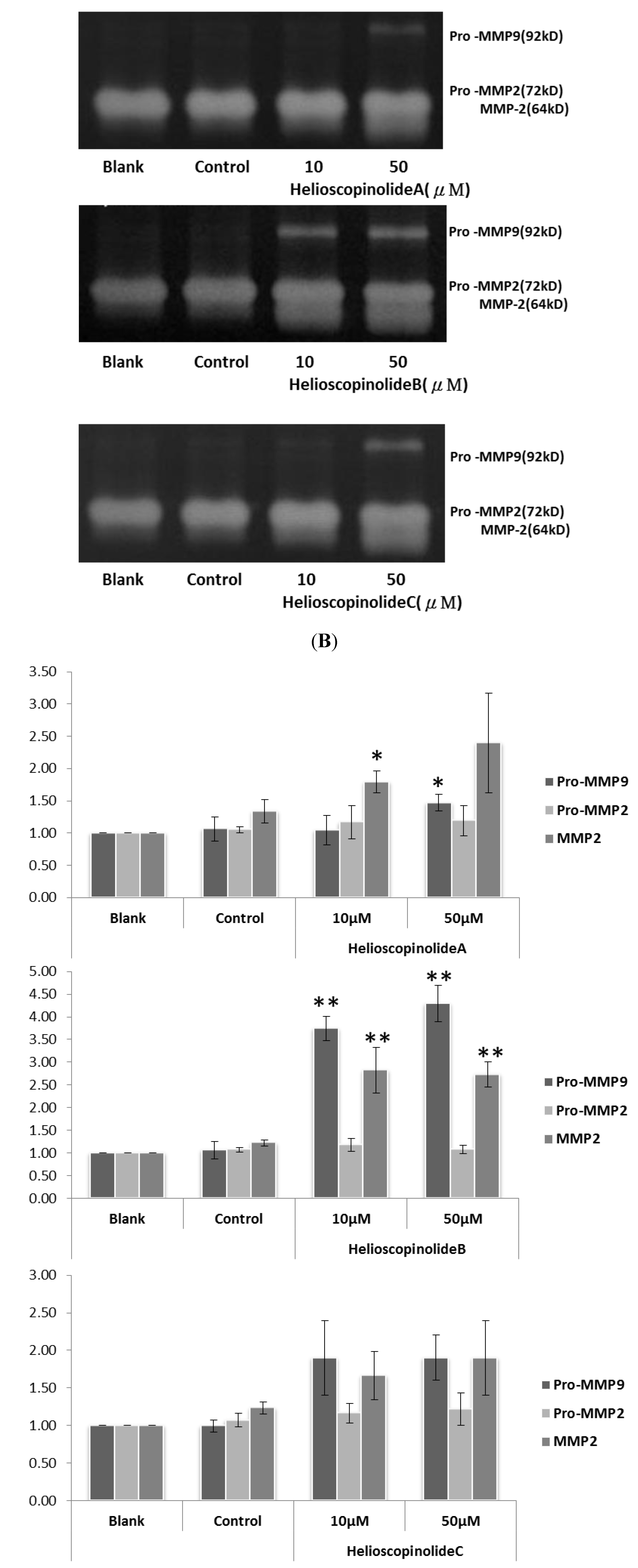
2. Results and Discussion

The aerial parts of E. formosana were extracted initially using methanol, and further partitioned between CHCl3 and H2O. The CHCl3 layer was then subjected to gravity silica column separation followed by HPLC purification to give two new diterpenoids 1 and 2, along with helioscopinolide A (3) [9], helioscopinolide B (4) [10], helioscopinolide C (5) [11] and ent-(5β,8α,9β,10α,12α)-12-hydroxyatis-16-ene-3,14-dione (6) [12].
Compound 1 was isolated as colorless oil, and its molecular formula, C21H30O5, was established through analysis of its 13C-NMR and HR-ESI-MS data. The IR spectrum of 1 exhibited the presence of a hydroxyl group (3470 cm−1), a carbonyl group (1729 cm−1) and a double bond (1665 cm−1). The 1H-NMR data (Table 1) of 1 revealed signals for four methyl groups at δH 1.04 (s, H3-19), 1.23 (s, H3-17), 1.29 (s, H3-18) and 1.81 (s, H3-20), five methylene groups at δΗ 1.50 (m, H-11a), 1.52 (m, H-6a), 1.78 (m, H-6b), 1.86 (m, H-1a), 2.12 (m, H-7a), 2.17 (m, H-2a), 2.44 (td, J = 3.3, 13.1 Hz, H-7b), 2.55 (dd, J = 5.8, 13.5 Hz, H-11b), 2.60 (ddd, J = 4.6, 11.1, 13.5 Hz, H-2b) and 2.75 (ddd, J = 4.6, 11.1, 13.2 Hz, H-1b), four methine protons at δΗ 1.69 (dd, J = 2.7, 12.4 Hz, H-5), 2.33 (d, J = 8.4 Hz, H-9), 4.88 (dd, J = 6.1, 13.5 Hz, H-12) and 6.26 (s, H-14) and one methoxyl proton at 3.66 (s, H3-21).
Table 1. 1H-NMR data of 1 and 2 [in CDCl3, 500 MHz, δ in ppm (mult., J in Hz)].
No.1 2
13C1H 13C1H
1a33.1 t1.86 (m)32.9 t1.80 (m)
1b2.75 (ddd, 4.6, 11.1, 13.2) 1.98 (m)
2a28.8 t2.17 (m) 26.9 t1.62 (m)
2b2.60 (ddd, 4.6, 11.1, 13.5) 1.96 (m)
3174.8 s 75.6 d3.40 (br s)
475.2 s 38.1 s
551.8 d1.69 (dd, 2.7, 12.4) 41.2 d2.33 (dd, 2.5, 13.5)
6a27.4 t1.52 (m) 31.7 t1.82 (td, 2.5, 13.5)
6b1.78 (m) 1.59 (m)
7a36.8 t2.12 (m) 72.3 d4.41 (br s)
7b2.44 (td, 3.3, 13.1)
8151.7 s 153.8 s
945.2 d2.33 (d, 8.4) 47.4 d2.82 (br d, 9.0)
1045.5 s 42.4 s
11a27.4 t1.50 (m) 28.2 t1.37 (dt, 9.0, 13.5)
11b2.55 (dd, 6.0, 13.5) 2.57 (dd, 6.5, 13.5)
1275.9 d4.88 (dd, 6.0, 13.5) 76.7 d4.89 (dd, 6.5, 13.5)
13155.9 s 156.7 s
14113.9 d6.26 (s) 115.8 d6.52 (br s)
15116.6 s 118.4 s
16175.2 s 174.9 s
1727.6 q1.23 (s) 29.4 q0.94 (s)
1834.8 q1.29 (s) 22.8 q0.86 (s)
1920.4 q1.04 (s) 16.6 q0.96 (s)
208.3 q1.81 (s) 8.5 q1.78 (d, 1.5)
2151.7 q3.66 (s)
The 13C-NMR spectrum coupled with DEPT analysis displayed 21 signals including four methyl carbons at δC 8.3 (C-20), 20.4 (C-19), 27.6 (C-17) and 34.8 (C-18), five methylene carbons at δC 27.4 (C-6 and -11), 28.8 (C-2), 33.1 (C-1) and 36.8 (C-7), four methine carbons at δC 45.2 (C-9), 51.8 (C-5), 75.9 (C-12) and 113.9 (C-14), seven quaternary carbons at δC 45.5 (C-10), 75.2 (C-4), 116.6 (C-15), 151.7 (C-8), 155.9 (C-13), 174.8 (C-3) and 175.2 (C-16), and one methoxyl carbon at δC 51.7 (C-21) (Table 1). Among the quaternary carbons, resonances at δ(C) 174.8 (C-3) and 175.2 (C-16) were assigned as two ester carbonyl groups. On account of the molecular formula, C21H30O5, since the index of hydrogen deficiency of 1 was seven, including two ester carbonyls and two olefinic functionalities, thus there should be three rings in 1. Analysis of COSY (Figure 2) and HSQC spectral data allowed the assignments of three spin systems including two aliphatic chains, (–H-5–H2-6–H2-7–) and (–H-9–H2-11–H-12–), one two-resonance unit, (–H2-1–H2-2–), and five germinal-coupled methylene functionalities, (H2-1, H2-2, H2-6, H2-7 and H2–11).
Figure 2. COSY of 1.
Molecules 17 02082 g002
In the HMBC spectrum of 1 (Figure 3), key long range proton-carbon correlations including H-7/C-14, H-12/C-13, -14 and -15, H3-19/C-1, -5, -9 and -10, H3-17/C-5 and -18, H3-20/C-13, -15 and -16 and H3-21/C-3 coupled by above interpretations established that 1 was quite similar to helioscopinolide A (3), except for an opened A ring, a methoxyl group attached at C-3 and a hydroxyl group borne by its C-4. Accordingly, the structure of 1 was elucidated to be as shown (Figure 1) and was named seco-helioscopinolide. The methyl ester at C-3 of 1 was not an artifact on the methanolic extraction process, as evidenced by further HPLC analyses of an acetonic extract of the original materials.
Figure 3. Selected HMBC of 1.
Molecules 17 02082 g003
Compound 2, obtained as amorphous white powder, was deduced to have a molecular formula of C20H28O4 by its HR-ESI-MS and 13C-NMR. The IR spectrum of 2 revealed the presence of a hydroxyl group (3411 cm−1), a γ-lactone carbonyl (1731 cm−1) and a double bond (1667 cm−1). The 1H-NMR data of 2 revealed signals compatible with those of helioscopinolide B (4), except that a carbinol group was present at C-7 in 2 (Table 1). The 1H-NMR differences between 2 and 4 were also reflected in their 13C-NMR, in which δC 37.0 of C-7 in 4 downfield shifted to δC 72.3 in 2. The structure of 2 was thus determined to be 7-hydroxyhelioscopinolide B. The relative configuration of both H-3 and H-7 was determined to be equatorial, due to their distinctive small coupling constants and chemical shifts (Table 1), which were further confirmed by comparison with its H-3 axial-oriented isomer, 3α,7β-dihydroxy-ent-abieta-8,13-diene-12,16-olide [4]. The chemical shifts of H-3 in 2H-3 3.40) and its isomer (δH-3 3.25) also corroborated that their H-3s were equatorial- and axial-oriented, respectively, as judged by the anisotropic effect. Compound 2 adopted an ent-abietane-type diterpenoid skeleton as evidenced from a positive Cotton effect at 250 nm and a negative Cotton effect at 215 nm in the circular dichroism spectrum of 2 and comparison with the literature [13]. After considering all the spectroscopic data of 2, its structure was thus elucidated as shown in Figure 1, and was named 3β,7β-dihydroxy-ent-abieta-8,13-diene-12,16-olide.
The MMP-2 and -9 modulating activity of the pure isolated compounds 16 in human fibrosarcoma cell line HT1080 were compared at the concentrations of 10 and 50 μM with blank group (Figure 4A). Among them, 3-hydroxyl ent-abietane compounds 3-5 exhibited significantly up-regulated the expressions of MMP-2, -9 according to gelatin zymography analysis (Figure 4B). The MMPs activity is regulated by transcriptional and post-transcriptional levels. The post-transcriptional activation of MMPs requires specific conditions, such as proteolytic removal of the propeptide, disruption of the bond between the cysteine sulhydryl moiety, and even their dephosphorylation [14,15] The intensive mode of action of 35 on posttranscriptional MMPs should remain to be further investigated.
Figure 4. Effects of 35 on MMP-2, -9 activities. (A1-3) Results of gelatin zymography. Lane 1: blank; Lane 2: control; Lane 3-4: different concentration of compounds 35 (B1-3) Activities of MMP-2, -9 expressed as multiplication product of zone area and average gray value. Data represent the mean ± SD (n = 3). * p < 0.05, ** p < 0.01.
Molecules 17 02082 g004

3. Experimental

3.1. General

Optical rotations were measured on a JASCO P-1020 digital polarimeter (Tokyo, Japan). 1H- and 13C-NMR were acquired on a Bruker DMX-500 SB spectrometer (Ettlingen, Germany). High resolution and low resolution mass spectra were obtained using a Waters LCT Premier XE (Waters, Manchester, UK) and ABI API 4000 Q-TRAP (Foster City, CA, USA), respectively. IR spectra were recorded on a JASCO FT/IR 4100 spectrometer (Tokyo, Japan). UV and CD spectra were measured on a Thermo Helios α spectrophotometer (Waltham, MA, USA) and JASCO J-720 circular dichroism spectrometer (Tokyo, Japan), respectively. Silica gel (40–63 μm, Merck, Darmstadt, Germany) was used for gravity column chromatography. Pre-coated silica gel plates (Si 60 F254, 0.2 mm, Merck, Darmstadt, Germany) were used for analytical TLC. HPLC was performed using a semi-preparative column (Hibar® Fertigäute, 10 × 250 mm, Merck).

3.2. Plant Material

Aerial parts of Euphorbia formosana Hayata was collected from Changhua County in July, 2004 and was identified by Dr. S. Y. Chen in the Council of Agriculture, Executive Yuan. Voucher Specimens (No. CKL07012004) have been deposited in the School of Pharmacy, Taipei Medical University, Taipei, Taiwan.

3.3. Extraction and Isolation

The dried aerial parts (11.8 kg) of E. formosana was extracted three times with MeOH (100 L), then partitioned between CHCl3 and water (2 L, 1:1, v/v). Subsequently, the dried CHCl3 layer (300 g) was pre-absorbed using 400 g silica gel (70–230 mesh), applied onto a 4,950 g silica gel open column (230–400 mesh) and then eluted by mixtures of n-hexane, ethyl acetate and methanol in a stepwise gradient mode. Each fraction collected (500 mL) was checked by TLC, and dipping in 10% H2SO4 in ethanol were used in the detection of compounds. Subsequently, 348 fractions were combined into 15 portions. Portion #10 was purified by HPLC on a semi-preparative normal-phase column with n-hexane/ethyl acetate/acetone/dichloromethane (25:6:4:10, v/v/v/v) as eluent, 3 mL/min, afforded 3 (9.0 mg). The same portion was purified using the same HPLC column with n-hexane/acetone (5:1, v/v) as eluent, 3 mL/min, gave 1 (9.6 mg), 4 (8.4 mg), 5 (7.8 mg) and 6 (4.5 mg). The portion #15 was purified using the same HPLC column with n-hexane/acetone (7:3, v/v) as eluent, 3 mL/min, obtained 2 (22.5 mg).

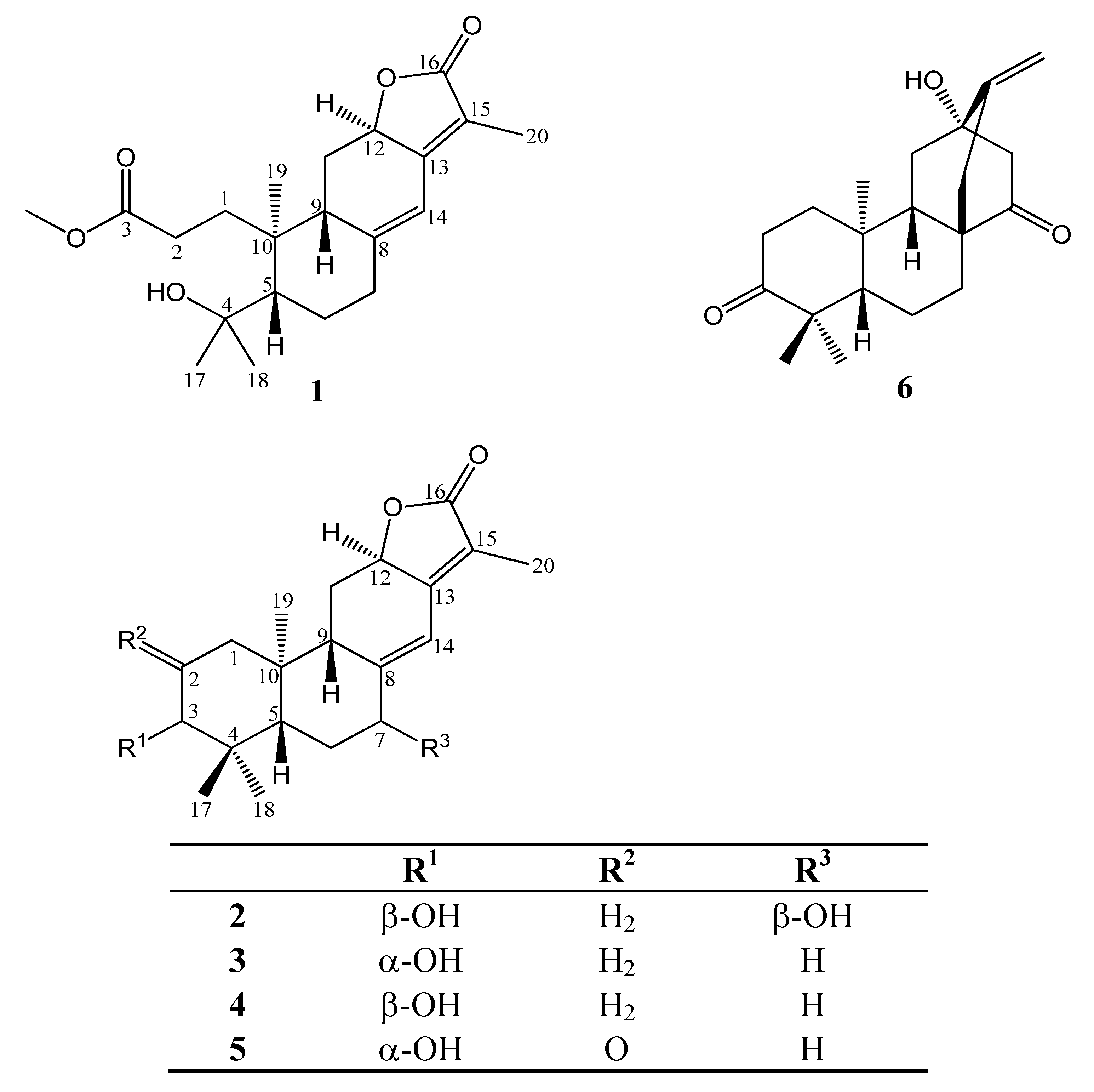
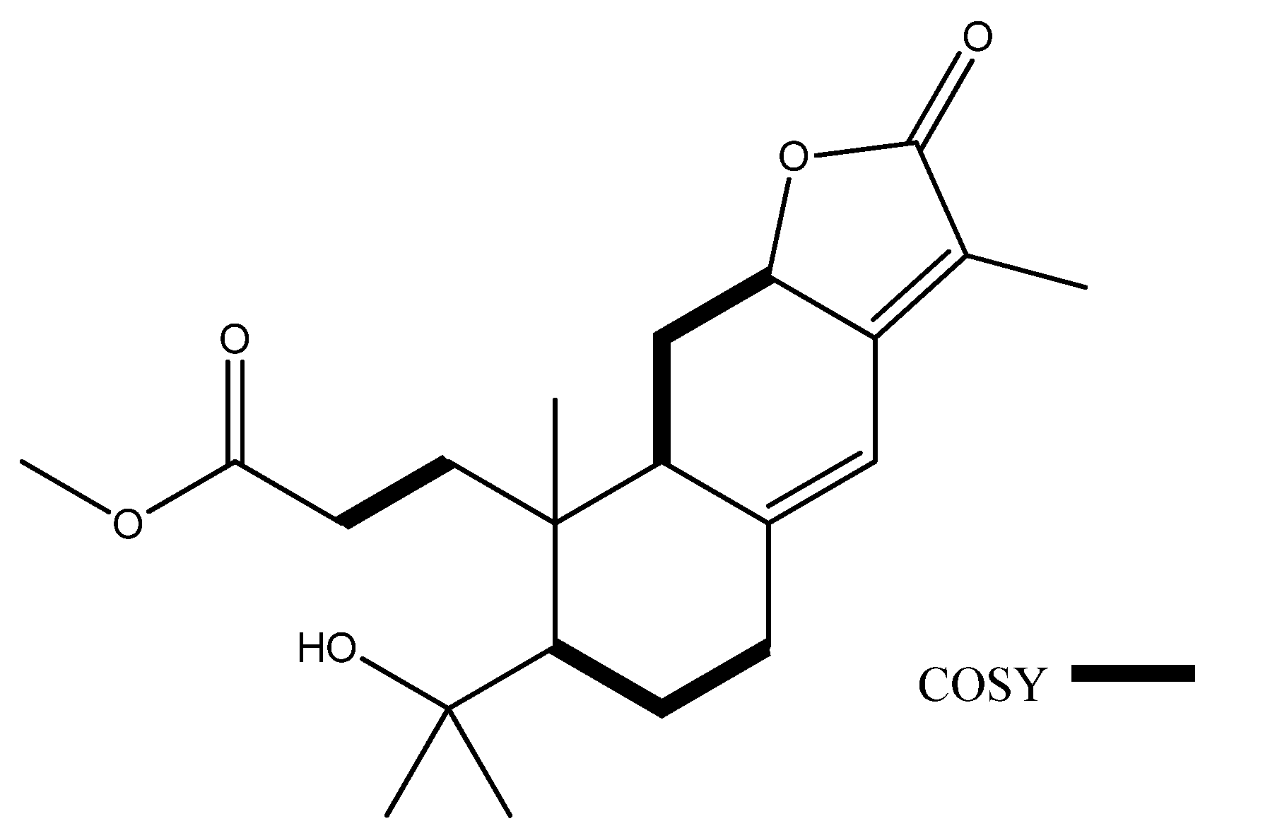
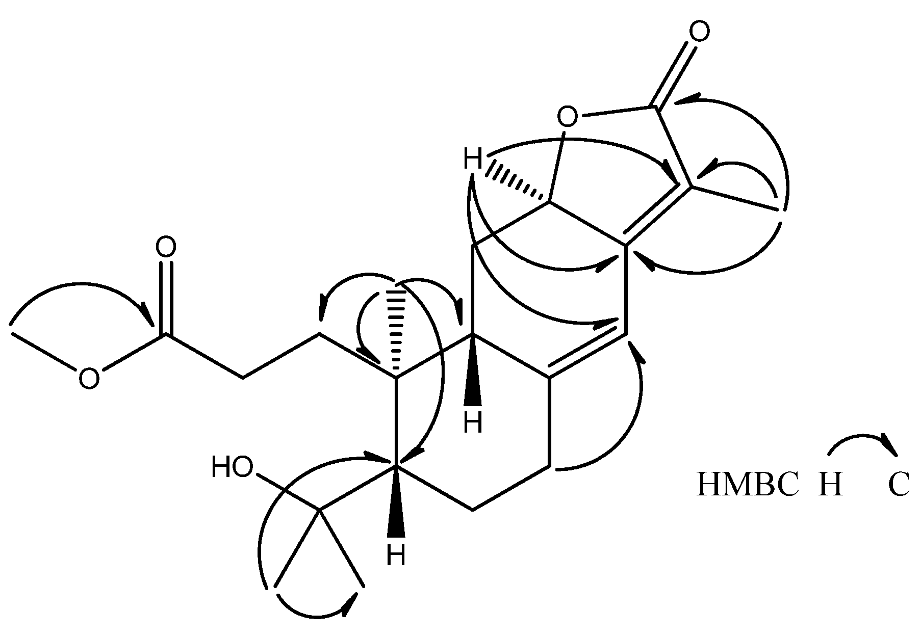
3.4. seco-Helioscopinolide (1)

Colorless oil. [α]25D = +50.8 (c = 0.1, CHCl3). UV (MeOH): 276 (3.9). IR (KBr): 3470 (−OH), 2928, 2877, 1729 (C=O), 1665 (C=C), 1438, 1024. 1H-NMR and 13C-NMR: see Table 1. ESI-MS: 363 [M+H]+. HR-ESI-MS: 363.2181 ([M+H]+, C21H31O5, calc.363.2171).

3.5. 3β,7β-Dihydroxy-ent-abieta-8,13-diene-16,12-olide (2)

Amorphous white powder. [α]25D = +180.4 (c = 0.1, MeOH). UV (MeOH): 272 (4.0). IR (KBr): 3411 (−OH), 2927, 1731 (C=O), 1667 (C=C), 1451, 1385, 1301, 1168, 1090, 1030. 1H-NMR and 13C-NMR: see Table 1. ESI-MS: 333 [M+H]+. HR-ESI-MS: 331.1905 ([M−H], C20H27O4, calc. 331.1909).

3.6. Cell Culture

HT1080 human fibrosarcoma cells which were from American Type Culture Collection (ATCC: CCL-121) were cultured in RPMI-1640 medium (Gibco) supplemented with 10% fetal bovine serum (Gibco), 100 U/mL penicillin, 100 mg/mL streptomycin. Cultures were maintained in a humidified incubator at 37 °C in 5% CO2/95% air.

3.7. Gelatin Zymography

Gelatin zymography was used to determine expression and activities of MMP-2 and -9 [16]. HT1080 cell line were seeded in 24-well plates using serum free medium for 24hr cell adhesion and growth. After 48 h of test compounds with different concentrations incubation, conditioned medium was collected to analyzed the activities of MMP-2 and -9.

4. Conclusions

In this report, we have identified six ent-abietane-type diterpenoids including two new ones from Euphorbia formosana Hayata. Ent-abietane-type diterpenoids such as compounds 35 significantly up-regulated the expressions and activation of MMP-2 and -9 in human fibrosarcoma cell line HT1080, and could potentially be leads for the development of novel wound-healing drugs.

Acknowledgements

This research was supported by the grant from National Science Council (NSC 98-2320-B-038-015-MY3), Taipei Medical University Hospital (98TMU-TMUH-04-2) and Center of Excellence for Clinical Trial and Research in Neuroscience, WanFang Hospital. We are grateful to Shwu-Huey Wang for the NMR data acquisition in the Instrumentation Center of Taipei Medical University and the YungShin Group for supported MS instrumentation.

References and Notes

  1. Goihman-Yahr, M. Skin aging and photoaging: An outlook. Clin. Dermatol. 1996, 14, 153–160. [Google Scholar] [CrossRef]
  2. Gill, S.E.; Parks, W.C. Metalloproteinases and their inhibitors: regulators of wound healing. Int. J. Biochem. Biol. 2008, 40, 1334–1347. [Google Scholar] [CrossRef]
  3. Chang, H.-C. Medicinal Herbs; Recreation Press: Taipei, Taiwan, 1990; p. 58. [Google Scholar]
  4. Yan, R.-Y.; Tan, Y.-X.; Cui, X.-Q.; Chen, R.-Y.; Yu, D.-Q. Diterpenoids from the roots of Suregada glomerulata. J. Nat. Prod. 2008, 71, 195–198. [Google Scholar] [CrossRef]
  5. Radulović, N.; Denić, M.; Stojanović-Radić, Z. Antimicrobial phenolic abietane diterpene from Lycopus europaeus L. (Lamiaceae). Bioorg. Med. Chem. Lett. 2010, 20, 4988–4991. [Google Scholar] [CrossRef]
  6. Perez-Hernandez, N.; Ponce-Monter, H.; Medina, J.A.; Joseph-Nathan, P. Spasmolytic effect of constituents from Lepechinia caulescens on rat uterus. J. Ethanopharmacol. 2008, 115, 30–35. [Google Scholar] [CrossRef]
  7. Kabouche, A.; Kabouche, Z.; Öztürk, M.; Kolak, U.; Topçu, G. Antioxidant abietane diterpenoids from Salvia barrelieri. Food Chem. 2007, 102, 1281–1287. [Google Scholar] [CrossRef]
  8. Rodríguez, J.A.; Theoduloz, C.; Yáñez, T.; Becerra, J.; Schmeda-Hirschmann, G. Gastroprotective and ulcer healing effect of ferruginol in mice and rats: Assessment of its mechanism of action using in vitro models. Life Sci. 2006, 78, 2503–2509. [Google Scholar] [CrossRef]
  9. Shzuri, Y.; Kosemura, S.; Yamamura, S.; Ohba, S.; Ito, M.; Saito, Y. Isolation and structures of helioscopinolide, new diterpenoids from Euphorbia helioscopia L. Chem. Lett. 1983, 12, 65–68. [Google Scholar]
  10. Crespi-Perellino, N.; Garofano, L.; Arlandini, E.; Pinciroli, V.; Minghetti, A.; Vincieri, F.F. Danieli, B. Identification of new diterpenoids from Euphorbia calyptrate cell culture. J. Nat. Prod. 1996, 59, 773–776. [Google Scholar] [CrossRef]
  11. Borghi, D.; Baumer, L.; Ballabio, M.; Arlandini, E.; Perellino, N.C.; Minghetti, A.; Vincieri, F.F. Structure elucidation of helioscopinolides D and E from Euphorbia calyptrata cell cultures. J. Nat. Prod. 1991, 54, 1503–1508. [Google Scholar] [CrossRef]
  12. He, F.; Pu, J.X.; Huang, S.X.; Xiao, W.L.; Yang, L.B.; Li, X.N.; Zhao, Y.; Ding, J.; Xu, C.H.; Sun, H.D. Eight new diterpenoids from the roots of Euphorbia nematocypha. Helv. Chim. Acta 2008, 91, 2139. [Google Scholar] [CrossRef]
  13. Lee, C.L.; Chang, F.R.; Hsieh, P.W.; Chiang, M.Y.; Wu, C.C.; Huang, Z.Y.; Lan, Y.H.; Chen, M.; Lee, K.H.; Yen, H.F.; et al. Cytotoxic ent-abietane diterpenes from Gelonium aequoreum. Phytochemistry 2008, 69, 276–287. [Google Scholar] [CrossRef]
  14. van Wart, H.E.; Birkedal-Hansen, H. The cysteine switch: A principle of regulation of metalloproteinase activity with potential applicability to the entire matrix metalloproteinase gene family. Proc. Natl. Acad. Sci. USA 1990, 87, 5578–5582. [Google Scholar] [CrossRef]
  15. Sariahmetoglu, M.; Crawford, B.D.; Leon, H.; Sawicka, J.; Li, L.; Ballermann, B.J.; Holmes, C.; Berthiaume, L.G.; Holt, A.; Sawicki, G.; et al. Regulation of matrix metalloproteinase-2 (MMP-2) activity by phosphorylation. FASEB J. 2007, 21, 2486–2496. [Google Scholar] [CrossRef]
  16. Fang, Q.; Liu, X.; Al-Mugotir, M.; Kobayashi, T.; Abe, S.; Kohyama, T.; Rennard, S.I. Thrombin and TNF-α/IL-1β synergistically induce fibroblast-mediated collagen gel degradation. Am. J. Respir. Cell Mol. Biol. 2006, 35, 714–721. [Google Scholar] [CrossRef]
  • Sample Availability: Samples of all compounds are available from authors.

