Heteroaryl Chalcones: Design, Synthesis, X-ray Crystal Structures and Biological Evaluation

Abstract
:1. Introduction
2. Results and Discussion

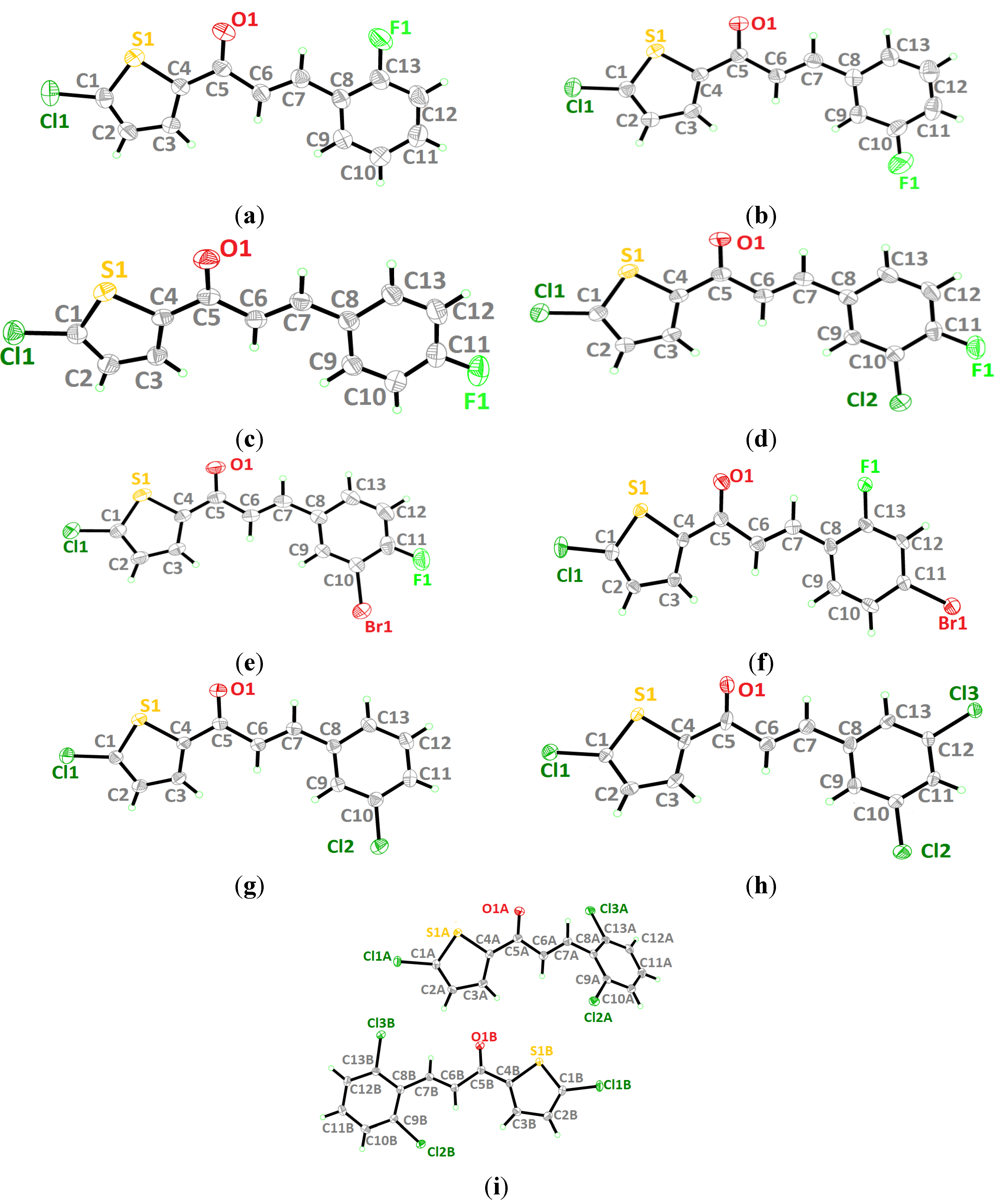
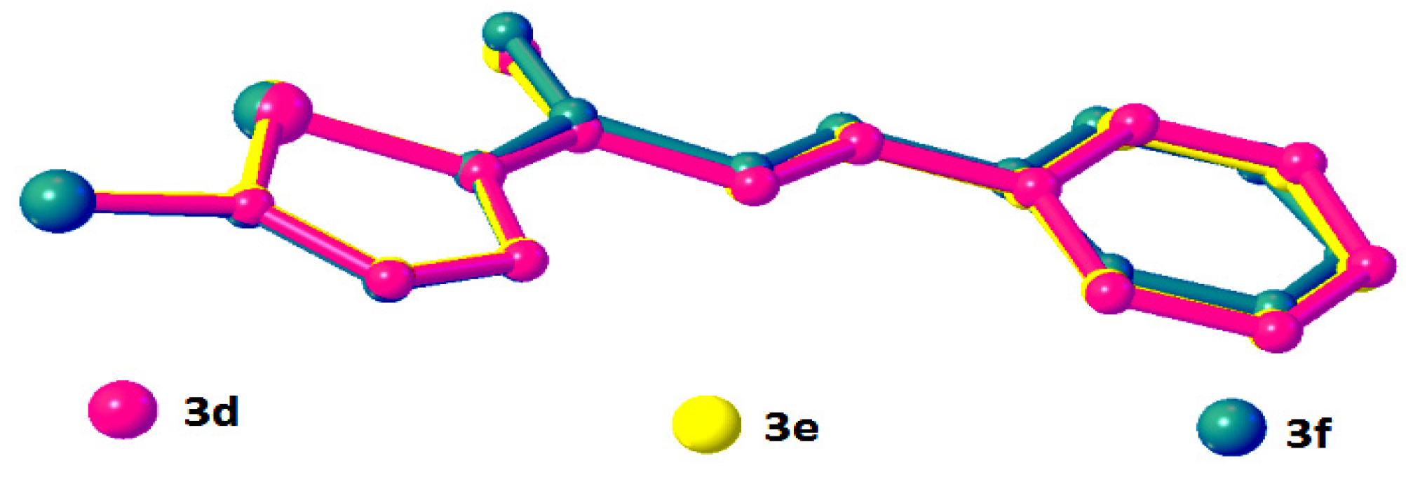
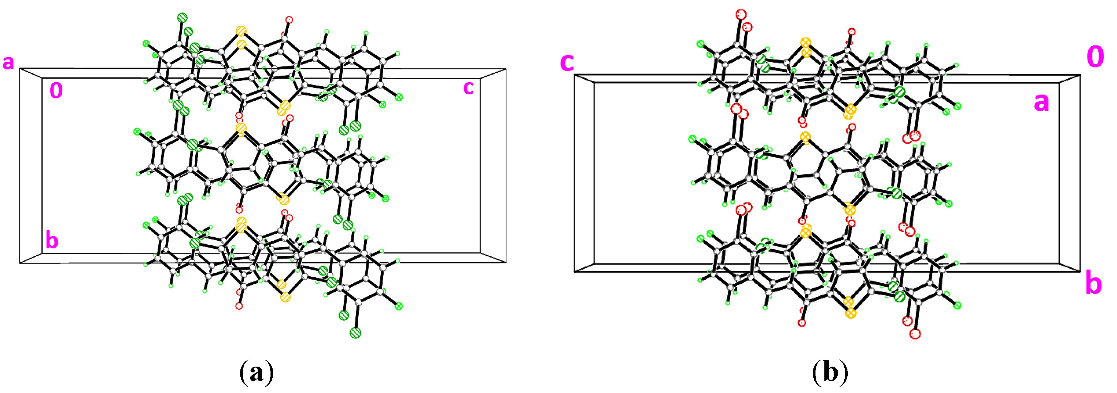
2.1. X-Ray Crystal Structure Description for Compounds 3a–3i
 ring motifs [22].

| Compound | 3a | 3b | 3c | 3d | 3e | 3f | 3g | 3h | 3i |
|---|---|---|---|---|---|---|---|---|---|
| CCDC deposition number | 939875 | 939876 | 939877 | 942741 | 942742 | 948856 | 944072 | 946445 | 948855 |
| Molecular formula | C13H8ClFOS | C13H8ClFOS | C13H8ClFOS | C13H7Cl2FOS | C14H7BrClFOS | C13H7BrClFOS | C13H8Cl2OS | C13H7Cl3OS | C13H7Cl3OS |
| Molecular weight | 266.70 | 266.70 | 266.70 | 301.15 | 345.61 | 345.61 | 283.15 | 317.60 | 317.60 |
| Crystal system | Orthorhombic | Monoclinic | Orthorhombic | Orthorhombic | Orthorhombic | Monoclinic | Monoclinic | Monoclinic | Monoclinic |
| Space group | Pca21 | P21/c | Pbca | Pbca | Pbca | P21/c | P21/c | P21/c | P21/c |
| a (Å) | 27.963 (9) | 14.282 (6) | 7.9809 (8) | 7.4870 (9) | 7.476 (2) | 3.8704 (7) | 14.5437 (10) | 14.9864(10) | 30.3418 (16) |
| b (Å) | 3.9233 (15) | 7.698 (4) | 11.0207 (11) | 11.6186 (14) | 11.724 (3) | 31.953 (6) | 7.5227 (6) | 3.8085 (3) | 3.7985 (2) |
| c (Å) | 11.146 (4) | 11.376 (5) | 27.518 (3) | 29.239 (4) | 30.009 (8) | 10.273 (2) | 11.7604 (9) | 24.6297 (13) | 23.4311 (12) |
| α (°) | 90 | 90 | 90 | 90 | 90 | 90 | 90 | 90 | 90 |
| β (°) | 90 | 104.430 | 90 | 90 | 90 | 95.243 (3) | 104.721 (1) | 115.264 (3) | 109.297 (1) |
| γ (°) | 90 | 90 | 90 | 90 | 90 | 90 | 90 | 90 | 90 |
| V (Å3) | 1222.8 (7) | 1211.3 (9) | 2420.3 (4) | 2543.4 (5) | 2630.4 (13) | 1265.2 (4) | 1244.44 (16) | 1271.30 (15) | 2548.8 (2) |
| Z | 4 | 4 | 8 | 8 | 8 | 4 | 4 | 4 | 8 |
| Dcalc (g cm−3) | 1.449 | 1.462 | 1.464 | 1.573 | 1.745 | 1.814 | 1.511 | 1.659 | 1.655 |
| Crystal dimensions (mm) | 0.93 × 0.18 × 0.05 | 0.25 × 0.25 × 0.05 | 0.79 × 0.37 × 0.04 | 0.75 × 0.52 × 0.10 | 0.58 × 0.28 × 0.09 | 0.89 × 0.12 × 0.09 | 0.50 × 0.23 × 0.08 | 0.51 × 0.10 × 0.05 | 0.60 × 0.10 × 0.10 |
| Colour | Colourless | Colourless | Colourless | Colourless | Colourless | Colourless | Colourless | Colourless | Colourless |
| μ(mm−1) | 0.47 | 0.48 | 0.48 | 0.67 | 3.48 | 3.62 | 0.67 | 0.87 | 0.86 |
| Radiation λ (Å) | 0.71073 | 0.71073 | 0.71073 | 0.71073 | 0.71073 | 0.71073 | 0.71073 | 0.71073 | 0.71073 |
| Tmin/Tmax | 0.668/0.975 | 0.889/0.975 | 0.704/0.980 | 0.633/0.935 | 0.237/0.752 | 0.141/0.744 | 0.733/0.946 | 0.665/0.955 | 0.624/0.922 |
| Reflections measured | 8618 | 9071 | 24679 | 16026 | 16116 | 2855 | 11650 | 11375 | 20108 |
| Ranges/indices (h, k, l) | −37, 39; −5, 5; −15, 15 | −16, 16; −9, 9; −13, 13 | −11, 11; −15, 15; −38, 37 | −10, 10; −14, 16; −41, 37 | −10, 9; −12, 16; −42, 31 | −5, 5; −41, 41; −1, 13 | −18, 18; −9, 9; −15, 15 | −19, 19; −4, 4; −31, 31 | −39, 39; −4, 4; −30, 30 |
| θ limit (°) | 2.3–22.4 | 3.0–19.5 | 3.2–25.3 | 2.8–26.4 | 2.7–25.3 | 2.8–29.8 | 2.9–29.0 | 2.8–30.1 | 2.9–29.9 |
| Unique reflections | 3368 | 2126 | 3540 | 3738 | 3816 | 2855 | 2829 | 2894 | 5762 |
| Observed reflections (I > 2σ(I)) | 1899 | 1133 | 2016 | 2486 | 1935 | 2624 | 2312 | 2607 | 4784 |
| Parameters | 154 | 154 | 154 | 163 | 163 | 164 | 154 | 163 | 325 |
| Goodness of fit on F2 | 0.99 | 1.00 | 1.03 | 1.03 | 1.01 | 1.09 | 1.04 | 1.05 | 1.03 |
| R1, wR2 [I ≥ 2σ(I)] | 0.049, 0.136 | 0.059, 0.191 | 0.047, 0.135 | 0.041, 0.135 | 0.044, 0.136 | 0.051, 0.127 | 0.030, 0.101 | 0.046, 0.120 | 0.033, 0.091 |
| Compound | Dihedral angle between two rings (°) |
|---|---|
| 3a | 5.5 (2) |
| 3b | 15.1 (3) |
| 3c | 14.98 (13) |
| 3d | 9.45 (9) |
| 3e | 6.58 (16) |
| 3f | 1.2 (2) |
| 3g | 16.40 (8) |
| 3h | 2.07 (15) |
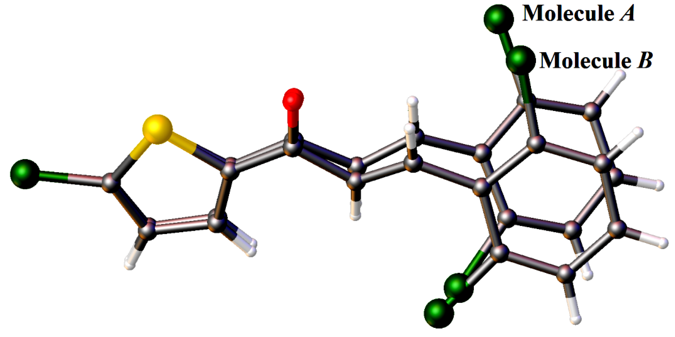
| 3i | Molecule A-45.68 (11) Molecule B-24.00 (11) |

| D–H···A | d(D–H) (Å) | d(H···A)(Å) | d(D···A)(Å) | Angle(D–H···A)(°) |
|---|---|---|---|---|
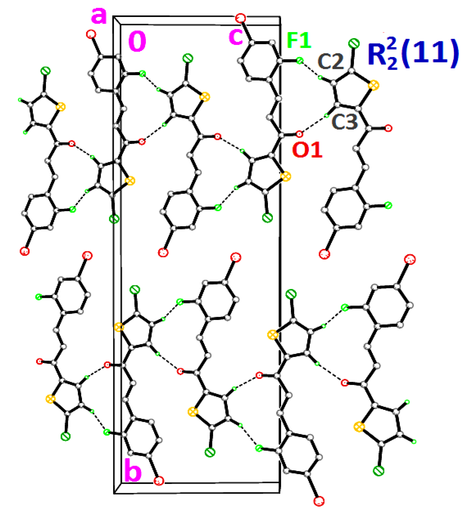
| 3c | ||||
| C3—H3A···O1 i | 0.93 | 2.56 | 3.438(3) | 157 |
| C9—H9A···O1 i | 0.93 | 2.54 | 3.474(3) | 179 |
| 3f | ||||
| C2—H2A···F1 ii | 0.95 | 2.40 | 3.295 (5) | 156 |
| C3—H3A···O1 ii | 0.95 | 2.48 | 3.381 (5) | 158 |
| 3h | ||||
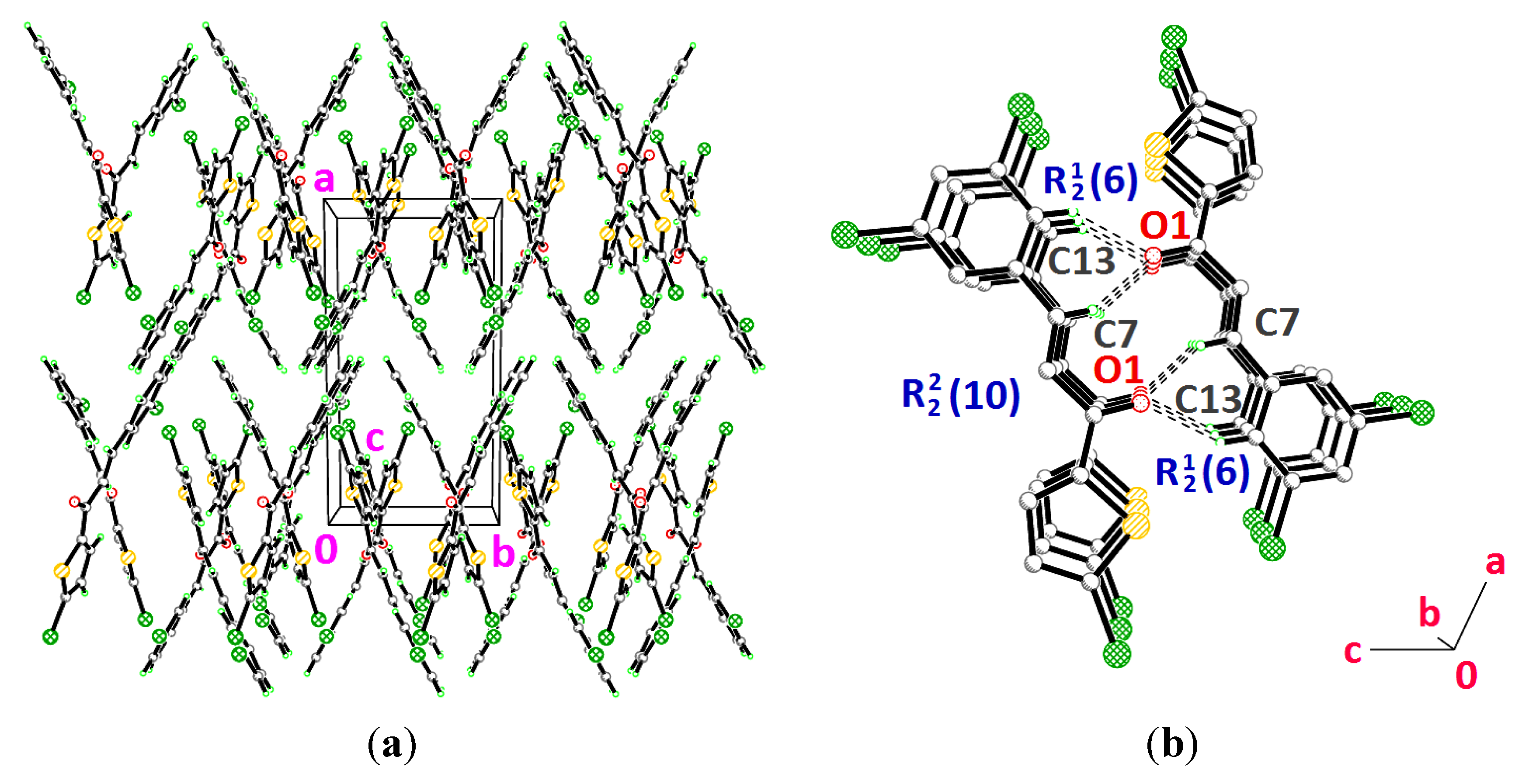
| C7—H7A···O1 iii | 0.93 | 2.49 | 3.337(4) | 151 |
| C13—H13A···O1 iii | 0.93 | 2.48 | 3.300(4) | 148 |
| 3i | ||||
| C2B—H2BA···O1A iv | 0.95 | 2.50 | 3.209(3) | 131 |


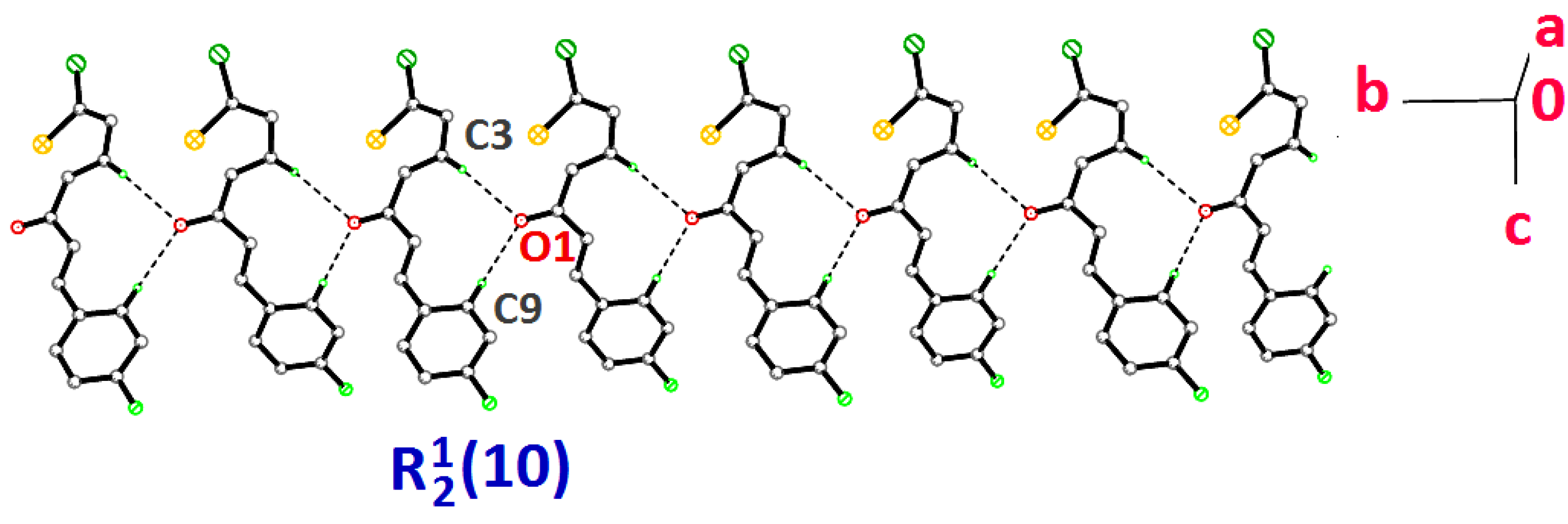
 ring motifs [22].


 ring motifs [22] via intermolecular C7—H7A···O1 hydrogen bonds (Table 3). These sets of dimers are further connected by intermolecular C13—H13A···O1 hydrogen bonds (Table 3) into another two
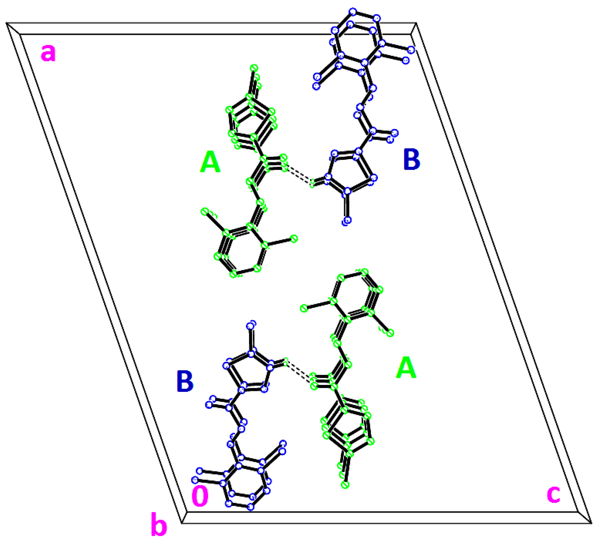
  ring motifs. The molecules stacked along the b-axis. In the crystal structure of 3i (Figure 11), molecules A highlighted in green are joined with the adjacent molecules B highlighted in blue through intermolecular C2B—H2BA···O1A hydrogen bonds (Table 3).


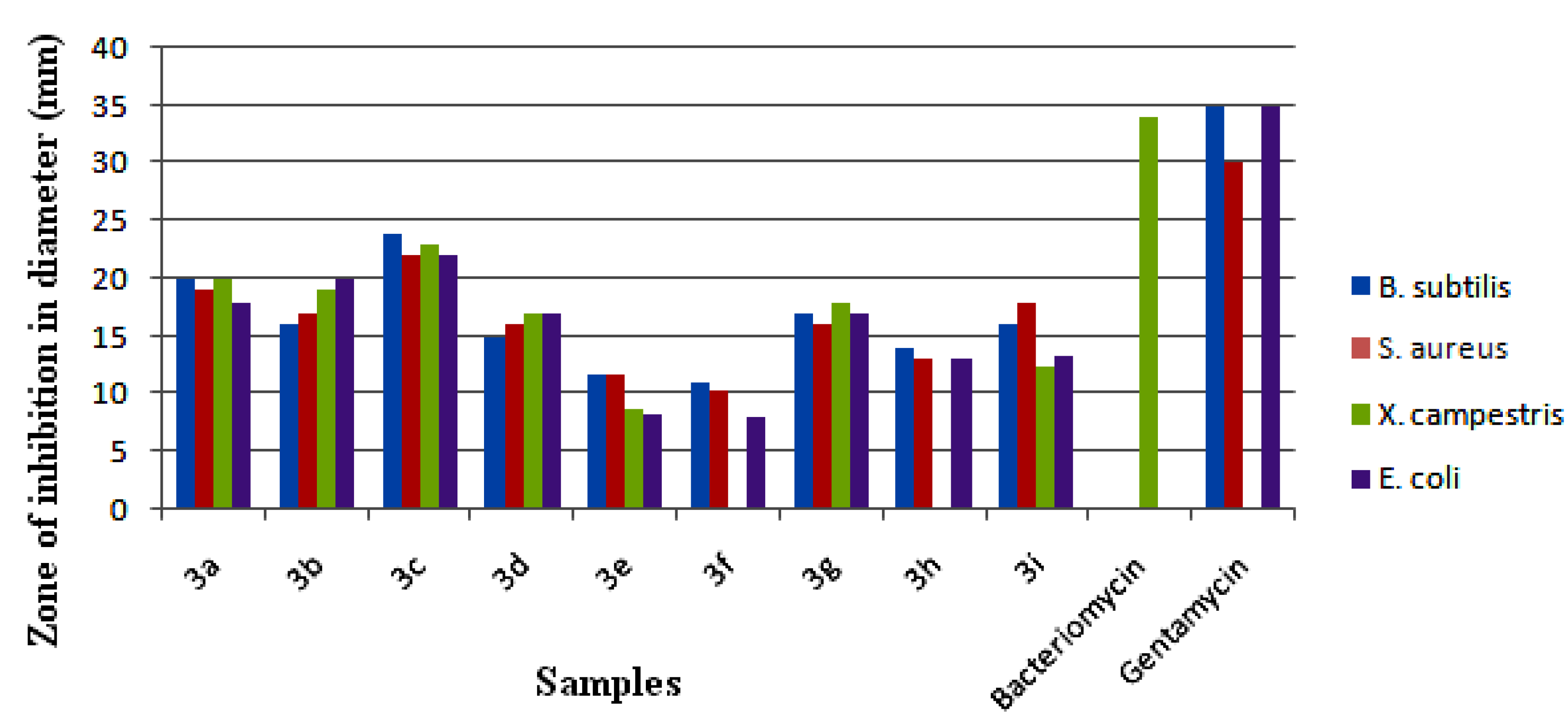
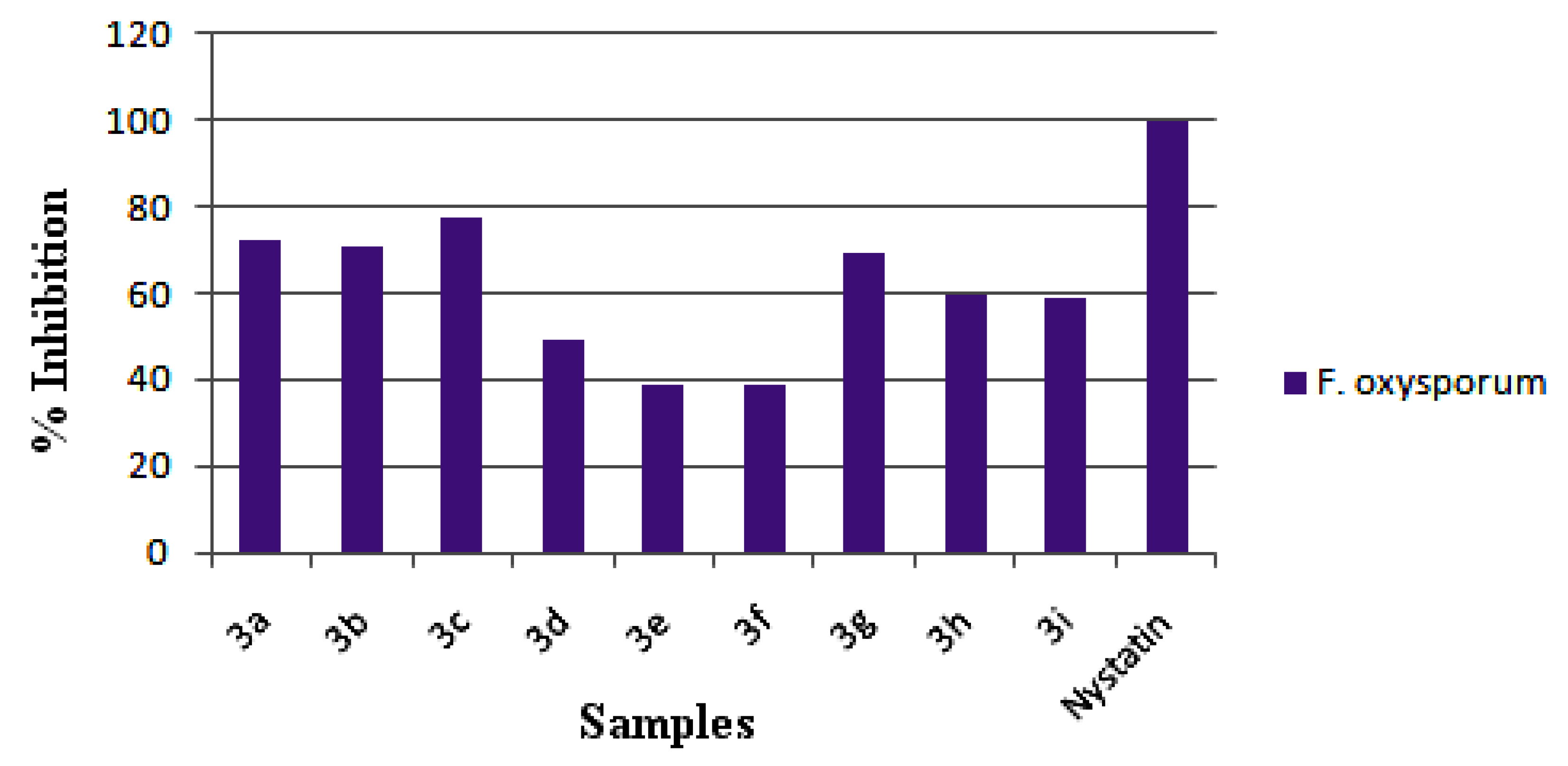
2.2. In Vitro Antimicrobial Activity


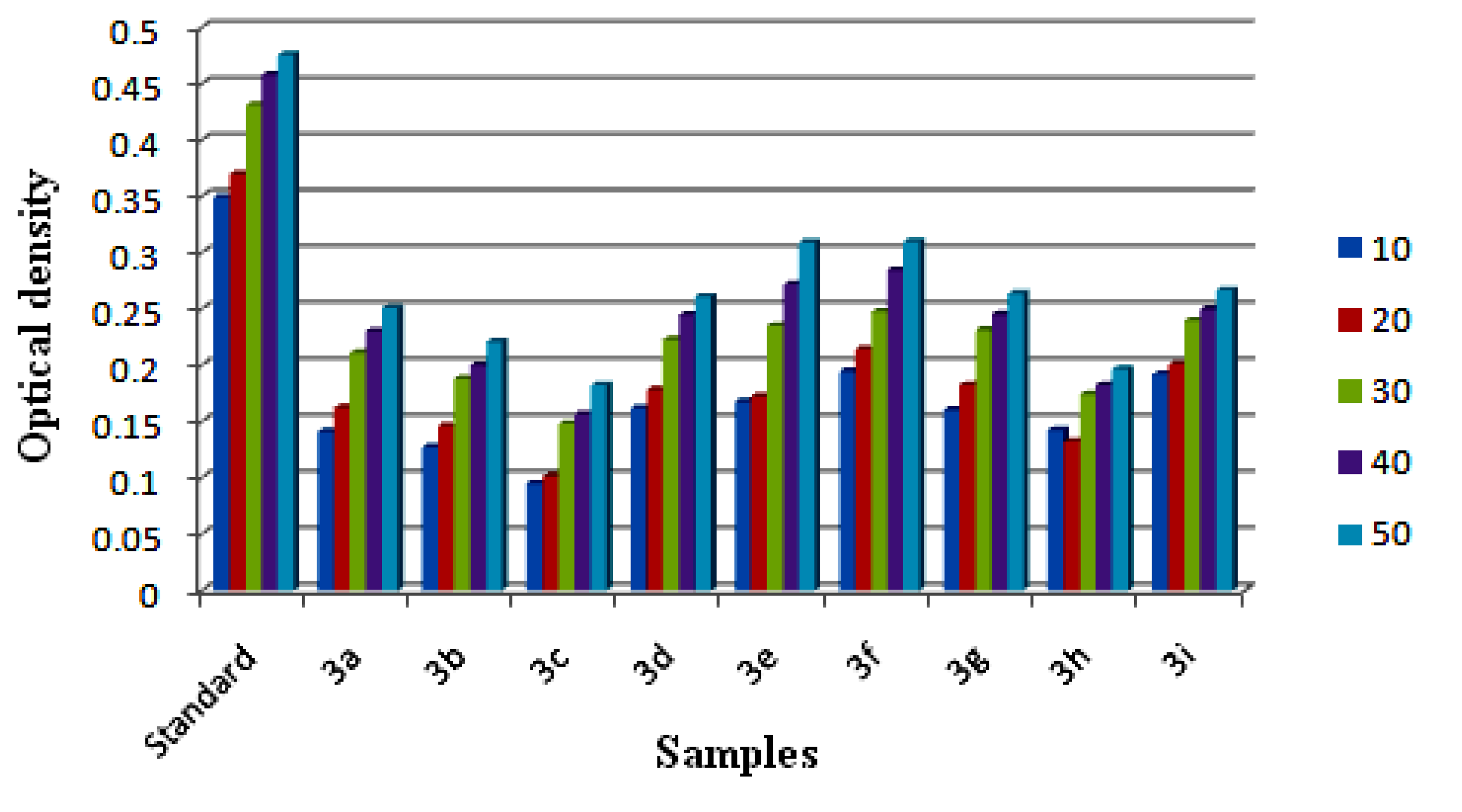
2.3. Reducing Power Ability
2.3.1. Ferric Reducing Antioxidant Power (FRAP) Assay

2.3.2. Cupric Ion Reducing Antioxidant Capacity (CUPRAC) Assay

3. Experimental
3.1. Materials and Method
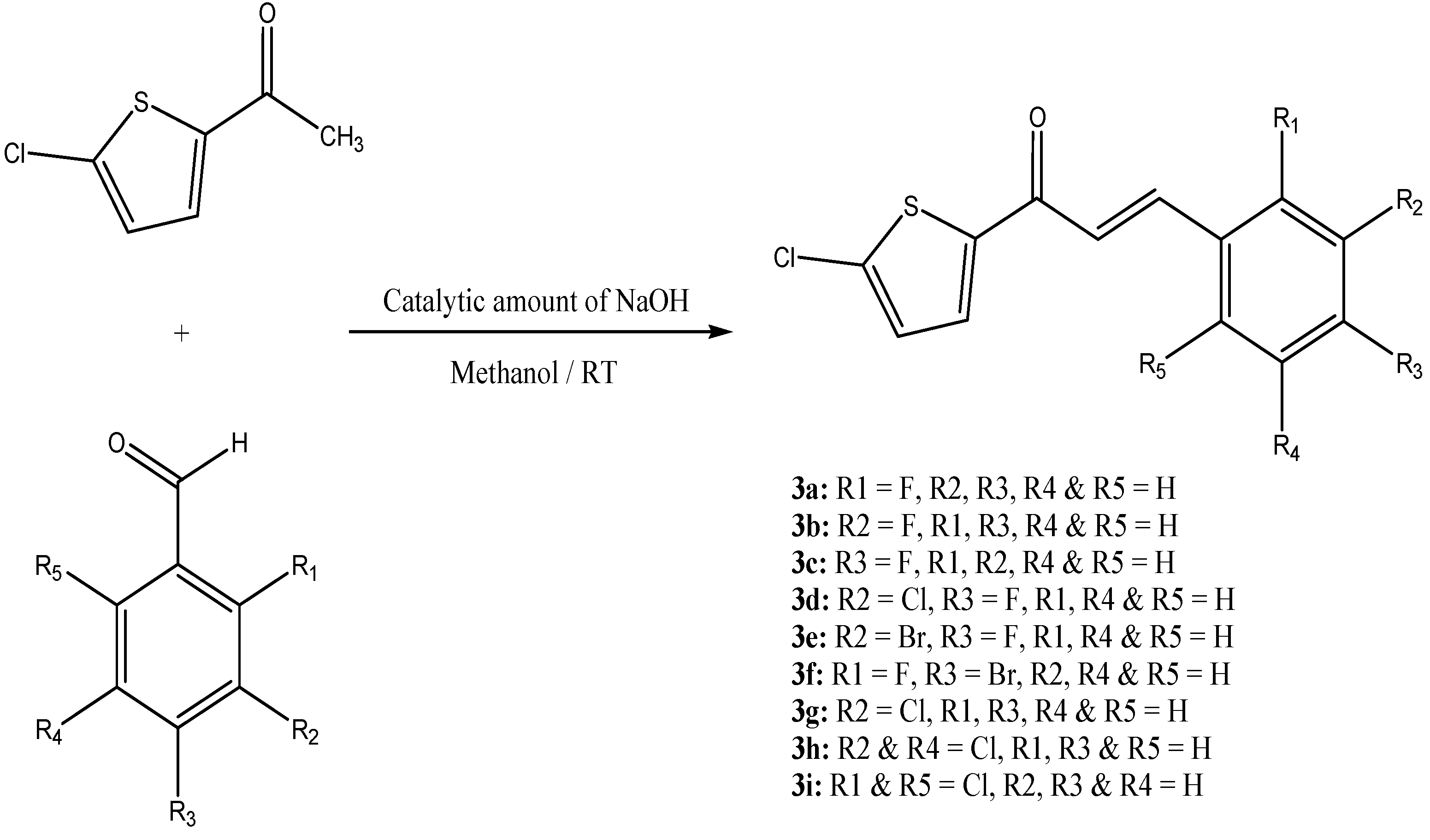
3.2. General Procedure for the Synthesis of Chalcones 3(a–i)
3.3. In Vitro Antimicrobial Activities
3.3.1. Antibacterial Activity
3.3.2. Antifungal Activity
3.3.3. Ferric Ion Reducing Antioxidant Power (FRAP) Assay
3.3.4. Cupric Ion Reducing Antioxidant Capacity (CUPRAC) Assay
3.3.5. Statistical Analysis
4. Conclusions
Supplementary Materials
Acknowledgments
Conflicts of Interest
References
- Alcaraz, L.E.; Blanco, S.E.; Puig, O.N.; Tomas, F.; Ferretti, F.H. Antibacterial activity of flavonoids against methicillin-resistant Staphylococcus aureus strains. J. Theor. Biol. 2000, 205, 231–240. [Google Scholar] [CrossRef]
- Baviskar, B.A.; Baviskar, B.; Shiradkar, M.R.; Deokate, U.A.; Khadabadi, S.S. Synthesis and antimicrobial activity of some novel benzimidazolyl chalcones. J. Chem. 2009, 6, 196–200. [Google Scholar]
- Echeverria, C.; Santibañez, J.F.; Donoso-Tauda, O.; Escobar, C.A.; Ramirez-Tagle, R. Structural antitumoral activity relationships of synthetic chalcones. Int. J. Mol. Sci. 2009, 10, 221–231. [Google Scholar]
- Modzelewska, A.; Pettit, C.; Achanta, G.; Davidson, N.E.; Huang, P.; Khan, S.R. Anticancer activities of novel chalcone and bis-chalcone derivatives. Bioorg. Med. Chem. 2006, 14, 3491–3495. [Google Scholar]
- Vogel, S.; Heilmann, J. Synthesis, cytotoxicity, and antioxidative activity of minor prenylated chalcones from Humulus lupulus. J. Nat. Prod. 2008, 71, 1237–1241. [Google Scholar] [CrossRef]
- Babasaheb, P.B.; Sachin, A.P.; Rajesh, N.G. Synthesis and biological evaluation of nitrogen containing chalcones as possible anti-inflammatory and antioxidant agents. Bioorg. Med. Chem.Lett. 2010, 20, 730–733. [Google Scholar]
- Kim, Y.H.; Kim, J.; Park, H.; Kim, H.P. Anti-inflammatory activity of the synthetic chalcone derivatives: Inhibition of inducible nitric oxide synthase-catalyzed nitric oxide production from lipopolysaccharide-treated RAW 264.7 cells. Biol. Pharm Bull. 2007, 30, 1450–1455. [Google Scholar] [CrossRef]
- Ballesteros, J.F.; Sanz, M.J.; Ubeda, A.; Miranda, M.A.; Iborra, S.; Paya, M.; Alcaraz, M.J. Synthesis and pharmacological evaluation of 2'-hydroxychalcones and flavones as inhibitors of inflammatory mediators generation. J. Med. Chem. 1995, 38, 2794–2797. [Google Scholar] [CrossRef]
- Vogel, S.; Barbic, M.; Jürgenliemk, G.; Heilmann, J. Synthesis, cytotoxicity, anti-oxidative and anti-inflammatory activity of chalcones and influence of A-ring modifications on the pharmacological effect. Eur. J. Med. Chem. 2010, 45, 2206–2213. [Google Scholar] [CrossRef]
- Lopez, S.N.; Castelli, M.V.; Zacchino, S.A.; Domnguez, J.N.; Lobo, G.; Charris-Charris, J.; Cortes, J.C.G.; Ribas, J.C.; Devia, C.; Rodrguez, A.M.; et al. In vitro antifungal evaluation and structure-activity relationships of a new series of chalcone derivatives and synthetic analogues, with inhibitory properties against polymers of the fungal cell wall. Bioorg. Med. Chem. 2001, 9, 1999–2013. [Google Scholar] [CrossRef]
- Beom-Tae, K.; Kwang-Joong, O.; Jae-Chul, C.; Ki-Jun, H. Synthesis of dihydroxylated chalcone derivatives with diverse substitution patterns and their radical scavenging ability toward DPPH free radicals. Bull. Korean Chem. Soc. 2008, 29, 1125–1130. [Google Scholar] [CrossRef]
- Doan, T.N.; Tran, T.-D. Synthesis, antioxidant and antimicrobial activities of a novel series of chalcones, pyrazolic chalcones, and allylic chalcones. Pharmacol. Pharm. 2011, 2, 282–288. [Google Scholar] [CrossRef]
- Go, M.L.; Wu, X.; Liu, X.L. Chalcones: An update on cytotoxic and chemoprotective properties. Curr. Med. Chem. 2005, 12, 481–499. [Google Scholar]
- Sivakumar, P.M.; Prabhakar, P.K.; Doble, M. Synthesis, antioxidant evaluation, and quantitative structure-activity relationship studies of chalcones. Med. Chem. Res. 2011, 20, 482–492. [Google Scholar] [CrossRef]
- Vogel, S.; Ohmayer, S.; Brunner, G.; Heilmann, J. Natural and non-natural prenylated chalcones: Synthesis, cytotoxicity and anti-oxidative activity. Bioorg. Med. Chem. 2008, 16, 4286–4293. [Google Scholar]
- Lemar, K.M.; Turner, M.P.; Lloyd, D. Garlic (Allium sativum) as an anti-Candida agent: A comparison of the efficacy of fresh garlic and freeze-dried extracts. J. Appl. Microbiol. 2002, 93, 398–405. [Google Scholar] [CrossRef]
- Tomar, V.; Bhattacharjee, G.; Kamaluddina, K. Synthesis and antimicrobial evaluation of newchalcones containing piperazine or 2,5-dichlorothiophene moiety. Bioorg. Med. Chem. Lett. 2007, 17, 5321–5324. [Google Scholar] [CrossRef]
- Bag, D.; Ramar, S.; Degani, M.S. Synthesis and biological evaluation of a, b-unsaturated ketone as potential antifungal agents. Med. Chem. Res. 2009, 18, 309–316. [Google Scholar]
- Tran, T.D.; Nguyen, T.T.; Do, T.H.; Huynh, T.N.; Tran, C.D.; Thai, K.M. Synthesis and antibacterial activity of some heterocyclic chalcone analogues alone and in combination with antibiotics. Molecules 2012, 17, 6684–6696. [Google Scholar] [CrossRef]
- Ranganathan, K.; Arulkumaran, R.; Kamalakkannan, D.; Sundararajan, R.; Sakthinathan, S.P.; Vijayakumar, S.; Suresh, R.; Vanangamudi, G.; Thirumurthy, K.; Mayavel, P.; et al. Silica-H2SO4 catalyzed environmentally benign crossed aldol condensation: Synthesis, spectral studies and biological activities of some 5-chloro-2-thienyl chalcones. Int. J. Pharm. Med. Biol. Sci. 2012, 1, 62–85. [Google Scholar]
- Kumar, C.S.C.; Loh, W.-S.; Ooi, C.W.; Quah, C.K.; Fun, H.-K. Sructural correlation of some heterocyclic chalcone analogues and evaluation of their antioxidant potential. Molecules 2013, 18, 11996–12011. [Google Scholar] [CrossRef]
- Bernstein, J.; Davis, R.E.; Shimoni, L.; Chang, N.L. Patterns in hydrogen bonding: Functionality and graph set analysis in crystals. Angew. Chem. Int. Edit. Engl. 1995, 34, 1555–1573. [Google Scholar] [CrossRef]
- Bruker. APEX2, SAINT and SADABS. Bruker AXS Inc.: Madison, WI, USA, 2009. [Google Scholar]
- Sheldrick, G.M. A short history of SHELX. Acta Cryst. 2008, A64, 112–122. [Google Scholar]
- Dolomanov, O.V.; Bourhis, L.J.; Gildea, R.J.; Howard, J.A.K.; Puschmann, H. OLEX2: A complete structure solution, refinement and analysis program. J. Appl. Cryst. 2009, 42, 339–341. [Google Scholar] [CrossRef]
- Bauer, W.M.; Kirby, J.C.; Sherris; Truck, M. Antibiotic susceptibility testing by a standardized single disk method. Am. J. Clin. Pathol. 1966, 45, 493–496. [Google Scholar]
- Satish, S.; Mohana, D.C.; Raghavendra, M.P.; Raveesha, K.A. Antifungal activity of some plant extracts against important seed borne pathogens of Aspergillus sp. J. Agric. Technol. 2007, 3, 109–119. [Google Scholar]
- Oyaizu, M. Studies on products of browning reaction prepared from glucosamine. Jpn. J. Nutr. 1986, 44, 307–315. [Google Scholar] [CrossRef]
- Apak, R.; Guclu, K.; Ozyurek, M.; Celik, S.E. Mechanism of antioxidant capacity assays and the CUPRAC (cupric ion reducing antioxidant capacity) assay. Microchim. Acta 2008, 160, 413–419. [Google Scholar] [CrossRef]
- Sample Availability: Samples of the compounds are available from the authors.
© 2013 by the authors; licensee MDPI, Basel, Switzerland. This article is an open access article distributed under the terms and conditions of the Creative Commons Attribution license (http://creativecommons.org/licenses/by/3.0/).
Share and Cite
Kumar, C.S.C.; Loh, W.-S.; Ooi, C.W.; Quah, C.K.; Fun, H.-K. Heteroaryl Chalcones: Design, Synthesis, X-ray Crystal Structures and Biological Evaluation. Molecules 2013, 18, 12707-12724. https://doi.org/10.3390/molecules181012707
Kumar CSC, Loh W-S, Ooi CW, Quah CK, Fun H-K. Heteroaryl Chalcones: Design, Synthesis, X-ray Crystal Structures and Biological Evaluation. Molecules. 2013; 18(10):12707-12724. https://doi.org/10.3390/molecules181012707
Chicago/Turabian StyleKumar, C. S. Chidan, Wan-Sin Loh, Chin Wei Ooi, Ching Kheng Quah, and Hoong-Kun Fun. 2013. "Heteroaryl Chalcones: Design, Synthesis, X-ray Crystal Structures and Biological Evaluation" Molecules 18, no. 10: 12707-12724. https://doi.org/10.3390/molecules181012707