Bioactive Quinic Acid Derivatives from Ageratina adenophora

Abstract
:1. Introduction

2. Results and Discussion

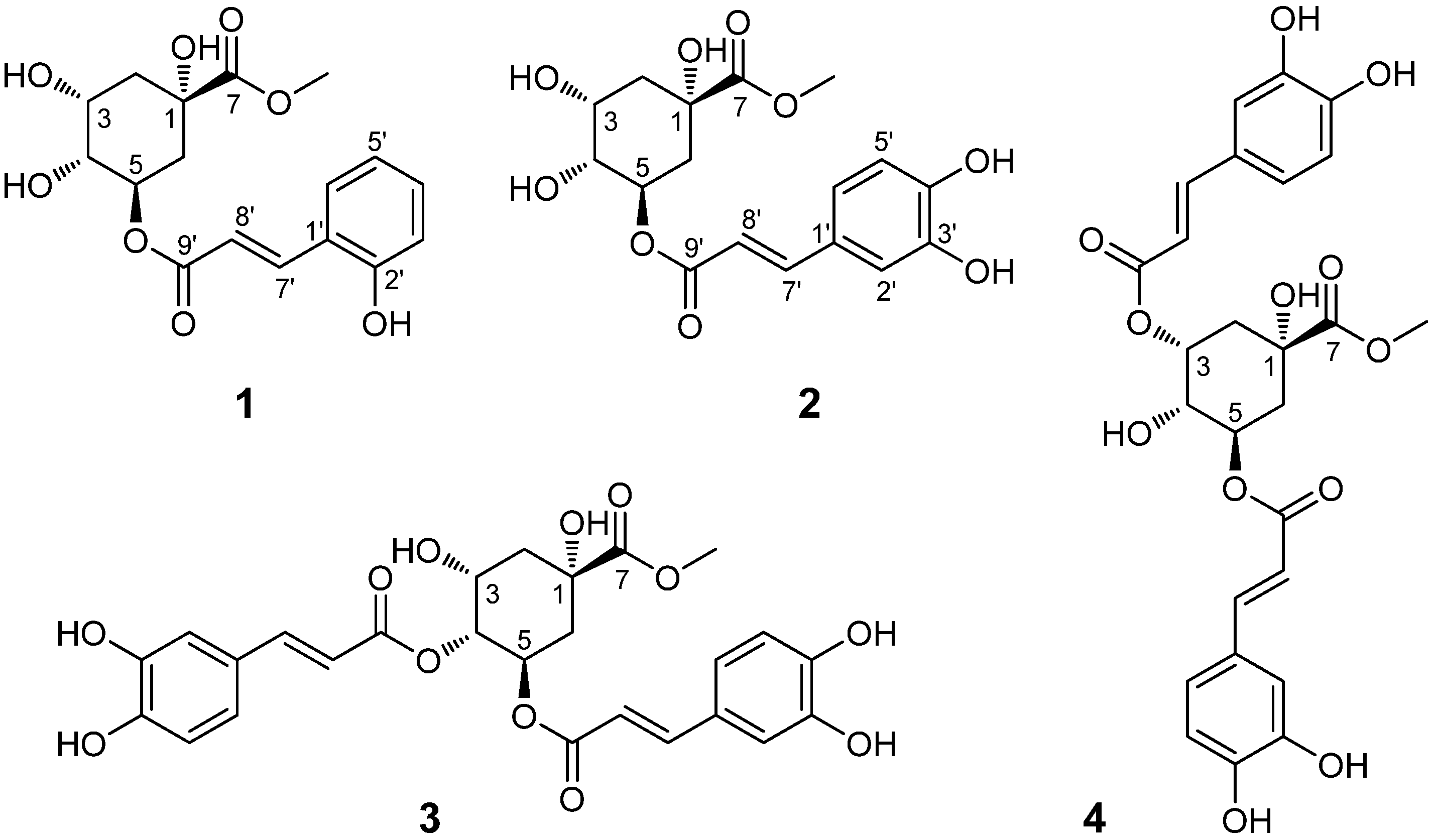
| Position | δC (1) | δH (1) | δC (2) | δH (2) |
|---|---|---|---|---|
| 1 | 75.8 | 75.8 | ||
| 2α | 38.0 | 2.00 (dd, 13.6, 6.8) | 38.0 | 1.99 (dd, 13.6, 6.8) |
| 2β | 2.19 (overlapped) | 2.19 (overlapped) | ||
| 3 | 70.3 | 4.13 (m) | 70.3 | 4.13 (m) |
| 4 | 72.5 | 3.74 (m) | 72.5 | 3.72 (dd, 7.2, 3.2) |
| 5 | 72.1 | 5.28 (m) | 72.1 | 5.26 (m) |
| 6α | 37.7 | 2.18 (overlapped) | 37.7 | 2.18 (overlapped) |
| 6β | 2.18 (overlapped) | 2.18 (overlapped) | ||
| 7 | 175.4 | 175.4 | ||
| 7-OCH3 | 53.0 | 3.69 (s) | 53.0 | 3.68 (s) |
| 1' | 122.5 | 127.6 | ||
| 2' | 158.4 | 115.1 | 7.03 (d, 2.0) | |
| 3' | 117.0 | 6.83 (overlapped) | 146.8 | |
| 4' | 132.7 | 7.20 (td, 8.0, 1.2) | 149.7 | |
| 5' | 120.8 | 6.83 (overlapped) | 116.5 | 6.77 (d, 8.0) |
| 6' | 130.4 | 7.45 (dd, 8.0, 1.2) | 123.0 | 6.94 (dd, 8.0, 2.0) |
| 7' | 142.7 | 7.91 (d, 16.0) | 147.2 | 7.51 (d, 16.0) |
| 8' | 118.4 | 6.58 (d, 16.0) | 115.0 | 6.21 (d, 16.0) |
| 9' | 168.6 | 168.3 |
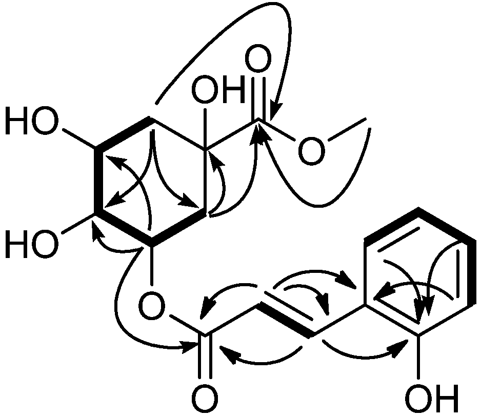
 ) and COSY (
  ) correlations of compound 1.
| Compounds | Staphylococcus aureus | Bacillus thuringiensis | Escherichia coli | Salmonella enterica | Shigella dysenteriae |
|---|---|---|---|---|---|
| 1 | 88.8 | 88.8 | 88.8 | 88.8 | 177.6 |
| 2 | 84.8 | 84.8 | 84.8 | 84.8 | 169.8 |
| 3 | 29.4 | 59.0 | 59.0 | 14.7 | 117.9 |
| 4 | 59.0 | 59.0 | 59.0 | 7.4 | 117.9 |
| KS | 6.7 | 6.7 | 3.4 | 3.4 | 3.4 |
3. Experimental
3.1. General
3.2. Plant Materials
3.3. Extraction and Isolation
 −22.2 (c 0.09, CH3OH); IR (KBr) νmax 3,411, 1,733, 1,622, 1,259 cm−1; UV (MeOH) λmax (log ε) nm: 212 (3.86), 276 (3.92); ESI-MS (+) m/z: 353 [M+H]+, 375 [M+Na]+; ESIMS (−) m/z 351 [M−H]−; HR-ESI-MS (pos.) m/z 375.1049 [M+Na]+ (calcd for C17H20NaO8, 375.1056); 1H-NMR (400 MHz, CD3OD) and 13C-NMR (100 MHz, CD3OD) data: see Table 1.
3.4. Antibacterial Assay
3.5. Antifungal Assay
3.6. Determination of Antioxidant Activities
4. Conclusions
Acknowledgments
Conflicts of Interest
References
- Qiang, S. The history and status of the study on crofton weed (Eupatorium adenophorum Spreng.), a worst worldwide weed. J. Wuhan Bot. Res. 1998, 16, 366–372. [Google Scholar]
- Wang, J.J. The crofton weed, Ageratina adenophora. In Biology and Management of Invasive Alien Species in Agriculture and Forestry; Wan, F.H., Zheng, X.B., Guo, J.Y., Eds.; Scinece Press: Beijing, China, 2005; pp. 650–661. [Google Scholar]
- Sun, X.Y.; Lu, Z.H.; Sang, W.G. Review on studies of Eupatorium adenophorum—An important invasive species in China. J. For. Res. 2004, 15, 319–322. [Google Scholar] [CrossRef]
- Wan, F.H.; Guo, J.Y.; Wang, D.H. Alien invasive species in China: their damages and management strategies. Biodivers. Sci. 2002, 10, 119–125. [Google Scholar]
- Wan, F.H.; Liu, W.X.; Guo, J.Y.; Qiang, S.; Li, B.P.; Wang, J.J.; Yang, G.Q.; Niu, H.B.; Gui, F.R.; Huang, W.K.; et al. Invasive mechanism and control strategy of Ageratina adenophora (Sprengel). Sci. China Life Sci. 2010, 53, 1291–1298. [Google Scholar] [CrossRef]
- Yan, Q.S.; Yang, J.; Li, H.M.; Cao, A.C.; Chen, Q.H.; Wen, Y.Q.; He, L. Advances in the studies on the chemical components and bioactivity of Eupatorium adenophorum Spreng as a intruding species. J. Beijing Normal Univ. 2006, 42, 70–73. [Google Scholar]
- Li, Y.M.; Li, Z.Y.; Ye, M. The chemical compositions and their bioactivities in the different parts of Eupatorium adenophorum Spreng. J. Yunnan Agric. Univ. 2008, 23, 42–46. [Google Scholar]
- He, L.; Hou, J.; Gan, M.L.; Shi, J.G.; Chantrapromma, S.; Fun, H.K.; Williams, I.D.; Sung, H.H.Y. Cadinane sesquiterpenes from the leaves of Eupatorium adenophorum. J. Nat. Prod. 2008, 71, 1485–1488. [Google Scholar] [CrossRef]
- Yang, G.Q.; Wan, F.H.; Liu, W.X.; Zhang, X.W. Physiological effects of allelochemicals from leachates of Ageratina adenophora Spreng on rice seedlings. Allelopathy J. 2006, 18, 237–245. [Google Scholar]
- Zhao, X.; Zheng, G.W.; Niu, X.M.; Li, W.Q.; Wang, F.S.; Li, S.H. Terpenes from Eupatorium adenophorum and their allelopathic effects on Arabidopsis seeds germination. J. Agric. Food Chem. 2009, 57, 478–482. [Google Scholar]
- Zheng, G.W.; Jia, Y.X.; Zhao, X.; Zhang, F.J.; Luo, S.H.; Li, S.H.; Li, W.Q. o-Coumaric acid from invasive Eupatorium adenophorum is a potent phytotoxin. Chemoecology 2012, 22, 131–138. [Google Scholar] [CrossRef]
- Shi, W.; Luo, S.H.; Li, S.H. Defensive sesquiterpenoids from leaves of Eupatorium adenophorum. Chin. J. Chem. 2012, 30, 1331–1334. [Google Scholar] [CrossRef]
- Zhu, X.D.; Dong, Y.F.; Wang, Y.F.; Ju, P.; Luo, S.D. Phenolic compounds from Viburnum cylindricum. Helv. Chim. Acta 2005, 88, 339–342. [Google Scholar] [CrossRef]
- Yang, Y.J.; Liu, X.; Wu, H.R.; He, X.F.; Bi, Y.R.; Zhu, Y.; Liu, Z.L. Radical scavenging activity and cytotoxicity of active quinic acid derivatives from Scorzonera divaricata roots. Food Chem. 2013, 138, 2057–2063. [Google Scholar] [CrossRef]
- Chen, M.; Wu, W.W.; Shen, G.Q.; Luo, S.Q.; Li, H.T. Chemical constituents of Lonicera Macranthoides Hand.-Mazz Part V. Isolation and stuctures of macranthoin F and G. Acta Pharm. Sin. 1994, 29, 617–620. [Google Scholar]
- Basnet, P.; Matsushige, K.; Hase, K.; Kadota, S.; Namba, T. Four di-O-caffeoyl quinic acid derivatives from propolis. Potent hepatoprotective activity in experimental liver injury models. Bio. Pharm. Bull. 1996, 19, 1479–1484. [Google Scholar] [CrossRef]
- Lee, S.Y.; Moon, E.; Kim, S.Y.; Lee, K.R. Quinic acid derivatives from Pimpinella brachycarpa exert anti-neuroinflammatory activity in lipopolysaccharide-induced microglia. Bioorg. Med. Chem. Lett. 2013, 23, 2140–2144. [Google Scholar]
- Nakamura, S.; Fujimoto, K.; Matsumoto, T.; Nakashima, S.; Ohta, T.; Ogawa, K.; Matsuda, H.; Yoshikawa, M. Acylated sucroses and acylated quinic acids analogs from the flower buds of Prunus mume and their inhibitory effect on melanogenesis. Phytochemistry 2013, 92, 128–136. [Google Scholar] [CrossRef]
- Li, Y.B.; Xu, H.; Shi, L.; Li, Z.Y. Allelopathic effects of Eupatorium adenophorum on five species of the family Gesneriaceae. Biodiver. Sci. 2007, 15, 486–491. [Google Scholar] [CrossRef]
- Zheng, L.; Feng, Y.L. Allelopathic effects of Eupatorium adenophorum Spreng. on seed germination and seedling growth in ten herbaceous species. Acta Ecol. Sin. 2005, 25, 2782–2787. [Google Scholar]
- Recent Research Developments in Phytochemistry; Pandalai, S.G. (Ed.) Research Signpost: Kerala, India, 2000; Chapter 4; pp. 143–152.
- Sarker, S.D.; Nahar, L.; Kumarasamy, Y. Microtitre plate-based antibacterial assay incorporating resazurin as an indicator of cell growth, and its application in the in vitro antibacterial screening of phytochemicals. Methods 2007, 42, 321–324. [Google Scholar] [CrossRef]
- Rahman, M.M.; Gray, A.I. A benzoisofuranone derivative and carbazole alkaloids from Murraya koenigii and their antimicrobial activity. Phytochemistry 2005, 66, 1601–1606. [Google Scholar] [CrossRef]
- He, S.; Wu, B.; Pan, Y.; Jiang, L. Stilbene oligomers from Parthenocissus laetevirens: Isolation, biomimetic synthesis, absolute configuration, and implication of antioxidative defense system in the plant. J. Org. Chem. 2008, 73, 5233–5241. [Google Scholar] [CrossRef]
- Sample Availability: Samples of the compounds are available from the authors.
© 2013 by the authors; licensee MDPI, Basel, Switzerland. This article is an open access article distributed under the terms and conditions of the Creative Commons Attribution license (http://creativecommons.org/licenses/by/3.0/).
Share and Cite
Zhang, M.; Liu, W.-X.; Zheng, M.-F.; Xu, Q.-L.; Wan, F.-H.; Wang, J.; Lei, T.; Zhou, Z.-Y.; Tan, J.-W. Bioactive Quinic Acid Derivatives from Ageratina adenophora. Molecules 2013, 18, 14096-14104. https://doi.org/10.3390/molecules181114096
Zhang M, Liu W-X, Zheng M-F, Xu Q-L, Wan F-H, Wang J, Lei T, Zhou Z-Y, Tan J-W. Bioactive Quinic Acid Derivatives from Ageratina adenophora. Molecules. 2013; 18(11):14096-14104. https://doi.org/10.3390/molecules181114096
Chicago/Turabian StyleZhang, Mei, Wan-Xue Liu, Meng-Fei Zheng, Qiao-Lin Xu, Fang-Hao Wan, Jing Wang, Ting Lei, Zhong-Yu Zhou, and Jian-Wen Tan. 2013. "Bioactive Quinic Acid Derivatives from Ageratina adenophora" Molecules 18, no. 11: 14096-14104. https://doi.org/10.3390/molecules181114096