2. Results and Discussion
The extract of fermented broth of
Myrothecium sp. GS-17 was fractionated by repeated column chromatography to yield five compounds
1–
5 (
Figure 1). Their structures were elucidated based on spectroscopic methods.
Figure 1.
The structures of compounds 1–5.
Figure 1.
The structures of compounds 1–5.
Compound
1 was obtained as colorless clustered crystals, and its molecular formula was determined as C
16H
18O
5 on the basis of the HRESI-MS (
m/z 291.1229, calcd. 291.1232 [M+H]
+). The
1H-NMR (600 MHz, DMSO-
d6) spectrum (
Table 1) of
1 exhibited a pair of
meta-coupled protons in an aromatic ring at
δ 6.28 (1H, brs), and 6.25 (1H, brs). It also displayed the signals of one methoxyl at
δ 3.59 (3H, s), two methyls at
δ 2.10 (3H, s) and 1.80 (3H, s), and a phenolic hydroxyl at
δ 9.51 (1H, s). In addition, it revealed the presence of an oxygenated methylene according to the protons at
δ 4.97 (1H, dd,
J = 2.5 Hz, 12.2 Hz), 4.83 (1H, d,
J = 12.2 Hz), and a hydroxymethyl group according to the protons at
δ 4.59 (1H, t,
J = 5.3 Hz), 4.17 (2H, d,
J = 5.3 Hz). The
13C-NMR spectrum (150 MHz, DMSO-
d6) showed sixteen carbon signals including ten sp
2 C-atoms at
δ 159.5, 154.9, 146.9, 146.5, 142.4, 120.7, 117.3, 116.3, 99.6, and 98.0, indicating the presence of one aromatic ring and one furan ring. The
1H- and
13C-NMR data (
Table 1) were assigned based on the HSQC and HMBC experiments.
Table 1.
1H- and 13C-NMR data of compound 1 (DMSO-d6, 600 and 150 MHz, resp., δ in ppm, J in Hz).
Table 1.
1H- and 13C-NMR data of compound 1 (DMSO-d6, 600 and 150 MHz, resp., δ in ppm, J in Hz).
| Position | δ(H) | δ(C) | Position | δ(H) | δ(C) |
|---|
| 1 | 4.97, dd (12.2, 2.5) | 72.1 (t) | 1' | | 146.5 (s) |
| 4.83, d (12.2) |
| 3 | 6.02, d (2.5) | 75.2 (d) | 3' | | 146.9 (s) |
| 4 | | 116.3 (s) | 4' | | 120.7 (s) |
| 5 | | 154.9 (s) | 5' | | 117.3 (s) |
| 6 | 6.25, brs | 98.0 (d) | 3'-CH3 | 2.10, s | 11.6 (q) |
| 7 | | 159.5 (s) | 4'-CH2OH | 4.17, d (5.3) | 53.2 (t) |
| 8 | 6.28, brs | 99.6 (d) | 4'-CH2OH | 4.59, t (5.3) | |
| 9 | | 142.4 (s) | 5'-CH3 | 1.80, s | 7.7 (q) |
| 5-OCH3 | 3.59, s | 55.3 (q) | | | |
| 7-OH | 9.51, s | | | | |
The HMBC spectrum showed the correlations of H-8 with C-1, C-6, C-4, C-7, of H-6 with C-8, C-4, C-5, C-7, of H-3 with C-1, C-4 and C-9, and of H-1 with C-3, C-4, C-8 and C-9, which revealed the presence of isobenzofuran fragment. The location of 7-OH and 5-OCH
3 were determined by the HMBC correlations of 7-OH (
δH 9.51) with C-6, C-7, C-8, and of 5-OCH
3 (
δH 3.59) with C-5. Based on the above evidence, the moiety A was elucidated as shown in
Figure 2.
Figure 2.
Key HMBC (H→C) correlations of compound 1.
Figure 2.
Key HMBC (H→C) correlations of compound 1.
In the HMBC spectrum, the correlations of the methyl 5'-CH
3 at
δH 1.80 with C-5', C-4' and C-1', of 3'-CH
3 at
δH 2.10 with C-4' and C-3' and of 4'-CH
2O- (
δH 4.17) with C-5', C-4' and C-3' suggested the presence of 3',5'-dimethyl-4'-hydroxymethylfuran fragment (moiety B). In addition, the HMBC correlations of H-1 with C-1', and of 5'-CH
3 (
δH 1.80) with C-3, indicated that the moieties A and B linked together by C-3 and C-1' to form the structure as shown in
Figure 2. X-ray crystallographic data further confirmed the structure of
1 to be as shown in
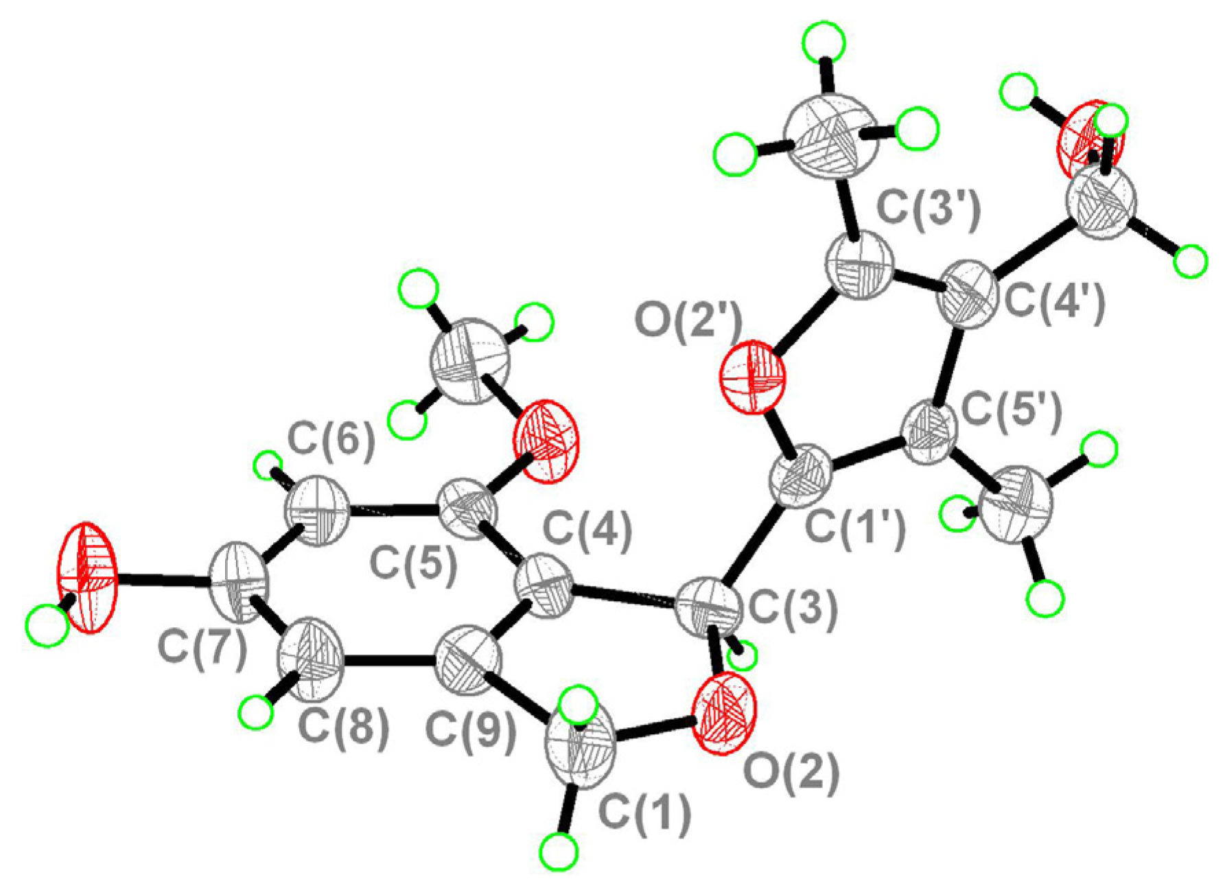
Figure 3, and it was named as myrothecol. Due to the fact the crystallographic data collected by Mo Kα radiation, the absolute configuration was not determined.
Figure 3.
X-ray crystallographic diagram of compound 1.
Figure 3.
X-ray crystallographic diagram of compound 1.
Compound
2 was obtained as a colorless oil, and its molecular formula was determined as C
7H
10O
4 on the basis of the HRESI-MS (
m/z 181.0474, calcd. 181.0477 [M+Na]
+). The IR spectrum indicated the presence of an
α,
β-unsaturated
γ-lactone (1,752 cm
−1). The
1H-NMR (300 MHz, DMSO-
d6) spectrum (
Table 2) of
2 revealed the presence of a methyl at
δH 1.82 attached to a quaternary olefinic C-atom, a oxymethine at
δH 6.13 (s, H-5) and a 1-hydroxyethyl at
δH 1.29 (d,
J = 6.7 Hz, H-8) and 4.62 (q,
J = 6.7 Hz, H-7).
Table 2.
1H- and 13C-NMR data of compounds 2–5 (DMSO-d6, 300 and 75 MHz, resp., δ in ppm, J in Hz).
Table 2.
1H- and 13C-NMR data of compounds 2–5 (DMSO-d6, 300 and 75 MHz, resp., δ in ppm, J in Hz).
| Position | 2 | 3 | 4 | 5 |
|---|
| δ(H) | δ(C) | δ(H) | δ(C) | δ(H) | δ(C) | δ(H) | δ(C) |
|---|
| 2 | | 172.4 (s) | | 170.5 (s) | | 171.5 (s) | | 171.5 (s) |
| 3 | | 123.8 (s) | | 131.0 (s) | | 128.1 (s) | | 123.7 (s) |
| 4 | | 162.0 (s) | | 157.6 (s) | | 156.6 (s) | | 160.4 (s) |
| 5 | 6.13, s | 97.9 (d) | 6.05, s | 97.9 (d) | | 107.8 (s) | | 107.3 (s) |
| 6 | 1.82, s | 8.6 (q) | 4.52, q (6.6) | 61.4 (d) | 1.93, s | 8.9 (q) | 1.81, s | 8.5 (q) |
| 7 | 4.62, q (6.7) | 62.9 (d) | 1.26, d (6.6) | 22.2 (q) | 4.46, s | 57.0 (t) | 4.30, s | 55.8 (t) |
| 8 | 1.29, d (6.7) | 21.9 (q) | 2.01, s | 11.3 (q) | 1.63, s | 22.9 (q) | 1.52, s | 24.8 (q) |
| MeO | | | | | 3.21, s | 51.2 (q) | | |
| 5-OH | | | 7.71 brs | | | | | |
| 6-OH | | | 5.17 s | | | | | |
The
13C-NMR (75 MHz, DMSO-
d6) (
Table 2) and HSQC spectra of
2 revealed seven C-atom signals, including one ester carbonyl group at
δC 172.4, two olefinic C-atoms at
δC 123.8 and 162.0, one hemiacetal C-atom at
δC 97.9, one oxymethine at
δC 62.9, and two methyl groups at
δC 8.6 and 21.9. The above data suggested the presence of a furan-2(5
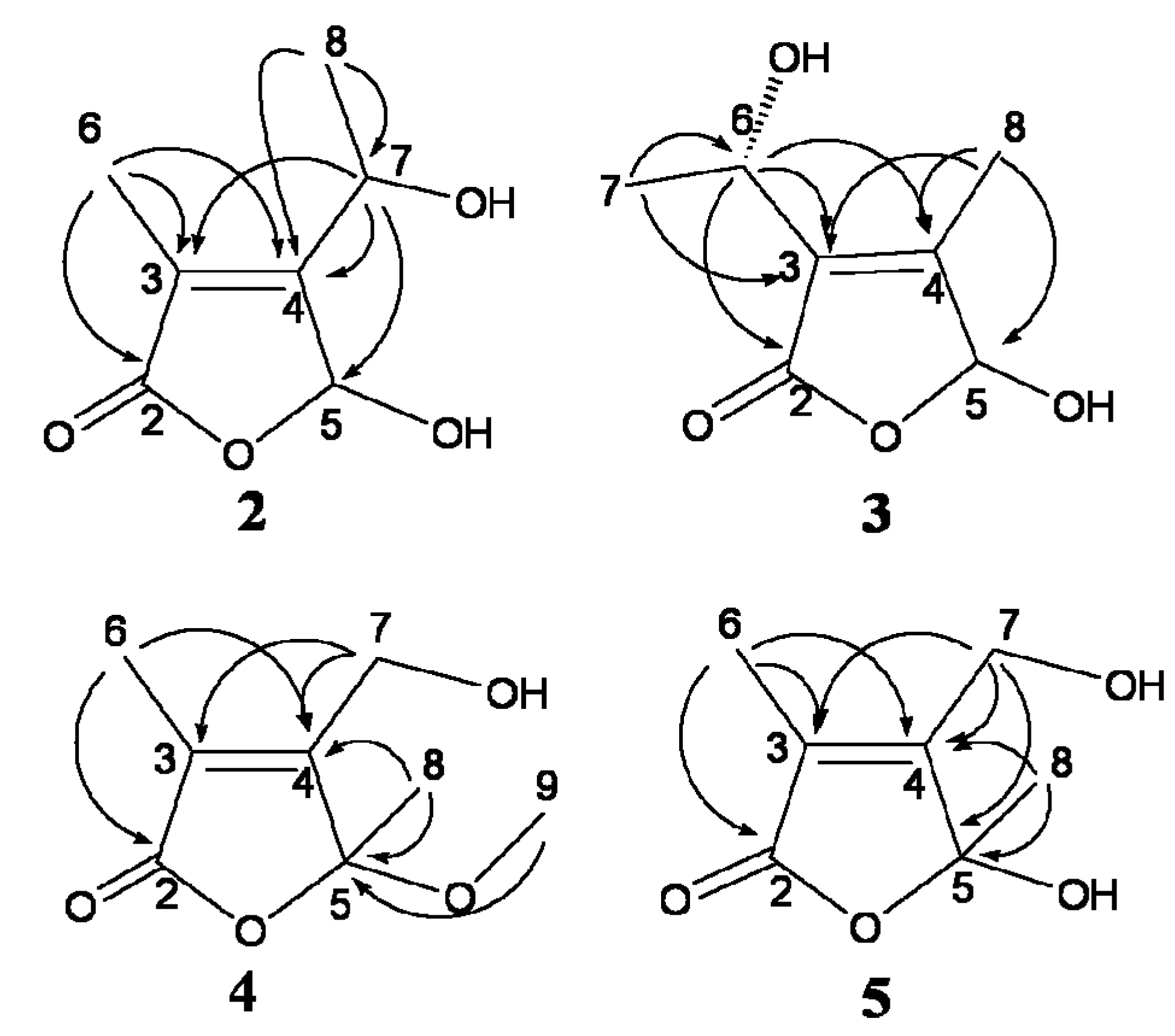
H)-one skeleton. The methyl group was located at C-3 by the HMBC (
Figure 4) correlations from H-6 (
δH 1.82) to C-2 (
δC 172.4), C-3 (
δC 123.8) and C-4 (
δC 162.0). The orientation of the 1-hydroxyethyl group was determined by the correlations from H-7 (
δH 4.62) to C-3, C-4 and C-5 (
δC 97.9), and from H-8 (
δH 1.29) to C-7 (
δC 62.9) and C-4. Thus, the structure of
2 was elucidated as 5-hydroxy-3-methyl-4-(1-hydroxyethyl)-furan-2(5
H)-one.
Figure 4.
Key HMBC (H→C) correlations of compounds 2–5.
Figure 4.
Key HMBC (H→C) correlations of compounds 2–5.
Compound
3 was obtained as a colorless oil, and its molecular formula was determined as C
7H
10O
4 on the basis of the HRESI-MS (
m/z 181.0474, calcd. 181.0477 [M+Na]
+). The absolute configuration at C-6 was deduced as
S on the basis of its value of specific optical rotation ([α]
20D = −17.65) [
6]. By comparison of its spectroscopic data with those reported previously, compound
3 was identified as 5-hydroxy-3-[(1
S)-1-hydroxyethyl]-4-methylfuran-2(5
H)-one [
6].
Compound
4 was obtained as a colorless oil, and its molecular formula was determined as C
8H
12O
4 on the basis of HRESI-MS (
m/
z 195.0623, calcd. 195.0633 [M+Na]
+). Compound
4 was a racemate due to its value of specific optical rotation ([α]
20D = 0). By comparison of its spectroscopic data with those reported previously, the structure of
4 was elucidated as 3,5-dimethyl-4-hydroxymethyl-5-methoxy- furan-2(5
H)-one [
7].
Compound
5 was obtained as a colorless oil, and its molecular formula was determined as C
7H
10O
4 on the basis of the HRESI-MS (
m/
z 181.0466, calcd. 181.0477 [M+Na]
+). Compound
5 was also determined to be a racemate by its value of specific optical rotation ([α]
20D = 0). By comparison of its spectroscopic data with those reported previously, the structure of
5 was elucidated as 3,5-dimethyl-4-hydroxymethyl-5-hydroxyfuran-2(5
H)-one [
7].
3. Experimental
3.1. General Procedures
UV Spectra were measured on a UV-260 spectrophotometer (Shimadzu, Kyoto, Japan) IR (KBr) spectra were obtained on a Avatar 360-ESP spectrophotometer (Thermo Nicolet, Boston, MA, USA). ESI-MS Spectra were recorded on an Agilent 1100 ion trap spectrometer (Agilent Technologies, Palo Alto, CA, USA). HRESI-MS Spectra were recorded on a QFT-ESI mass spectrometer (Varian, Palo Alto, CA, USA). 1D- and 2D-NMR experiments were performed on Bruker ARX-300 and AV-600 NMR spectrometers (Bruker, Ettlingen, Germany) using DMSO-d6 as an internal standard. Optical rotations were measured on a Perkin-Elmer 241 polarimeter (Perkin-Elmer, Boston, MA, USA). RP-HPLC was carried out on a C18 column (10 mm × 250 mm, 10 μm; YMC, Kyoto, Japan) on a Hitachi (Tokyo, Japan) L-2000 HPLC system equipped with a Hitachi L2400 UV-detector. Silica gel (100–200, 200–300 mesh, Qingdao Ocean Chemical Co., Qingdao, China), ODS (50 µm, YMC) and Sephadex LH-20 (GE Healthcare, Uppsala, Sweden) for column chromatography as well as silica gel GF254 (10–40 μm, Qingdao Ocean Chemical Co) for TLC were used.
3.2. Fungal Material and Culture
The halotolerant fungus Myrothecium sp. GS-17 was obtained from the soil samples obtained from a saline field located in Gansu Province in China. On the basis of its macroscopic appearance and 18S rDNA gene sequence, GS-17 was identified as Myrothecium sp., and preserved in Department of Natural Products Chemistry, Shenyang Pharmaceutical University, P. R. China. A small loop of spores growing on a Martin slant was inoculated into a 250 mL Erlenmeyer flask containing 75 mL sea-water-based culture medium (maltose 2%, monosodium glutamate 1%, KH2PO4 0.05%, MgSO4·7H2O 0.03%, glucose 1%, yeast extract 0.3%, corn steep liquor 0.1%, mannitol 2%, sea water, pH 6.5) and cultured at 28 °C for 2 days on a rotary shaker at 180 r·min−1. Then, 10 mL of the resultant seed culture was inoculated into a 500 mL Erlenmeyer flask containing the above culture medium (150 mL) and incubated (500 flasks) for 8 days under the same conditions.
3.3. Extraction and Isolation
The fermented broth (56 L) was filtered through cheesecloth to yield the supernatant and mycelia. The supernatant was subjected to HPD100 macroporous absorption resin chromatography, eluted in gradient with EtOH/H2O 30%, 70%, 100% to yield three fractions (Frs. 1–3). Fr. 2 (20 g) was fractioned on a silica gel column eluting with CH2Cl2–MeOH (from 100:0 to 0:100) to afford sixty fractions (SubFrs. 1–60). SubFr. 2 was further separated by a flash silica gel column eluting with petroleum ether–acetone (from 11:1 to 3:1) to give three fractions (SubFrs. 2A–2C). SubFr. 2A was further purified by RP-HPLC (MeOH–H2O: from 10% to 30%) to yield compounds 2 (7.0 mg), 3 (8.1 mg), 4 (14.2 mg) and 5 (10.0 mg). SubFr. 29 was purified by recrystallization from methanol to yield 1 (5.8 mg).
3.3.1. Myrothecol (1)
Colorless clustered crystals. IR (KBr)
νmax: 3332, 3154, 1609, 1477, 1117 cm
−1.
1H-NMR (600 MHz, DMSO-
d6) and
13C-NMR (150 MHz, DMSO-
d6): see
Table 1. HRESI-MS:
m/z 291.1229 ([M+H]
+, C
16H
19O
5; calcd. 291.1232).
3.3.2. 5-Hydroxy-3-methyl-4-(1-hydroxyethyl)-furan-2(5H)-one (2)
Colorless oil. [α]
20D = –7.53 (
c 0.11, MeOH). UV (MeOH)
λmax: 208 nm. IR (KBr)
νmax: 3425, 1752, 1052, 1027, 1006 cm
−1.
1H-NMR (300 MHz, DMSO-
d6) and
13C-NMR (75 MHz, DMSO-
d6): see
Table 2. ESI-MS: 156.9 [M–H]
−. HRESI-MS: 181.0474 ([M+Na]
+, C
7H
10NaO
4+; calcd. 181.0477).
3.3.3. 5-Hydroxy-3-[(1S)-1-hydroxyethyl]-4-methylfuran-2(5H)-one (3)
Colorless oil. [α]
20D = –17.65 (
c 0.07, MeOH). UV (MeOH)
λmax: 205 nm. IR (KBr)
νmax: 3401, 2983, 1749, 1050, 1029 cm
−1.
1H-NMR (300 MHz, DMSO-
d6) and
13C-NMR (75 MHz, DMSO-
d6): see
Table 2. ESI-MS: 156.9 [M–H]
−. HRESI-MS: 181.0474 ([M+Na]
+, C
7H
10NaO
4+; calcd. 181.0477).
3.3.4. 3,5-Dimethyl-4-hydroxymethyl-5-methoxyfuran-2(5H)-one (4)
Colorless oil. [α]
20D = 0 (
c 0.20, MeOH). UV (MeOH)
λmax: 210 nm. IR (KBr)
νmax: 3399, 2938, 1758, 1071, 1026 cm
−1.
1H-NMR (300 MHz, DMSO-
d6) and
13C-NMR (75 MHz, DMSO-
d6): see
Table 2. ESI-MS: 173.1 [M+H]
+. HRESI-MS: 195.0623 ([M+Na]
+, C
8H
12NaO
4+; calcd. 195.0633).
3.3.5. 3,5-Dimethyl-4-hydroxymethyl-5-hydroxyfuran-2(5H)-one (5)
Colorless oil. [α]
20D = 0 (
c = 0.50, MeOH). UV (MeOH)
λmax: 207 nm. IR (KBr)
νmax:3368, 2993, 2933, 1749, 1199, 1044 cm
−1.
1H-NMR (300 MHz, DMSO-
d6) and
13C-NMR (75 MHz, DMSO-
d6): see
Table 2. ESI-MS: 156.9 [M–H]
−. HRESI-MS: 181.0466 ([M+Na]
+, C
7H
10NaO
4+; calcd. 181.0477).
3.4. Single Crystal X-ray Structure Determination of Myrothecol (1)
The crystal structure of
1 was determined using data collected on a SMART APEX II CCD diffractometer (Mo Kα radiation). A suitable colorless crystal (0.18 × 0.14 × 0.12 mm) of
1 for diffraction was obtained from methanol solution. Crystal data: C
16H
18O
5 orthorhombic, a = 7.180(5) Å, b = 9.723(5) Å, c = 10.924(5) Å,
V = 722.6(7) Å
3,
Z = 2, D
calcd 1.334 mg/m
3, λ = 0.71069 Å,
F(000) = 308,
T = 293(2) K. A total of 2433 reflections were collected, of which 1545 unique reflections (R
int = 0.0532) with I > 2σ (
I) were used for the analysis. The data was solved using direct method SHELXL-97, and the structure was refined full-matrix least-squares on
F2 values. The refined structure model converged to a final
R1 0.0476,
wR2 = 0.1205 with Goodness-of-fit = 0.951. CCDC 969517 contains the supplementary crystallographic data for
1. These data can be obtained free of charge via
http://www.ccdc.cam.ac.uk/conts/retrieving.html (or from the CCDC, 12 Union Road, Cambridge CB2 1EZ, UK; Fax: +44 (0) 1223 336033; E-mail:
[email protected]).

 ,
, 


