Synthesis and Structure-Activity Relationships of Novel Ecdysteroid Dioxolanes as MDR Modulators in Cancer

 ,
,
Abstract
:1. Introduction
2. Results and Discussion
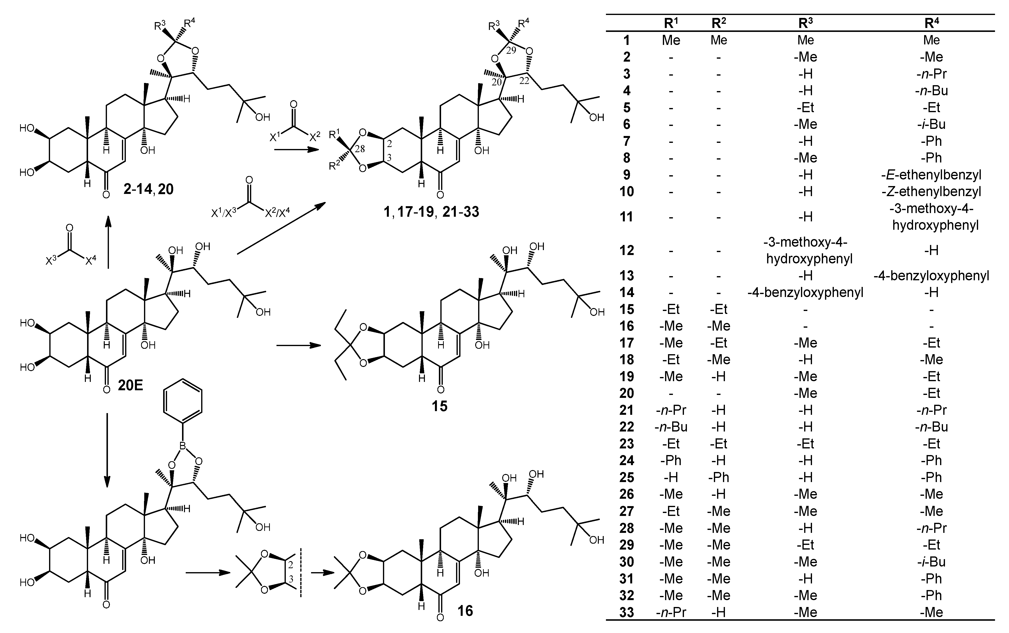
2.1. Semi-Synthesis


| Atom no. | 4 | 6 | 22 | 30 | 33 | 9 | 10 | |||||||
|---|---|---|---|---|---|---|---|---|---|---|---|---|---|---|
| H | C | H | C | H | C | H | C | H | C | H | C | H | C | |
| 1α β | 1.43 1.79 | 37.5 | 1.43 1.79 | 37.5 | 1.17 2.00 | 39.6 | 1.22 1.98 | 38.8 | 1.16 1.99 | 39.6 | 1.43 1.79 | 37.5 | 1.44 1.79 | 37.5 |
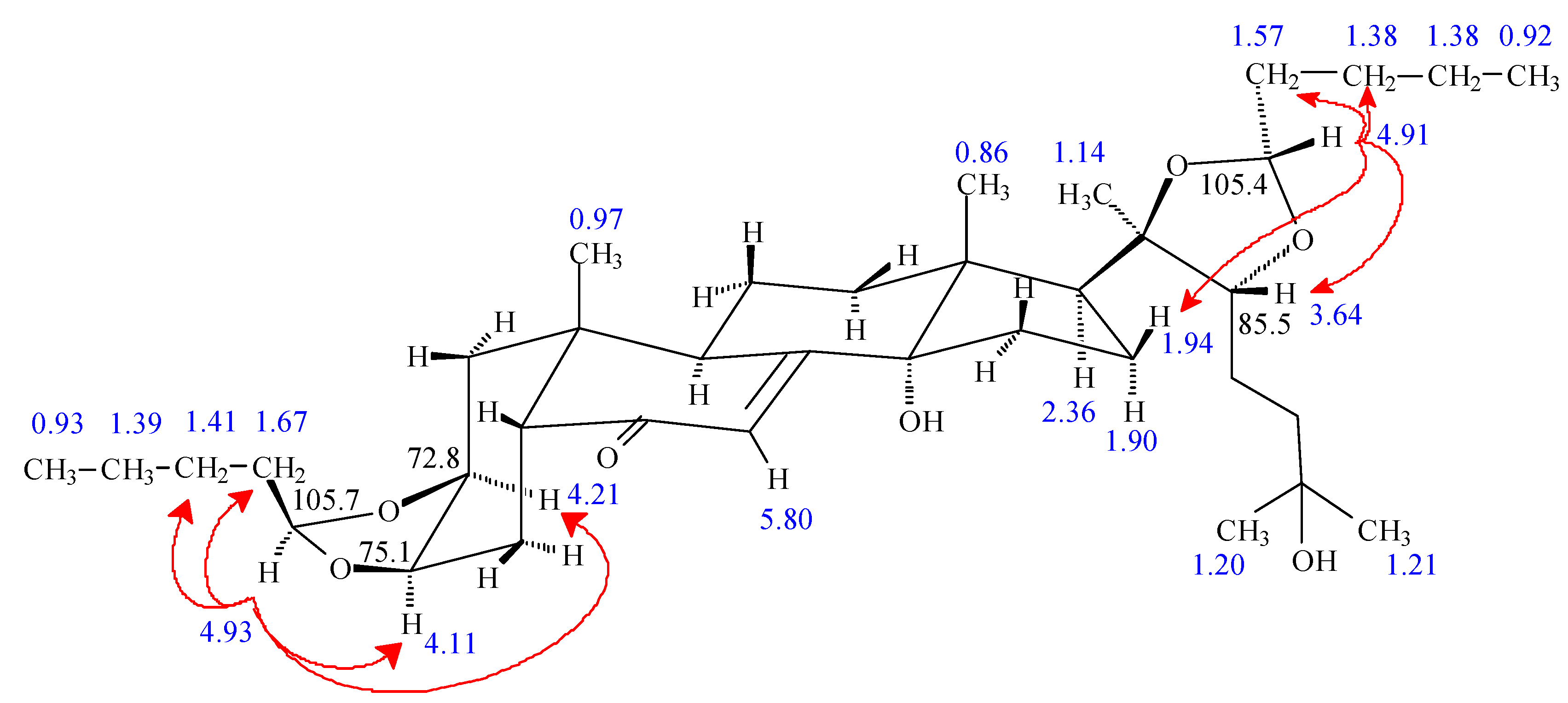
| 2 | 3.84 | 68.8 | 3.84 | 68.8 | 4.21 | 72.8 | 4.26 | 73.6 | 4.21 | 72.8 | 3.84 | 68.8 | 3.84 | 68.8 |
| 3 | 3.95 | 68.6 | 3.95 | 68.6 | 4.11 | 75.0 | 4.30 | 73.2 | 4.12 | 75.1 | 3.95 | 68.6 | 3.95 | 68.6 |
| 4α β | 1.74 1.70 | 32.9 | 1.75 1.70 | 32.9 | 2.01 1.97 | 27.8 | 1.97 1.97 | 27.8 | 2.00 2.00 | 27.8 | 1.74 1.71 | 32.9 | 1.74 1.71 | 33.0 |
| 5 | 2.39 | 51.8 | 2.39 | 51.8 | 2.24 | 52.6 | 2.24 | 52.6 | 2.24 | 52.6 | 2.39 | 51.9 | 2.39 | 51.9 |
| 6 | - | 206.4 | - | 206.5 | - | 205.4 | - | 205.7 | - | 205.5 | - | 206.4 | - | 206.5 |
| 7 | 5.81 | 122.2 | 5.82 | 122.2 | 5.80 | 121.9 | 5.80 | 121.9 | 5.80 | 121.9 | 5.82 | 122.3 | 5.82 | 122.3 |
| 8 | - | 167.6 | - | 167.7 | - | 167.0 | - | 167.0 | - | 167.1 | - | 167.6 | - | 167.6 |
| 9 | 3.15 | 35.2 | 3.15 | 35.2 | 2.94 | 36.3 | 2.93 | 35.8 | 2.94 | 36.3 | 3.16 | 35.3 | 3.16 | 35.3 |
| 10 | - | 39.0 | - | 39.3 | - | 38.8 | - | 38.9 | - | 38.8 | - | 39.3 | - | 39.4 |
| 11α β | 1.69 1.80 | 21.6 | 1.69 1.80 | 21.6 | 1.69 1.78 | 21.6 | 1.66 1.77 | 21.7 | 1.70 1.79 | 21.8 | 1.68 1.80 | 21.6 | 1.71 1.81 | 21.6 |
| 12α β | 1.84 2.11 | 32.3 | 1.86 2.12 | 32.4 | 1.84 2.10 | 32.3 | 1.85 2.11 | 32.4 | 1.83 2.11 | 32.4 | 1.86 2.13 | 32.3 | 1.87 2.13 | 32.3 |
| 13 | - | 48.4 | - | 48.6 | - | 48.7 | - | 48.8 | - | 48.8 | - | 48.4 | - | 48.5 |
| 14 | - | 85.3 | - | 85.4 | - | 85.3 | - | 85.3 | - | 85.4 | - | 85.3 | - | 85.3 |
| 15α β | 1.95 1.62 | 31.9 | 1.95 1.62 | 31.8 | 1.96 1.61 | 31.7 | 1.95 1.61 | 31.7 | 1.95 1.59 | 31.7 | 1.97 1.63 | 31.9 | 2.01 1.64 | 31.9 |
| 16α β | 1.94 1.89 | 22.7 | 2.03 1.89 | 22.5 | 1.94 1.90 | 22.7 | 2.03 1.86 | 22.5 | 2.03 1.86 | 22.5 | 2.00 1.94 | 22.8 | 1.93 1.90 | 22.9 |
| 17 | 2.35 | 51.4 | 2.32 | 50.6 | 2.36 | 51.4 | 2.31 | 50.6 | 2.31 | 50.6 | 2.40 | 51.4 | 2.40 | 51.4 |
| 18 | 0.86 | 17.8 | 0.83 | 17.9 | 0.86 | 17.8 | 0.82 | 17.9 | 0.82 | 17.8 | 0.89 | 17.8 | 0.92 | 17.8 |
| 19 | 0.97 | 24.6 | 0.96 | 24.6 | 0.97 | 24.2 | 0.96 | 24.1 | 0.96 | 24.1 | 0.96 | 24.6 | 0.97 | 24.6 |
| 20 | - | 85.5 | - | 85.7 | - | 84.9 | - | 85.7 | - | 85.9 | - | 85.8 | - | 85.9 |
| 21 | 1.14 | 23.7 | 1.17 | 23.0 | 1.14 | 23.7 | 1.17 | 23.0 | 1.18 | 22.7 | 1.24 | 23.5 | 1.24 | 23.5 |
| 22 | 3.63 | 85.0 | 3.65 | 82.4 | 3.64 | 85.5 | 3.65 | 82.4 | 3.69 | 83.4 | 3.73 | 85.7 | 3.69 | 85.8 |
| 23 | 1.57 | 24.7 | 1.73 | 24.8 | 1.55 | 24.8 | 1.50 | 24.8 | 1.53 | 24.8 | 1.62 | 24.8 | 1.63 | 24.8 |
| 24 | 1.76 1.50 | 42.3 | 1.73 1.50 | 42.3 | 1.77 1.51 | 42.3 | 1.73 1.48 | 42.3 | 1.73 1.50 | 42.3 | 1.79 1.53 | 42.2 | 1.79 1.53 | 42.3 |
| 25 | - | 71.2 | - | 71.2 | - | 71.2 | - | 71.2 | - | 71.2 | - | 71.2 | - | 71.2 |
| 26 | 1.20 | 29.1 | 1.20 | 29.1 | 1.20 | 29.1 | 1.19 | 29.1 | 1.20 | 29.1 | 1.21 | 29.1 | 1.21 | 29.1 |
| 27 | 1.21 | 29.6 | 1.21 | 29.6 | 1.21 | 29.6 | 1.21 | 29.6 | 1.21 | 29.6 | 1.22 | 29.7 | 1.22 | 29.6 |
| 28 | - | - | - | - | - | 105.9 | - | 109.6 | - | 105.7 | - | - | - | - |
| 29 | - | 105.6 | - | 109.8 | - | 105.6 | - | 109.8 | - | 108.1 | - | 105.2 | - | 100.6 |
| R1 1 2 3 4 | - | - | - | - | 1.67 1.41 1.39 0.93 | 36.3 27.5 23.8 14.5 | 1.47 - | 28.9 - | 1.63 1.46 0.97 | 38.8 18.8 14.5 | - | - | - | - |
| R2 1 | - | - | - | - | 4.93 | - | 1.32 | 26.8 | 4.94 | - | - | - | - | - |
| R3 1 | 4.91 | - | 1.29 | 25.1 | 4.91 | - | 1.29 | 25.1 | 1.32 | 27.3 | 5.41 | - | 5.47 | - |
| R4 1 2 3 4 | 1.59 1.38 1.38 0.92 | 36.4 27.5 23.8 14.5 | 1.64 1.45 1.80 0.94 0.97 | 52.3 26.1 25.0 24.0 | 1.59 1.38 1.38 0.92 | 36.4 27.6 23.8 14.5 | 1.65 1.44 1.80 0.94 0.97 | 52.2 26.2 25.0 24.0 | 1.39 | 29.5 | 6.13 6.74 | 128.5 135.7 | 5.67 6.78 | 130.9 135.8 |
| Atom no. | 16 | 26 | 27 | 17 | 18 | 19 | 20 | |||||||
|---|---|---|---|---|---|---|---|---|---|---|---|---|---|---|
| H | C | H | C | H | C | H | C | H | C | H | C | H | C | |
| 1α β | 1.22 1.99 | 38.9 | 1.16 2.02 | 39.8 | 1.22 1.98 | 39.0 | 1.25 1.99 | 39.0 | 1.22 1.98 | 39.0 | 1.17 2.02 | 39.8 | 1.43 1.79 | 37.5 |
| 2 | 4.26 | 73.6 | 4.21 | 73.1 | 4.27 | 73.2 | 4.25 | 73.6 | 4.27 | 73.2 | 4.21 | 73.1 | 3.84 | 68.8 |
| 3 | 4.31 | 73.3 | 4.13 | 75.3 | 4.33 | 72.8 | 4.27 | 73.3 | 4.33 | 73.8 | 4.13 | 75.3 | 3.95 | 68.6 |
| 4α β | 1.97 1.97 | 27.8 | 2.00 2.00 | 27.9 | 1.98 1.98 | 27.8 | 1.97 1.97 | 27.8 | 1.98 1.98 | 27.8 | 2.00 2.00 | 27.8 | 1.76 1.72 | 33.0 |
| 5 | 2.24 | 52.6 | 2.25 | 52.7 | 2.23 | 52.6 | 2.25 | 52.5 | 2.22 | 52.7 | 2.25 | 52.6 | 2.39 | 51.9 |
| 6 | - | 205.7 | - | 205.8 | - | 205.7 | - | 205.6 | - | 205.7 | - | 205.5 | - | 206.5 |
| 7 | 5.79 | 121.9 | 5.80 | 121.9 | 5.79 | 121.9 | 5.79 | 121.9 | 5.79 | 121.9 | 5.80 | 121.9 | 5.82 | 122.2 |
| 8 | - | 167.3 | - | 167.1 | - | 167.0 | - | 166.9 | - | 167.0 | - | 167.0 | - | 167.7 |
| 9 | 2.94 | 35.9 | 2.94 | 36.2 | 2.93 | 36.1 | 2.93 | 35.9 | 2.93 | 36.1 | 2.94 | 36.2 | 3.15 | 35.2 |
| 10 | - | 39.8 | - | 38.9 | - | 38.9 | - | 38.9 | - | 38.9 | - | 38.9 | - | 39.3 |
| 11α β | 1.67 1.78 | 21.7 | 1.67 1.77 | 21.7 | 1.68 1.78 | 21.8 | 1.68 1.77 | 21.7 | 1.68 1.78 | 21.7 | 1.66 1.78 | 21.8 | 1.70 1.81 | 21.6 |
| 12α β | 1.87 2.12 | 32.6 | 1.85 2.10 | 32.4 | 1.84 2.10 | 32.4 | 1.85 2.11 | 32.5 | 1.83 2.10 | 32.3 | 1.85 2.11 | 32.5 | 1.86 2.12 | 32.5 |
| 13 | - | 49.0 | - | 49.3 | - | 48.8 | - | 48.8 | - | 48.7 | - | 48.8 | - | 48.7 |
| 14 | - | 85.3 | - | 85.3 | - | 85.9 | - | 85.3 | - | 85.4 | - | 85.3 | - | 85.4 |
| 15α β | 1.97 1.59 | 31.8 | 1.95 1.61 | 31.7 | 1.96 1.61 | 31.7 | 1.96 1.61 | 31.7 | 1.95 1.61 | 31.7 | 1.97 1.62 | 31.7 | 1.97 1.61 | 31.8 |
| 16α β | 1.98 1.72 | 21.6 | 2.03 1.87 | 22.5 | 2.03 1.86 | 22.7 | 2.04 1.88 | 22.6 | 1.94 1.88 | 22.7 | 2.04 1.87 | 22.6 | 2.03 1.88 | 22.6 |
| 17 | 2.39 | 50.7 | 2.31 | 50.6 | 2.31 | 50.6 | 2.32 | 50.7 | 2.35 | 51.4 | 2.32 | 50.7 | 2.32 | 50.7 |
| 18 | 0.88 | 18.1 | 0.82 | 17.8 | 0.82 | 17.8 | 0.83 | 17.8 | 0.85 | 17.7 | 0.83 | 17.8 | 0.83 | 17.8 |
| 19 | 0.96 | 24.1 | 0.96 | 24.1 | 0.96 | 24.2 | 0.97 | 24.2 | 0.96 | 24.1 | 0.96 | 24.1 | 0.96 | 24.6 |
| 20 | - | 78.0 | - | 85.3 | - | 85.3 | - | 85.5 | - | 85.5 | - | 85.6 | - | 85.6 |
| 21 | 1.19 | 21.2 | 1.18 | 22.7 | 1.18 | 22.7 | 1.16 | 231 | 1.15 | 23.8 | 1.16 | 23.0 | 1.16 | 23.0 |
| 22 | 3.33 | 78.5 | 3.68 | 83.4 | 3.69 | 83.4 | 3.71 | 83.1 | 3.65 | 83.8 | 3.71 | 83.1 | 3.71 | 83.1 |
| 23 | 1.66 1.27 | 27.5 | 1.52 | 24.8 | 1.53 | 24.8 | 1.52 | 24.5 | 1.56 | 24.8 | 1.52 | 24.8 | 1.53 | 24.8 |
| 24 | 1.78 1.44 | 42.5 | 1.73 1.49 | 42.3 | 1.73 1.48 | 42.3 | 1.72 1.49 | 42.3 | 1.74 1.50 | 42.3 | 1.73 1.50 | 42.3 | 1.74 1.50 | 42.3 |
| 25 | - | 71.4 | - | 71.2 | - | 71.2 | - | 71.2 | - | 71.2 | - | 71.2 | - | 71.2 |
| 26 | 1.19 | 29.1 | 1.20 | 29.1 | 1.20 | 29.1 | 1.20 | 29.2 | 1.20 | 29.0 | 1.20 | 29.1 | 1.20 | 29.1 |
| 27 | 1.20 | 29.8 | 1.21 | 29.6 | 1.21 | 29.6 | 1.21 | 29.6 | 1.21 | 29.6 | 1.21 | 29.6 | 1.21 | 29.5 |
| 28 | - | 109.6 | - | 102.7 | - | 111.5 | - | 111.7 | - | 111.5 | - | 102.8 | - | - |
| 29 | - | - | - | 108.0 | - | 108.1 | - | 110.0 | - | 102.5 | - | 110.0 | - | 110.0 |
| R1 1 2 | 1.47 | 28.9 | 1.38 | 21.9 | 1.75 0.99 | 35.7 9.4 | 1.41 | 25.8 | 1.73 0.99 | 35.7 9.4 | 1.38 | 21.9 | - | - |
| R2 1 2 | 1.32 | 26.7 | 5.09 | - | 1.27 | 23.6 | 1.61 0.91 | 39.5 9.1 | 1.27 | 23.6 | 5.10 | - | - | - |
| R3 1 | - | - | 1.32 | 27.3 | 1.32 | 27.6 | 1.27 | 24.2 | 5.06 | - | 1.27 | 24.2 | 1.27 | 24.2 |
| R4 1 2 | - | - | 1.39 | 29.4 | 1.39 | 29.5 | 1.63 0.96 | 36.2 9.6 | 1.30 | 22.1 | 1.64 0.95 | 36.2 9.6 | 1.65 0.95 | 36.2 9.6 |
| Atom no. | 11 | 12 | 13 | 14 | 31 | 32 | ||||||
|---|---|---|---|---|---|---|---|---|---|---|---|---|
| H | C | H | C | H | C | H | C | H | C | H | C | |
| 1α β | 1.43 1.80 | 37.5 | 1.43 1.80 | 37.5 | 1.43 1.79 | 37.5 | 1.43 1.80 | 37.5 | 1.22 1.97 | 38.8 | 1.23 1.99 | 38.8 |
| 2 | 3.84 | 68.8 | 3.84 | 68.8 | 3.84 | 68.8 | 3.84 | 68.8 | 4.27 | 73.6 | 4.26 | 73.6 |
| 3 | 3.95 | 68.2 | 3.95 | 68.6 | 3.95 | 68.6 | 3.95 | 68.6 | 4.30 | 73.2 | 4.30 | 73.3 |
| 4α β | 1.72 1.72 | 33.0 | 1.72 1.72 | 33.0 | 1.72 1.72 | 32.9 | 1.72 1.72 | 33.0 | 1.97 1.97 | 27.8 | 1.98 1.98 | 27.8 |
| 5 | 2.38 | 51.9 | 2.38 | 51.9 | 2.39 | 51.8 | 2.38 | 51.9 | 2.24 | 52.5 | 2.26 | 52.5 |
| 6 | - | 206.5 | - | 206.5 | - | 206.5 | - | 206.5 | - | 205.5 | - | 205.7 |
| 7 | 5.82 | 122.3 | 5.81 | 122.3 | 5.82 | 122.3 | 5.80 | 122.3 | 5.81 | 121.9 | 5.83 | 122.0 |
| 8 | - | 167.7 | - | 167.6 | - | 167.6 | - | 167.6 | - | 166.9 | - | 167.0 |
| 9 | 3.16 | 35.3 | 3.16 | 35.2 | 3.15 | 35.2 | 3.16 | 35.2 | 2.95 | 35.9 | 2.94 | 35.9 |
| 10 | - | 39.3 | - | 39.3 | - | 39.1 | - | 39.3 | - | 38.9 | - | 39.0 |
| 11α β | 1.71 1.81 | 21.6 | 1.70 1.82 | 21.6 | 1.67 1.79 | 21.6 | 1.70 1.82 | 21.6 | 1.67 1.77 | 21.7 | 1.67 1.75 | 21.7 |
| 12α β | 1.88 2.14 | 32.3 | 1.90 2.17 | 32.5 | 1.87 2.14 | 32.2 | 1.90 2.17 | 32.5 | 1.87 2.13 | 32.3 | 1.82 2.11 | 32.4 |
| 13 | - | 48.8 | - | 48.7 | - | 48.5 | - | 48.5 | - | 48.7 | - | 48.8 |
| 14 | - | 85.3 | - | 85.4 | - | 85.3 | - | 85.4 | - | 85.2 | - | 85.4 |
| 15α β | 2.00 1.66 | 31.9 | 1.94 1.60 | 31.7 | 1.98 1.65 | 31.9 | 1.92 1.59 | 31.7 | 1.99 1.65 | 31.8 | 2.03 1.67 | 31.8 |
| 16α β | 2.07 1.99 | 22.9 | 2.06 1.88 | 22.1 | 2.07 1.98 | 22.9 | 2.03 1.85 | 22.0 | 2.07 2.00 | 22.9 | 2.18 1.95 | 22.5 |
| 17 | 2.44 | 51.7 | 2.45 | 51.0 | 2.44 | 51.6 | 2.44 | 50.9 | 2.45 | 51.6 | 2.36 | 50.7 |
| 18 | 0.89 | 17.8 | 0.88 | 17.9 | 0.87 | 17.8 | 0.86 | 17.9 | 0.89 | 17.8 | 0.96 | 17.9 |
| 19 | 0.96 | 24.6 | 0.96 | 24.5 | 0.95 | 24.6 | 0.96 | 24.5 | 0.96 | 24.2 | 0.98 | 24.1 |
| 20 | - | 85.7 | - | 86.7 | - | 85.8 | - | 86.8 | - | 86.0 | - | 86.5 |
| 21 | 1.31 | 23.7 | 1.33 | 19.7 | 1.30 | 23.8 | 1.33 | 19.6 | 1.29 | 23.7 | 0.90 | 21.9 |
| 22 | 3.85 | 86.2 | 379 | 84.3 | 3.85 | 86.2 | 3.78 | 84.3 | 3.88 | 86.3 | 3.86 | 84.1 |
| 23 | 1.65 | 24.8 | 1.58 | 25.6 | 1.64 | 24.8 | 1.59 | 25.6 | 1.65 | 24.8 | 1.53 | 25.1 |
| 24 | 1.78 1.54 | 42.3 | 1.79 1.51 | 42.4 | 1.78 1.54 | 42.2 | 1.78 1.50 | 42.4 | 1.75 1.55 | 42.2 | 1.85 1.52 | 42.3 |
| 25 | - | 71.2 | - | 71.2 | - | 71.2 | - | 71.2 | - | 71.2 | - | 71.3 |
| 26 | 1.20 | 29.1 | 1.21 | 29.1 | 1.20 | 29.1 | 1.20 | 29.1 | 1.21 | 29.1 | 1.21 | 29.2 |
| 27 | 1.21 | 29.6 | 1.22 | 29.6 | 1.21 | 29.6 | 1.21 | 29.6 | 1.22 | 29.6 | 1.22 | 29.6 |
| 28 | - | - | - | - | - | - | - | - | - | 109.5 | - | 109.6 |
| 29 | - | 105.3 | - | 102.8 | - | 105.0 | - | 102.6 | - | 105.7 | - | 108.5 |
| R1 1 | - | - | - | - | - | - | - | - | 1.47 | 29.0 | 1.47 | 28.9 |
| R2 1 | - | - | - | - | - | - | - | - | 1.32 | 26.8 | 1.32 | 26.7 |
| R3 1 2 3 4 5 6 | 5.73 | - | - 7.00 - - 6.79 6.89 | 133.4 111.1 149.1 148.6 116.0 120.4 | 5.75 | - | - 7.36 6.99 - 6.99 7.36 | 134.3 128.8 115.8 160.7 115.8 128.8 | 5.80 | - | 1.53 | 30.0 |
| R4 1 2 3 4 5 6 | - 7.03 - - 6.78 6.93 | 131.7 111.6 148.6 n.d. 115.9 121.5 | 5.92 | - | - 7.38 6.98 - 6.98 7.38 | 132.7 129.4 115.7 160.8 115.7 129.4 | 5.94 | - | - 7.47 7.36 7.36 7.36 7.47 | 140.4 128.0 129.3 130.1 129.3 128.0 | - 7.47 7.28 7.22 7.28 7.47 | 148.0 125.9 128.8 1283 128.8 125.9 |

2.2. Anti-Proliferative Effect of Ecdysteroid Derivatives on PAR and MDR Mouse Lymphoma Cells
2.3. Inhibition of the ABCB1 Pump of MDR Mouse Lymphoma Cells (Rhodamine 123 Accumulation Assay)
| Compound | IC50 (µM) | FAR | ||
|---|---|---|---|---|
| PAR | MDR | 2 µM | 20 µM | |
| 20E | >150 | >150 | 1.70 | 1.76 [7] |
| 3 | 40.6 ± 3.6 | >150 | 1.02 | 4.04 |
| 4 | 21.4 ± 1.4 | 57.8 ± 5.7 | 2.40 | 40.76 |
| 5 | 77.0 ± 1.5 | 92.7 ± 2.0 | 1.42 | 1.40 |
| 6 | 18.5 ± 1.8 | 35.7 ± 0.5 | 1.53 | 98.74 |
| 7 | 50.0 ± 3.4 | >150 | 1.04 | 1.69 |
| 8 | 34.1 ± 3.3 | 38.8 ± 0.4 | 1.21 | 94.56 |
| 9 | 36.7 ± 0.8 | 89.4 ± 14.4 | 0.77 | 1.01 |
| 10 | 45.0 ± 9.6 | >150 | 0.76 | 0.91 |
| 11 | 92.3 ± 23.1 | >150 | 0.87 | 7.85 |
| 12 | ~98.9 | >150 | 0.87 | 0.87 |
| 13 | 31.6 ± 5.3 | 43.2 ± 2.7 | 1.11 | * |
| 14 | 41.0 ± 6.8 | 59.5 ± 5.8 | 1.00 | 109.40 |
| 15 | 98.0 ± 1.1 | 87.1 ± 9.3 | 43.43 | 14.23 |
| 16 | 75.13 ± 0.7 | 85.8 ± 10.6 | 0.79 | 3.68 |
| 17 | 51.2 ± 1.5 | 56.0 ± 6.4 | 29.96 | 45.35 |
| 18 | 99.6 ± 5.2 | >150 | 6.47 | 53.49 |
| 19 | 52.6 ± 1.5 | 87.8 ± 10.9 | 10.00 | 55.81 |
| 20 | 68.9 ± 2.1 | >150 | 0.95 | 1.04 |
| 21 | 19.5 ± 2.6 | 30.6 ± 1.4 | 43.81 | * |
| 22 | 19.9 ± 0.03 | 25.5 ± 3.4 | 114.64 | * |
| 23 | 52.6 ± 12.9 | 49.7 ± 3.9 | 14.71 | * |
| 24 | 20.3 ± 0.8 | 22.4 ± 0.8 | 51.97 | * |
| 25 | 30.2 ± 1.2 | 38.3 ± 1.1 | 1.08 | 82.68 |
| 26 | >150 | >150 | 3.88 | 14.07 |
| 27 | >150 | >150 | 1.41 | 17.67 |
| 28 | 75.2 ± 12.1 | 64.4 ± 13.7 | 1.68 | 60.67 |
| 29 | 77.5 ± 20.7 | 75.9 ± 3.1 | 2.21 | 68.46 |
| 30 | 72.2 ± 9.8 | 66.8 ± 4.7 | 51.67 | 75.17 |
| 31 | 42.0 ± 18.9 | 42.7 ± 2.6 | 10.98 | 67.78 |
| 32 | 41.6 ± 6.7 | 46.5 ± 7.0 | 61.67 | * |
| 33 | 62.6 ± 16.8 | 64.7 ± 7.3 | 3.47 | 63.22 |
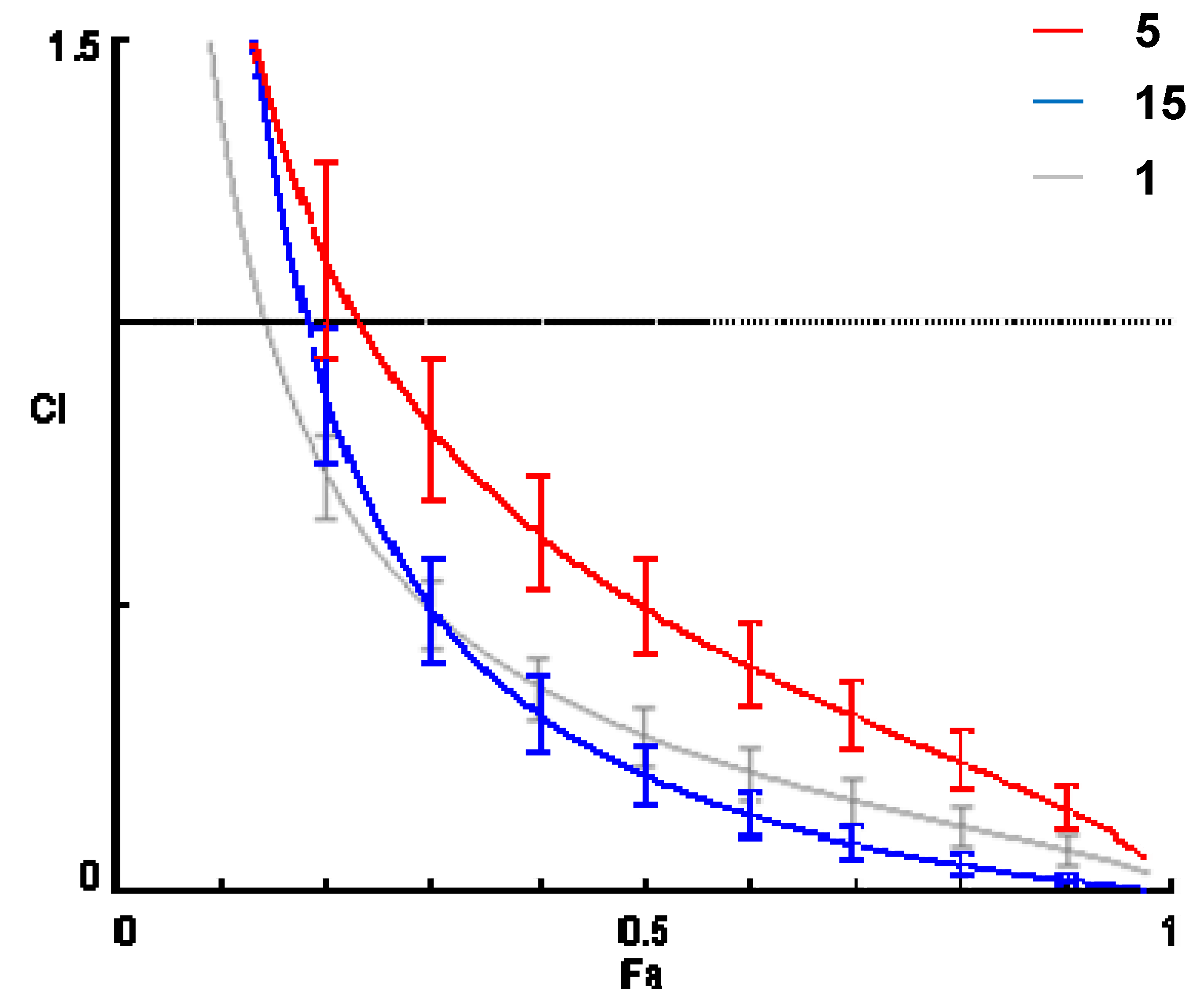
2.4. Combination Studies: Effect of Ecdysteroid Derivatives on the Activity of Doxorubicin on MDR Mouse Lymphoma Cells
| Compound | Drug Ratio | CI Values at | Dm | m | r | CIavg | ||
|---|---|---|---|---|---|---|---|---|
| ED50 | ED75 | ED90 | ||||||
| 20E [7] | 20.4:1 | 2.00 | 2.02 | 2.04 | 35.52 | 2.855 | 0.997 | 2.03 |
| 40.8:1 | 1.86 | 1.97 | 2.10 | 54.05 | 2.487 | 0.978 | 2.02 | |
| 81.5:1 | 1.80 | 1.93 | 2.08 | 76.90 | 2.376 | 0.978 | 1.99 | |
| 1 [7] | 20.4:1 | 0.28 | 0.14 | 0.07 | 11.68 | 3.246 | 0.964 | 0.13 |
| 40.8:1 | 0.26 | 0.14 | 0.08 | 14.73 | 3.167 | 0.996 | 0.13 | |
| 81.5:1 | 0.39 | 0.22 | 0.12 | 26.60 | 3.859 | 0.970 | 0.20 | |
| 2 [7] | 20.4:1 | 0.84 | 0.54 | 0.35 | 20.51 | 1.933 | 0.955 | 0.49 |
| 40.8:1 | 1.22 | 0.86 | 0.61 | 48.83 | 1.766 | 0.947 | 0.79 | |
| 81.5:1 | 1.11 | 0.77 | 0.54 | 64.94 | 1.920 | 0.916 | 0.71 | |
| 3 | 20.4:1 | 0.87 | 0.43 | 0.22 | 35.28 | 2.44 | 0.985 | 0.40 |
| 40.8:1 | 0.76 | 0.31 | 0.13 | 45.44 | 3.54 | 0.962 | 0.29 | |
| 81.6:1 | 0.91 | 0.31 | 0.11 | 71.98 | 5.78 | 0.989 | 0.31 | |
| 4 | 20.4:1 | 0.61 | 0.56 | 0.80 | 15.50 | 1.69 | 0.907 | 0.69 |
| 40.8:1 | 0.56 | 0.58 | 0.78 | 18.06 | 1.95 | 0.870 | 0.68 | |
| 81.6:1 | 1.08 | 0.91 | 0.88 | 40.46 | 5.02 | 0.921 | 0.92 | |
| 5 | 20.4:1 | 0.64 | 0.43 | 0.29 | 23.22 | 1.56 | 0.986 | 0.39 |
| 40.8:1 | 0.50 | 0.26 | 0.14 | 29.50 | 2.25 | 0.959 | 0.24 | |
| 81.6:1 | 0.61 | 0.44 | 0.33 | 52.58 | 1.32 | 0.989 | 0.41 | |
| 6 | 20.4:1 | 0.57 | 0.52 | 0.50 | 6.38 | 1.15 | 0.992 | 0.52 |
| 40.8:1 | 0.99 | 0.59 | 0.36 | 13.92 | 2.38 | 0.981 | 0.54 | |
| 81.6:1 | 0.70 | 0.44 | 0.28 | 11.21 | 2.34 | 0.936 | 0.40 | |
| 7 | 20.4:1 | 0.80 | 0.86 | 1.20 | 31.47 | 1.04 | 0.945 | 1.02 |
| 40.8:1 | 0.84 | 0.81 | 0.91 | 40.24 | 1.36 | 0.967 | 0.87 | |
| 81.6:1 | 1.16 | 0.96 | 0.86 | 62.14 | 1.96 | 0.996 | 0.94 | |
| 8 | 20.4:1 | 0.63 | 0.47 | 0.61 | 10.85 | 2.04 | 0.820 | 0.57 |
| 40.8:1 | 0.84 | 0.64 | 0.69 | 20.03 | 3.68 | 0.990 | 0.70 | |
| 81.6:1 | 0.76 | 0.68 | 0.74 | 22.38 | 3.99 | 0.949 | 0.72 | |
| 9 | 20.4:1 | 1.32 | 0.92 | 1.17 | 25.01 | 1.46 | 0.988 | 1.11 |
| 40.8:1 | 0.89 | 0.66 | 0.76 | 25.78 | 2.04 | 0.968 | 0.75 | |
| 81.6:1 | 0.82 | 0.68 | 0.74 | 32.71 | 2.66 | 0.950 | 0.73 | |
| 10 | 20.4:1 | 1.12 | 0.84 | 0.91 | 26.31 | 1.84 | 0.980 | 0.92 |
| 40.8:1 | 0.92 | 0.81 | 0.94 | 31.74 | 2.01 | 0.991 | 0.89 | |
| 81.6:1 | 1.24 | 0.98 | 0.92 | 55.74 | 4.10 | 0.988 | 0.99 | |
| 11 | 20.4:1 | 0.95 | 0.53 | 0.34 | 23.61 | 2.03 | 0.945 | 0.51 |
| 40.8:1 | 1.04 | 0.71 | 0.56 | 44.10 | 1.76 | 0.990 | 0.69 | |
| 81.6:1 | 0.89 | 0.65 | 0.55 | 59.01 | 1.95 | 0.997 | 0.64 | |
| 12 | 20.4:1 | 1.13 | 0.80 | 0.57 | 19.20 | 1.09 | 0.982 | 0.74 |
| 40.8:1 | 0.86 | 0.48 | 0.27 | 26.10 | 1.44 | 0.958 | 0.44 | |
| 81.6:1 | 1.32 | 0.53 | 0.21 | 66.47 | 2.79 | 0.997 | 0.50 | |
| 13 | 20.4:1 | 0.66 | 0.61 | 0.70 | 12.53 | 1.75 | 0.954 | 0.66 |
| 40.8:1 | 0.87 | 0.74 | 0.73 | 21.71 | 2.79 | 0.961 | 0.76 | |
| 81.6:1 | 0.85 | 0.93 | 1.12 | 25.07 | 2.02 | 0.898 | 1.01 | |
| 14 | 20.4:1 | 0.20 | 0.25 | 0.33 | 6.21 | 1.70 | 0.946 | 0.28 |
| 40.8:1 | 0.26 | 0.30 | 0.35 | 10.74 | 2.43 | 0.992 | 0.32 | |
| 81.6:1 | 0.28 | 0.34 | 0.42 | 13.24 | 2.33 | 0.979 | 0.37 | |
| 15 | 20.4:1 | 0.33 | 0.12 | 0.05 | 16.86 | 2.25 | 0.971 | 0.12 |
| 40.8:1 | 0.27 | 0.10 | 0.04 | 24.03 | 2.61 | 1.000 | 0.10 | |
| 81.6:1 | 0.20 | 0.06 | 0.02 | 26.98 | 4.12 | 0.969 | 0.07 | |
| 16 | 20.4:1 | 0.64 | 0.53 | 0.44 | 13.16 | 1.31 | 0.993 | 0.50 |
| 40.8:1 | 0.67 | 0.35 | 0.19 | 23.63 | 3.09 | 0.989 | 0.33 | |
| 81.6:1 | 0.34 | 0.24 | 0.17 | 18.98 | 1.92 | 0.967 | 0.22 | |
| 17 | 20.4:1 | 0.31 | 0.25 | 0.24 | 5.14 | 3.21 | 0.999 | 0.25 |
| 40.8:1 | 0.37 | 0.35 | 0.36 | 7.80 | 2.96 | 0.996 | 0.36 | |
| 81.6:1 | 0.54 | 0.56 | 0.60 | 12.84 | 2.85 | 0.987 | 0.58 | |
| 18 | 20.4:1 | 0.23 | 0.16 | 0.15 | 6.32 | 2.66 | 0.956 | 0.16 |
| 40.8:1 | 0.27 | 0.23 | 0.24 | 10.03 | 2.48 | 0.984 | 0.24 | |
| 81.6:1 | 0.40 | 0.37 | 0.39 | 17.83 | 2.61 | 0.998 | 0.39 | |
| 19 | 20.4:1 | 0.31 | 0.22 | 0.17 | 6.48 | 3.67 | 0.978 | 0.21 |
| 40.8:1 | 0.36 | 0.31 | 0.27 | 9.15 | 2.77 | 0.970 | 0.30 | |
| 81.6:1 | 0.52 | 0.46 | 0.42 | 14.59 | 2.81 | 0.977 | 0.45 | |
| 20 | 20.4:1 | 0.92 | 0.59 | 0.41 | 19.96 | 2.41 | 0.968 | 0.55 |
| 40.8:1 | 0.72 | 0.58 | 0.50 | 23.55 | 1.86 | 0.984 | 0.56 | |
| 81.6:1 | 1.06 | 0.65 | 0.42 | 46.17 | 4.64 | 0.970 | 0.60 | |
| 21 | 20.4:1 | 0.44 | 0.32 | 0.27 | 6.72 | 4.53 | 1.000 | 0.31 |
| 40.8:1 | 0.43 | 0.40 | 0.40 | 7.82 | 2.88 | 0.944 | 0.40 | |
| 81.6:1 | 0.58 | 0.61 | 0.67 | 11.75 | 2.46 | 0.963 | 0.63 | |
| 22 | 20.4:1 | 0.39 | 0.38 | 0.39 | 6.55 | 2.41 | 0.979 | 0.39 |
| 40.8:1 | 0.52 | 0.50 | 0.49 | 9.80 | 2.85 | 0.992 | 0.50 | |
| 81.6:1 | 0.71 | 0.65 | 0.60 | 14.25 | 3.64 | 0.980 | 0.63 | |
| 23 | 20.4:1 | 0.22 | 0.29 | 0.42 | 6.84 | 1.54 | 0.998 | 0.34 |
| 40.8:1 | 0.26 | 0.30 | 0.35 | 10.74 | 2.43 | 0.992 | 0.32 | |
| 81.6:1 | 0.28 | 0.34 | 0.42 | 13.24 | 2.33 | 0.979 | 0.37 | |
| 24 | 20.4:1 | 0.70 | 0.69 | 0.74 | 5.87 | 1.98 | 0.998 | 0.71 |
| 40.8:1 | 0.79 | 0.82 | 0.88 | 7.25 | 2.02 | 0.989 | 0.85 | |
| 81.6:1 | 0.56 | 0.88 | 1.42 | 5.30 | 1.17 | 0.942 | 1.10 | |
| 25 | 20.4:1 | 0.54 | 0.40 | 0.31 | 4.22 | 2.18 | 0.947 | 0.38 |
| 40.8:1 | 0.95 | 0.64 | 0.43 | 7.74 | 2.99 | 0.995 | 0.59 | |
| 81.6:1 | 1.01 | 0.72 | 0.52 | 8.37 | 2.62 | 0.985 | 0.67 | |
| 26 | 20.4:1 | 0.31 | 0.18 | 0.13 | 6.14 | 4.16 | 0.962 | 0.18 |
| 40.8:1 | 0.37 | 0.25 | 0.20 | 10.51 | 4.37 | 0.991 | 0.25 | |
| 81.6:1 | 0.56 | 0.42 | 0.36 | 20.13 | 4.72 | 0.992 | 0.41 | |
| 27 | 20.4:1 | 0.34 | 0.18 | 0.15 | 5.39 | 3.20 | 0.956 | 0.19 |
| 40.8:1 | 0.31 | 0.22 | 0.21 | 7.32 | 2.73 | 0.981 | 0.23 | |
| 81.6:1 | 0.38 | 0.32 | 0.33 | 12.04 | 2.85 | 0.969 | 0.33 | |
| 28 | 20.4:1 | 0.21 | 0.12 | 0.07 | 6.01 | 3.34 | 0.956 | 0.11 |
| 40.8:1 | 0.20 | 0.13 | 0.09 | 9.21 | 3.03 | 0.991 | 0.12 | |
| 81.6:1 | 0.20 | 0.14 | 0.10 | 12.73 | 3.14 | 0.973 | 0.13 | |
| 29 | 20.4:1 | 0.20 | 0.13 | 0.10 | 6.07 | 3.31 | 0.958 | 0.13 |
| 40.8:1 | 0.19 | 0.14 | 0.12 | 8.23 | 3.39 | 0.963 | 0.14 | |
| 81.6:1 | 0.22 | 0.21 | 0.22 | 12.36 | 2.46 | 0.981 | 0.22 | |
| 30 | 20.4:1 | 0.23 | 0.08 | 0.06 | 4.71 | 2.20 | 0.984 | 0.09 |
| 40.8:1 | 0.21 | 0.10 | 0.07 | 6.98 | 2.32 | 0.994 | 0.11 | |
| 81.6:1 | 0.21 | 0.14 | 0.13 | 9.92 | 1.86 | 0.995 | 0.15 | |
| 31 | 20.4:1 | 0.12 | 0.10 | 0.12 | 2.86 | 1.83 | 0.963 | 0.11 |
| 40.8:1 | 0.13 | 0.15 | 0.21 | 4.43 | 1.66 | 0.969 | 0.17 | |
| 81.6:1 | 0.22 | 0.23 | 0.29 | 8.97 | 2.26 | 0.998 | 0.26 | |
| 32 | 20.4:1 | 0.18 | 0.12 | 0.12 | 4.40 | 2.66 | 0.999 | 0.13 |
| 40.8:1 | 0.16 | 0.16 | 0.20 | 5.22 | 1.80 | 0.990 | 0.18 | |
| 81.6:1 | 0.23 | 0.24 | 0.29 | 9.18 | 2.15 | 0.971 | 0.26 | |
| 33 | 20.4:1 | 0.15 | 0.13 | 0.14 | 4.12 | 1.85 | 0.999 | 0.14 |
| 40.8:1 | 0.14 | 0.15 | 0.22 | 5.14 | 1.52 | 0.982 | 0.18 | |
| 81.6:1 | 0.19 | 0.27 | 0.44 | 9.18 | 1.36 | 0.992 | 0.34 | |


3. Experimental
3.1. General Information
3.2. Semi-Synthesis and Purification of Monosubstituted Ecdysteroid Dioxolane Derivatives 2–16
3.3. Semi-Synthesis and Purification of Disubstituted Ecdysteroid Derivatives 17–25 in One-Step
3.4. Semi-Synthesis and Purification of Disubstituted Ecdysteroid Derivatives 26–33 in Two-Steps
3.5. Further Experimental Data for the New Compounds
3.6. Preparation of the Compounds for the Bioassays
3.7. Cell Lines
3.8. Anti-proliferative Assay
3.9. Inhibition of ABCB1 Pump of MDR Mouse Lymphoma Cells (Rhodamine 123 Accumulation Assay)
3.10. Combination Assays
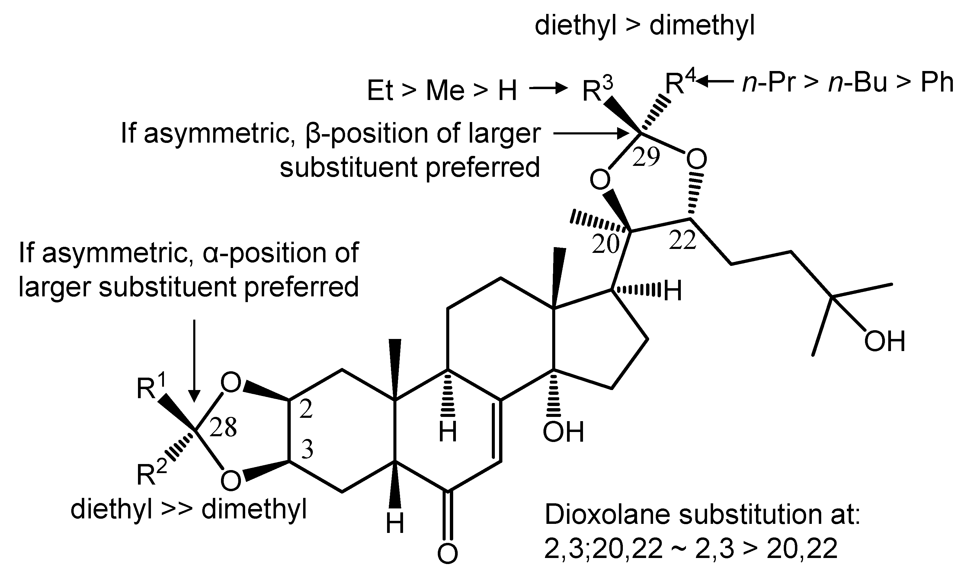
4. Conclusions
Acknowledgments
Conflicts of Interest
References
- Karlson, P. Mode of Action of Ecdysones. In Invertebrate Endocrinology and Hormonal Heterophylly; Burdette, W.B., Ed.; Springer: Berlin/Heidelberg, Germany, 1974; pp. 43–54. [Google Scholar]
- Zeleny, J.; Havelka, J.; Sláma, K. Hormonally mediated insect-plant relationships: Arthropod populations associated with ecdysteroid-containing plant, Leuzea carthamoides (Asteraceae). Eur J. Entomol. 1997, 94, 183–198. [Google Scholar]
- Dinan, L. A strategy for the identification of ecdysteroid receptor agonists and antagonists from plants. Eur. J. Entomol. 1995, 92, 271–283. [Google Scholar]
- Tóth, N.; Hunyadi, A.; Báthori, M.; Zádor, E. Phytoecdysteroids and vitamin D analogues—Similarities in structure and mode of action. Curr. Med. Chem. 2010, 17, 1974–1994. [Google Scholar] [CrossRef]
- Báthori, M.; Tóth, N.; Hunyadi, A.; Márki, Á.; Zádor, E. Phytoecdysteroids and anabolic-androgenic steroids. Structure and effects on humans. Curr. Med. Chem. 2008, 15, 75–91. [Google Scholar] [CrossRef]
- Dinan, L. The Karlson lecture. Phytoecdysteroids: What use are they? Arch. Insect Biochem. Physiol. 2009, 72, 126–141. [Google Scholar] [CrossRef]
- Martins, A.; Tóth, N.; Ványolós, A.; Béni, Z.; Zupkó, I.; Molnár, J.; Báthori, M.; Hunyadi, A. Significant activity of ecdysteroids on the resistance to doxorubicin in mammalian cancer cells expressing the human ABCB1 transporter. J. Med. Chem. 2012, 55, 5034–5043. [Google Scholar] [CrossRef]
- Möllmann, H.; Rohdewald, P.; Schmidt, E.W.; Salomon, V.; Derendorf, H. Pharmacokinetics of triamcinolone acetonide and its phosphate ester. Eur. J. Clin. Pharmacol. 1985, 29, 85–89. [Google Scholar] [CrossRef]
- Balázs, A.; Hunyadi, A.; Csábi, J.; Jedlinszki, N.; Martins, A.; Simon, A.; Tóth, G. 1H- and 13C-NMR investigation of 20-hydroxyecdysone dioxolane derivatives, a novel group of MDR modulator agents. Magn. Reson. Chem. 2013, 51, 830–836. [Google Scholar]
- Chou, T.-C. Theoretical basis, experimental design, and computerized simulation of synergism and antagonism in drug combination studies. Pharmacol. Rev. 2006, 58, 621–681. [Google Scholar]
- Pastan, I.; Gottesman, M.M.; Ueda, K.; Lovelace, E.; Rutherford, A.V.; Willingham, M.C. A retrovirus carrying an MDR1 cDNA confers multidrug resistance and polarized expression of P-glycoprotein in MDCK cells. Proc. Natl. Acad. Sci. USA 1988, 85, 4486–4490. [Google Scholar]
- Choi, K.; Frommel, T.O.; Stern, R.K.; Perez, C.F.; Kriegler, M.; Tsuruo, T.; Roninson, I.B. Multidrug resistance after retroviral transfer of the human MDR1 gene correlates with P-glycoprotein density in the plasma membrane and is not affected by cytotoxic selection. Proc. Natl. Acad. Sci. USA 1991, 88, 7386–7390. [Google Scholar]
- Mosmann, T. Rapid colorimetric assay for cellular growth and survival: Application to proliferation and cytotoxicity assays. J. Immunol. Methods 1983, 65, 55–63. [Google Scholar] [CrossRef]
- Sample Availability: Samples of the compounds 1–33 are available from the authors.
© 2013 by the authors; licensee MDPI, Basel, Switzerland. This article is an open access article distributed under the terms and conditions of the Creative Commons Attribution license (http://creativecommons.org/licenses/by/3.0/).
Share and Cite
Martins, A.; Csábi, J.; Balázs, A.; Kitka, D.; Amaral, L.; Molnár, J.; Simon, A.; Tóth, G.; Hunyadi, A. Synthesis and Structure-Activity Relationships of Novel Ecdysteroid Dioxolanes as MDR Modulators in Cancer. Molecules 2013, 18, 15255-15275. https://doi.org/10.3390/molecules181215255
Martins A, Csábi J, Balázs A, Kitka D, Amaral L, Molnár J, Simon A, Tóth G, Hunyadi A. Synthesis and Structure-Activity Relationships of Novel Ecdysteroid Dioxolanes as MDR Modulators in Cancer. Molecules. 2013; 18(12):15255-15275. https://doi.org/10.3390/molecules181215255
Chicago/Turabian StyleMartins, Ana, József Csábi, Attila Balázs, Diána Kitka, Leonard Amaral, József Molnár, András Simon, Gábor Tóth, and Attila Hunyadi. 2013. "Synthesis and Structure-Activity Relationships of Novel Ecdysteroid Dioxolanes as MDR Modulators in Cancer" Molecules 18, no. 12: 15255-15275. https://doi.org/10.3390/molecules181215255