Review

Antioxidants Improve the Phenotypes of Dilated Cardiomyopathy and Muscle Fatigue in Mitochondrial Superoxide Dismutase-Deficient Mice

1
Department of Advanced Aging Medicine, Chiba University Graduate School of Medicine, Chiba 260-8670, Japan
2
Department of Orthopaedics, Juntendo University School of Medicine, Bunkyo-ku, Tokyo 113-8421, Japan
3
Molecular Gerontology, Tokyo Metropolitan Institute of Gerontology, Itabashi-ku, Tokyo 173-0015, Japan
4
Research Laboratories for Applied Technology of Food, Asahi Group Holdings, Ltd., Ibaraki 302-0106, Japan
5
Department of Ageing Control Medicine, Juntendo University Graduate School of Medicine, Bunkyo-ku, Tokyo 113-0033, Japan
*
Author to whom correspondence should be addressed.
Molecules 2013, 18(2), 1383-1393; https://doi.org/10.3390/molecules18021383
Submission received: 10 December 2012 / Revised: 14 January 2013 / Accepted: 16 January 2013 / Published: 24 January 2013

Abstract

:
Redox imbalance elevates the reactive oxygen species (ROS) level in cells and promotes age-related diseases. Superoxide dismutases (SODs) are antioxidative enzymes that catalyze the degradation of ROS. There are three SOD isoforms: SOD1/CuZn-SOD, SOD2/Mn-SOD, and SOD3/EC-SOD. SOD2, which is localized in the mitochondria, is an essential enzyme required for mouse survival, and systemic knockout causes neonatal lethality in mice. To investigate the physiological function of SOD2 in adult mice, we generated a conditional Sod2 knockout mouse using a Cre-loxP system. When Sod2 was specifically deleted in the heart and muscle, all mice exhibited dilated cardiomyopathy (DCM) and died by six months of age. On the other hand, when Sod2 was specifically deleted in the skeletal muscle, mice showed severe exercise disturbance without morphological abnormalities. These provide useful model of DCM and muscle fatigue. In this review, we summarize the impact of antioxidants, which were able to regulate mitochondrial superoxide generation and improve the phenotypes of the DCM and the muscle fatigue in mice.

1. Introduction

An imbalance between the oxidation by reactive oxygen species (ROS) and reduction by antioxidant systems induces intracellular oxidative stress leading to the initiation and progression of age-related diseases, including diabetes, hypertension, atherosclerosis, osteoporosis, and neuro-degenerative diseases. ROS include several harmful species, such as superoxide anion (O2•−), hydrogen peroxide (H2O2), and the hydroxyl radical (HO). They are physiologically generated by mitochondrial respiration, as well as cellular enzymatic reactions in response to environmental stimuli. In antioxidant systems, enzymes (dismutase, catalase, and peroxidase) and small molecules (glutathione and vitamins etc.) finally detoxify ROS to non-toxic metabolites, such as molecular oxygen and water [1]. Superoxide dismutases (SODs) are the main antioxidant enzymes that catalyze the conversion of superoxide anion (O2•− to hydrogen peroxide (H2O2) and protect cells and tissues from the reactive oxygen species (ROS) generated from endogenous and exogenous sources. Three SOD isoforms are expressed in mammalian cells: copper/zinc SOD (CuZn-SOD, SOD1), which is located in the cytoplasm [2]; manganese SOD (Mn-SOD, SOD2), which is localized in the mitochondrial matrix [3]; and extracellular SOD (EC-SOD, SOD3) [4]. A small fraction of CuZn-SOD has also been reported to be present in the intermembrane space of the mitochondria [5]. Mitochondria are both a major source of ROS production from the respiratory chain and a major target of ROS-induced cellular injury [6]. Therefore, mitochondrial Mn-SOD is thought to play an important role in the cellular defense against oxidative damage by ROS.
The pathological consequences of increased mitochondrial ROS production have been thoroughly analyzed in model animals, such as Mn-SOD-knockout mice. These mice die from severe dilated cardiomyopathy within several weeks of birth and exhibit striking lipid deposits in the liver, as well as neurodegeneration [7,8,9]. The mice also show a significant reduction in the activity of mitochodrial respiratory enzymes, including complexes I, II, and III and the TCA cycle enzyme, aconitase [10]. Furthermore, genomic DNA prepared from the hearts and the brains of Mn-SOD-deficient mice has been reported to show a significant accumulation of oxidative DNA damage [10]. Since Mn-SOD-deficient mice die within several weeks of birth, it is not possible to investigate the pathological consequences of oxidative damage in adult tissues or the physiological aging process in vivo. We have also argued that the phenotypes of such mice are too complex to obtain information about specific aging processes for each tissue in vivo. In order to devise a mouse model in which the Mn-SOD deficiency could be directed to specific tissues, we designed a conditional knockout allele using the Cre-loxP system.

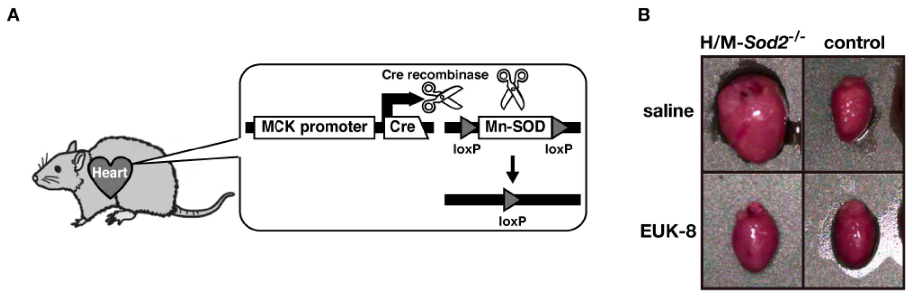
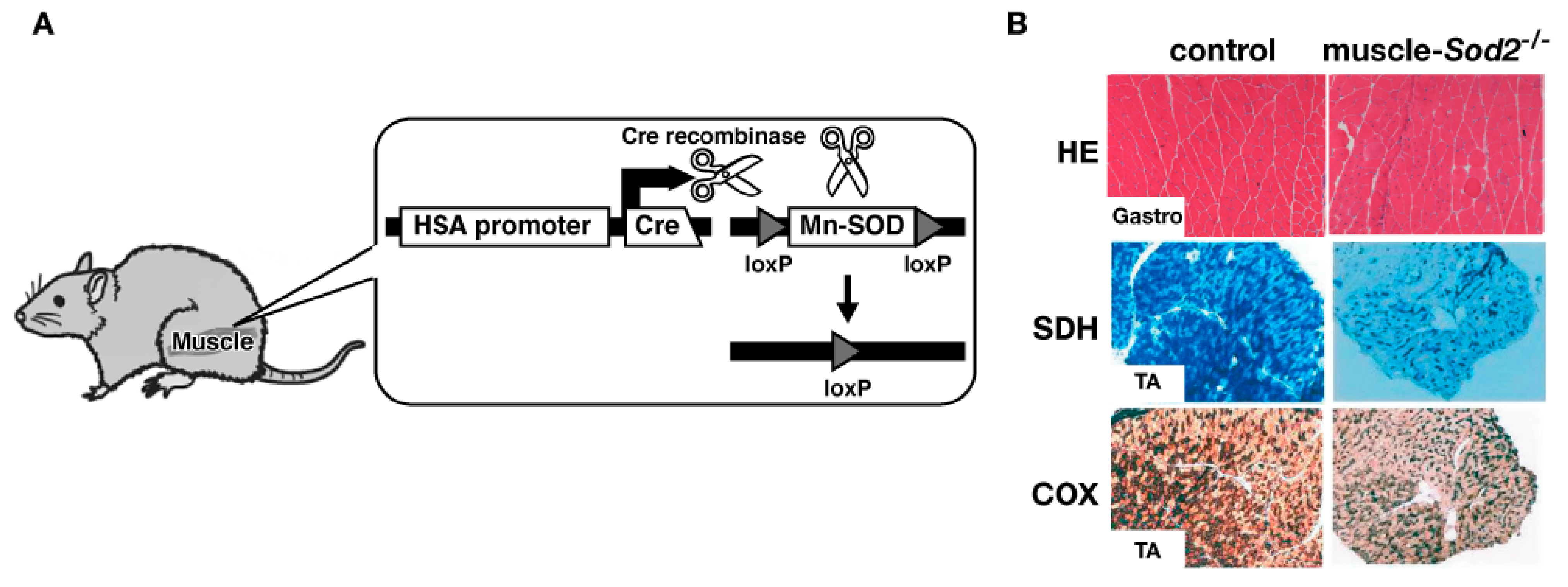
2. Heart and Muscle-Specific Mn-SOD-Deficient Mice
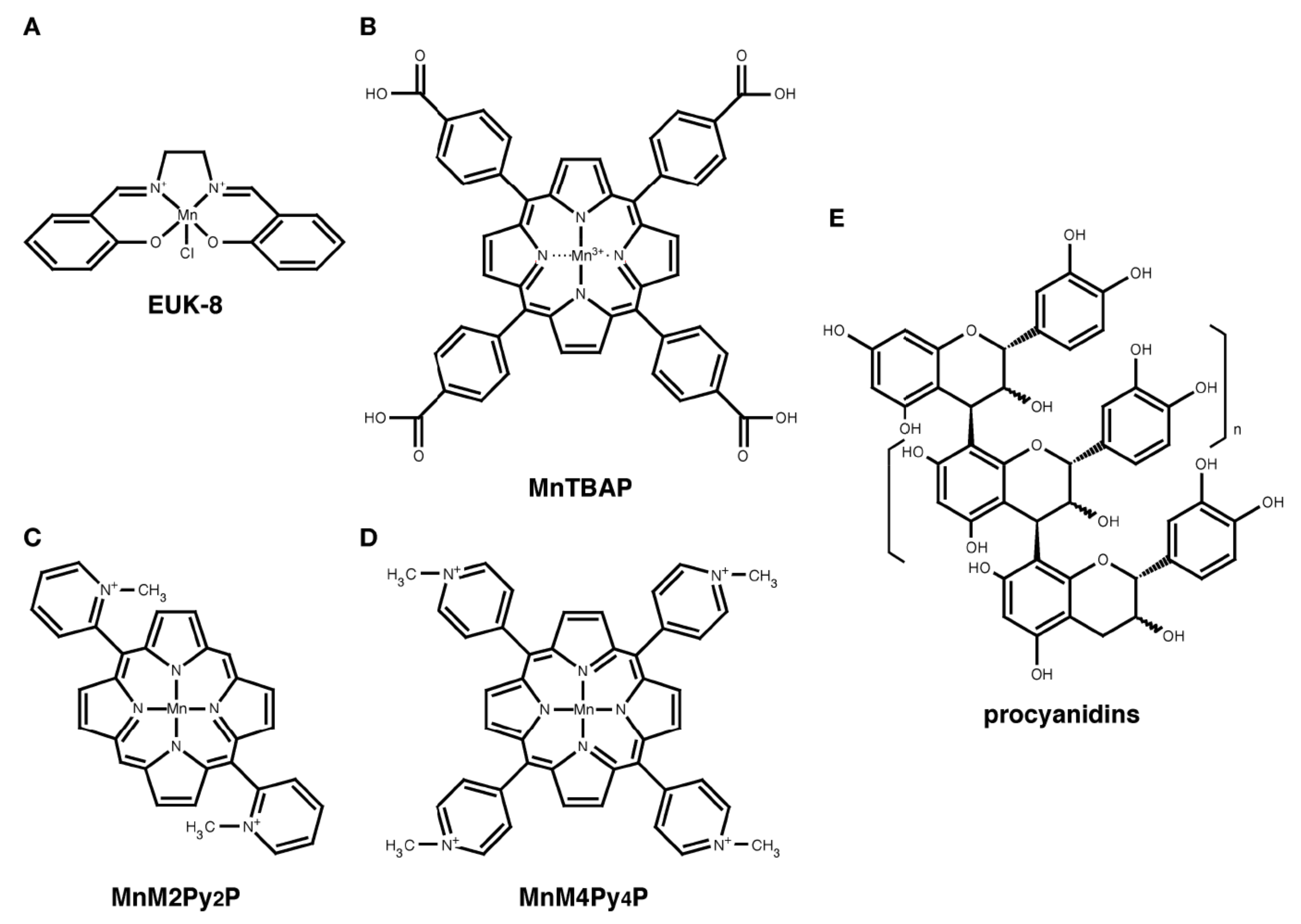
Oxidative stress plays a role in the development of heart failure [11,12]. To investigate the pathological role of mitochondrial ROS in the heart and muscle, we generated conditional Mn-SOD knockout mice by targeting these tissues (Figure 1A) [13]. We used muscle creatine kinase (MCK)-Cre transgenic mice to provide selective expression of the Cre protein in muscle tissues [14]. The Western blot analyses confirmed the specific loss of Mn-SOD expression in the heart and skeletal muscle of the heart and muscle-specific Mn-SOD-deficient mice (H/M-Sod2−/− mice), but no loss was observed in the control mice. In the neonatal stage, we were unable to find any differences in the appearance or body size between H/M-Sod2−/− and control mice. However, at two months of age, the H/M-Sod2−/− mice began to exhibit growth retardation. At four months of age, the H/M-Sod2−/− mice showed a 25% reduction in body weight compared to control mice, without any distinct muscle atrophy. By six months of age, all H/M-Sod2−/− mutant mice had died (the median survival time was about four months). When examined macroscopically, all of the hearts from the H/M-Sod2−/− mice showed cardiac enlargement at four months of age (Figure 1B). Transverse sections of the hearts from the H/M-Sod2−/− mice showed a marked dilation of both the left and right ventricles, which was compatible with the end stage of dilated cardiomyopathy. In histological sections with Azan staining, diffuse fibrotic scars surrounded the myocardial cells. Some of the thickened fibrotic foci were due to necrotic changes in the myocardium. These results indicate that the H/M-Sod2−/− mice progressively develop dilated cardiomyopathy.
To understand the biochemical alterations involved in the pathogenesis of cardiomyopathy, we examined the mitochondrial respiratory functions in the hearts of H/M-Sod2−/− mice. Using an enzymatic histochemical analysis, we observed that there was a specific loss of succinate dehydrogenase (SDH, Complex II) activity, but not cytochrome c oxidase (COX, Complex IV) in the mutant hearts [15]. Among the mitochondrial respiratory chain complexes, SDH is particularly vulnerable to O2•− because of its Fe-S center. Enhanced O2•− generation due to Mn-SOD deficiency might induce the degradation of the SDH protein. In the heart mitochondria, the H/M-Sod2−/− mice were previously shown to have significantly reduced ATP production. In fact, we revealed that the O2•− formation in the mutant heart mitochondria was increased to 1.87-fold the level of the control mice. Recently, a flow cytometric analysis also demonstrated increased ROS production in isolated cardiomyocytes from H/M-Sod2−/− mice [16]. In fibroblasts, Mn-SOD deficiency also markedly induced ROS generation, thus indicating that Mn-SOD depletion generally causes higher ROS levels in the cells (unpublished results).

3. EUK-8

EUK-8 is a synthetic salen-manganese complex, which has both SOD and catalase activities (Figure 2A) [17,18]. EUK-8 is a potent SOD/catalase mimetic and antioxidant. EUK-8 can dismutate O2•− to H2O2 and catalyze the breakdown of H2O2 to O2 and H2O. Some in vivo studies have shown that EUK-8 has protective effects in model organisms with ROS-induced diseases, such as heart mitochondrial dysfunction, aging, adrenergic hypertrophy, ischemia-reperfusion injury, and postischemic reperfusion arrhythmias [17,19,20,21,22]. Interestingly, EUK-8 was demonstrated to extend the lifespan of wild-type nematodes and systemic Sod2 knockout mice [23,24]. These results suggest that EUK-8 scavenges the ROS generated in the cytoplasm or organelles, including the mitochondria.
We have previously reported the effect of EUK-8 on H/M-Sod2−/− mice [25]. When EUK-8 was injected intraperitoneally (30 mg/kg/day) into the H/M-Sod2−/− mice for four weeks, cardiac dilation and pump failure were significantly prevented [25]. The ROS generation in heart mitochondria was also reduced by EUK-8 treatment, suggesting that EUK-8 is a mitochondria-targeting antioxidant. Interestingly, EUK-8 therapy also significantly reversed the established DCM-like phenotypes in H/M-Sod2−/− mice [25]. This was the first preclinical data indicating that EUK-8 is a useful new drug for the treatment of DCM. Furthermore, our findings revealed that the mimetic treatment normalized the molecular changes due to DCM, such as those in connexin43, telomerase reverse transcriptase (mTERT), telomeric repeat binding factor 2 (TRF2), phospho-AKT, insulin-like growth factor (IGF-1), endothelial nitric oxide synthase (eNOS), SIRT1, and FOXO3a [25,26]. These results suggest that the administration of antioxidants significantly restores cardiac contractility, as well as normalizing the molecular alterations in H/M-Sod2−/− mice.

4. MnTBAP

MnTBAP is a metalloporphyrin that is widely used as an SOD/catalase mimetic worldwide (Figure 2B) [27]. We examined the effect of MnTBAP on H/M-Sod2−/− mice [15]. Although the heart weight/body weight of H/M-Sod2−/− mice was not rescued by the intraperitoneal administration of MnTBAP, the cardiac function of H/M-Sod2−/− mice was significantly improved by MnTBAP treatment. MnTBAP treatment also recovered the daily running distances and the rotarod tasks in the H/M-Sod2−/− mice [15]. These data suggested that MnTBAP treatment significantly rescued the impaired cardiac contractility, as well as the physical disabilities, of the H/M-Sod2−/− mice [15].

5. Manganese Porphyrins

Manganese (Mn) porphyrins are water-soluble SOD/catalase mimic complex and have antioxidant activity under some conditions [28,29,30]. Recently, Hayakawa et al. reported that the Mn-porphyrins, 5,15-bis(2-methylpyridyl)porphinato manganese (MnM2Py2P), and 5,10,15,20-tetrakis(4-methyl-pyridyl)porphinato manganese (MnM4Py4P) (Figure 2C,D) [30,31], decreased the oxidative stress and heart weight of H/M-Sod2−/− mice [32]. MnM2Py2P could suppress the pathology of DCM more effectively than MnM4Py4P, and is expected to be an effective drug against DCM. Taken together with the data about EUK-8 and MnTBAP, antioxidants with SOD activity may protect hearts from mitochondrial ROS in bioavailability- and drug metabolism-dependent manners, rather than by their antioxidative activity, in vivo.

6. Procyanidins

Polyphenols are a structural class of mainly natural, but also synthetic, organic chemicals characterized by the presence of large and multiple phenol structural units. Polyphenols comprise several groups of compounds, such as anthocyanins, flavonols, and phenolic acids, and are accumulated in plants as well as fruits [33]. Resveratrol is a well-known polyphenol, which can extend the lifespan of yeast, worms, flies, and mice [34,35,36]. Several studies have reported that dietary polyphenols from vegetables and fruits prevent oxidative stress in vivo [37,38].
The apple polyphenols (AP) have various physiological functions, such as anti-oxidant, anti-tumor, anti-allergy, and anti-obesity effects [16,39,40,41]. The major polyphenols contained in apple are procyanidins (PC), which are composed of (–)-epicatechins and (+)-catechins (Figure 2E) [42]. PC are also contained in other fruits, such as grape seeds and blueberries [43].
Previously, we tested the effect of PC on the lifespan of Caenorhabditis elegans [44]. Treatment with AP and PC could significantly extend the mean lifespan of wild-type N2 and fem-1 mutant worms. However, PC could not extend the longevity of mev-1 and sir-2.1 mutant worms. The mev-1 strain is a short-lived mutant that suffers from excessive oxidative stress [45]. This suggested that PC extends the lifespan in C. elegans in an antioxidant-independent manner, although PC showed a strong antioxidant activity in vitro [46]. The sir-2.1 worm is a mutant that lacks histone deacetylase SIR-2 activity [35]. The findings in this strain indicated that PC prolongs the lifespan of worms in a sir-2-dependent manner. In an in vitro HDAC assay, PC failed to activate SIRT1. It has been reported that the up regulation of the sir-2 gene extends the survival in worms [47]. Therefore, it seems possible that PC might regulate sir-2 expression. Since NAD is essential for SIR-2 activity, it is possible that PC controls NAD metabolism. Wilson et al. reported that PC from blueberries extended the lifespan of worms in a sir-2-independent manner [48]. Therefore, it is possible that the PC from apples and blueberries extends the lifespan of worms by different mechanisms.
Recently, we examined the effect of AP on H/M-Sod2−/− mice [16]. When AP were administered to H/M-Sod2−/− mice in drinking water containing 0.1% AP, the AP significantly extended the lifespan of the H/M-Sod2−/− mice, and was associated with an attenuation of the enlarged heart from 22 weeks to 29 weeks of age. AP treatment also mitigated the accumulation of the plasma creatine phosphokinase (CPK) level, histopathology, and fibrosis in the heart of H/M-Sod2−/− mice. The biochemical analyses using ROS-reacting fluorescent reagents revealed that AP supplementation reduced the ROS production in the cardiomyocytes of mutant hearts in vivo. In vitro experiments also demonstrated that AP and PC treatment significantly decreased the ROS production in fibroblasts isolated from systemic conditional Sod2−/− mice, indicating that AP and PC improved the DCM-like phenotypes of H/M-Sod2−/− mice by an antioxidative action [16].

7. Antioxidants for Human DCM

Though we have described about the effects of antioxidants on the DCM model mice, some antioxidative drugs have been already used to human DCM patients. Coenzyme Q10 (CoQ10) is a small redox active lipid, which is containing in all membrane and is an electron transporter in the respiratory chain of mitochondria [49,50,51]. Manzoli et al. reported that the beneficial effect of CoQ10 on DCM patients [52]. The administration of CoQ10 could improve ejection fraction, telediastolic volume, and reduced CoQ10 plasmatic level of DCM patients [52]. Carvedilol, one of β-adrenoceptor blockers has an antioxidant activity and is used to therapy for human DCM [53,54,55]. Carvedilol therapy reduced the overall mortality rate and the risk of hospitalization [54]. These clinical data suggest that antioxidants are useful to the therapy for DCM patients.

8. Effects of EUK-8 on Muscle

We have reported the effect of EUK-8 on muscle specific Sod2 knockout (muscle-Sod2−/−) mice [56]. The muscle-Sod2−/− mice express Cre recombinase under the control of the human skeletal actin promoter (Figure 3A) [57]. Although muscle-Sod2−/− mice showed the loss of SOD2 in their skeletal muscle, there was no difference in their outer appearance, body size, or food intake compared with control mice. Interestingly, muscle-Sod2−/− mice showed centronuclear muscle with increased serum creatine phosphokinase activity, which is a typical hallmark of regenerative muscle, in spite of the fact that they exhibited no loss of muscle mass (Figure 3B) [56]. The biochemical analyses revealed significantly increased oxidative DNA damage, reduced respiratory enzyme activities, including NADH dehydrogenase (Complex I) and SDH (Complex II), and a reduced ATP level in the skeletal muscles of muscle-Sod2−/− mice (Figure 3B) [56]. Intriguingly, the muscle-Sod2−/− mice showed normal spontaneous activity during free movement in a cage, while they exhibited severe exercise disturbance on a forced running task using treadmill. When a single dose of EUK-8 was injected intraperitoneally into muscle-Sod2−/− mice, the decreased ATP content in the skeletal muscle and running disturbance in the mutant mice were markedly attenuated at 24 hours after the injection [56]. These data suggest that EUK-8 is useful for protecting the mitochondrial function in skeletal muscle from the ROS generated by the mitochondria in vivo, and demonstrate that the muscle-Sod2−/− mouse is a beneficial model for investigate the cellular mechanism(s) of muscle fatigue.

9. Conclusions

In this review, we introduced the tissue-specific Mn-SOD knockout mice and summarized our recent studies on antioxidants using these mice. The H/M-Sod2−/− and muscle-Sod2−/− mice are useful mouse models for DCM and muscle fatigue, respectively. Antioxidants such as EUK-8, MnTBAP, Mn-porphyrins, and procyanidins efficiently improved the phenotypes of H/M-Sod2−/− mice and/or muscle-Sod2−/− mice. These data suggest that these antioxidative compounds are beneficial for the treatment and/or prevention of some diseases caused by ROS-mediated mitochondrial dysfunction.

Acknowledgments

This study was supported in part by the Program for the Promotion of Basic Research Activities for Innovative Biosciences and Grants-in-Aid for Scientific Research (B), (C) from the Ministry of Education, Culture, Sports, Science, and Technology of the Japanese Government.

References

  1. Dröge, W. Free radicals in the physiological control of cell function. Physiol. Rev. 2002, 82, 47–95. [Google Scholar] [CrossRef] [PubMed]
  2. McCord, J.M.; Fridovich, I. Superoxide dismutase. An enzymic function for erythrocuprein (hemocuprein). J. Biol. Chem. 1969, 244, 6049–6055. [Google Scholar] [PubMed]
  3. Weisiger, R.A.; Fridovich, I. Mitochondrial superoxide dismutase. Site of synthesis and intramitochondrial localization. J. Biol. Chem. 1973, 248, 4793–4796. [Google Scholar] [PubMed]
  4. Marklund, S.L. Human copper-containing superoxide dismutase of high molecular weight. Proc. Natl. Acad. Sci. USA 1982, 79, 7634–7638. [Google Scholar] [CrossRef] [PubMed]
  5. Okado-Matsumoto, A.; Fridovich, I. Subcellular distribution of superoxide dismutases (SOD) in rat liver: Cu,Zn-SOD in mitochondria. J. Biol. Chem. 2001, 276, 38388–38393. [Google Scholar] [CrossRef] [PubMed]
  6. Daiber, A. Redox signaling (cross-talk) from and to mitochondria involves mitochondrial pores and reactive oxygen species. Biochim. Biophys. Acta 2010, 1797, 897–906. [Google Scholar] [CrossRef] [PubMed]
  7. Lebovitz, R.M.; Zhang, H.; Vogel, H.; Cartwright, J., Jr.; Dionne, L.; Lu, N.; Huang, S.; Matzuk, M.M. Neurodegeneration, myocardial injury, and perinatal death in mitochondrial superoxide dismutase-deficient mice. Proc. Natl. Acad. Sci. USA 1996, 93, 9782–9787. [Google Scholar] [CrossRef] [PubMed]
  8. Li, Y.; Huang, T.T.; Carlson, E.J.; Melov, S.; Ursell, P.C.; Olson, J.L.; Noble, L.J.; Yoshimura, M.P.; Berger, C.; Chan, P.H.; et al. Dilated cardiomyopathy and neonatal lethality in mutant mice lacking manganese superoxide dismutase. Nat. Genet. 1995, 11, 376–381. [Google Scholar] [CrossRef] [PubMed]
  9. Melov, S.; Schneider, J.A.; Day, B.J.; Hinerfeld, D.; Coskun, P.; Mirra, S.S.; Crapo, J.D.; Wallace, D.C. A novel neurological phenotype in mice lacking mitochondrial manganese superoxide dismutase. Nat. Genet. 1998, 18, 159–163. [Google Scholar] [CrossRef] [PubMed]
  10. Melov, S.; Coskun, P.; Patel, M.; Tuinstra, R.; Cottrell, B.; Jun, A.S.; Zastawny, T.H.; Dizdaroglu, M.; Goodman, S.I.; Huang, T.T.; et al. Mitochondrial disease in superoxide dismutase 2 mutant mice. Proc. Natl. Acad. Sci. USA 1999, 96, 846–851. [Google Scholar] [CrossRef] [PubMed]
  11. Alameddine, F.M.; Zafari, A.M. Genetic polymorphisms and oxidative stress in heart failure. Congest. Heart Fail. 2002, 8, 157–164. [Google Scholar] [CrossRef] [PubMed]
  12. Givertz, M.M.; Colucci, W.S. New targets for heart-failure therapy: Endothelin, inflammatory cytokines, and oxidative stress. Lancet 1998, 352, SI34–SI38. [Google Scholar] [CrossRef]
  13. Ikegami, T.; Suzuki, Y.; Shimizu, T.; Isono, K.; Koseki, H.; Shirasawa, T. Model mice for tissue-specific deletion of the manganese superoxidedismutase (MnSOD) gene. Biochem. Biophys. Res. Commun. 2002, 296, 729–736. [Google Scholar] [CrossRef]
  14. Brüning, J.C.; Michael, M.D.; Winnay, J.N.; Hayashi, T.; Hörsch, D.; Accili, D.; Goodyear, L.J.; Kahn, C.R. A muscle-specific insulin receptor knockout exhibits features of the metabolic syndrome of NIDDM without altering glucose tolerance. Mol. Cell 1998, 2, 559–569. [Google Scholar] [CrossRef]
  15. Nojiri, H.; Shimizu, T.; Funakoshi, M.; Yamaguchi, O.; Zhou, H.; Kawakami, S.; Ohta, Y.; Sami, M.; Tachibana, T.; Ishikawa, H.; et al. Oxidative stress causes heart failure with impaired mitochondrial respiration. J. Biol. Chem. 2006, 281, 33789–33801. [Google Scholar] [CrossRef] [PubMed]
  16. Sunagawa, T.; Watanabe, K.; Ozawa, Y.; Nakashima, S.; Kanda, T.; Tagashira, M.; Sami, M.; Kaneko, T.; Tahara, S.; Nakaya, H.; et al. Apple polyphenols regulate mitochondrial superoxide generation and extend survival in a mouse model of dilated cardiomyopathy. Int. J. Life Sci. Med. Res. 2012, 2, 46–51. [Google Scholar] [CrossRef]
  17. Pucheu, S.; Boucher, F.; Sulpice, T.; Tresallet, N.; Bonhomme, Y.; Malfroy, B.; de Leiris, J. EUK-8 a synthetic catalytic scavenger of reactive oxygen species protects isolated iron-overloaded rat heart from functional and structural damage induced by ischemia/reperfusion. Cardiovasc. Drugs Ther. 1996, 10, 331–339. [Google Scholar] [CrossRef] [PubMed]
  18. Baker, K.; Marcus, C.B.; Huffman, K.; Kruk, H.; Malfroy, B.; Doctrow, S.R. Synthetic combined superoxide dismutase/catalase mimetics are protective as a delayed treatment in a rat stroke model: A key role for reactive oxygen species in ischemic brain injury. J. Pharmacol. Exp. Ther. 1998, 284, 215–221. [Google Scholar] [PubMed]
  19. Morten, K.J.; Ackrell, B.A.; Melov, S. Mitochondrial reactive oxygen species in mice lacking superoxide dismutase 2: Attenuation via antioxidant treatment. J. Biol. Chem. 2006, 281, 3354–3359. [Google Scholar] [CrossRef] [PubMed]
  20. Xu, Y.; Armstrong, S.J.; Arenas, I.A.; Pehowich, D.J.; Davidge, S.T. Cardioprotection by chronic estrogen or superoxide dismutase mimetic treatment in the aged female rat. Am. J. Physiol. Heart Circ. Physiol. 2004, 287, H165–H171. [Google Scholar] [CrossRef] [PubMed]
  21. Amin, J.K.; Xiao, L.; Pimental, D.R.; Pagano, P.J.; Singh, K.; Sawyer, D.B.; Colucci, W.S. Reactive oxygen species mediate alpha-adrenergic receptor-stimulated hypertrophy in adult rat ventricular myocytes. J. Mol. Cell Cardiol. 2001, 33, 131–139. [Google Scholar] [CrossRef] [PubMed]
  22. Tanguy, S.; Boucher, F.R.; Malfroy, B.; de Leiris, J.G. Free radicals in reperfusion-induced arrhythmias: Study with EUK 8, a novel nonprotein catalytic antioxidant. Free Radic. Biol. Med. 1996, 21, 945–954. [Google Scholar] [CrossRef]
  23. Melov, S.; Ravenscroft, J.; Malik, S.; Gill, M.S.; Walker, D.W.; Clayton, P.E.; Wallace, D.C.; Malfroy, B.; Doctrow, S.R.; Lithgow, G.J. Extension of life-span with superoxide dismutase/ catalase mimetics. Science 2000, 289, 1567–1569. [Google Scholar] [CrossRef] [PubMed]
  24. Melov, S.; Doctrow, S.R.; Schneider, J.A.; Haberson, J.; Patel, M.; Coskun, P.E.; Huffman, K.; Wallace, D.C.; Malfroy, B. Lifespan extension and rescue of spongiform encephalopathy in superoxide dismutase 2 nullizygous mice treated with superoxide dismutase-catalase mimetics. J. Neurosci. 2001, 21, 8348–8353. [Google Scholar] [CrossRef] [PubMed]
  25. Kawakami, S.; Matsuda, A.; Sunagawa, T.; Noda, Y.; Kaneko, T.; Tahara, S.; Hiraumi, Y.; Adachi, S.; Matsui, H.; Ando, K.; et al. Antioxidant, EUK-8, prevents murine dilated cardiomyopathy. Circ. J. 2009, 73, 2125–2134. [Google Scholar] [CrossRef] [PubMed]
  26. Makino, N.; Maeda, T.; Oyama, J.; Sasaki, M.; Higuchi, Y.; Mimori, K.; Shimizu, T. Antioxidant therapy attenuates myocardial telomerase activity reduction in superoxide dismutase-deficient mice. J. Mol. Cell. Cardiol. 2011, 50, 670–677. [Google Scholar] [CrossRef] [PubMed]
  27. Day, B.J.; Shawen, S.; Liochev, S.I.; Crapo, J.D. A metalloporphyrin superoxide dismutase mimetic protects against paraquat-induced endothelial cell injury, in vitro. J. Pharmacol. Exp. Ther. 1995, 275, 1227–1232. [Google Scholar] [PubMed]
  28. Asayama, S.; Mizushima, K.; Nagaoka, S.; Kawakami, H. Design of metalloporphyrin-carbohydrate conjugates for a new superoxide dismutase mimic with cellular recognition. Bioconjug. Chem. 2004, 15, 1360–1363. [Google Scholar] [CrossRef] [PubMed]
  29. Asayama, S.; Kawamura, E.; Nagaoka, S.; Kawakami, H. Design of manganese porphyrin modified with mitochondrial signal peptide for a new antioxidant. Mol. Pharm. 2006, 3, 468–470. [Google Scholar] [CrossRef] [PubMed]
  30. Asayama, S.; Nakajima, T.; Kawakami, H. New water-soluble Mn-porphyrin with catalytic activity for superoxide dismutation and peroxynitrite decomposition. Metallomics 2011, 3, 744–748. [Google Scholar] [CrossRef] [PubMed]
  31. Batinic-Haberle, I.; Benov, L.; Spasojevic, I.; Fridovich, I. The ortho effect makes manganese(III) meso-tetrakis(N-methylpyridinium-2-yl)porphyrin a powerful and potentially useful superoxide dismutase mimic. J. Biol. Chem. 1998, 273, 24521–24528. [Google Scholar] [CrossRef] [PubMed]
  32. Hayakawa, N.; Asayama, S.; Noda, Y.; Shimizu, T.; Kawakami, H. Pharmaceutical effect of manganese porphyrins on manganese superoxide dismutase deficient mice. Mol. Pharm. 2012, 9, 2956–2959. [Google Scholar] [CrossRef] [PubMed]
  33. Tsao, R. Chemistry and biochemistry of dietary polyphenols. Nutrients 2010, 2, 1231–1246. [Google Scholar] [CrossRef] [PubMed]
  34. Howitz, K.T.; Bitterman, K.J.; Cohen, H.Y.; Lamming, D.W.; Lavu, S.; Wood, J.G.; Zipkin, R.E.; Chung, P.; Kisielewski, A.; Zhang, L.L.; et al. Small molecule activators of sirtuins extend Saccharomyces cerevisiae lifespan. Nature 2003, 425, 191–196. [Google Scholar] [CrossRef] [PubMed]
  35. Wood, J.G.; Rogina, B.; Lavu, S.; Howitz, K.; Helfand, S.L.; Tatar, M.; Sinclair, D. Sirtuin activators mimic caloric restriction and delay ageing in metazoans. Nature 2004, 430, 686–689. [Google Scholar] [CrossRef] [PubMed]
  36. Baur, J.A.; Pearson, K.J.; Price, N.L.; Jamieson, H.A.; Lerin, C.; Kalra, A.; Prabhu, V.V.; Allard, J.S.; Lopez-Lluch, G.; Lewis, K.; et al. Resveratrol improves health and survival of mice on a high-calorie diet. Nature 2006, 444, 337–342. [Google Scholar] [CrossRef] [PubMed]
  37. Hertog, M.G.; Feskens, E.J.; Hollman, P.C.; Katan, M.B.; Kromhout, D. Dietary antioxidant flavonoids and risk of coronary heart disease: the Zutphen Elderly Study. Lancet 1993, 342, 1007–1011. [Google Scholar] [CrossRef]
  38. Mursu, J.; Voutilainen, S.; Nurmi, T.; Tuomainen, T.P.; Kurl, S.; Salonen, J.T. Flavonoid intake and the risk of ischaemic stroke and CVD mortality in middle-aged Finnish men: The Kuopio Ischaemic Heart Disease Risk Factor Study. Br. J. Nutr. 2008, 100, 890–895. [Google Scholar] [CrossRef] [PubMed]
  39. Miura, T.; Chiba, M.; Kasai, K.; Nozaka, H.; Nakamura, T.; Shoji, T.; Kanda, T.; Ohtake, Y.; Sato, T. Apple procyanidins induce tumor cell apoptosis through mitochondrial pathway activation of caspase-3. Carcinogenesis 2008, 29, 585–593. [Google Scholar] [CrossRef] [PubMed]
  40. Akiyama, H.; Sato, Y.; Watanabe, T.; Nagaoka, M.H.; Yoshioka, Y.; Shoji, T.; Kanda, T.; Yamada, K.; Totsuka, M.; Teshima, R.; et al. Dietary unripe apple polyphenol inhibits the development of food allergies in murine models. FEBS Lett. 2005, 579, 4485–4491. [Google Scholar] [CrossRef] [PubMed]
  41. Osada, K.; Funayama, M.; Fuchi, S.; Sami, M.; Ohta, Y.; Kanda, T.; Ikeda, M. Effects of dietary procyanidins and tea polyphenols on adipose tissue mass and fatty acid metabolism in rats on a high fat diet. J. Oleo Sci. 2006, 55, 79–89. [Google Scholar] [CrossRef]
  42. Ohnishi-Kameyama, M.; Yanagida, A.; Kanda, T.; Nagata, T. Identification of catechin oligomers from apple (Malus pumila cv. Fuji) in matrix-assisted laser desorption/ionization time-of-flight mass spectrometry and fast-atom bombardment mass spectrometry. Rapid Commun. Mass Spectrom. 1997, 11, 31–36. [Google Scholar] [CrossRef]
  43. Santos-Buelga, C.; Scalbert, A. Proanthocyanidins and tannin-like compounds: Nature, occurrence, dietary intake and effects on nutrition and health. J. Sci. Food Agric. 2000, 80, 1094–1117. [Google Scholar] [CrossRef]
  44. Sunagawa, T.; Shimizu, T.; Kanda, T.; Tagashira, M.; Sami, M.; Shirasawa, T. Procyanidins from apples (Malus pumila Mill.) extend the lifespan of Caenorhabditis elegans. Planta Med. 2011, 77, 122–127. [Google Scholar] [CrossRef] [PubMed]
  45. Senoo-Matsuda, N.; Yasuda, K.; Tsuda, M.; Ohkubo, T.; Yoshimura, S.; Nakazawa, H.; Hartman, P.S.; Ishii, N. A defect in the cytochrome b large subunit in complex II causes both superoxide anion overproduction and abnormal energy metabolism in Caenorhabditis elegans. J. Biol. Chem. 2001, 276, 41553–41558. [Google Scholar] [CrossRef] [PubMed]
  46. Leontowicz, H.; Gorinstein, S.; Lojek, A.; Leontowicz, M.; Cíz, M.; Soliva-Fortuny, R.; Park, Y.S.; Jung, S.T.; Trakhtenberg, S.; Martin-Belloso, O. Comparative content of some bioactive compounds in apples, peaches and pears and their influence on lipids and antioxidant capacity in rats. J. Nutr. Biochem. 2002, 13, 603–610. [Google Scholar] [CrossRef]
  47. Tissenbaum, H.A.; Guarente, L. Increased dosage of a sir-2 gene extends lifespan in Caenorhabditis elegans. Nature 2001, 410, 227–230. [Google Scholar] [CrossRef] [PubMed]
  48. Wilson, M.A.; Shukitt-Hale, B.; Kalt, W.; Ingram, D.K.; Joseph, J.A.; Wolkow, C.A. Blueberry polyphenols increase lifespan and thermotolerance in Caenorhabditis elegans. Aging Cell 2006, 5, 59–68. [Google Scholar] [CrossRef] [PubMed]
  49. Crane, F.L.; Hatefi, Y.; Lester, R.L.; Widmer, C. Isolation of a quinone from beef heart mitochondria. Biochim. Biophys. Acta 1957, 25, 220–221. [Google Scholar] [CrossRef]
  50. Ernster, L.; Dallner, G. Biochemical, physiological and medical aspects of ubiquinone function. Biochim. Biophys. Acta 1995, 1271, 195–204. [Google Scholar] [CrossRef]
  51. Turunen, M.; Olsson, J.; Dallner, G. Metabolism and function of coenzyme Q. Biochim. Biophys. Acta 2004, 1660, 171–199. [Google Scholar] [CrossRef] [PubMed]
  52. Manzoli, U.; Rossi, E.; Littarru, G.P.; Frustaci, A.; Lippa, S.; Oradei, A.; Aureli, V. Coenzyme Q10 in dilated cardiomyopathy. Int. J. Tissue React. 1990, 12, 173–178. [Google Scholar] [PubMed]
  53. Yue, T.L.; Cheng, H.Y.; Lysko, P.G.; McKenna, P.J.; Feuerstein, R.; Gu, J.L.; Lysko, K.A.; Davis, L.L.; Feuerstein, G. Carvedilol, a new vasodilator and beta adrenoceptor antagonist, is an antioxidant and free radical scavenger. J. Pharmacol. Exp. Ther. 1992, 263, 92–98. [Google Scholar] [PubMed]
  54. Packer, M.; Bristow, M.R.; Cohn, J.N.; Colucci, W.S.; Fowler, M.B.; Gilbert, E.M.; Shusterman, N.H. The effect of carvedilol on morbidity and mortality in patients with chronic heart failure. U.S. Carvedilol Heart Failure Study Group. N. Engl. J. Med. 1996, 334, 1349–1355. [Google Scholar] [CrossRef] [PubMed]
  55. Dandona, P.; Karne, R.; Ghanim, H.; Hamouda, W.; Aljada, A.; Magsino, C.H., Jr. Carvedilol inhibits reactive oxygen species generation by leukocytes and oxidative damage to amino acids. Circulation 2000, 101, 122–124. [Google Scholar] [CrossRef] [PubMed]
  56. Kuwahara, H.; Horie, T.; Ishikawa, S.; Tsuda, C.; Kawakami, S.; Noda, Y.; Kaneko, T.; Tahara, S.; Tachibana, T.; Okabe, M.; et al. Oxidative stress in skeletal muscle causes severe disturbance of exercise activity without muscle atrophy. Free Radic. Biol. Med. 2010, 48, 1252–1262. [Google Scholar] [CrossRef] [PubMed]
  57. Miniou, P.; Tiziano, D.; Frugier, T.; Roblot, N.; Le Meur, M.; Melki, J. Gene targeting restricted to mouse striated muscle lineage. Nucleic Acids Res. 1999, 27, e27. [Google Scholar] [CrossRef] [PubMed]
Figure 1. Heart and muscle-specific Mn-SOD-deficient mice. (A) Cre recombinase is expressed under the control of a heart and muscle-specific muscle creatine kinase (MCK) promoter. Cre recombinase recombines the loxP sites selectively to generate heart and muscle-specific Mn-SOD-deficient mice; (B) Isolated hearts from a H/M-Sod2−/− mouse (top left) and a littermate control mouse (top right) at eight weeks of age. EUK-8 treatment improved the enlarged heart of a H/M-Sod2−/− mouse (bottom left).
Molecules 18 01383 g001
Figure 2. The chemical structures of the antioxidants described in this review. (A) EUK-8; (B) MnTBAP; (C) MnM2Py2P; (D) MnM2Py2P, and (E) procyanidins.
Molecules 18 01383 g002
Figure 3. Muscle-specific Mn-SOD-deficient mice. (A) Cre recombinase is expressed under the control of a skeletal muscle-specific human skeletal actin (HSA) promoter. Cre recombinase recombines the loxP sites selectively to generate muscle-specific Mn-SOD-deficient mice; (B) Sections of gastrocnemius (Gastro) and tibialis anterior (TA) muscles from control mice (left panels) and muscle-Sod2−/− mice (right panels). Examples of HE staining (top panels) and enzymatic staining for succinate dehydrogenase (SDH, middle panels) and cytochrome c oxidase activity (COX, bottom panels) are shown.
Molecules 18 01383 g003

