Cytotoxicity and Structure-Activity Relationships of Xanthone Derivatives from Mesua beccariana, Mesua ferrea and Mesua congestiflora towards Nine Human Cancer Cell Lines

Abstract
:1. Introduction

2. Results and Discussion

 | |||||||
|---|---|---|---|---|---|---|---|
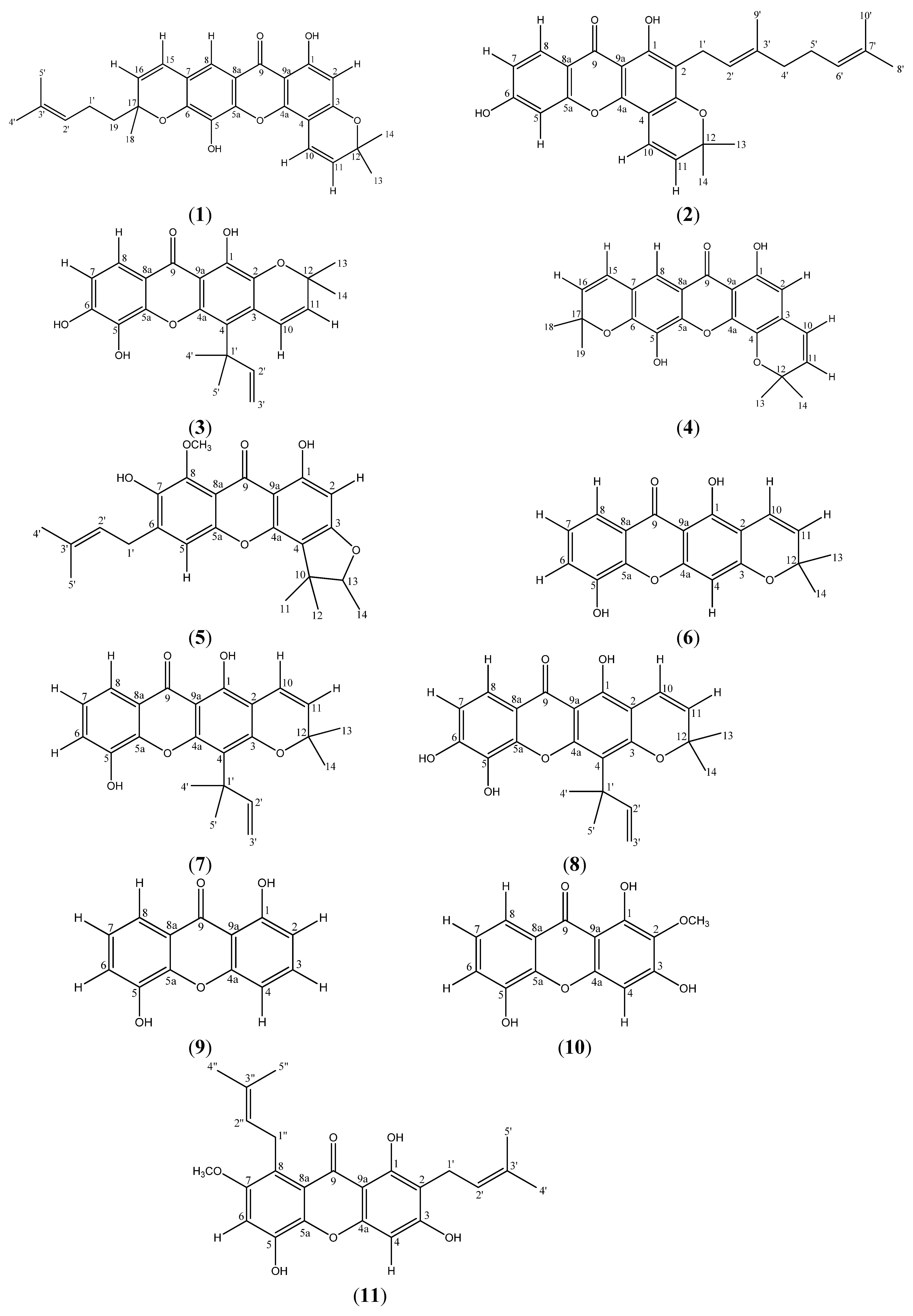
| Compds. | R2 | R3 | R4 | R5 | R6 | R7 | R8 |
| 1 | H |  | OH |  | H | ||
| 2 | geranyl | | H | OH | H | H | |
| 3 |  | prenyl B | OH | OH | H | H | |
| 4 | H |  | OH |  | H | ||
| 5 | H |  | H | prenyl A | OH | OCH3 | |
| 6 |  | H | OH | H | H | H | |
| 7 | | prenyl B | OH | H | H | H | |
| 8 | | prenyl B | OH | OH | H | H | |
| 9 | H | H | H | OH | H | H | H |
| 10 | OCH3 | OH | H | OH | H | H | H |
| 11 | prenyl A | OH | H | OH | H | OCH3 | prenyl A |
 ; prenyl B:
  ; geranyl:
  .
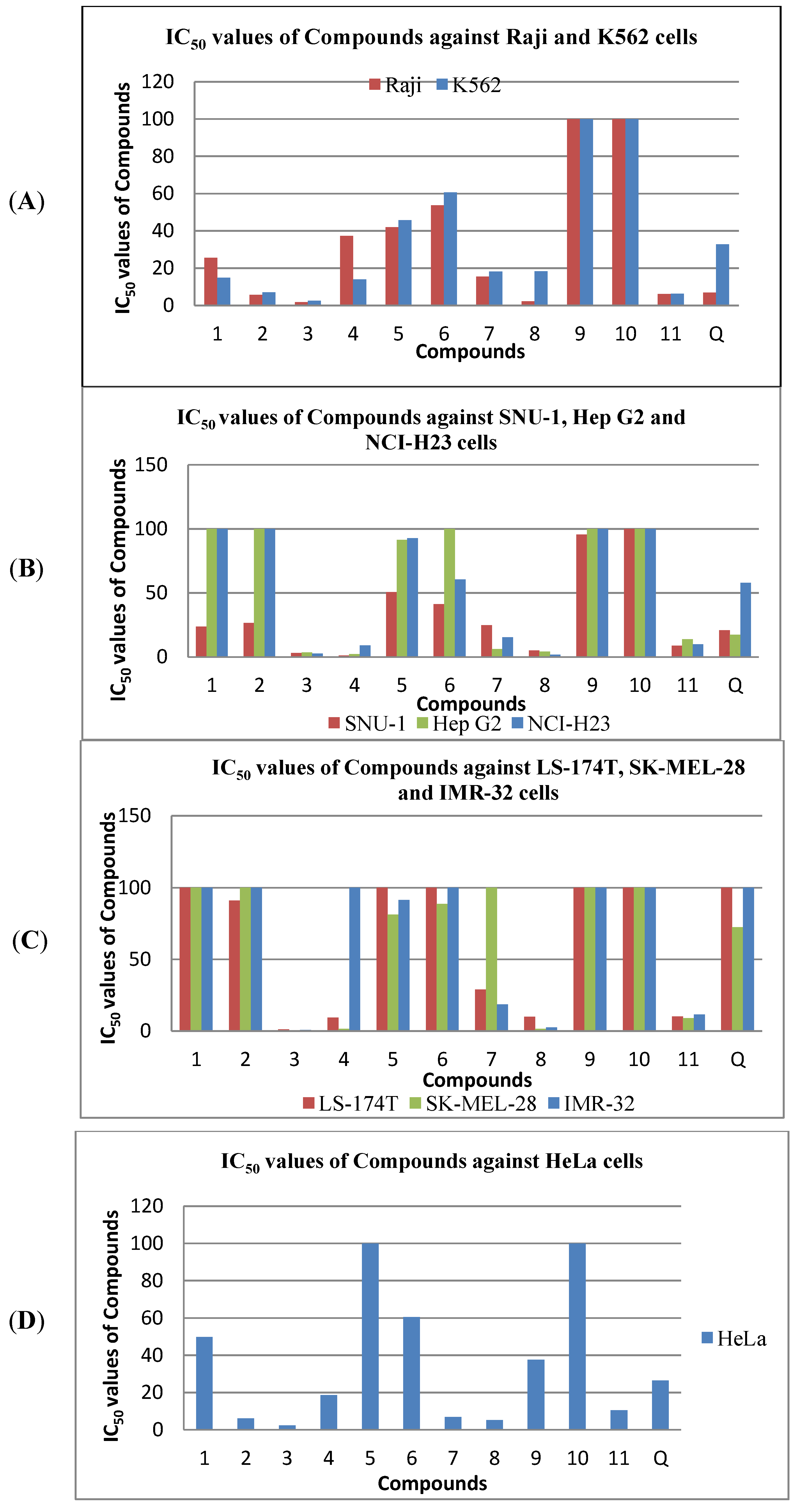
.| Compds. | Cell Lines with IC50 values (µM) | ||||||||
|---|---|---|---|---|---|---|---|---|---|
| Raji | SNU-1 | K562 | LS-174T | SK-MEL-28 | IMR-32 | HeLa | Hep G2 | NCI-H23 | |
| 1 | 25.46 ± 0.90 | 23.78 ± 1.33 | 14.93 ± 0.29 | - | - | - | 49.83 ± 1.31 | - | - |
| 2 | 5.61 ± 2.25 | 26.61 ± 0.58 | 7.02 ± 0.50 | 91.19 ± 1.45 | - | - | 6.12 ± 1.82 | - | - |
| 3 | 1.78 ± 0.57 | 3.17 ± 1.05 | 2.54 ± 2.00 | 1.17 ± 2.02 | 0.36 ± 2.38 | 0.79 ± 1.24 | 2.39 ± 1.07 | 3.68 ± 2.41 | 2.64 ± 2.95 |
| 4 | 37.24 ± 0.72 | 1.19 ± 1.36 | 13.95 ± 0.72 | 9.31 ± 2.36 | 1.48 ± 1.24 | - | 18.60 ± 1.52 | 2.32 ± 1.07 | 8.98 ± 1.32 |
| 5 | 41.90 ± 1.51 | 50.80 ± 1.50 | 45.73 ± 1.39 | - | 81.29 ± 2.30 | 91.46 ± 1.45 | - | 91.46 ± 1.04 | 92.68 ± 1.25 |
| 6 | 53.77 ± 1.11 | 41.29 ± 1.36 | 60.65 ± 0.64 | - | 88.71 ± 2.19 | - | 60.48 ± 1.59 | - | 60.48 ± 1.87 |
| 7 | 15.50 ± 1.99 | 24.79 ± 1.96 | 18.20 ± 1.32 | 28.94 ± 2.20 | - | 18.60 ± 1.03 | 6.88 ± 1.54 | 6.22 ± 1.49 | 15.42 ± 2.88 |
| 8 | 2.18 ± 1.87 | 5.08 ± 2.20 | 18.25 ± 1.25 | 9.90 ± 2.47 | 1.40 ± 1.69 | 2.49 ± 0.54 | 5.28 ± 2.04 | 4.24 ± 2.13 | 1.85 ± 1.76 |
| 9 | - | 95.53 ± 1.81 | - | - | - | - | 37.68 ± 2.53 | - | - |
| 10 | - | - | - | - | - | - | - | - | - |
| 11 | 6.10 ± 1.20 | 8.90 ± 1.06 | 6.34 ± 2.24 | 10.17 ± 1.68 | 8.88 ± 1.77 | 11.44 ± 2.45 | 10.49 ± 2.05 | 13.90 ± 1.24 | 10.00 ± 0.53 |
| Q | 6.89 ± 0.80 | 20.86 ± 1.93 | 32.75 ± 3.20 | - | 72.45 ± 2.07 | - | 26.49 ± 1.74 | 17.25 ± 1.85 | 57.95 ± 1.88 |

3. Experimental
3.1. General
3.2. Plant Material
3.3. Extraction and Isolation
3.4. Cytotoxicity Assay
4. Conclusions
Acknowledgments
- Sample Availability: Samples of the compounds 1–11 are available from the authors.
References
- Verotta, L. Hypericum perforatum, a source of neuroactivelead structures. Curr. Top. Med. Chem. 2003, 3, 187–201. [Google Scholar] [CrossRef]
- Verotta, L. Are acylphloroglucinols lead structures for the treatment of degenerative diseases? Phytochem. Rev. 2003, 1, 389–407. [Google Scholar]
- Verotta, L.; Lovaglio, E.; Vidari, G.; Finzi, P.V.; Neri, M.G.; Raimondi, A.; Parapini, S.; Taramelli, D.; Riva, A.; Bombardelli, E. 4-Alkyl- and 4-phenylcoumarins from Mesua ferrea as promising multidrug resistant antibacterials. Phytochemistry 2004, 65, 2867–2879. [Google Scholar] [CrossRef]
- Niu, S.-L.; Li, Z.-L.; Ji, F.; Liu, G.-Y.; Zhao, N.; Liu, X.-Q.; Jing, Y.-K.; Hua, H.-M. Xanthones from the stem bark of Garcinia bracteata with growth inhibitory effects against HL-60 cells. Phytochemistry 2012, 77, 280–286. [Google Scholar] [CrossRef]
- Yi, B.; Hu, L.; Mei, W.; Zhou, K.; Wang, H.; Luo, Y.; Wei, X.; Dai, H. Antioxidant Phenolic Compounds of Cassava (Manihot esculenta) from Hainan. Molecules 2010, 16, 10157–10167. [Google Scholar]
- Jantan, I.; Juriyati, J.; Warif, N.A. Inhibitory effects of xanthones on platelet activating factor receptor binding in vitro. J. Ethnopharmacol. 2001, 75, 287–290. [Google Scholar] [CrossRef]
- Jantan, I.; Jalil, J.; Warif, N.M.A. Platelet Activating Factor (PAF) antagonistic activities of compounds isolated from Guttiferae species. Pharm. Biol. 2001, 39, 243–246. [Google Scholar] [CrossRef]
- Hay, A.-E.; Helesbeux, J.-J.; Duval, O.; Labaied, M.; Grellier, P.; Richomme, P. Antimalarialxanthones from Calophyllum caledonicum and Garcinia vieillardii. Life Sci. 2004, 75, 3077–3085. [Google Scholar] [CrossRef] [Green Version]
- Khan, M.T.H.; Orhan, I.; Senol, F.S.; Kartal, M.; Sener, B.; Dvorska, M.; Smejkal, K.; Slapetova, T. Cholinesterase inhibitory activities of some flavonoid derivatives and chosen xanthone and their molecular docking studies. Chem. Biol. Interact. 2009, 181, 383–389. [Google Scholar] [CrossRef]
- Lee, B.W.; Lee, J.H.; Lee, S.-T.; Lee, H.S.; Lee, W.S.; Jeong, T.-S.; Park, K.H. Antioxidant and cytotoxic activities of xanthones from Cudrania tricuspidata. Bioorg. Med. Chem. Lett. 2005, 15, 5548–5552. [Google Scholar]
- Wabo, H.K.; Kikuchi, H.; Katou, Y.; Tane, P.; Oshima, Y. Xanthones and a benzophenone from the roots of Pentadesma butyracea and their antiproliferative activity. Phytochem. Lett. 2010, 3, 104–107. [Google Scholar] [CrossRef]
- Menasria, F.; Azebaze, A.G.B.; Billard, C.; Faussat, A.M.; Nkengfack, A.E.; Meyer, M.; Kolb, J.P. Apoptotic effects on B-cell chronic lymphocytic leukemia (B-CLL) cells of heterocyclic compounds isolated from Guttiferaes. Leuk. Res. 2008, 32, 1914–1926. [Google Scholar]
- Krajarng, A.; Nilwarankoon, S.; Suksamrarn, S.; Watanapokasin, R. Antiproliferative effect of α-mangostin on Canine Osteosarcoma cells. Res. Vet. Sci. 2012, 93, 788–794. [Google Scholar] [CrossRef]
- Teh, S.S.; Ee, G.C.L.; Rahmani, M.; Sim, W.C.; Mah, S.H.; Teo, S.H. Two new pyranoxanthones from Mesua beccariana. Molecules 2010, 15, 6733–6742. [Google Scholar]
- Iinuma, M.; Tosa, H.; Tanaka, T.; Yonemori, S. Two new xanthones in the underground part of Calophyllum inophyllum. Heterocycles 1994, 37, 833–838. [Google Scholar] [CrossRef]
- Teh, S.S.; Ee, G.C.L.; Rahmani, M.; Yap, Y.H.T.; Go, R.; Mah, S.H. Pyranoxanthones from Mesua ferrea. Molecules 2011, 16, 5647–5654. [Google Scholar] [CrossRef]
- Ee, G.C.L.; Teh, S.S.; Rahmani, M.; Yap, Y.H.T.; Go, R.; Mah, S.H. A new furanoxanthone from the root bark of Mesua ferrea. Lett. Org. Chem. 2012, 9, 457–459. [Google Scholar] [CrossRef]
- Iinuma, M.; Tosa, H.; Tanaka, T.; Yonemori, S. Two xanthones from root bark of Calophyllum inophyllum. Phytochemistry 1994, 35, 527–532. [Google Scholar] [CrossRef]
- Locksley, H.D.; Murray, I.G. Xanthones from the heartwood of Ochrocarpos odoratus. Phytochemistry 1971, 10, 3179–3183. [Google Scholar] [CrossRef]
- Mesquita, A.A.L.; Oliveira, G.; Neiva, R.M.T. Xanthones from Tovomita pyrifolium. Phytochemistry 1975, 14, 803–806. [Google Scholar] [CrossRef]
- Yates, P.; Stout, G.H. The Structure of Mangostin. J. Am. Chem. Soc. 1958, 80, 1691–1700. [Google Scholar] [CrossRef]
- Mosmann, T. Rapid colorimetric assay for cellular growth and survivals: Application to proliferation and cytotoxic assays. J. Immunol. Methods 1983, 65, 55–63. [Google Scholar] [CrossRef]
© 2013 by the authors; licensee MDPI, Basel, Switzerland. This article is an open access article distributed under the terms and conditions of the Creative Commons Attribution license (http://creativecommons.org/licenses/by/3.0/).
Share and Cite
Teh, S.S.; Ee, G.C.L.; Mah, S.H.; Lim, Y.M.; Ahmad, Z. Cytotoxicity and Structure-Activity Relationships of Xanthone Derivatives from Mesua beccariana, Mesua ferrea and Mesua congestiflora towards Nine Human Cancer Cell Lines. Molecules 2013, 18, 1985-1994. https://doi.org/10.3390/molecules18021985
Teh SS, Ee GCL, Mah SH, Lim YM, Ahmad Z. Cytotoxicity and Structure-Activity Relationships of Xanthone Derivatives from Mesua beccariana, Mesua ferrea and Mesua congestiflora towards Nine Human Cancer Cell Lines. Molecules. 2013; 18(2):1985-1994. https://doi.org/10.3390/molecules18021985
Chicago/Turabian StyleTeh, Soek Sin, Gwendoline Cheng Lian Ee, Siau Hui Mah, Yang Mooi Lim, and Zuraini Ahmad. 2013. "Cytotoxicity and Structure-Activity Relationships of Xanthone Derivatives from Mesua beccariana, Mesua ferrea and Mesua congestiflora towards Nine Human Cancer Cell Lines" Molecules 18, no. 2: 1985-1994. https://doi.org/10.3390/molecules18021985