Yellow Pigments, Fomitellanols A and B, and Drimane Sesquiterpenoids, Cryptoporic Acids P and Q, from Fomitella fraxinea and Their Inhibitory Activity against COX and 5-LO

Abstract
:1. Introduction


2. Results and Discussion


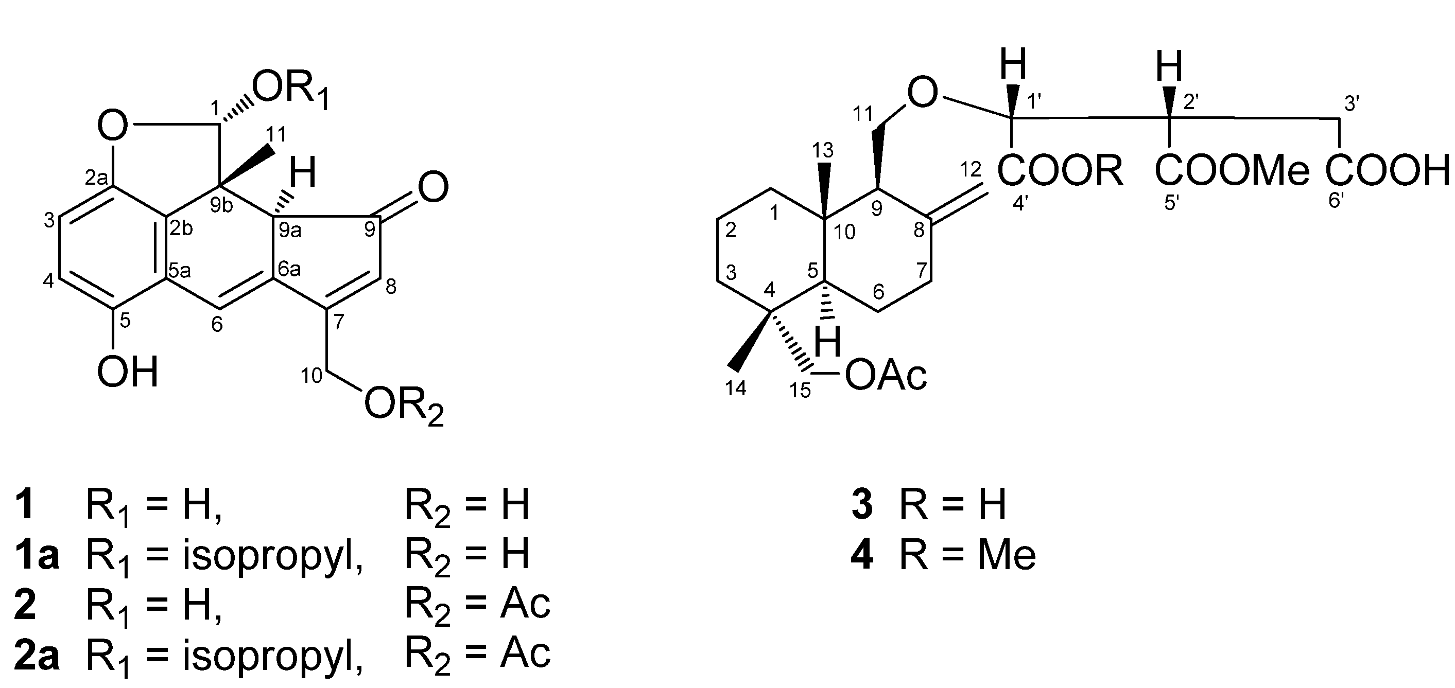
| Position | 1a | 2a | ||
|---|---|---|---|---|
| δC | δH (mult, J in Hz) | δC | δH (mult, J in Hz) | |
| 1 | 113.6 | 5.95 (s) | 113.4 | 5.93 (s) |
| 2a | 148.8 | 148.7 | ||
| 2b | 133.9 | 133.8 | ||
| 3 | 111.4 | 6.90 (d, J = 8.5) | 111.7 | 6.91 (d, J = 8.5) |
| 4 | 115.9 | 6.97 (d, J = 8.5) | 115.9 | 6.96 (d, J = 8.5) |
| 5 | 150.2 | 150.2 | ||
| 5a | 120.0 | 119.8 | ||
| 6 | 114.6 | 7.43 (d, J = 1.9) | 115.5 | 7.38 (d, J = 1.9) |
| 6a | 138.7 | 137.9 | ||
| 7 | 173.1 | 165.3 | ||
| 8 | 131.9 | 6.91 (t, J =1.4) | 133.2 | 6.54 (t, J =1.4) |
| 9 | 202.7 | 202.1 | ||
| 9a | 49.4 | 4.10 (br. d, J = 1.9) | 49.3 | 4.05 (br. s) |
| 9b | 46.2 | 46.2 | ||
| 10 | 58.9 | 4.94 (dt, J = 17.3, 1.4) | 60.4 | 5.27 (dt, J = 16.2, 1.4) |
| 5.07 (dt, J = 17.3, 1.4) | 5.38 (dt, J = 16.2, 1.4) | |||
| 11 | 16.5 | 1.13 (s) | 16.5 | 1.10 (s) |
| 1' | 72.1 | 4.26 (sept, J = 6.3) | 72.1 | 4.25 (sept, J = 6.3) |
| 2' | 22.2 | 1.24 (d, J = 6.3) | 22.2 | 1.24 (d, J = 6.3) |
| 3' | 23.5 | 1.31 (d, J = 6.3) | 23.5 | 1.31 (d, J = 6.3) |
| Ac | 20.4 | 2.08 (s) | ||
| 170.2 | ||||
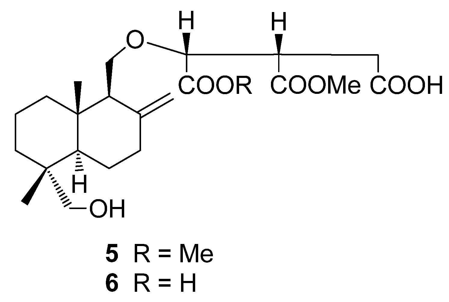
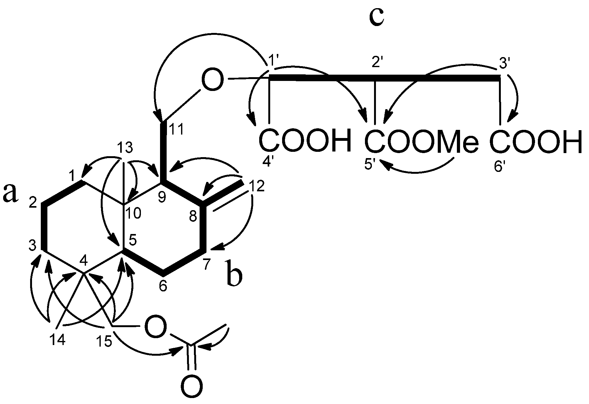
| Position | 3 | 4 | ||
|---|---|---|---|---|
| δC | δH (mult, J in Hz) | δC | δH (mult, J in Hz) | |
| 1 | 38.7 | 1.20 (m), 1.77 (m) | 38.6 | 1.15 (m), 1.72 (m) |
| 2 | 18.7 | 1.45 (m), 1.45 (m) | 18.6 | 1.45 (m), 1.45 (m) |
| 3 | 36.0 | 1.36 (m), 1.36 (m) | 35.9 | 1.34 (m), 1.34 (m) |
| 4 | 36.9 | 36.9 | ||
| 5 | 49.3 | 1.40 (dd, J = 12.6, 2.2) | 49.3 | 1.38 (m) |
| 6 | 24.0 | 1.27 (m), 1.51 (m) | 23.9 | 1.25 (m), 1.51 (m) |
| 7 | 37.6 | 2.01 (m), 2.32 (m) | 37.5 | 1.99 (m), 2.31 (m) |
| 8 | 147.1 | 147.0 | ||
| 9 | 56.2 | 2.21 (m) | 56.0 | 2.15 (m) |
| 10 | 38.9 | 38.8 | ||
| 11 | 68.2 | 3.81 (dd, J = 9.6, 3.3) | 68.6 | 3.70 (dd, J = 9.6, 3.3) |
| 4.33 (dd, J = 9.6, 8.2) | 4.14 (dd, J = 9.6, 8.0) | |||
| 12 | 108.9 | 5.02 (br. s) | 108.6 | 4.98 (br. s) |
| 5.31 (br. s) | 5.15 (br. s) | |||
| 13 | 15.9 | 0.79 (s) | 15.9 | 0.76 (s) |
| 14 | 17.5 | 0.75 (s) | 17.5 | 0.75 (s) |
| 15 | 73.0 | 3.74 (d, J = 10.7) | 73.0 | 3.73 (d, J = 11.0) |
| 3.94 (d, J = 10.7) | 3.93 (d, J = 11.0) | |||
| 1' | 80.1 | 4.70 (d, J = 4.7) | 79.5 | 4.57 (d, J = 4.4) |
| 2' | 45.7 | 4.14 (m) | 45.6 | 3.98 (m) |
| 3' | 33.4 | 3.26 (dd, J = 17.0, 4.1) | 33.6 | 3.09 (m) |
| 3.49 (dd, J = 17.0, 10.7) | 3.35 (dd, J = 17.3, 9.3) | |||
| 4' | 173.7 | 171.5 | ||
| 5' | 172.7 | 172.1 | ||
| 6' | 174.7 | 175.0 | ||
| 4'-OMe | 51.9 | 3.65 (s) | ||
| 5'-OMe | 51.9 | 3.73 (s) | 52.0 | 3.76 (s) |
| Ac | 20.8 | 2.07 (s) | 20.8 | 2.08 (s) |
| 170.1 | 170.1 | |||

| Compound | % Inhibition of at 10 μg/mL | IC50b,c | ||
|---|---|---|---|---|
| COX-1 | COX-2 | COX-1 | COX-2 | |
| Extract a | 96.4 | 59.0 | ||
| 1a | 90.5 | 7.5 | 47.7 | ND d |
| 3 | 40.2 | 1.7 | ND | ND |
| 4 | 66.5 | 10.3 | 78.8 | ND |
| 6 | 35.8 | 19.0 | ND | ND |
| aspirin | – | – | 79.0 | 150 |
| Compound | % Inhibition of at 10 μg/mL | IC50b,c |
|---|---|---|
| Extract a | 69.5 | – |
| 1a | 84.7 | 15.1 |
| 3 | 14.9 | ND d |
| 4 | 50.1 | 203.5 |
| 6 | 28.9 | ND |
| NDGA a | – | 0.4 |
3. Experimental
3.1. General
3.2. Materials
3.3. Extraction and Isolation
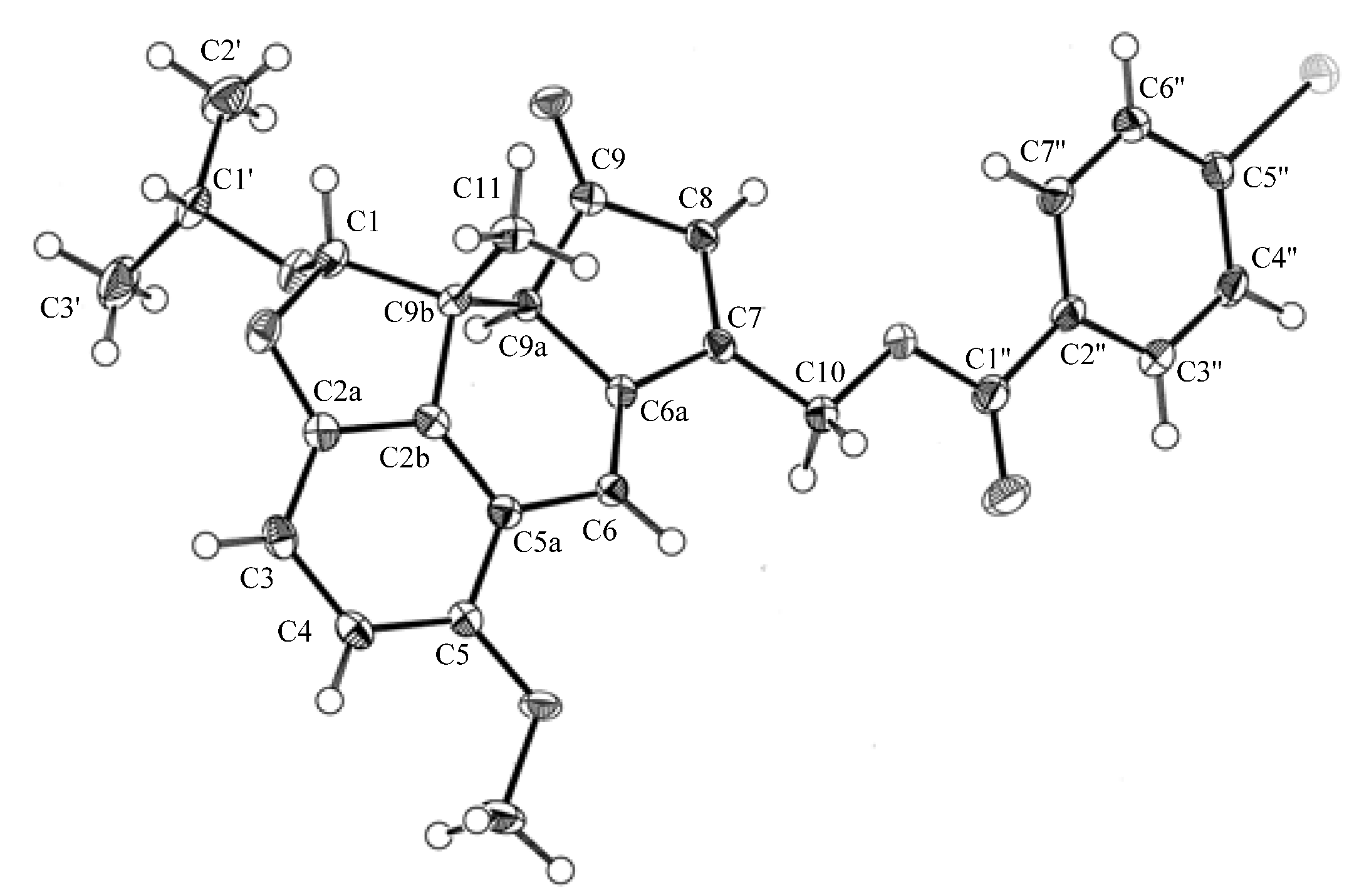
3.5. X-ray Crystallographic Data for 1b
3.6. Acetylations of 6 Giving 3 and of 5 Giving 4
3.7. COX-1 and COX-2-Catalyzed Prostaglandin Biosynthesis Assay in Vitro
3.8. Measurement of RBL-1 5-Lipoxygenase Activity
4. Conclusions
Supplementary Materials
Acknowledgments
References and Notes
- Yoshikawa, K.; Matsumoto, K.; Arihara, S. New lanostanoid glycosidesfrom the fruit body of Laetiporus versisporus. J. Nat. Prod. 1999, 69, 543–545. [Google Scholar] [CrossRef]
- Yoshikawa, K.; Bando, S.; Arihara, S.; Matsumura, E.; Katayama, S. A benzofuran glycoside and an acetylenic acid from the fungus Laetiporus sulphureus var. miniatus. Chem. Pharm. Bull. 2001, 49, 327–329. [Google Scholar]
- Yoshikawa, K.; Nishimura, N.; Bando, S.; Arihara, S.; Matsumura, E.; Katayama, S. New lanostanoids, elfvingic acids A-H, from the fruit body of Elfvingia applanat. J. Nat. Prod. 2002, 65, 548–552. [Google Scholar]
- Yoshikawa, K.; Inoue, M.; Matsumoto, Y.; Sakakibara, C.; Miyataka, H.; Matsumoto, H.; Arihara, S. Lanostane triterpenoids and triterpene glycosides from the fruit body of Fomitopsis pinicola and their inhibitory activity against COX-1 and COX-2. J. Nat. Prod. 2005, 68, 69–73. [Google Scholar] [CrossRef]
- Yoshikawa, K.; Koso, K.; Takahashi, J.; Matsuda, A.; Okazoe, M.; Umeyama, A.; Arihara, S. Cytotoxic constituents of the fruit body of Daedalea dickisii. J. Nat. Prod. 2005, 68, 911–914. [Google Scholar] [CrossRef]
- Imazeki, R.; Hongo, T. Colored Illustrations of Mushrooms of Japan, 2nd ed; Hoikusha Press: Osaka, Japan, 1989. [Google Scholar]
- Shirokawa, S. Polypores Mushroom Illustrated Book, 1st ed; Kanagawa, K., Ed.; Chikyusha Press: Tokyo, Japan, 1996; p. 124. [Google Scholar]
- Tanaka, N.; Kitamura, A.; Mizushina, Y.; Sugawara, F.; Sakaguchi, K. Fomitellic acids, triterpenoid inhibitors of eukaryotic DNA polymerases from a basidiomycete, Fomitella fraxinea. J. Nat. Prod. 1998, 61, 193–197. [Google Scholar] [CrossRef]
- Cho, S.-M.; Yun, B.-S.; Yoo, I.-D.; Koshino, H. Structure of fomitellan A, a mannofucogalactan from the fruiting bodies of Fomitella fraxinea. Bioorg. Med. Chem. Lett. 2011, 21, 204–206. [Google Scholar] [CrossRef]
- Hashimoto, T.; Tori, M.; Mizuno, Y.; Asakawa, Y. Cryptoporic acids A and B, novel bitter drimane sesquiterpenoid ethers of isocitric acid, from the fungus Cryptoporus volvatus. Tetrahedron Lett. 1987, 28, 6303–6304. [Google Scholar] [CrossRef]
- Asakawa, Y.; Hashimoto, T.; Mizuno, Y.; Tori, M.; Fukazawa, Y. Cryptoporic acids A-G, drimane-type sesquiterpenoid ethers of isocitric acid from the fungus Cryptoporus volvatus. Phytochemistry 1992, 31, 579–592. [Google Scholar] [CrossRef]
- Wu, W.; Zhao, F.; Bao, L.; Lu, J.-C.; Liu, H.-W. Two new cryptoporic acid derivatives from the fruiting bodies of Cryptoporus sinensis. Helv. Chim. Acta, 2011; 94, 2020–2026. [Google Scholar]
- Crystallographic data (excluding structural factors) for the structure of 1b have been deposited in the Cambridge Crystallographic Data Centre as supplementary publication number CCDC 924637. Copies of the data can be obtained, free of charge, on application to CCDC, 12 Union Road, Cambridge CB2 1EZ UK (Fax: +44(0)-1223–336033; E-mail: [email protected].
- Futaki, N.; Takahashi, S.; Yokoyama, M.; Arai, I.; Higuchi, S.; Omoto, S. NS-398, a new anti-inflammatory agent, Selectively inhibits prostaglandin G/H synthase/cyclooxygenase (COX-2) activity in vitro. Prostaglandins 1994, 47, 55–59. [Google Scholar]
- Blackham, A.; Griffiths, R.J.; Hallman, C.; Mann, J.; Mitchell, P.D.; Norris, A.A.; Simpson, W.T. FPL 62064, a topically active 5-lipoxygenase/cyclooxygenase inhibitor. Agents Actions 1990, 30, 432–442. [Google Scholar]
- Sample Availability: Samples of the compounds 1a, 2a, 3–6 are available from the authors.
© 2013 by the authors; licensee MDPI, Basel, Switzerland. This article is an open-access article distributed under the terms and conditions of the Creative Commons Attribution license (http://creativecommons.org/licenses/by/3.0/).
Share and Cite
Yoshikawa, K.; Koso, K.; Shimomura, M.; Tanaka, M.; Yamamoto, H.; Imagawa, H.; Arihara, S.; Hashimoto, T. Yellow Pigments, Fomitellanols A and B, and Drimane Sesquiterpenoids, Cryptoporic Acids P and Q, from Fomitella fraxinea and Their Inhibitory Activity against COX and 5-LO. Molecules 2013, 18, 4181-4191. https://doi.org/10.3390/molecules18044181
Yoshikawa K, Koso K, Shimomura M, Tanaka M, Yamamoto H, Imagawa H, Arihara S, Hashimoto T. Yellow Pigments, Fomitellanols A and B, and Drimane Sesquiterpenoids, Cryptoporic Acids P and Q, from Fomitella fraxinea and Their Inhibitory Activity against COX and 5-LO. Molecules. 2013; 18(4):4181-4191. https://doi.org/10.3390/molecules18044181
Chicago/Turabian StyleYoshikawa, Kazuko, Kazuaki Koso, Masumi Shimomura, Masami Tanaka, Hirofumi Yamamoto, Hiroshi Imagawa, Shigenobu Arihara, and Toshihiro Hashimoto. 2013. "Yellow Pigments, Fomitellanols A and B, and Drimane Sesquiterpenoids, Cryptoporic Acids P and Q, from Fomitella fraxinea and Their Inhibitory Activity against COX and 5-LO" Molecules 18, no. 4: 4181-4191. https://doi.org/10.3390/molecules18044181