Communication

A Series of Asymmetrical Phthalocyanines: Synthesis and Near Infrared Properties

1
State Key Laboratory on Integrated Optoelectronics, College of Electronic Science and Engineering, Jilin University, Changchun 130012, China
2
Department of Chemistry, Northeast Normal University, Changchun 130024, China
*
Authors to whom correspondence should be addressed.
Molecules 2013, 18(4), 4628-4639; https://doi.org/10.3390/molecules18044628
Submission received: 30 January 2013 / Revised: 7 April 2013 / Accepted: 8 April 2013 / Published: 19 April 2013

Abstract

:
We report here the preparation of asymmetrical phthalocyanine dimers 1a3a, which are endowed with novel charge transfer bands at 1,151–1,154 nm and strong NIR luminescences at 840–860 nm and 1,600–1,650 nm. Through H-bonding interaction, 1a3a are inclined to self-assemble into hexrod nanotubes at the interface of CHCl3 and CH3OH. Our results provide further insights into the interaction in molecular dimers, and suggest that 1a3a have potential application in magnets and supramolecular architectures.

Graphical Abstract

1. Introduction

Considerable efforts have been made to develop various types of supramolecular architectures formed by self-organization between functional chromophores, because these supramolecular architectures have better photophysical and electronic properties as a result of excitonic interactions [1,2,3,4,5,6,7,8]. Phthalocyanines (Pcs), as a type of functional chromophores, have potential application in supermolecular assemblies, charge transfer properties, and magnets [9], such as NIR-absorbing Pc supermolecules with some unique electronic spectrum and electrochemical behaviors [10,11,12,13,14,15,16]. Among them, double-decker Pc dimers have been of specially interest in numerous contexts because they are inclined to generate intermolecular π-π interactions [17,18]. Especially those with new CT bands in the range of 1,100–1,280 nm (in oxidized state) have potential applications in emerging photoelectric technologies [19,20]. Some self-organization structures of Pcs were reported by other groups [21].
Of particular interest in a number of contexts are Pcs which self-assemble and self-organize into 1D nanostructures. Recently, Pcs that self-organize by H-bonding, electrostatic, labile metal-ligand bonds and flat π-π interaction into highly ordered supramolecular structures with controlled dimensions and size, have attracted great interest, such as Nolte and co-workers’ reports of a Pc with four crown ether moieties, which are self-organized into helical, micrometer-long fibers in chloroform solution [22]. Ultralong nanowires with new crystal structured are formed by CuPc [23]. Although scientists studying Pcs have reported some 1D nanostructures [24,25], 1D nano-tubular structures produced by self-assembly methods have seldom been reported.
Our previous research work on the design and synthesis of metallo-Pcs and Pc 1D tubular structures [26,27,28], helped us find and obtain A3B asymmetrical Ni (II) Pcs 1a3a, which are connected with three phenoxy groups and one benzyloxy group. It is found that 1a3a are self-assembled into hexo-nanotubes by H-bonding interactions between Pc molecules. The mechanism of formation of the hexotubular structures is proposed.

2. Results and Discussion
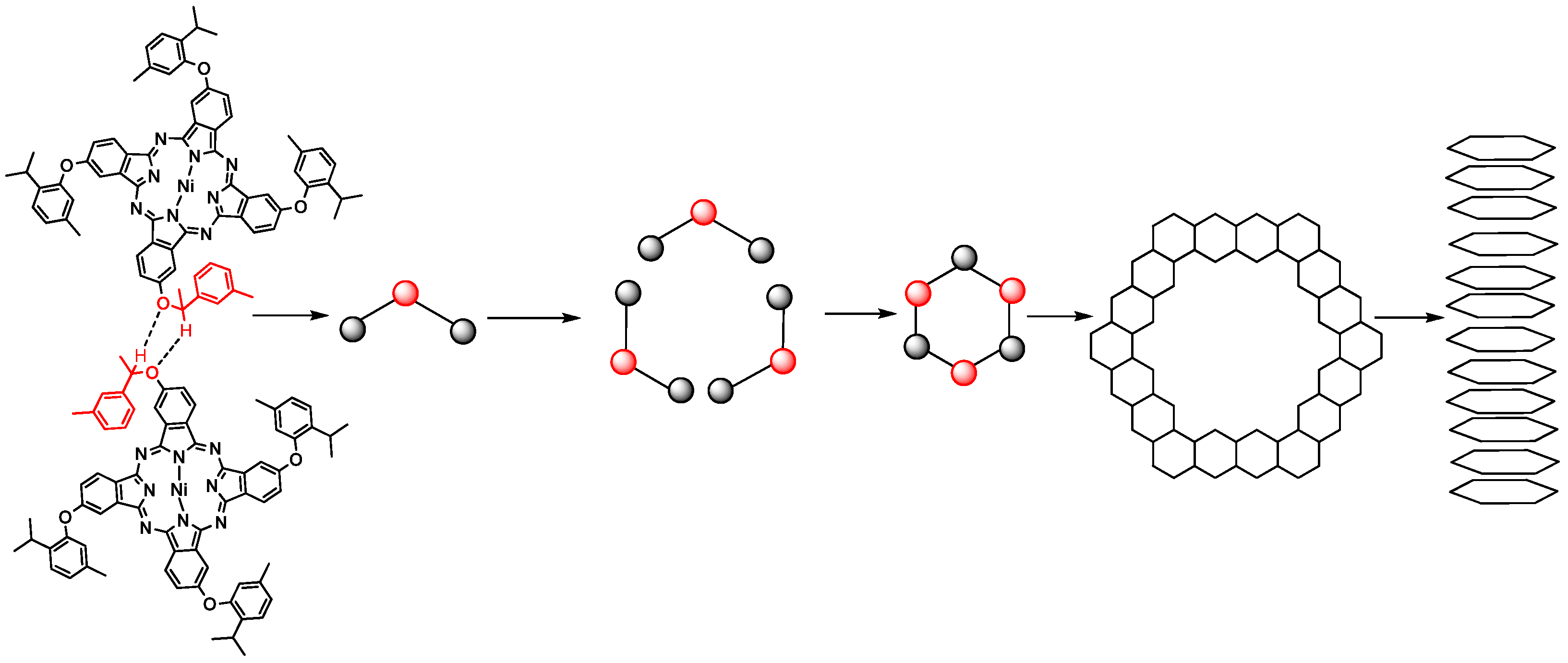
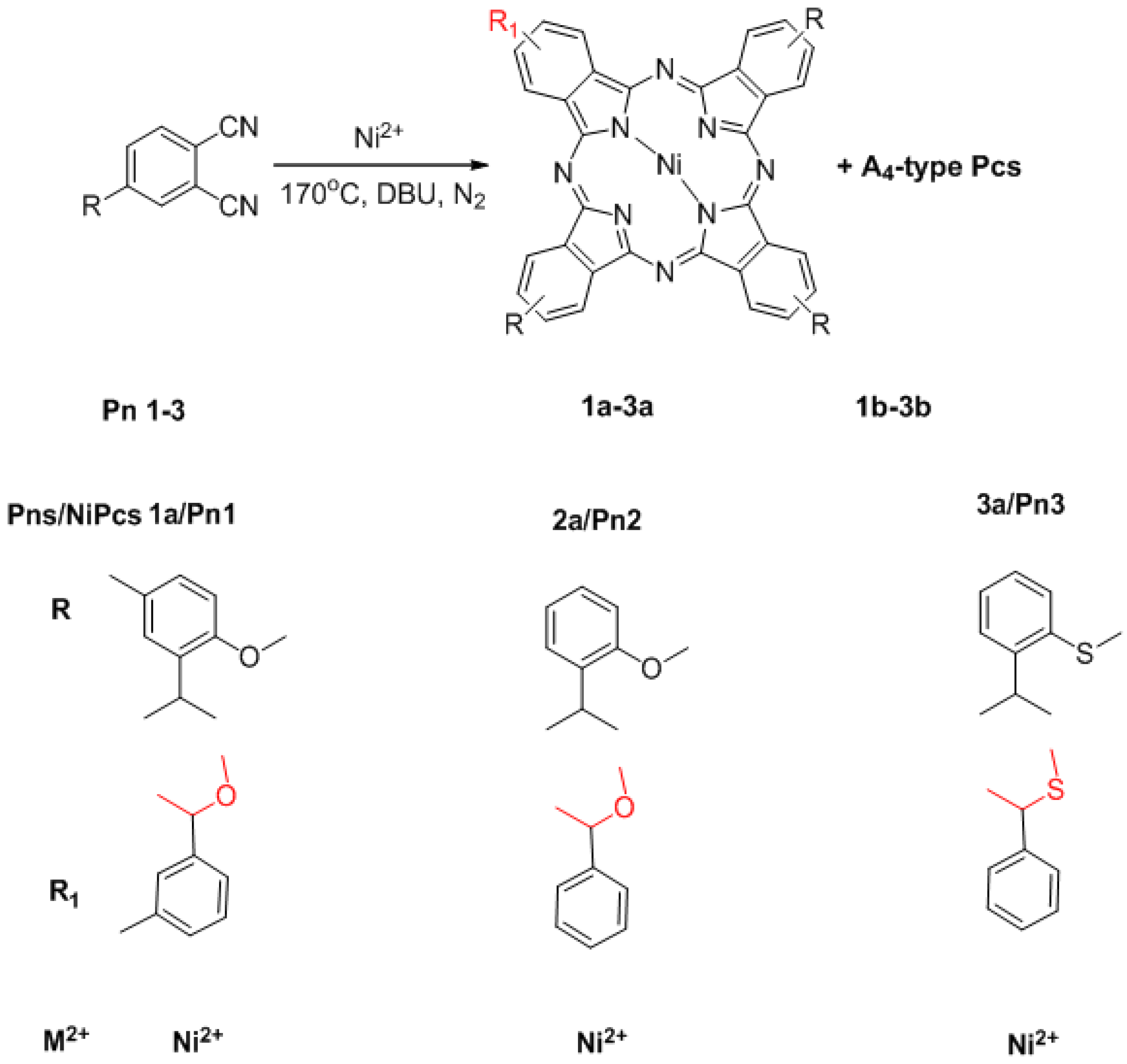
2.1. Synthesis

The synthesis of 1a3a is summarized in Scheme 1. Only the cyclic tetramerization of single Pn1-3 (Scheme S1 and Figure S1) in 1-octanol in the presence of 1,8-diaza-bicyclo[5.4.0] undec-7-ene (DBU) and at higher temperature of 170 °C (the traditional method use 140 °C), yields 1a3a and 1b3b (A3B-type Pcs and A4-type Pcs).
Scheme 1. Synthesis of 1a3a.
Molecules 18 04628 g010
Reagents and Conditions: 1-octanol (5 mL), DBU (2 mL), 170 °C, NiCl2·6H2O, 8 h.
The yield of 1a is 15%, and the 2a and 3a is 13% and 8% respectively. This method overcomes many of the separation difficulties typically encountered in the traditional syntheses of asymmetrical Pcs [29] and only uses a single substituted phthalonitrile to synthesize asymmetrical Pcs (the traditional methods use two substituted phthalonitriles). A possible mechanism for the synthesis of 1a3a is provided in the Supporting Information (Scheme S2). Compounds 1a3a were characterized by UV-Vis-NIR (Figure S2), 1H-13C-NMR (Figure S3), high resolution MS and MALDA-TOF MS (Figure S4), IR (Figure S5). Characterization of 1a, as an example of 1a3a, is discussed below. The data of 2a3a are provided in the Supporting Information.

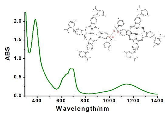
2.2. High Resolution MS
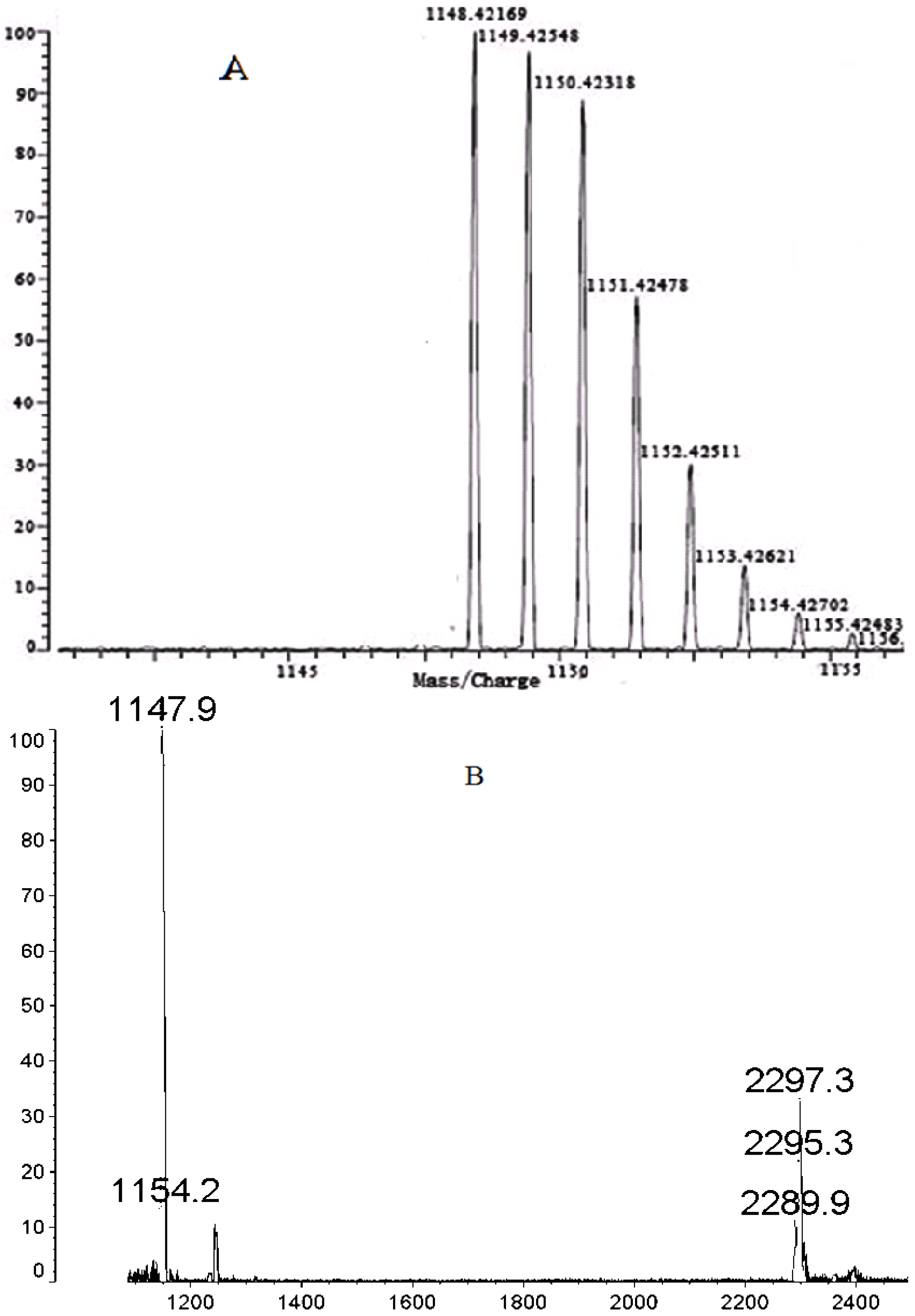
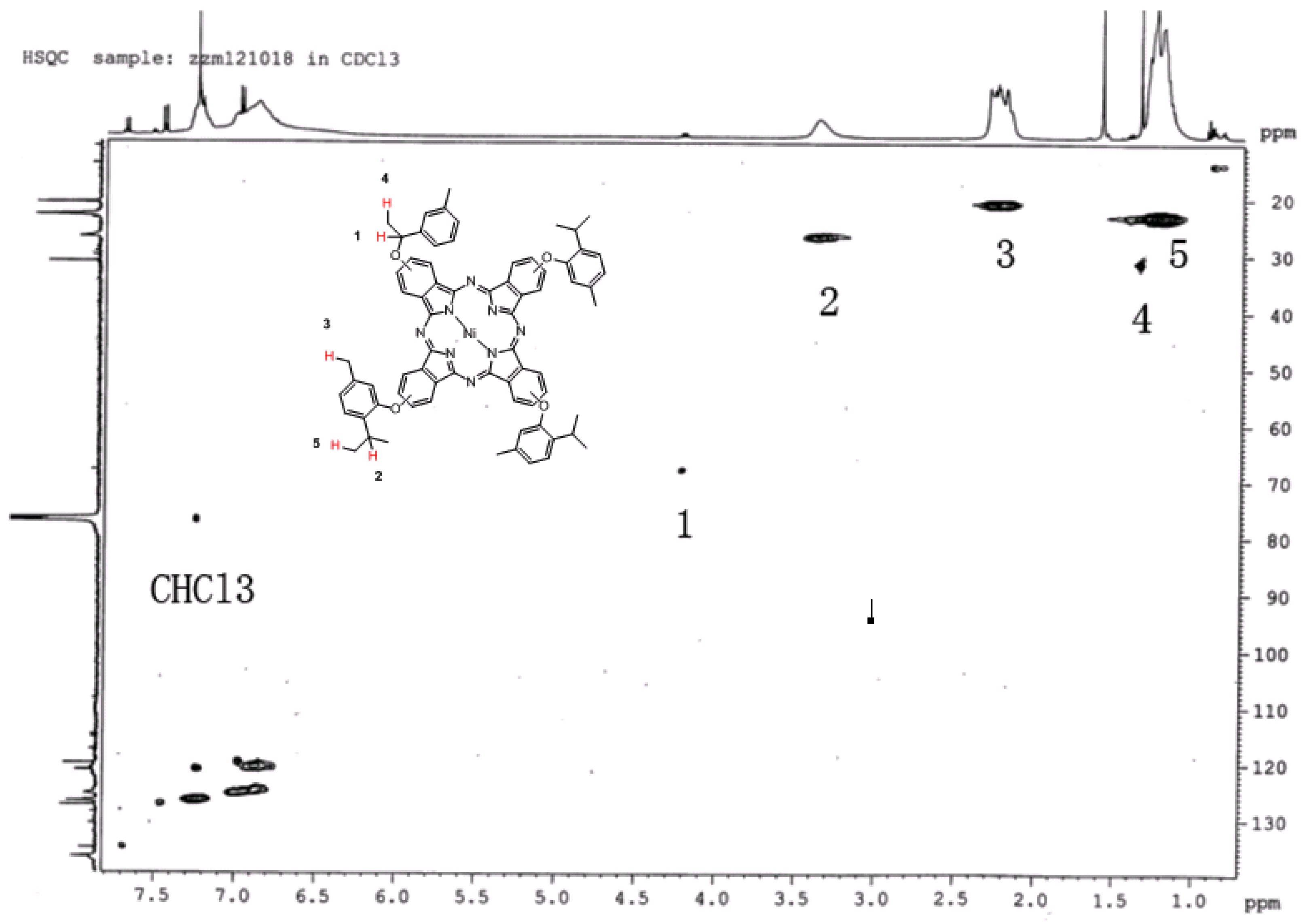
Figure 1A shows the clear m/z peaks ([M+H+] or M+) of 1a obtained in high resolution MS. It is found that the molecular weight of 1a is 1,148.42169, which is in line with the real molecular weight. The m/z peak at 2,297.3 provides direct evidence that 1a tends to form a 1a dimer (Figure 1B).
Figure 1. (A) Hi-Res MS spectrum of 1a; (B) MALDI-TOF MS spectrum of 1a dimer.
Molecules 18 04628 g001

2.3. 1H-13C-NMR

Because of the magnetism of 1a, the peak area is influenced. The structure of 1a is characterized by 1H-13C-NMR (in CDCl3) in Figure 2. We can see that label 1 is assigned to the C and H atoms in the benzyloxy group, label 2 is assigned to the C and H atoms in the isopropyl group, label 3 is the C and H atoms of the methyl group linked to the benzene ring, label 4 is the C and H atoms of the hypo-methyl group in the benzyloxy structure and the label 5 is the C and H atom of methyl group in isopropyl. The structure of 1a was confirmed by 1H-13C-NMR and high resolution MS.
Figure 2. The 1H-13C-NMR of 1a.
Molecules 18 04628 g002

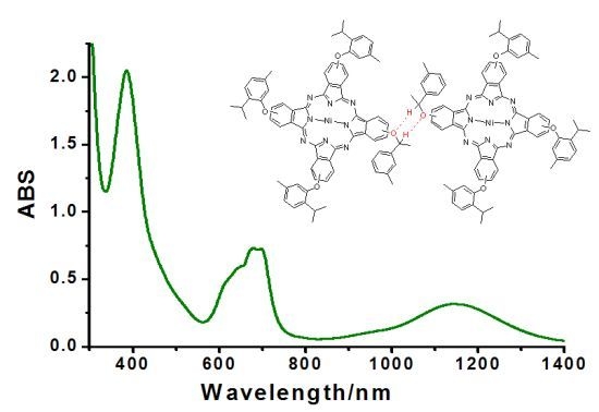
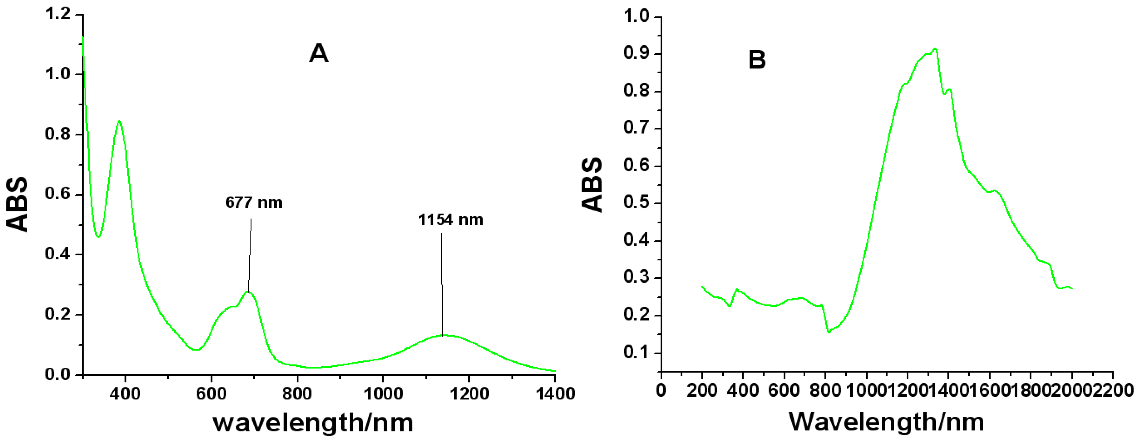
2.4. UV-Vis-NIR Spectroscopy

The same conclusion concerning the aggregation properties of the molecule were also obtained using UV-Vis-NIR spectroscopy (Figure 3). The B, Q and shoulder bands of 1a in CHCl3 (1 × 10⁻⁴ mol/L) are observed around 385–390, 610 and 677 nm, respectively. It is noteworthy that the Q band of 1a broadens considerably relative to the symmetrical one (Figure S6), but its molar extinction coefficient decreases dramatically from log ε 5.35 to log ε 2.89 (Figure 3A), which are just the characteristic absorptions of Pc dimers and oligomers [30]. Additionally, a broad intensity-strong band at around 1,154 nm is attributed to 1a dimer, which is assignable to a CT consequence of electronic coupling due to strong H-bonding interaction in-between the 1a dimer units (Figure 4). Compared with 2a, the Q band and the NIR band of 3a is red shifted from 1,148 nm to 1,230 nm, because the donating electron ability of phenylthiol group is better than that of a phenoxy group. The solid state UV-Vis-NIR spectrum of 1a dimer shows that the molecule strongly NIR-absorbs light from 900 to 2,000 nm (Figure 3B), which hints that 1a may exist in a face-to-tail J-type aggregates in the solid state and 1a film might be improved as an available NIR material in optical communication field in the future.
Figure 3. (A) UV-Vis/NIR spectrum of 1a in CHCl3 (1.0 × 10⁻⁴ mol/dm3) and (B) Solid state UV-Vis/NIR Spectrum of 1a.
Molecules 18 04628 g003
Figure 4. 1a dimer in H-bonding interaction.
Molecules 18 04628 g004

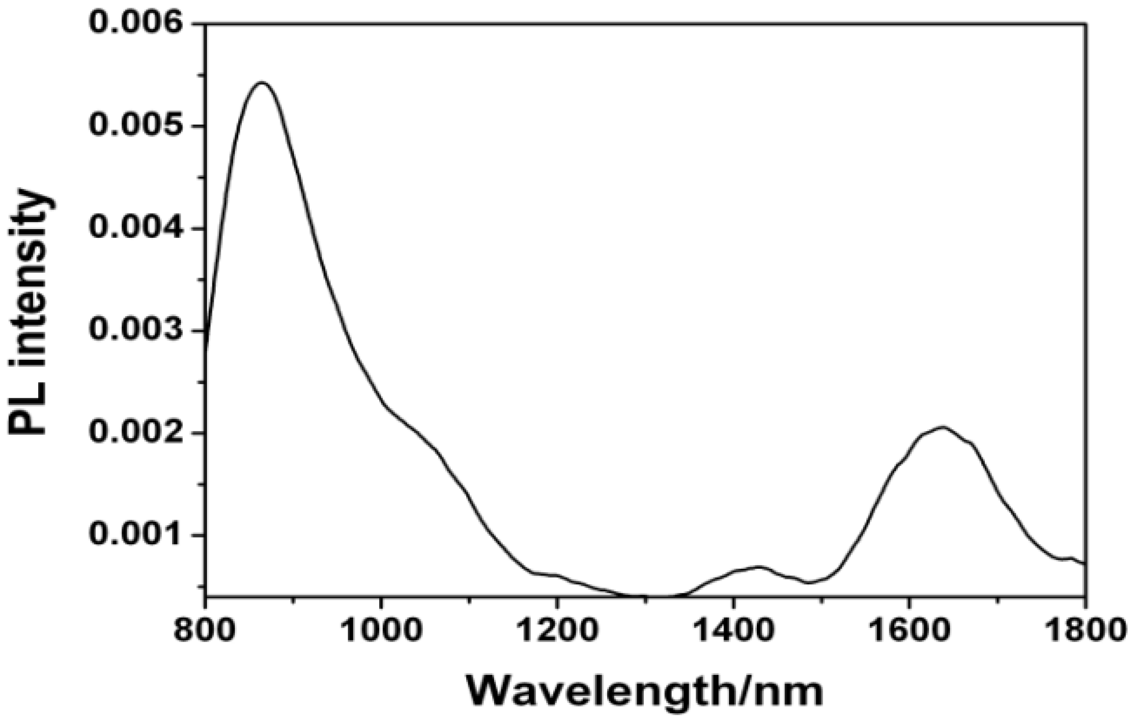
2.5. NIR Luminescence

To further confirm the CT capacity of dimer, we tested the NIR luminescence properties of 1a by a 325 nm He-Cd laser as the excitation source and Ge detector (Figure 5). When 1a is excited in KBr, it showed strong NIR luminescence at 840–860 nm and 1,600–1,650 nm, respectively. The NIR luminescence at 840–860 nm results from the fluorescent radiation transition (S1→S0) in the monomer, while the luminescence at 1,600–1,650 nm comes from the fluorescent radiation monomer-to-monomer transition. The results provide direct evidence of CT action in 1a dimer.
Figure 5. Emission spectrum of 1a at room temperature.
Molecules 18 04628 g005

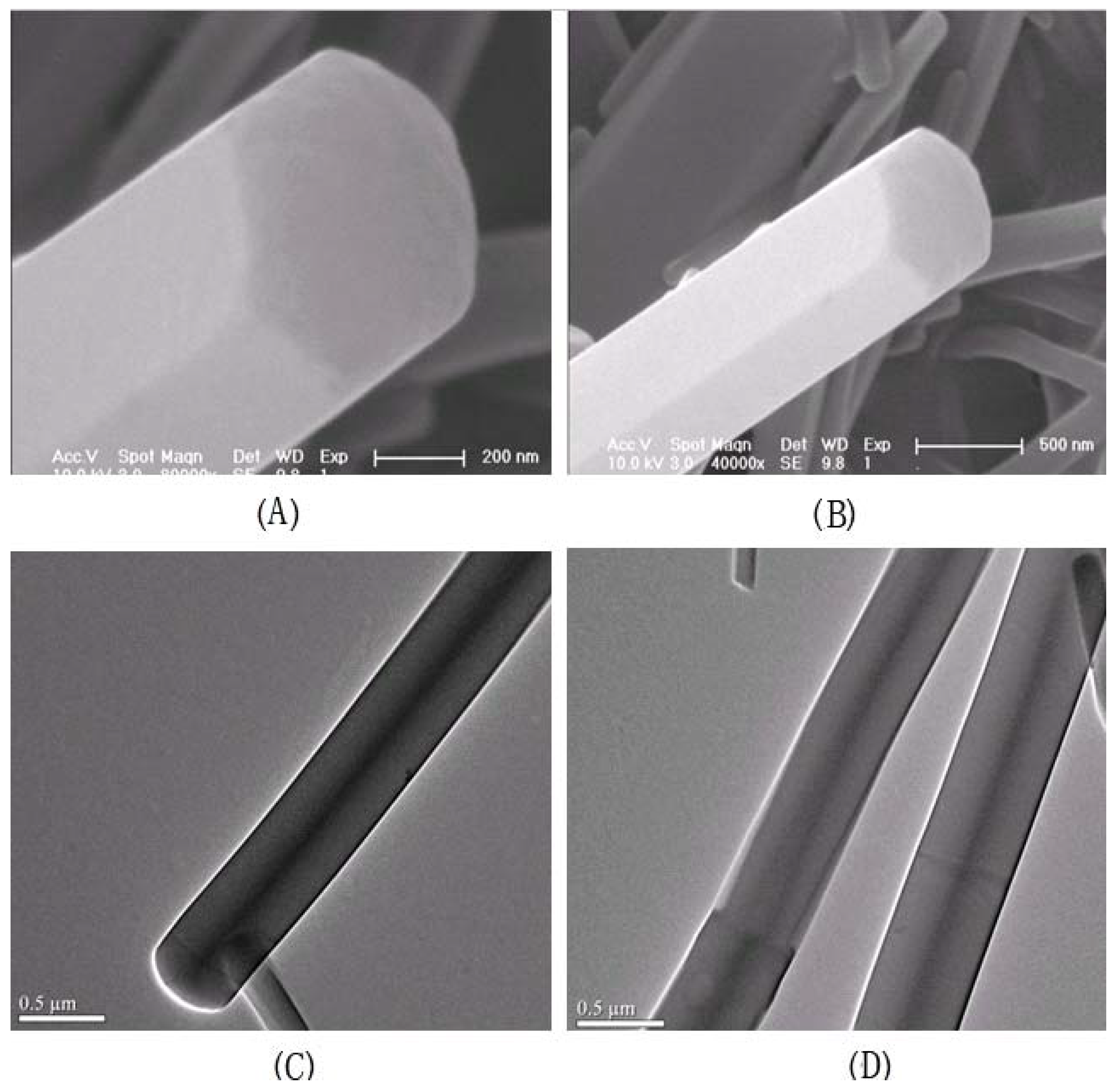
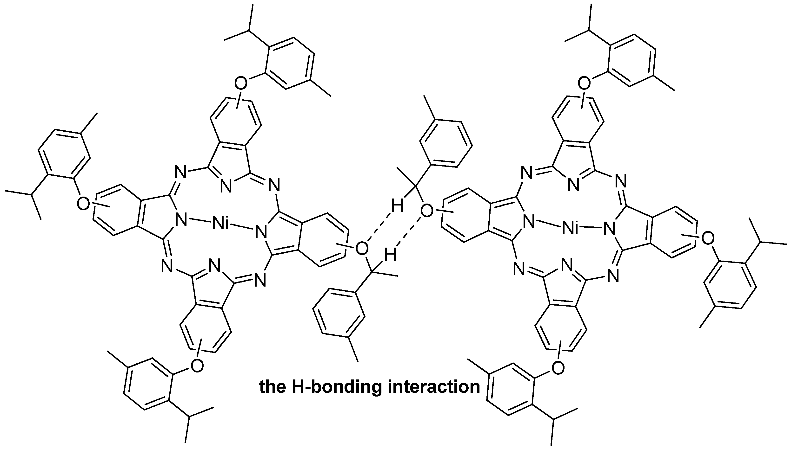
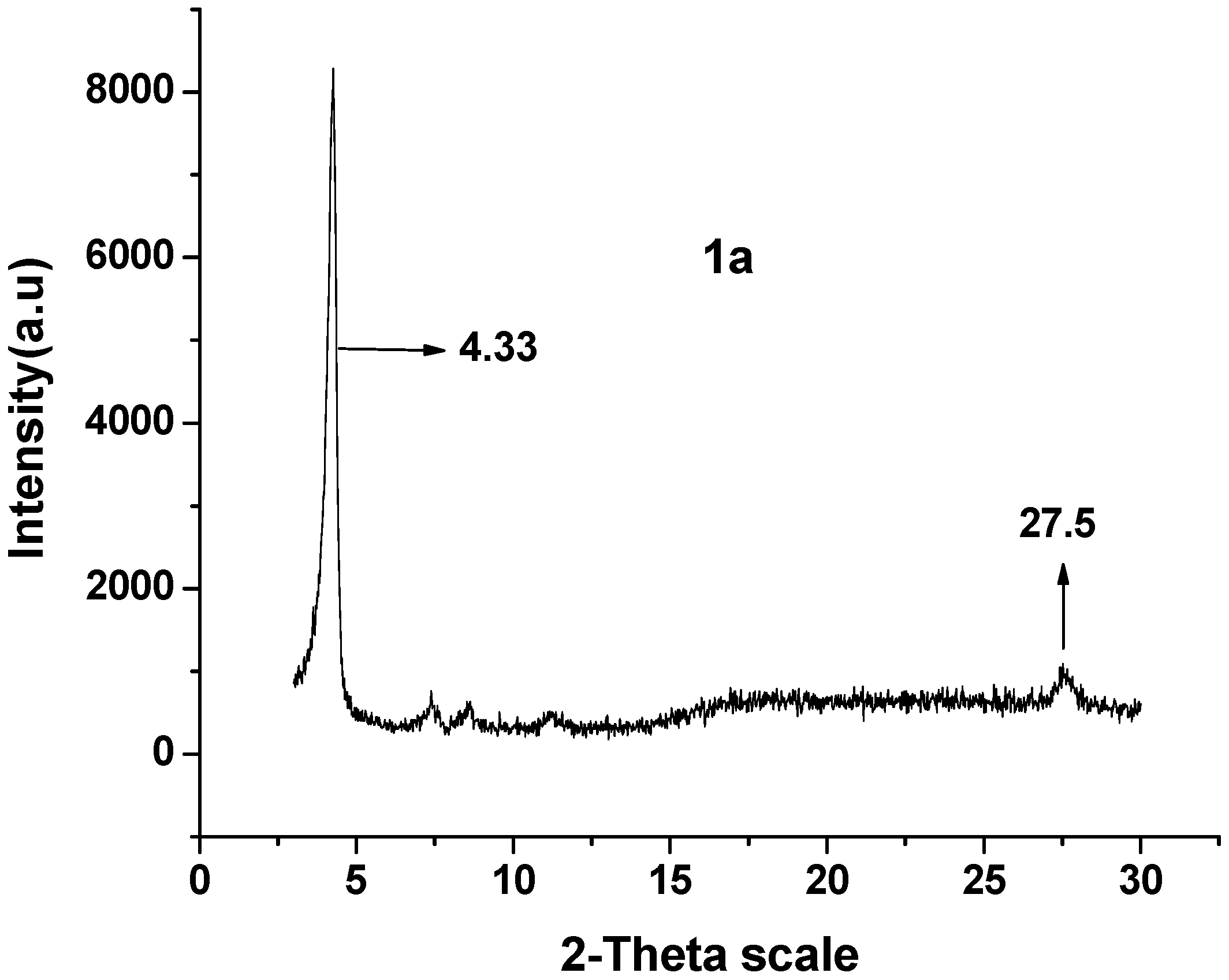
2.6. 1-D Molecular Morphology and XRD

Figure 6 shows 1-D molecular morphology of 1a, which was tested by scanning electron microscope (SEM, Figure 6A,B) and high resolution transmission electron microscope (HR-TEM, Figure 6C,D). The SEM images show 1a is self-assembled into hexrod nanotubes at the CHCl3 and CH3OH interface, and the nanotubes differ in length and thickness. The HR-TEM images show the hexrod-nanotubes are hollow, and the black lines in the HR-TEM images is the edges of nanotubes. These nanotubes indicate strong intermolecular aggregation in-between 1a units.
Figure 6. (A,B) SEM images of 1a nanotubes and (C,D) HR-TEM micrographs of 1a nanotubes.
Molecules 18 04628 g006
The XRD spectrum of 1a (Figure 7) shows two clear diffraction peaks at 4.33° and 27.5°, whose plane separations are approximately 21.45 Å (d1) and 3.098 Å (d2), respectively. The two peaks of the nanotubes shows that the 1a molecules have been stacked in highly long-range ordered lamellar structure [31], d1 and d2 are the interlayer spacing of the nanotubes [32].
Figure 7. XRD data of 1a.
Molecules 18 04628 g007
Connecting the molecular morphology with the XRD data of 1a, the possible formation of 1a nanotubes can be described as shown in Figure 8: (1) the H-bonding interaction in-between 1a is self-assembled into 1a dimer. The angle of 1a dimer is 120°, which is similar to the H-bonding interaction of H2O; (2) according to “proximity compatibility principle”, the 1a dimer is self-assembled into a hexacyclic compound; (3) through π-π interaction in J-type aggregates [33], the hexacyclic compound forms nanotubes differing in length and thickness.
Figure 8. The possible formation of 1a hexrod nanotubes.
Molecules 18 04628 g008

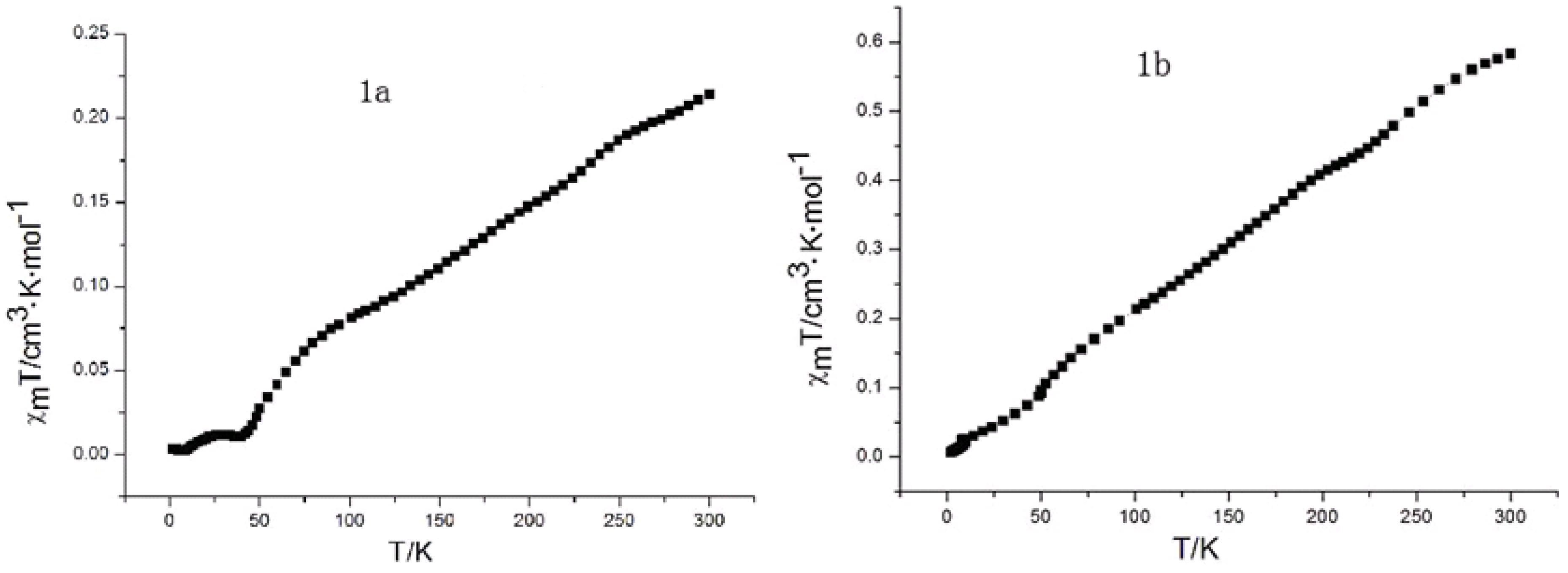
2.7. Magnetic Susceptibility

Magnetic susceptibility data of 1a and 1b were recorded using a Quantum Design SQUID MPMS XL-5 magnetometer. Variable temperature susceptibility measurements were carried out in the temperature range of 2–300 K at a magnetic field of 1,000 Oe on polycrystalline samples. Since Ni(II)Pc is diamagnetic due to the electronic configuration (b2g)2(eg)4(a1g)2, the paramagnetism of 1a and 1b should arise from the spin polarization of electrons on the aromatic macrocycle [34,35]. Both 1a and 1b with Ni(II) ion appear as planar molecules with no axial ligand, so their crystals are formed from various stacking columnar structures with different tilting angles and interplanar distances. As seen in Figure 9, upon continuously lowering temperature from 300 K to 2 K, the χmT values of 1a and 1b smoothly decreases to a zero value; this is because the antiferromagnetic exchange interaction occurs between the radical aromatic macrocycle complexes in the solid state.
Figure 9. χmT curves of magnetic measurements for 1a and 1b.
Molecules 18 04628 g009

3. Experimental

3.1. Experimental Materials and Equipment

Chloroform was distilled from CaH2 under nitrogen. DMSO was distilled from molecular sieves. The other reagents were purchased from commercial sources and used without further treatment. 1H-NMR and 13C-NMR spectra were recorded on a Varian 500 MHz instrument at 25 °C. Mass spectra were recorded on a Hi-Res MALDI and a LDI-1700 MALDI-TOF MS spectrometer. IR spectra (KBr) were recorded on a Magna-560 FT-IR spectrophotometer in the 400~4,000 cm−1 range. UV/VIS/NIR spectra were recorded on a Cary 500UV-VIS-NIR spectrophotometer.

3.2. Synthesis of Substrates and Pc Derivatives

3.2.1. General Procedure for the Synthesis of Substrates Pn1Pn3

Lithium hydroxide hydrate (4.20 g, 0.10 mol) was interfused at room temperature into a stirred mixture of the appropriate phenol/thiophenol (2-isopropyl-5-methylphenol, 6.08 g, 0.04 mol for Pn1, 2-isopropylphenol, 5.44 g, 0.04 mol for Pn-2 or 2-isopropylphenylthiol, 6.08 g, 0.04 mol for Pn-3) and 4-nitrophthalonitrile (6.92 g, 0.04 mol) in dimethylsulfoxide (80 mL) over a 2 h period. The mixture was stirred unceasingly and the reaction was monitored by thin-layer chromatography. After 24 h, the reactant was poured into NaCl solution (400 mL, 10%) and stirred till precipitate appeared. The product was collected by vacuum filtration and purified by column chromatography (silica gel/petroleum ether–ethyl ether 1:1) to afford Pn-1-Pn-3.
4-(2-Isopropyl-5-methylphenoxy)phthalonitrile (Pn1). Yellow solid (8.77 g, 0.032 mol, 79% yield). 1H-NMR (CDCl3): δ = 7.71 (d, J = 8.5 Hz, 1 H; Ar-H), 7.30 (d, J = 8.5 Hz, 1 H; Ar-H), 7.22 (s, 1 H; Ar-H), 7.19 (d, J = 8 Hz, 1 H; Ar-H), 7.11 (d, J = 8 Hz, 1 H; Ar-H), 6.75 (s, 1 H; Ar-H), 2.96 (m, 1 H; C-H), 2.33 (s, 3 H; ArCH3), 1.15 (d, J = 6.5 Hz, 6 H; CH(CH3)2). TOF-MS: m/z calcd: 299.1 [M+Na]+, 315.1 [M+K]+; found: 299.1, 314.9. IR (KBr): C-O-C 1242 cm−1, CN 2230 cm−1. Elemental analysis calcd (%) for C18H16N2O [M = 276.13 g·mol−1]: C 78.24, H 5.84, N 10.14; found: C 78.28%, H 5.91%, N 10.02%.
4-(2-Isopropylphenoxy)phthalonitrile (Pn2). Light yellow solid (8.38 g, 0.032 mol, 79% yield). 1H-NMR (CDCl3): δ =7.54 (q, J = 8.5 Hz, 1 H; Ar-H), 7.42 (t, J = 8.5 Hz, 1 H; Ar-H), 7.286 (t, J = 8.5 Hz, 2 H; Ar-H), 7.07 (t, J = 8 Hz, 1 H; Ar-H), 7.05 (t, J = 8 Hz, 2 H; Ar-H), 2.95 (m, J = 7 Hz, 1 H; C–H), 1.27 (d, 6H; J = 7 Hz, CH(CH3)2). TOF-MS: m/z calcd: 299.1 [M+Na]+, 315.1 [M+K]+; found: 299.1, 314.9. IR (KBr): C–O–C 1242 cm−1, –CN 2230 cm−1. Elemental analysis calcd (%) for C17H14N2O [M = 262.1 g·mol−1]: C 77.84, H 5.38, N 10.68; found: C 78.80%, H 5.42%, N 10.66%.
4-(2-Isopropylphenylthio)phthalonitrile (Pn3). Light yellow solid (1.01 g, 0.036 mol, 89% yield). 1H-NMR (CDCl3): δ = 7.54 (q, J = 8.5 Hz, 1 H; Ar-H), 7.42(t, J = 8.5 Hz, 1 H; Ar-H), 7.286 (t, J = 8.5 Hz, 2 H; Ar-H), 7.07 (t, J = 8 Hz, 1 H; Ar-H), 7.05 (t, J = 8 Hz, 2 H; Ar-H), 2.95 (m, J = 7 Hz, 1 H; C–H), 1.27 (d, J =7 Hz, 6 H; CH(CH3)2). TOF-MS: m/z calcd: 301.38 [M+Na]+, 315.1 [M+K]+; found: 299.1, 314.9. IR (KBr): C–S–C 1240 cm−1, –CN 2230 cm−1. Elemental analysis calcd (%) for C17H14N2O [M = 262.1 g·mol−1]: C 67.75, H 4.68, N 10.64; found: C 67.73%, H 4.68%, N 10.66%.

3.2.2. Synthesis of 1a and 1b

NiCl2·6H2O (95 mg, 0.4 mmol) and Pn1 (440 mg, 1.6 mmol) were added to 1-octanol (5 mL) and stirred for 0.5 h in succession. After DBU (2 mL) was added, the mixture was sequentially stirred for 21 h and then heated to 160 °C over 8 h under a nitrogen atmosphere. After cooling down, 1-octanol was removed under reduced pressure. The collected solid was purified by column chromatography (silica gel/CHCl3–MeOH 20:1) to afford pure 1a as a green powder in 15% yield (70 mg, 0.05 mmol) and 1b as a blue powder in 37% yield (172 mg, 0.15 mmol), respectively.
1a: MS(CHCl3): found: m/z = 1162.9 (M); FT-IR (KBr) cm−1: 3080, 3050, 2961, 2924, 2869, 1770, 1735, 1699, 1650, 1613, 1573, 1559, 1535, 1502, 1471, 1412, 1337, 1248 (C–O–C), UV-Vis B band: 299, 332nm; Q band: 609, 675nm. Anal. Calcd: C 73.68%; H 5.73 %; N 9.48%; calculated: C 74.29%; H 5.54%; N 9.63%.
1b: 1H-NMR (500 MHz, CDCl3): δ = 7.87 (brs, 12 H; Pc-Ar-H), 7.41 (brs, 4 H; O-Ar-H), 7.10 (brs, 8 H; O-Ar-H), 3.58 (brs, 4 H; tert-C-H), 2.37 (brs, 12 H; Ar-CH3), 1.41 (brs, 24 H; isopropyl-CH3). TOF-MS: m/z calcd: 1162.4 [M]+; found: 1162.9. UV/Vis (CHCl3): λmax (logε) = 677 nm (5.35). IR (KBr): C–O–C 1249 cm−1. Elemental analysis calcd (%) for C72H64N8O4Ni [M = 1162.44 g·mol−1]: C 74.29%, H 5.54%, N 9.63%; found: C 74.68%, H 5.73%, N 9.48%.
The synthesis of Pcs 2a3a are the same as above; the yields of 2a and 3a were 14% and 11%, respectively.

3.3. Synthesis of 1a3a Nanotubes

The 1a3a molecules are self-organized into nanotubes via a simple solvent diffusion method [36,37]. When methanol was added dropwise into 1a3a chloroform solution, the 1a3a molecules are self-assembled into nanotubes. The tubes were then washed carefully by ethanol to remove chloroform.

4. Conclusions

In this paper, through H-bonding interaction a series of 1a3a dimers were constructed by self-assembly as a result of surface accumulation. The dimers are endowed with a novel CT band at around 1d154 nm. Images of 1a nanotubes are observed by electron microscopy. Our results provide insights into the interaction of molecular dimers and the direct possibility to synthesize materials having potential application in magnets and supramolecular architectures.

Supplementary Materials

Supplementary materials can be accessed at: https://www.mdpi.com/1420-3049/18/4/4628/s1.

Acknowledgments

The work at the Jilin University was supported by National Nature Science Foundation of China (Nos. 20472014) and the Fundamental Research Funds for the Central Universities (No. DUT11LK40).

Conflicts of Interest

The authors declare no conflict of interest.

References

  1. Nabiev, I.; Rakovich, A.; Sukhanova, A.; Lukashev, E.; Zagidullin, V.; Pachenko, V.; Rakovich, Y.P.; Donegan, J.F.; Rubin, A.B.; Govorov, A.O. Fluorescent quantum dots as artificial antennas for enhanced light harvesting and energy transfer to photosynthetic reaction centers. Angew. Chem. Int. Ed. 2010, 49, 7217–7221. [Google Scholar] [CrossRef]
  2. Kurreck, H.; Huber, M. Model reactions for photosynthesis-photoinduced charge and energy transfer between covalently linked porphyrin and quinone units. Angew. Chem. Int. Ed. 1995, 34, 849–866. [Google Scholar] [CrossRef]
  3. Hill, J.P.; Jin, W.; Kosaka, A.; Fukushima, T.; Ichihara, H.; Shimomura, T.; Ito, K.; Hashizume, T.; Ishii, N. Self-assembled hexa-peri-hexabenzocoronene graphitic nanotube. Science 2004, 304, 1481–1483. [Google Scholar] [CrossRef] [PubMed]
  4. Brand, J.D.; Geerts, Y.; Mullen, K.; van de Craats, A.M.; Warman, J.M. Rapid charge transport along self-assembling graphitic nano-wires. Adv. Mater. 1998, 10, 36–38. [Google Scholar] [CrossRef]
  5. Ishikawa, N.; Kaizu, Y. Synthetic, spectroscopic and theoretical study of novel supramolecular structures composed of lanthanide phthalocyanine double-decker complexes. Coord. Chem. Rev. 2002, 226, 93–101. [Google Scholar] [CrossRef]
  6. Wang, Y.F.; Ge, X.; Manzano, C.; Kröger, J.; Berndt, R.; Hofer, W.A.; Tang, H.; Cerda, J. Supramolecular patterns controlled by electron interference and direct intermolecular interactions. J. Am. Chem. Soc. 2009, 131, 10400–10402. [Google Scholar] [CrossRef] [PubMed]
  7. Cui, S.; Liu, H.B.; Gan, L.B.; Li, Y.L.; Zhu, D. Fabrication of low-dimension nanostructures based on organic conjugated molecules. Adv. Mater. 2008, 20, 2918–2925. [Google Scholar] [CrossRef]
  8. Liu, H.B.; Xu, J.L.; Li, Y.J.; Li, Y.L. Aggregate nanostructures of organic molecular materials. Acc. Chem. Res. 2010, 43, 1496–1508. [Google Scholar] [CrossRef] [PubMed]
  9. Ishikawa, N. Phthalocyanine-based magnets. Funct. Phthalocyanine Mol. Mater. 2010, 135, 211–228. [Google Scholar]
  10. Nyokong, T. Electronic spectral and electrochemical behavior of near infrared absorbing metallophthalocyanines. Funct. Phthalocyanine Mol. Mater. 2010, 135, 45–88. [Google Scholar]
  11. Zangmeister, R.A.P.; Molenyak, S.P.E.; Drager, A.S.; O’Brien, D.F.; Armstrong, N.R. Transfer of rodlike aggregate phthalocyanines to hydrophobized gold and silicon surfaces: Effect of phenyl-terminated surface modifiers on thin film transfer efficiency and molecular orientation. Langmuir 2001, 17, 7071–7078. [Google Scholar] [CrossRef]
  12. Ge, X.; Manzano, C.; Berndt, R.; Anger, L.T.; Herges, R. Controlled formation of an axially bonded co-phthalocyanine dimer. J. Am. Chem. Soc. 2009, 131, 6096–6098. [Google Scholar] [CrossRef] [PubMed]
  13. Gonidec, M.; Luis, F.; Esquena, J.; Amabilino, D.B.; Veciana, J. A liquid-crystalline single-molecule magnet with variable magnetic properties. Angew. Chem. Int. Ed. 2010, 49, 1623–1626. [Google Scholar] [CrossRef]
  14. Yoshimoto, S.; Sawaguchi, T.; Su, W.; Jiang, J.; Kobayashi, N. Superstructure formation and rearrangement in the adlayer of a rare-earth-metal triple-decker sandwich complex at the electrochemical interface. Angew. Chem. Int. Ed. 2007, 46, 1071–1074. [Google Scholar] [CrossRef]
  15. Fukada, T.; Sugita, I.; Kobayashi, N. Cation-induced molecular orbital modulations and supramolecular formation of a 15-crown-5-substituted tribenzotetraazachlorin-C60 conjugate. Chem. Commun. 2009, 3449–3451. [Google Scholar] [CrossRef]
  16. D’Souza, F.; Maligaspe, E.; Ohkubo, K.; Zandler, M.E.; Subbaiyan, N.K.; Fukuzumi, S. Photosynthetic reaction center mimicry: Low reorganization energy driven charge stabilization in self-assembled cofacial zinc phthalocyanine dimer-fullerene conjugate. J. Am. Chem. Soc. 2009, 131, 8787–8797. [Google Scholar] [CrossRef] [PubMed]
  17. De la Escosura, A.; Martínez-Díaz, M.V.; Thordarson, P.; Rowan, A.E.; Nolte, R.J.M.; Torres, T. Donor-acceptor phthalocyanine nanoaggregates. J. Am. Chem. Soc. 2003, 125, 12300–12308. [Google Scholar] [CrossRef] [PubMed]
  18. Sessler, J.L.; Jayawickramarajah, J.; Gouloumis, A.; Pantos, G.D.; Torres, T.; Guldi, D.M. Guanosine and fullerene derived de-aggregation of a new phthalocyanine-linked cytidine derivative. Tetrahedron 2006, 62, 2123–2131. [Google Scholar] [CrossRef]
  19. Torres, T.; Gouloumis, A.; Sanchez-Garcia, D.; Jayawickramarajah, J.; Seitz, W.; Guldi, D.M.; Sessler, J.L. Photophysical characterization of a cytidine-guanosine tethered phthalocyanine-fullerene dyad. Chem. Commun. 2007, 292–294. [Google Scholar] [CrossRef]
  20. Liu, L.Y.; Lo, P.C.; Ng, D.K.P. Phthalocyanine-containing supramolecular arrays. Funct. Phthalocyanine Mol. Mater. 2010, 135, 169–210. [Google Scholar]
  21. Fukuda, T.; Hata, K.; Ishikawa, N. Observation of exceptionally low-lying π–π* excited states in oxidized forms of quadruple-decker phthalocyanine complexes. J. Am. Chem. Soc. 2012, 134, 14698–14701. [Google Scholar] [CrossRef] [PubMed]
  22. Engelkamp, H.; Middelbeek, S.; Nolte, R.J.M. Self-assembly of disk-shaped molecules to coiled-coil aggregates with tunable helicity. Science 1999, 284, 785–788. [Google Scholar] [CrossRef] [PubMed]
  23. Wang, H.; Mauthoor, S.; Din, S.; Gardener, J.A.; Chang, R.; Warner, M.; Aeppli, G.; McComb, D.W.; Ryan, M.P.; Wu, W.; et al. Ultralong copper phthalocyanine nanowires with new crystal structure and broad optical absorption. ACS Nano 2010, 4, 3921–3926. [Google Scholar] [CrossRef] [PubMed]
  24. Guldi, D.M.; Gouloumis, A.; Vazquez, P.; Torres, T.; Georgakilas, V.; Prato, M. Nanoscale organization of a phthalocyanine-fullerene system: Remarkable stabilization of charges in photoactive 1-D nanotubules. J. Am. Chem. Soc. 2005, 127, 5811–5813. [Google Scholar] [CrossRef] [PubMed]
  25. Wang, C.-Y.; Cho, C.-P.; Perng, T.-P. Structural transformation and crystallization of amorphous copper phthalocyanine nanostructures. Thin Solid Films 2010, 518, 6720–6278. [Google Scholar] [CrossRef]
  26. Luo, Y.; Gao, J.S.; Cheng, C.W.; Sun, Y.F.; Du, X.G.; Xu, G.Y.; Wang, Z.L. Fabrication micro-tube of substituted Zn-phthalocyanine in large scale by simple solvent evaporation method and its surface photovoltaic properties. Org. Electron. 2008, 9, 466–472. [Google Scholar] [CrossRef]
  27. Gao, J.S.; Zhao, Q.H.; Shi, T.Z.; Huang, G.Q.; Zhang, H.Q. Study on synthesis, characterization and self-assemble nanotubes of a new asymmetrical nickel phthalocyanine. J. Optoelectron. Adv. Mater. 2010, 12, 1562–1565. [Google Scholar]
  28. Hou, X.K.; Du, X.G.; Ma, C.Y.; Li, Y. Surface photovoltaic properties of an expanded aza H2Pc containing four 1,10-phenanthroline subunits. Synth. Met. 2005, 150, 305–308. [Google Scholar] [CrossRef]
  29. De la Torre, G.; Bottari, G.; Hahn, U.; Torres, T. Functional phthalocyanines: Synthesis, nanostructuration, and electro-optical applications. Funct. Phthalocyanine Mol. Mater. 2010, 135, 1–44. [Google Scholar]
  30. Kobayashi, N.; Togashi, M.; Osa, T.; Ishii, K.; Yamauchi, S.; Hino, H. Low symmetrical phthalocyanine analogues substituted with three crown ether voids and their cation-induced supermolecules. J. Am. Chem. Soc. 1996, 118, 1073–1085. [Google Scholar] [CrossRef]
  31. Li, X.Y.; Sinks, L.E.; Rybtchinski, B.; Wasielewski, M.R. Ultrafast aggregate-to-aggregate energy transfer within self-assembled light-harvesting columns of zinc phthalocyanine tetrakis(perylenediimide). J. Am. Chem. Soc. 2004, 126, 10810–10811. [Google Scholar] [CrossRef] [PubMed]
  32. Cao, L.; Chen, H.Z.; Zhu, L.; Zhang, X.B.; Wang, M. Optical absorption and structural studies of erbium biphthalocyanine sublimed films. Mater. Lett. 2003, 57, 4309–4314. [Google Scholar] [CrossRef]
  33. Tong, W.Y.; Djurisic, A.B.; Xie, M.H.; Ng, A.C.M.; Cheung, K.Y.; Chan, W.K.; Leung, Y.H.; Lin, H.W.; Gwo, S. Metal phthalocyanine nanoribbons and nanowires. J. Phys. Chem B 2006, 110, 17406–17413. [Google Scholar] [CrossRef] [PubMed]
  34. Liao, M.S.; Watts, J.D.; Huang, M.J. DFT study of unligated and ligated manganeseii porphyrins and phthalocyanines. Inorg. Chem. 2005, 44, 1941–1948. [Google Scholar] [CrossRef] [PubMed]
  35. Wu, W.; Kerridge, A.; Harker, A.H.; Fisher, A.J. Structure-dependent exchange in the organic magnets Cu(II)Pc and Mn(II)Pc. J. Phys. Rev. B 2008, 77, 184403–184415. [Google Scholar] [CrossRef] [Green Version]
  36. Cheng, C.W.; Gao, J.S.; Xu, G.Y.; Zhang, H.Q. Fabricating photoswitches and field-effect transistors from self-assembled tetra(2-isopropyl-5-methyphenoxy) copper phthalocyanines nanowires. J. Nanosci. Nanotechnol. 2009, 9, 2836–2840. [Google Scholar] [CrossRef] [PubMed]
  37. Alejandro, L.B.; Stefan, C.B.M.; Colin, R.; Jessica, M.H.; Xiong, Y.J. Perylenediimide nanowires and their use in fabricating field-effect transistors and complementary inverters. Nano Lett. 2007, 7, 2847–2853. [Google Scholar] [CrossRef] [PubMed]
  • Sample Availability: Contact the authors.

