Arabinoxylan Microspheres: Structural and Textural Characteristics

 , , and
Abstract
:1. Introduction

2. Results and Discussion
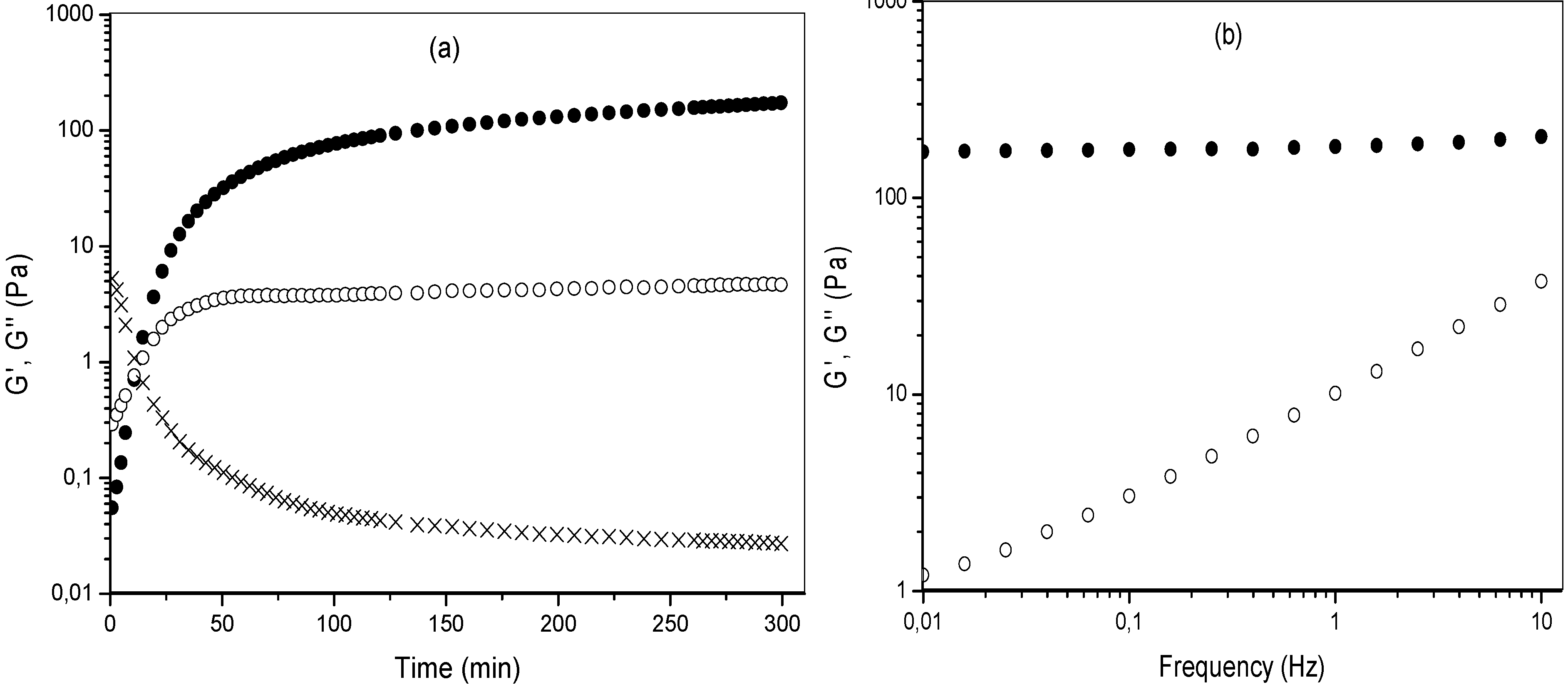
2.1. MBAX Cross-Linking
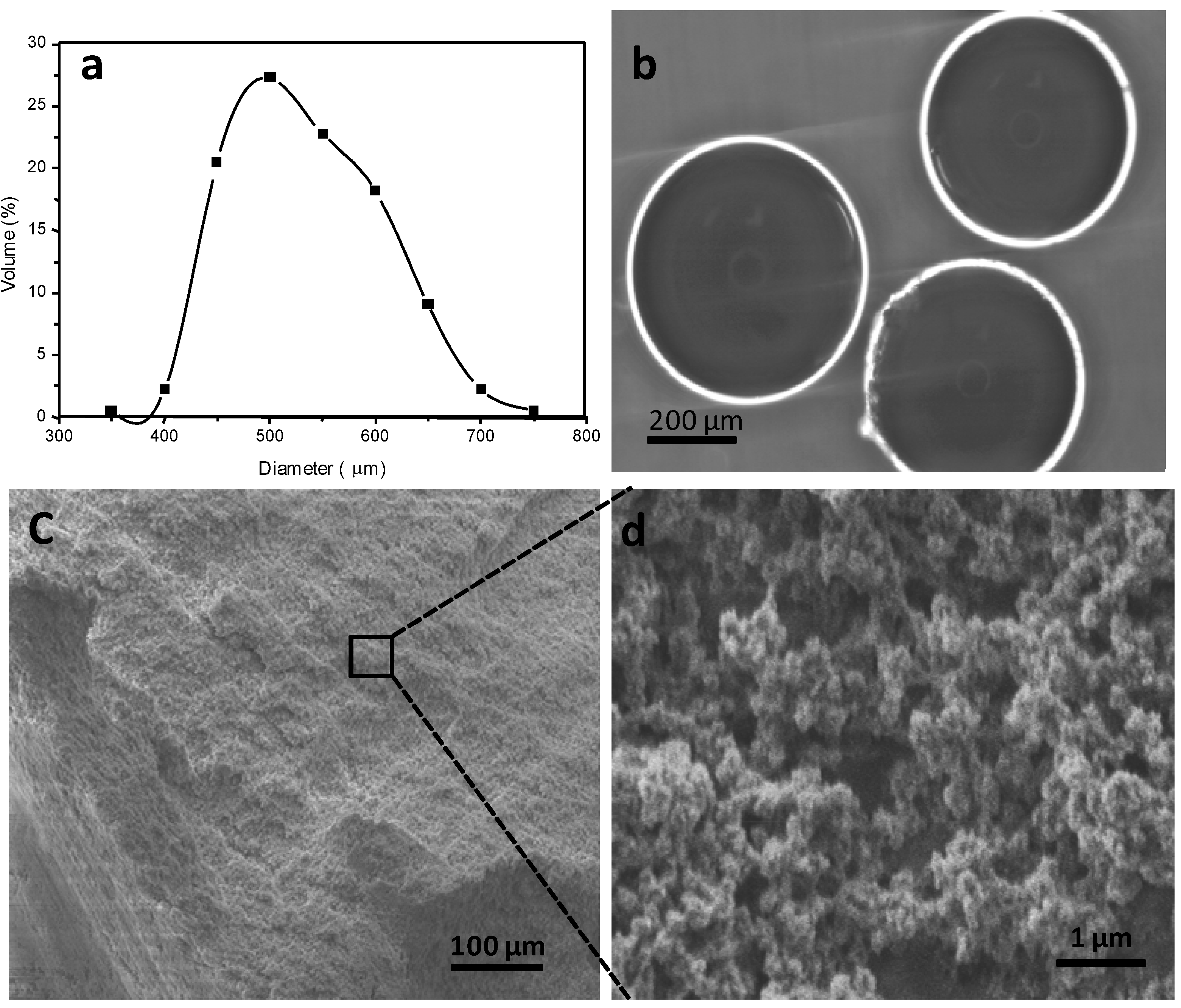
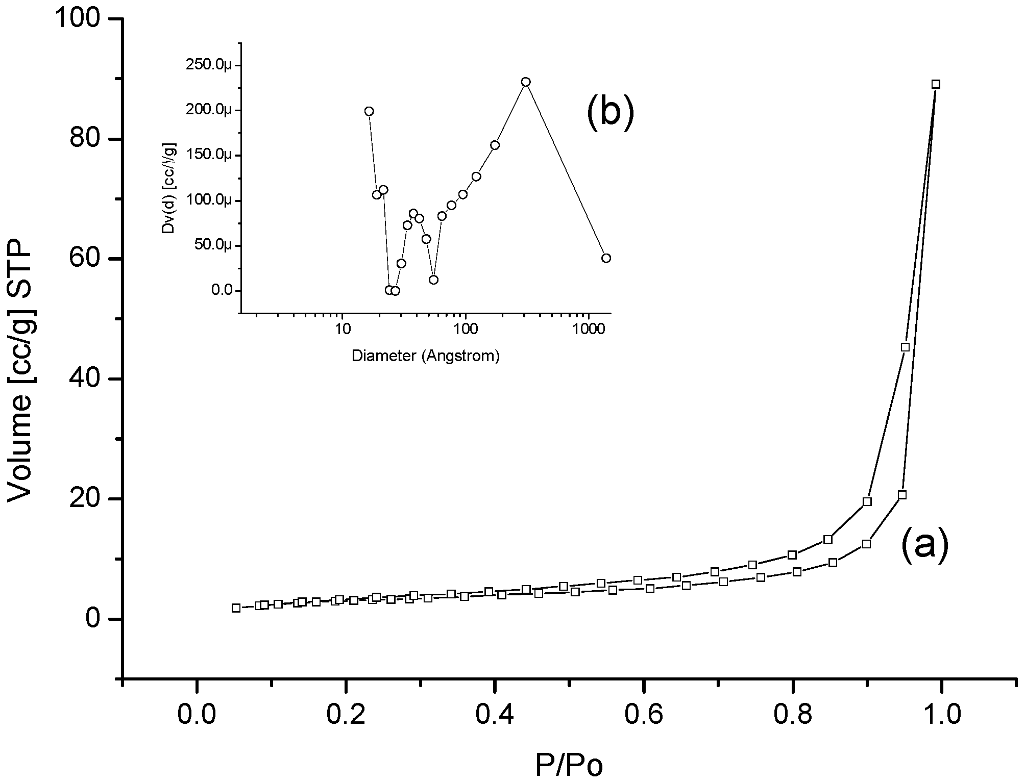
2.2. MBAX Microspheres


| Characteristic | t = 0 h | t = 6 h |
|---|---|---|
| FA (μg/mg MBAX) | 0.255 ± 0.017 | 0.045 ± 0.002 |
| di-FA (μg/mg MBAX) | 0.135 ± 0.011 | 0.03 ± 0.001 |
| tri-FA (μg/mg MBAX) | 0.064 ± 0.010 | Traces |
| Mc a ×10 3 (g/mol) | - | 24 ± 0.4 |
| ρc b × 10−6 (mol/cm3) | - | 59 ± 0.2 |
| ξ c (nm) | - | 52 ± 1.1 |
3. Experimental
3.1. Materials
3.2. Methods
3.2.1. MBAX Gelation
3.2.1.1. Rheological Tests
3.2.1.2. Covalent Cross-Links Content
3.2.2. MBAX Microspheres
3.2.2.1. Phase-Contrast Optical Microscopy
3.2.2.2. Swelling and Structural Parameters
3.2.2.3. Scanning Electron Microscopy (SEM)
3.2.2.4. Textural Analysis
3.2.3. Statistical Analysis
4. Conclusions
Acknowledgments
References
- Dai, C.; Wang, B.; Zhao, H. Microencapsulation peptide and protein drugs delivery system. Colloids Surf. B Biointerfaces 2005, 41, 117–120. [Google Scholar]
- Zhang, H.; Alsarra, I.A.; Neau, S.H. An in vitro evaluation of a chitosan-containing multiparticulate system for macromolecule delivery to the colon. Int. J. Pharm. 2002, 239, 197–205. [Google Scholar] [CrossRef]
- Yang, L.; Chu, J.S.; Fix, J. Colon-specific drug delivery: New approaches and in vitro/in vivo evaluation. Int. J. Pharm. 2002, 235, 1–15. [Google Scholar] [CrossRef]
- Liu, Z.; Jiao, Y.; Wang, Y.; Zhou, C.; Zhang, Z. Polysaccharides-based nanoparticles as drug delivery systems. Adv. Drug Delivery Rev. 2008, 60, 1650–1662. [Google Scholar] [CrossRef]
- Sinha, V.R.; Singla, A.K.; Wadhawan, S.; Kaushik, R.; Kumria, R.; Bansal, K.; Dhawan, S. Chitosan microspheres as a potential carrier for drugs. Int. J. Pharm. 2004, 274, 1–33. [Google Scholar] [CrossRef]
- Sinha, V.R.; Kumria, R. Polysaccharides in colon-specific drug delivery. Int. J. Pharm. 2001, 224, 19–38. [Google Scholar] [CrossRef]
- Silva, C.M.; Ribeiro, J.; Vit, I. Alginate microspheres prepared by internal gelation: Development and effect on insulin stability. Int. J. Pharm. 2006, 311, 1–10. [Google Scholar] [CrossRef]
- Sinha, V.R.; Trehan, A. Biodegradable microspheres for protein delivery. J. Control. Release 2003, 90, 261–280. [Google Scholar] [CrossRef]
- Peppas, N.A. Hydrogels and drug delivery. Curr. Opin. Colloid Interface Sci. 1997, 2, 531–537. [Google Scholar] [CrossRef]
- Izydorczyk, M.S.; Biliaderi, C.G. Cereal arabinoxylans: Advances in structure and physicochemical properties. Carbohydr. Polym. 1995, 28, 33–48. [Google Scholar] [CrossRef]
- Smith, M.M.; Hartley, R.D. Occurrence and nature of ferulic acid substitution of cell-wall polysaccharides in graminaceous plants. Carbohydr. Res. 1983, 118, 65–80. [Google Scholar] [CrossRef]
- Vansteenkiste, E.; Babot, C.; Rouau, X.; Micard, V. Oxidative gelation of feruloylated arabinoxylan as affected by protein. Influence on protein enzymatic hydrolysis. Food Hydrocolloid. 2004, 18, 557–564. [Google Scholar] [CrossRef]
- Schooneveld-bergmans, M.E.F.; Dignum, M.J.W.; Grabber, J.H.; Beldman, G. Studies on the oxidative cross-linking of feruloylated arabinoxylans from wheat flour and wheat bran. Carbohydr. Polym. 2008, 38, 309–317. [Google Scholar] [CrossRef]
- Martínez-López, A.L.; Carvajal-Millan, E.; Lizardi-Mendoza, J.; López-Franco, Y.L.; Rascón-Chu, A.; Salas-Muñoz, E.; Barron, C.; Micard, V. The peroxidase/H2O2 system as a free radical-generating agent for gelling maize bran arabinoxylans: Rheological and structural properties. Molecules 2011, 16, 8410–8418. [Google Scholar] [CrossRef] [Green Version]
- Carvajal-Millan, E.; Rascón-Chu, A.; Márquez-Escalante, J.A.; Micard, V.; León, N.P.D.; Gardea, A. Maize bran gum: Extraction, characterization and functional properties. Carbohydr. Polym. 2007, 69, 280–285. [Google Scholar] [CrossRef]
- Carvajal-Millan, E.; Guilbert, S.; Morel, M.-H.; Micard, V. Impact of the structure of arabinoxylan gels on their rheological and protein transport properties. Carbohydr. Polym. 2005, 60, 431–438. [Google Scholar] [CrossRef]
- Carvajal-Millan, E.; Landillon, V.; Morel, M.-H.; Rouau, X.; Doublier, J.-L.; Micard, V. Arabinoxylan gels: Impact of the feruloylation degree on their structure and properties. Biomacromolecules 2005, 6, 309–317. [Google Scholar] [CrossRef]
- Bunzel, M.; Ralph, J.; Funk, C.; Steinhart, H. Structural elucidation of new ferulic acid-containing phenolic dimers and trimers isolated from maize bran. Tetrahedron Lett. 2005, 46, 5845–5850. [Google Scholar] [CrossRef]
- Carvajal-Millan, E.; Guilbert, S.; Doublier, J.-L.; Micard, V. Arabinoxylan/protein gels: Structural, rheological and controlled release properties. Food Hydrocolloid. 2006, 20, 53–61. [Google Scholar] [CrossRef]
- Berlanga-Reyes, C.M.; Carvajal-Millán, E.; Lizardi-Mendoza, J.; Rascón-Chu, A.; Marquez-Escalante, J.A.; Martínez-López, A.L. Maize arabinoxylan gels as protein delivery matrices. Molecules 2009, 14, 1475–1482. [Google Scholar] [CrossRef]
- Iravani, S.; Fitchett, C.S.; Georget, D.M.R. Physical characterization of arabinoxylan powder and its hydrogel containing a methyl xanthine. Carbohydr. Polym. 2011, 85, 201–207. [Google Scholar] [CrossRef]
- Hernández-Espinoza, A.B.; Piñón-Muñiz, M.I.; Rascón-Chu, A.; Santana-Rodríguez, V.M.; Carvajal-Millan, E. Lycopene/arabinoxylan gels: Rheological and controlled release characteristics. Molecules 2012, 17, 2428–2436. [Google Scholar] [CrossRef]
- Martínez-López, A.L.; Carvajal-Millan, E.; Lizardi-Mendoza, J.; López-Franco, Y.L.; Rascón-Chu, A.; Micard, V. Laccase Induced Cross-Linking Arabinoxylans Microspheres. In Proceedings of the 11th International Food Hydrocolloids Conference: Biofunctionality and Technofunctionality of Hydrocolloids, Purdue, IN, USA, 15–18 May 2012.
- Berlanga-Reyes, C.M.; Carvajal-Millán, E.; Caire Juvera, G.; Rascón-Chu, A.; Marquez-Escalante, J.A.; Martinez-Lopez, A.L. Laccase induced maize bran arabinoxylans gels: Structural and rheological properties. Food Sci. Biotechnol. 2009, 18, 1027–1029. [Google Scholar]
- Niño-Medina, G.; Carvajal-Millán, E.; Lizardi, J.; Rascon-Chu, A.; Marquez-Escalante, J.A.; Gardea, A.; Martinez-Lopez, A.L.; Guerrero, V. Maize processing waste water arabinoxylans: Gelling capability and cross-linking content. Food Chem. 2009, 115, 1286–1290. [Google Scholar] [CrossRef]
- Lapierre, C.; Pollet, B.; Ralet, M.C.; Saulnier, L. The phenolic fraction of maize bran: Evidence for lignin-heteroxylan association. Phytochemistry 2001, 57, 765–772. [Google Scholar] [CrossRef]
- Fundueanu, G.; Esposito, E.; Mihai, D.; Carpov, A.; Desbrieres, J.; Rinaudo, M.; Nastruzzi, C. Preparation and characterization of Ca-alginate microspheres by a new emulsification method. Int. J. Pharm. 1998, 170, 11–21. [Google Scholar] [CrossRef]
- Grishechko, L.I.; Amaral-Labat, G.; Szczurek, A.; Fierro, V.; Kuznetsov, B.N.; Pizzi, A.; Celzard, A. New tannin-lignin aerogels. Ind. Crops Prod. 2013, 41, 347–355. [Google Scholar] [CrossRef]
- Chen, F.; Li, J. Synthesis and structural characteristics of organic aerogels with different content of lignin. Adv. Mater. Res. 2010, 113–116, 1837–1840. [Google Scholar]
- Chen, F.; Xu, M.; Wang, L.; Li, J. Preparation and characterization of organic aerogels from a lignin–resorcinol–formaldehyde copolymer. Bioresources 2011, 6, 1261–1272. [Google Scholar]
- Al-Muhtaseb, S.A.; Ritter, J.A. Preparation and properties of resorcinol-Formaldehyde organic and carbon gels. Adv. Mater. 2003, 15, 101–114. [Google Scholar] [CrossRef]
- Raschip, I.E.; Hitruc, E.G.; Oprea, A.M.; Popescu, M.C.; Vasile, C. In vitro evaluation of the mixed xanthan/lignin hydrogels as vanillin carriers. J. Mol. Struct. 2011, 1003, 67–74. [Google Scholar]
- Barakat, A.; Putaux, J.-L.; Saulnier, L.; Chabbert, B.; Cathala, B. Characterization of arabinoxylans-dehydrogenation (synthetic lignin polymer) polymer nanoparticles. Biomacromolecules 2007, 8, 1236–1245. [Google Scholar] [CrossRef]
- Berlanga-Reyes, C.M.; Carvajal-Millan, E.; Lizardi-Mendoza, J.; Islas-Rubio, A.R.; Rascón-Chu, A. Enzymatic cross-linking of alkali extracted arabinoxylans: Gel rheological and structural characteristics. Int. J. Mol. Sci. 2011, 12, 5853–5861. [Google Scholar] [CrossRef]
- Martinez-Lopez, A.L.; Carvajal-Millan, E.; Lizardi-Mendoza, J.; Rascón-Chu, A.; López-Franco, Y.L.; Salas-Muñoz, E. Ferulated Arabinoxylans as By-Product from Maize Wet-Milling Process: Chacterization and Gelling Capability. In Maize: Cultivation, Uses and Health Benefits; Jimenez-Lopez, J.C., Ed.; Nova Science Publisher: Granade, Spain, 2012; pp. 65–74. [Google Scholar]
- Gregg, S.J.; Sing, K.S.W. Adsortion, Surface Area and Porosimetry, 2nd ed; Academic Press Inc.: London, UK, 1991. [Google Scholar]
- Flory, P.J.; Rehner, J. Statistical mechanics of cross-linked polymer networks I. rubberlike elasticity. J. Chem. Phys. 1943, 11, 512. [Google Scholar] [CrossRef]
- Peppas, N.A.; Merrill, E.W. Poly(vinyl alcohol) hydrogels: Reinforcement of radiation-crosslinked networks by crystallization. J. Polym. Sci. Polym. Chem. 1976, 14, 441–457. [Google Scholar] [CrossRef]
- Peppas, N.A. Analysis of Fickian and non-Fickian drug release from polymers. Phram. Acta Helv. 1985, 60, 110–111. [Google Scholar]
- Brunauer, S.; Emmett, P.H.; Teller, E. Asorption of gases in multimolecular layers. J. Am. Chem. Soc. 1938, 60, 309–319. [Google Scholar] [CrossRef]
- Sample Availability: Not available.
© 2013 by the authors; licensee MDPI, Basel, Switzerland. This article is an open access article distributed under the terms and conditions of the Creative Commons Attribution license (http://creativecommons.org/licenses/by/3.0/).
Share and Cite
Martínez-López, A.L.; Carvajal-Millan, E.; Miki-Yoshida, M.; Alvarez-Contreras, L.; Rascón-Chu, A.; Lizardi-Mendoza, J.; López-Franco, Y. Arabinoxylan Microspheres: Structural and Textural Characteristics. Molecules 2013, 18, 4640-4650. https://doi.org/10.3390/molecules18044640
Martínez-López AL, Carvajal-Millan E, Miki-Yoshida M, Alvarez-Contreras L, Rascón-Chu A, Lizardi-Mendoza J, López-Franco Y. Arabinoxylan Microspheres: Structural and Textural Characteristics. Molecules. 2013; 18(4):4640-4650. https://doi.org/10.3390/molecules18044640
Chicago/Turabian StyleMartínez-López, Ana L., Elizabeth Carvajal-Millan, Mario Miki-Yoshida, Lorena Alvarez-Contreras, Agustín Rascón-Chu, Jaime Lizardi-Mendoza, and Yolanda López-Franco. 2013. "Arabinoxylan Microspheres: Structural and Textural Characteristics" Molecules 18, no. 4: 4640-4650. https://doi.org/10.3390/molecules18044640