Article

Phenolic Glucosides from Dendrobium aurantiacum var. denneanum and Their Bioactivities

1
State Key Laboratory Breeding Base of Systematic Research, Development and Utilization of Chinese Medicine Resources, Sichuan Province and Ministry of Science and Technology, Chengdu 610075, Sichuan, China
2
Pharmacy College, Chengdu University of Traditional Chinese Medicine, Chengdu 610075, Sichuan, China
3
Sichuan Wan'an Dendrobe Industry Development Co., Ltd., Chengdu 610072, Sichuan, China
*
Authors to whom correspondence should be addressed.
Molecules 2013, 18(6), 6153-6160; https://doi.org/10.3390/molecules18066153
Submission received: 15 April 2013 / Revised: 16 May 2013 / Accepted: 20 May 2013 / Published: 23 May 2013
(This article belongs to the Section Natural Products Chemistry)

Abstract

:
A new 8,4'-oxyneolignane glucoside 1 has been isolated from the stems of Dendrobium aurantiacum var. denneanum together with six known phenolic glucosides 27. The structure of the new compound, including its absolute configuration, was determined by spectroscopic and chemical methods as (−)-(7S,8R,7'E)-4-hydroxy-3,3',5,5'-tetramethoxy-8,4'-oxyneolign-7'-ene-7,9,9'-triol 7,9'-bis-O-β-D-glucopyranoside (1). In the in vitro assays, compound 1 and (−)-syringaresinol-4,4'-bis-O-β-D-glucopyranoside (2) showed evident activity against glutamate-induced neurotoxicity in PC12 cells. Shashenoside I (4) showed a selective cytotoxic activity with the IC50 value of 4.17 μM against the acute myeloid leukemia cell line MV4-11, while it was inactive against 10 other human tumor cell lines.

1. Introduction

Dendrobium aurantiacum Rchb.f. var. denneanum (Kerr.) Z. H. Tsi (Orchidaceae) is widely distributed and cultivated in southern China, Burma, Laos and South Asia. The stem of the plant, commonly referred to as “Shihu” or “Huangcao” in Chinese, has been used in traditional Chinese medicine for its antipyretic, eyesight improving, immunomodulatory, antioxidant and anti-aging effects [1,2]. Previous chemical investigations on this plant have resulted in more than twenty phenolic secondary metabolites, including bibenzyls, phenanthrenes, fluorenones, phenylpropanoids, flavones and coumarins [3,4,5,6]. In the course of our search for bioactive natural products, the stems of D. aurantiacum var. denneanum were shown to afford seven phenolic glucosides, including a new 8,4'-oxyneolignane 7-O-glucoside 1 and six known phenolic glucosides 27. Based on IUPAC recommendations for the nomenclature of lignans and neolignans [7], 1 was identified as (−)-(7S,8R,7'E)-4-hydroxy-3,3',5,5'-tetramethoxy-8,4'-oxyneolign-7'-ene-7,9,9'-triol 7,9'-bis-O-β-D-glucopyranoside. All the compounds were assessed for their neuroprotective activity against glutamate-induced toxicity in PC12 cells and cytotoxic activity against 11 kinds of human tumor cells. This paper describes the isolation, structure elucidation, and biological assays of these compounds.

2. Results and Discussion
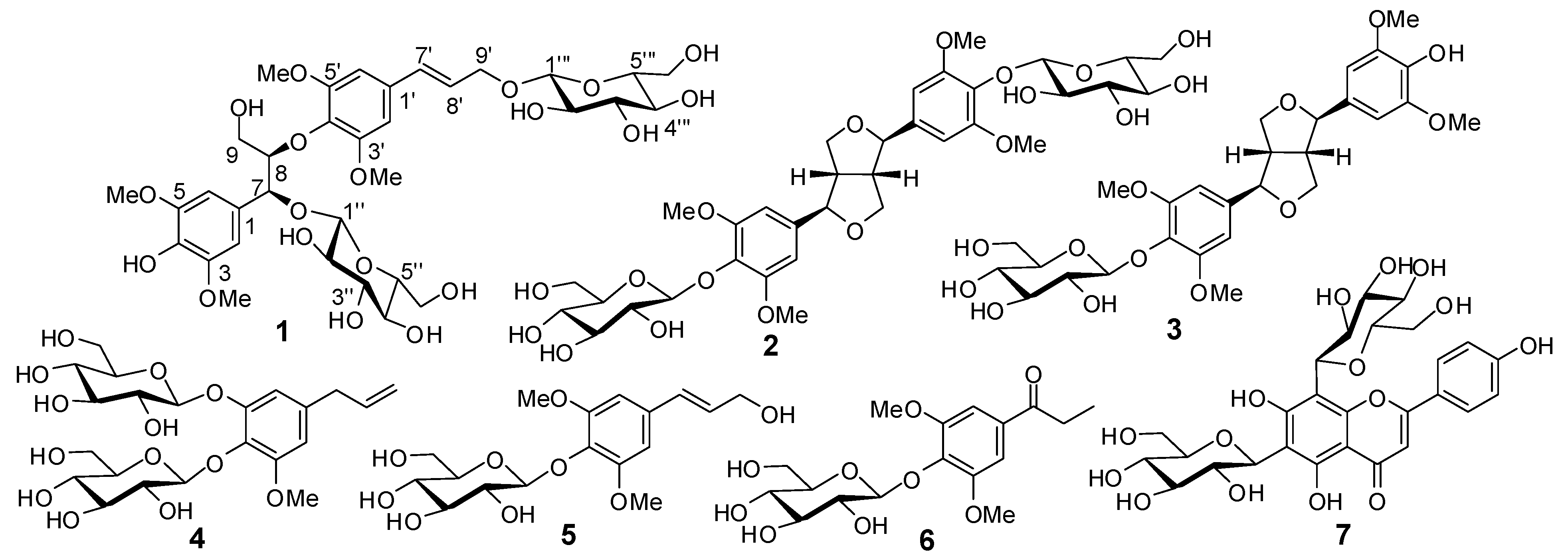
The EtOH extract of the stem of D. aurantiacum var. denneanum was suspended in water and successively partitioned with EtOAc and n-BuOH. Separation of the n-BuOH fraction by column chromatography provided compounds 17 (Figure 1). The known compounds 27 were identified as (−)-syringaresinol-4,4'-bis-O-β-D-glucopyranoside (2) [8], syringaresinol-4'-O-D-monoglucopyranoside (3) [9], shashenoside I (4) [10], syringin (5) [11], 1-[4-(β-D-glucopyranosyloxy)-3,5-dimethoxyphenyl]-1-propanone (6) [12], and vicenin-2 (7) [13] by comparing their spectroscopic data with those reported in the corresponding literature.
Figure 1. Structures of compounds 17.
Molecules 18 06153 g001
Compound 1 was obtained as a colorless gum, and the presence of OH (3,374 cm−1) and aromatic (1,588 and 1,502 cm−1) groups were indicated by its IR spectrum. The molecular formula of C34H48O19 was indicated by a HRESIMS peak at m/z 783.2681 [M+Na]+. The 1H-NMR spectrum of 1 (Table 1) showed signals attributable to two symmetrically 1,3,4,5-tetrasubstituted aromatic rings at δ 6.77 (H-2', 6') and 6.72 (H-2, 6), together with two six-proton aromatic methoxy singlets at δ 3.78 and 3.73. Signals for two olefinic methines and an oxymethylene at δ 6.59 (d, J = 16.0 Hz, H-7'), 6.35 (dt, J = 16.0, 6.0 Hz, H-8'), 4.43 (dd, J = 13.0, 6.0 Hz, H-9'a), and 4.20 (dd, J = 13.0, 6.0 Hz, H-9'b) suggested the presence of a trans-arylpropenoxy unit. In addition, an arylpropanediyloxy unit was indicated by signals of a vicinal coupling system attributed to two oxymethines at δ 5.11 (d, J = 3.5 Hz, H-7) and 4.27 (H-8), and an oxymethylene at δ 3.59 (H-9a) and 3.17 (H-9b). Two diagnostic doublets attributed to anomeric protons at δ 4.34 (d, J = 7.5 Hz, H-1'') and 4.22 (1H, d, J = 8.0 Hz, H-1'''), together with partially overlapped signals assigned to oxymethylene and oxymethine protons between δ 3.00 and 3.70, suggested the presence of two β-glycopyranosyl units in 1 [14]. The 13C-NMR and DEPT spectra of 1 revealed carbon signals corresponding to the above units and quaternary aromatic carbons (Table 1). These spectroscopic features suggested that 1 was a syringylglycerol-8-O-4'-sinapyl alcohol ether (SGSE) β-diglucopyranoside [15]. Comparison of the 13C-NMR data of 1 with those of erythro-SGSE showed that the resonances for C-7 and C-9' in 1 were deshielded significantly by Δδ +6.0 and +6.7 ppm, whereas the C-1, C-8, and C-8' resonances were shielded by Δδ −4.5, −2.6, and −3.7 ppm, respectively. This revealed that two β-glycopyranosyl units were attached to C-4 and C-9', which was further confirmed by the HMBC correlations of H-1'' with C-4 and H-1''' with C-9' (Figure 2).
Table 1. 1H- (500 MHz) and 13C-NMR (125 MHz) data of 1 (in DMSO-d6, δ in ppm, J in Hz).
No.δHδCNo.δHδC
1129.81''4.34 d (7.5)103.1
26.72 s106.12''3.18 m75.2
3148.23''3.06 m77.8
4135.64''3.03 m71.1
5148.25''3.02 m78.3
66.72 s106.16''a3.66 (overlapped)62.1
75.11 d (3.5)79.56''b3.41 (overlapped)
84.27 m85.01'''4.22 d (8.0)103.2
9a3.59 m61.22'''3.00 m74.5
9b3.17 m3'''3.13 m77.4
1'133.14'''3.12 m70.9
2'6.77 s104.85'''3.23 m77.9
3'153.76'''a3.62 (overlapped)61.9
4'136.76'''b3.40 (overlapped)
5'153.73/5-OMe3.73 s57.0
6'6.77 s104.83'/5'-OMe3.78 s57.0
7'6.59 d (16.0)132.2
8'6.35 dt (16.0, 6.0)126.8
9'a4.43 dd (13.0, 6.0)69.6
9'b4.20 dd (13.0, 6.0)
Enzymatic hydrolysis of 1 produced the aglycone 1a and a sugar, which was further identified as D-glucose by the positive optical rotation ([α]20D +45.5) [16,17,18] and TLC comparison with an authentic sugar sample. Comparison the 1H-NMR and HREIMS data of 1a with those of known 8,4'-oxyneolignane led characterization of 1a as erythro-SGSE [15]. The CD spectra of 1 and 1a showed negative Cotton effects at 235 nm (Δε −0.18) and 239 nm (Δε −0.22), respectively, indicating an 8R configuration for 1 and 1a [18,19,20]. Thus, compound 1 was determined to be (−)-(7S,8R,7'E)-4-hydroxy-3,3',5,5'-tetramethoxy-8,4'-oxyneolign-7'-ene-7,9,9'-triol 7,9'-bis-O-β-D-glucopyranoside.
Figure 2. Key HMBC correlations of 1.
Molecules 18 06153 g002
The isolated compounds were assessed for their cytotoxic activity against 11 kinds of human tumor cells, including the lung cancer cell lines H1975, H358 and A549, hepatocellular carcinoma cell lines HepG-2 and SMMC7721, colorectal carcinoma cell line HCT116, mammary carcinoma cell lines MDA-MB-231 and MCF-7, melanoma cell line A2058, pancreatic cancer cell line PANC-1, and acute myeloid leukemia cell line MV4-11 in in vitro bioassays. Among them, compound 4 showed a selective cytotoxic activity against acute myeloid leukemia cell line MV4-11, with the IC50 value of 4.17 μM. Other compounds were inactive against the tested human tumor cells at the concentration of 10 μM.
In addition, the protective activity of the compounds against glutamate-induced neurotoxicity in PC12 cells was evaluated by an MTT assay [18,21]. The results showed that glutamate induced an inhibition of MTT reduction, while compounds 1 and 2 showed neuroprotective activity at a concentration of 10 μM, with the relative protection of 25.7 ± 2.2% and 19.3 ± 5.6%, respectively (the positive control MK-801, 85.9 ± 3.2%). Thus, lignan glucosides 1 and 2 may be effective in neurodegenerative disorders.

3. Experimental

3.1. General

NMR spectra were recorded on a INOVA-500 spectrometer. HRESIMS were measured with Waters Synapt G2 HDMS. IR were recorded on a Vector 22 FT-IR spectrometer. UV spectra were obtained on a Shimadzu UV-260 spectrophotometer. Optical rotations were measured with a Perkin-Elmer 341 plus. CD spectra were recorded on a JASCO J-815 CD spectrometer. Column chromatography was performed with silica gel (200–300 mesh, Yantai Institute of Chemical Technology, Yantai, China) and Sephadex LH-20 (Amersham Pharmacia Biotech AB, Uppsala, Sweden). HPLC separation was performed on an instrument consisting of a Cometro 6000LDS pump and a Cometro 6000PVW UV/VIS detector with an Ultimate (250 × 10 mm) preparative column packed with C18 (5 μm).

3.2. Plant Material

The stem of Dendrobium aurantiacum var. denneanum was collected in April of 2011 from a culture field in Shuangliu, Sichuan Province, China. Plant identity was verified by Prof. Min Li (Chengdu University of TCM, Sichuan, China). A voucher specimen (SSF-20110410) was deposited at the School of Pharmacy, Chengdu University of TCM, Chengdu, China.

3.3. Extraction and Isolation

The air-dried stem of D. aurantiacum var. denneanum (10 kg) was powdered and extracted three times with 95% EtOH (30 L) for 3 h under reflux. The EtOH extract was evaporated under reduced pressure to yield a dark brown residue (530 g), which was suspended in H2O (2.5 L) and then successively partitioned with EtOAc and n-BuOH (6 × 2.5 L). The n-BuOH extract (110 g) was applied to a D-101 macroporous adsorbent resin (1.5 Kg) column. Successive elution of the column with H2O, 10% EtOH, 30% EtOH, 50% EtOH, and 95% EtOH (4 L each) yielded five portions. The portion (48 g) eluted by 30% EtOH was separated by MPLC over reversed-phase silica gel eluting with a gradient of increasing MeOH (5−90%) in H2O to give eight fractions (A-H). Fraction 7 (185 mg) was precipitated from the fraction D in MeOH. Subsequent separation of fraction F (6.5 g) over Sephadex LH-20 eluted with MeOH−H2O (1:1) gave five subfractions (F1−F5). Subfraction F2 was further fractionated via silica gel CC, eluting with CHCl3−MeOH (8:1), to yield six fractions (F2-1−F2-6). Separation of F2-2 with RP semipreparative HPLC (37% MeOH in H2O) yielded 1 (6.5 mg) and 2 (38.0 mg). F2-5 was further separated by Sephadex LH−20 (MeOH−H2O, 3:7), and then purified by RP semipreparative HPLC (34% MeOH in H2O), to yield 4 (31 mg). Subfraction F4 was fractioned by silica gel CC, eluting with a gradient of increasing MeOH (5−50%) in CHCl3, to afford seven fractions (F4-1−F4-7). F4-3 was purified by RP preparative HPLC (35% MeOH in H2O) to afford 3 (9 mg), 5 (36 mg), and 6 (11 mg).
(−)-(7S,8R,7'E)-4-hydroxy-3,3',5,5'-tetramethoxy-8,4'-oxyneolign-7'-ene-7,9,9'-triol 7,9'-bis-O-β-D glucopyranoside (1): White gum, Molecules 18 06153 i001 = −12.2 (c = 0.20, MeOH); IR (KBr) νmax: 3374, 2920, 1588, 1502, 1462, 1420, 1331, 1226, 1124, 1071, 1023, 835, 706, 615 cm−1; UV (MeOH) λmax (log ε): 204 (4.35), 226 (4.03, sh), 271 (3.67) nm; CD (MeOH): 222 (Δε +0.16), 235 (Δε −0.18), 251 (Δε +0.03), 266 (Δε −0.12), 290 (Δε +0.23) nm; ESI-MS m/z783 [M+Na]+; HRESI-MS: m/z 783.2681 [M+Na]+ (calcd for C34H48O19Na, 783.2687); 1H- and 13C-NMR data see Table 1.

3.4. Enzymatic Hydrolysis of 1

A solution of compound 1 (2 mg) in H2O (10 mL) was hydrolyzed with β-glucosidase (15 mg) at 37 °C for 96 h. The reaction mixture was extracted with EtOAc (3 × 10 mL) to yield the individual EtOAc extract and H2O phase after removing the solvents. The aqueous phases were subjected to preparative TLC eluted with MeCN−H2O (8:1) to yield the sole sugar, which could be identified as D-glucose by the sign of its positive optical rotation. The EtOAc extracts were purified by preparative TLC using CHCl3-MeOH (12:1) to afford 1a (0.2 mg): Molecules 18 06153 i001 = +13.2 (c = 0.02, MeOH); CD (MeOH) 221 (Δε +1.25), 239 (Δε −0.22), 270 (Δε +0.43) nm; 1H-NMR (CD3OD, 500 MHz) δ: 6.76 (2H, s, H-2', 6'), 6.68 (2H, s, H-2, 6), 6.55 (1H, d, J = 16.0 Hz, H-7'), 6.38 (1H, dt, J = 16.0, 6.0 Hz, H-8'), 4.99 (1H, d, J = 3.5 Hz, H-7), 4.27 (2H, d, J = 6.0 Hz, H2-9'), 4.19 (1H, m, H-8), 3.88 and 3.83 (each 6H, s, OMe-3, 5, 3', 5'), 3.87 (1H, m, H-9a), 3.48 (1H, m, H-9b); ESI-MS m/z: 459 [M+Na]+; HR-ESI-MS m/z: 459.1624 [M+Na]+ (calcd for C22H28O9Na, 459.1631).

3.5. Cell Culture and Assessment of Cytotoxic Activity against Human Tumor Cells

The human lung cancer cell lines H1975, H358 and A549, human hepatocellular carcinoma cell lines HepG-2 and SMMC7721, human colorectal carcinoma cell line HCT116, human mammary carcinoma cell lines MDA-MB-231 and MCF-7, human melanoma cell line A2058, human pancreatic cancer cell line PANC-1, human acute myeloid leukemia cell line MV4-11 were obtained from the American Type Culture Collection (ATCC) and grown in RPMI1640, DMEM or IMDM containing 10% fetal bovine serum (v/v) in 5% CO2 at 37 °C. Cells (2 × 10³–10 × 103) were seeded in 96-well plates and cultured for 24 h, followed by the test compounds treatment at concentrations of 0.625–20 µM for 72 h. After the culture period, 20 µL of MTT (5 mg/mL) was added per well and incubated for 4 h at 37 °C, then 50 μL of 20% acidified SDS was used to lyse the cells. Finally, absorbance was measured at 570 nm using a microplate reader. Each assay was replicated three times. The effect of the compounds on tumor cells viability was calculated and expressed by IC50 of each cell line.

3.6. Cell Culture and Assessment of Neuroprotective Activity

PC12 cells at a density of 5 × 10³ cells per well in 96-well plates were suspended in Dulbecco’s Modified Eagle’s Medium (DMEM, Gibco) media supplemented with 5% fetal bovine serum (FBS, Hyclone) and 5% horse serum, penicillin (100 IU/mL), streptomycin (100 μg/mL), and L-glutamine (2 μM) and incubated in a CO2 incubator (5%) at 37 °C for 24 h. The cells were pre-treated with test compounds and MK-801 for 1 h, respectively, and then exposed to glutamate (50 μM). After incubation for an additional 24 h, MTT (0.5 mg/mL) was added to the medium and incubated for 4 h. Absorbance was measured at 570 nm using a microplate reader, the cell viability was evaluated by relative protection, which was calculated as 100 × [optical density (OD) of test compound + glutamate-treated culture − OD of glutamate-treated culture]/[OD of control culture − OD of glutamate-treated culture].

4. Conclusions

Several Dendrobium species are not only ornamental plants, but are also used as Traditional Chinese Medicines. To search for bioactive natural products from the botanical drugs, we carried out an examination of the n-BuOH soluble portion of the ethanolic extract of the stems of D. aurantiacum var. denneanum. A new 8,4'-oxyneolignane 7-O-glucoside, (−)-(7S,8R,7'E)-4-hydroxy-3,3',5,5'-tetramethoxy-8,4'-oxyneolign-7'-ene-7,9,9'-triol 7,9'-bis-O-β-D-glucopyranoside (1) was isolated, together with two 7,9':7',9-diepoxylignan glucosides 23, four phenylpropanoid glycosides 46 and a flavone C-glycoside 7. The results showed that the types of compounds of D. aurantiacum var. denneanum were similar to other species of Dendrobium, including D. chrysanthum, D. loddigesii, D. nobile, D. moniliforme, D. trigonopus, and D. aphyllum. Previous phytochemical studies of these species have also led to the isolation of lignans [22,23,24,25,26,27], phenylpropanoids [28], and flavonoids [26,27]. However, to fully elucidate the systematic correlation of these Dendrobium plants, further phytochemical investigations are necessary. In addition, it deserves to be mentioned that most of the lignans from Dendrobium plants were 7,9':7',9-diepoxylignans, while 8,4'-oxyneolignans were rarely reported. To the best of our knowledge, in Dendrobium 8,4'-oxyneolignans have been only reported from D. chrysanthum so far, which implied chemotaxonomic significance between D. aurantiacum var. denneanum and D. chrysanthum. In the in vitro assays, compounds 1 [(−)-(7S,8R,7'E)-4-hydroxy-3,3',5,5'-tetramethoxy-8,4'-oxyneolign-7'-ene-7,9,9'-triol 7,9'-bis-O-β-D-glucopyranoside] and 2 [(−)-syringaresinol-4,4'-bis-O-β-D-glucopyranoside] showed neuroprotective activity in PC12 cells, while 4 (shashenoside I) had selective cytotoxic activity against the acute myeloid leukemia cell line MV4-11.

Acknowledgments

Financial support from the National Technology R&D Program for the “Twelfth Five-Year” Plan of China (grant No. 2011BAI13B02-8) is acknowledged.

Conflicts of Interest

The authors declare no conflict of interest.

References

  1. Commission of Chinese Pharmacopoeia, Pharmacopoeia of the People’s Republic of China; Chemical Industry Press: Beijing, China, 2010; Volume 1, pp. 85–87.
  2. Jiangsu New Medical College, Dictionary of Traditional Chinese Medicine; Shanghai Science and Technology Publishing House: Shanghai, China, 1995; pp. 586–590.
  3. Zheng, W.P.; Tang, Y.P.; Lou, F.C.; Zhi, F. Studies on the constituents of Dendrobium chryseum Rolfe. J. China Pharm. Univ. 2000, 31, 5–7. [Google Scholar]
  4. Yang, L.; Wang, Z.T.; Xu, L.S. Phenols and a triterpene from Dendrobium aurantiacum var. denneanum (Orchidaceae). Biochem. Syst. Ecol. 2006, 34, 658–660. [Google Scholar] [CrossRef]
  5. Liu, Y.; Jiang, J.H.; Zhang, Y.; Chen, Y.G. Chemical constituents of Dendrobium aurantiacum var. denneanum. Chem. Nat. Compd. 2009, 45, 525–527. [Google Scholar] [CrossRef]
  6. Yang, L.; Han, H.F.; Nakamura, N.; Hattori, M.; Wang, Z.T.; Xu, L.S. Bio-guided isolation of antioxidants from the stems of Dendrobium aurantiacum var. denneanum. Phytother. Res. 2007, 21, 696–698. [Google Scholar] [CrossRef]
  7. Moss, G.P. Nomenclature of lignans and neolignans (IUPAC recommendations 2000). Pure Appl. Chem. 2000, 72, 1493–1523. [Google Scholar] [CrossRef]
  8. El Gamal, A.A.; Takeya, K.; Itokawa, H.; Halim, A.F.; Amer, M.M.; Saad, H.E.A. Lignan bis-glucosides from Galium sinaicum. Phytochemistry 1997, 45, 597–600. [Google Scholar] [CrossRef]
  9. Wang, C.Z.; Yu, D.Q. Lignan and acetylenic glycosides from Aster auriculatus. Phytochemistry 1998, 48, 711–717. [Google Scholar] [CrossRef]
  10. Kuang, H.X.; Shao, C.J.; Kasai, R.; Ohtani, K.; Tian, Z.K.; Xu, J.D.; Tanaka, O. Phenolic glycosides from roots of Adenophora tetraphylla collected in Heilongjiang, China. Chem. Pharm. Bull. 1991, 39, 2440–2442. [Google Scholar] [CrossRef]
  11. Sugiyama, M.; Nagayama, E.; Kikuchi, M. Lignan and phenylpropanoid glycosides from Osmanthus aslaticus. Phytochemistry 1993, 33, 1215–1219. [Google Scholar] [CrossRef]
  12. Takara, K.; Matsui, D.; Wada, K.; Ichiba, T.; Chinen, I.; Nakasone, Y. New phenolic compounds from Kokuto, non-centrifuged cane sugar. Biosci. Biotechnol. Biochem. 2003, 67, 376–379. [Google Scholar] [CrossRef]
  13. Siciliano, T.; De Tommasi, N.; Morelli, I.; Braca, A. A. Study of flavonoids of sechium edule (Jacq) Swartz (Cucurbitaceae) different edible organs by liquid chromatography photodiode array mass spectrometry. J. Agric. Food. Chem. 2004, 52, 6510–6515. [Google Scholar] [CrossRef]
  14. Wu, B.; Zhang, X.; Wu, X. New lignan glucosides with tyrosinase inhibitory activities from exocarp of Castanea henryi. Carbohydr. Res. 2012, 355, 45–49. [Google Scholar] [CrossRef]
  15. Lourith, N.; Katayama, T.; Suzuki, T. Stereochemistry and biosynthesis of 8-O-4' neolignans in Eucommia ulmoides: diastereoselective formation of guaiacylglycerol-8-O-4'-(sinapyl alcohol) ether. J. Wood Sci. 2009, 51, 370–378. [Google Scholar] [CrossRef]
  16. Hudson, C.S.; Dale, J.K. Studies on the forms of D-glucose and their mutarotation. J. Am. Chem. Soc. 1917, 39, 320–328. [Google Scholar] [CrossRef]
  17. Zi, J.C.; Li, S.; Liu, M.T.; Gan, M.L.; Lin, S.; Song, W.X.; Zhang, Y.L.; Fan, X.N.; Yang, Y.C.; Zhang, J.J.; Shi, J.G.; Di, D.L. Glycosidic constituents of the tubers of Gymnadenia conopsea. J. Nat. Prod. 2008, 71, 799–805. [Google Scholar] [CrossRef]
  18. Gan, M.L.; Zhang, Y.L.; Lin, S.; Liu, M.T.; Song, W.X.; Zi, J.C.; Yang, Y.C.; Fan, X.N.; Shi, J.G.; Hu, J.F.; Sun, J.D.; Chen, N.H. Glycosides from the root of Iodes cirrhosa. J. Nat. Prod. 2008, 71, 647–654. [Google Scholar] [CrossRef]
  19. Kim, K.H.; Kim, H.K.; Choi, S.U.; Moon, E.; Kim, S.Y.; Lee, K.R. Bioactive lignans from the rhizomes of Acorus gramineus. J. Nat. Prod. 2011, 74, 2187–2192. [Google Scholar] [CrossRef]
  20. Fang, J.M.; Lee, C.K.; Cheng, Y.S. Lignans from leaves of Juniperus chinensis. Phytochemistry 1992, 31, 3659–3661. [Google Scholar] [CrossRef]
  21. Yoon, J.S.; Lee, M.K.; Sung, S.H.; Kim, Y.C. Neuroprotective 2-(2-phenylethyl)chromones of Imperata cylindrica. J. Nat. Prod. 2006, 69, 290–291. [Google Scholar] [CrossRef]
  22. Ye, Q.H.; Zhao, W.M.; Qin, G.W. Lignans from Dendrobium chrysanthum. J. Asian Nat. Prod. Res. 2004, 6, 39–43. [Google Scholar] [CrossRef]
  23. Ito, M.; Matsuzaki, K.; Wang, J.; Daikonya, A.; Wang, N.L.; Yao, X.S.; Kitanaka, S. New phenanthrenes and stilbenes from Dendrobium loddigesii. Chem. Pharm. Bull. 2010, 58, 628–633. [Google Scholar] [CrossRef]
  24. Zhang, X.; Xu, J.K.; Wang, N.L.; Kurihara, H.; Yao, X.S. Antioxidant phenanthrenes and lignans from Dendrobium nobile. J. Chin. Pharm. Sci. 2008, 17, 314–318. [Google Scholar]
  25. Zhao, W.M.; Ye, Q.H.; Dai, J.Q.; Martin, M.T.; Zhu, J.P. allo-Aromadendrane- and picrotoxane-type sesquiterpenes from Dendrobium moniliforme. Planta Med. 2003, 69, 1136–1140. [Google Scholar] [CrossRef]
  26. Zhang, T.; Zhang, C.F.; Wang, Z.T.; Xu, L.S. Studies on chemical constituents of Dendrobium trigonopus Rchb. f. Chin. J. Nat. Med. 2005, 3, 28–30. [Google Scholar]
  27. Shao, L.; Huang, W.H.; Zhang, C.F.; Wang, L.; Zhang, M.; Wang, Z.T. Study on chemical constituents from stem of Dendrobium aphyllum. Zhongguo Zhongyao Zazhi 2008, 33, 1693–1695. [Google Scholar]
  28. Zhang, X.; Gao, H.; Wang, N.L.; Yao, X.S. Phenolic components from Dendrobium nobile. Zhongcaoyao 2006, 37, 652–655. [Google Scholar]
  • Sample Availability: Not available.

