Article

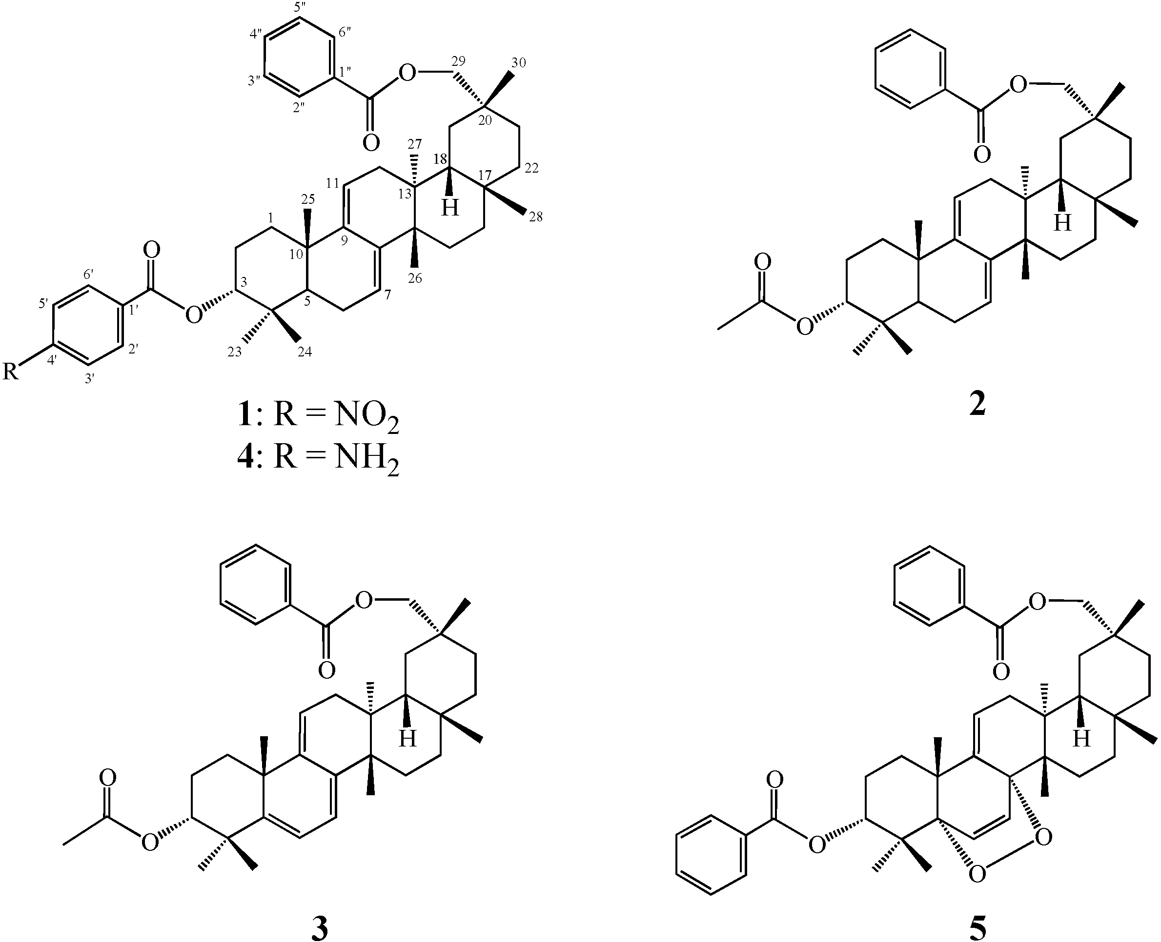
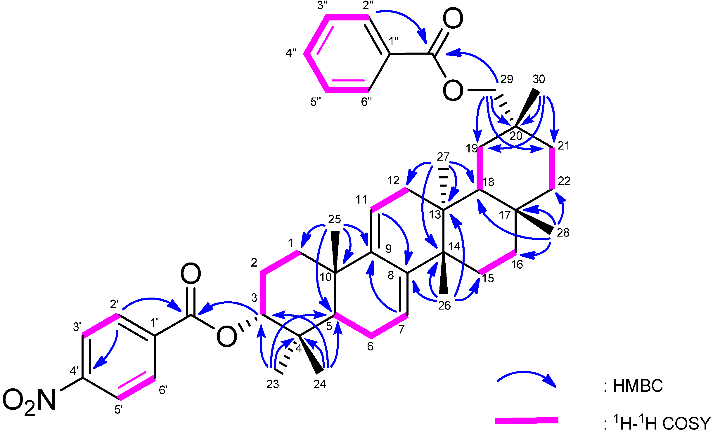
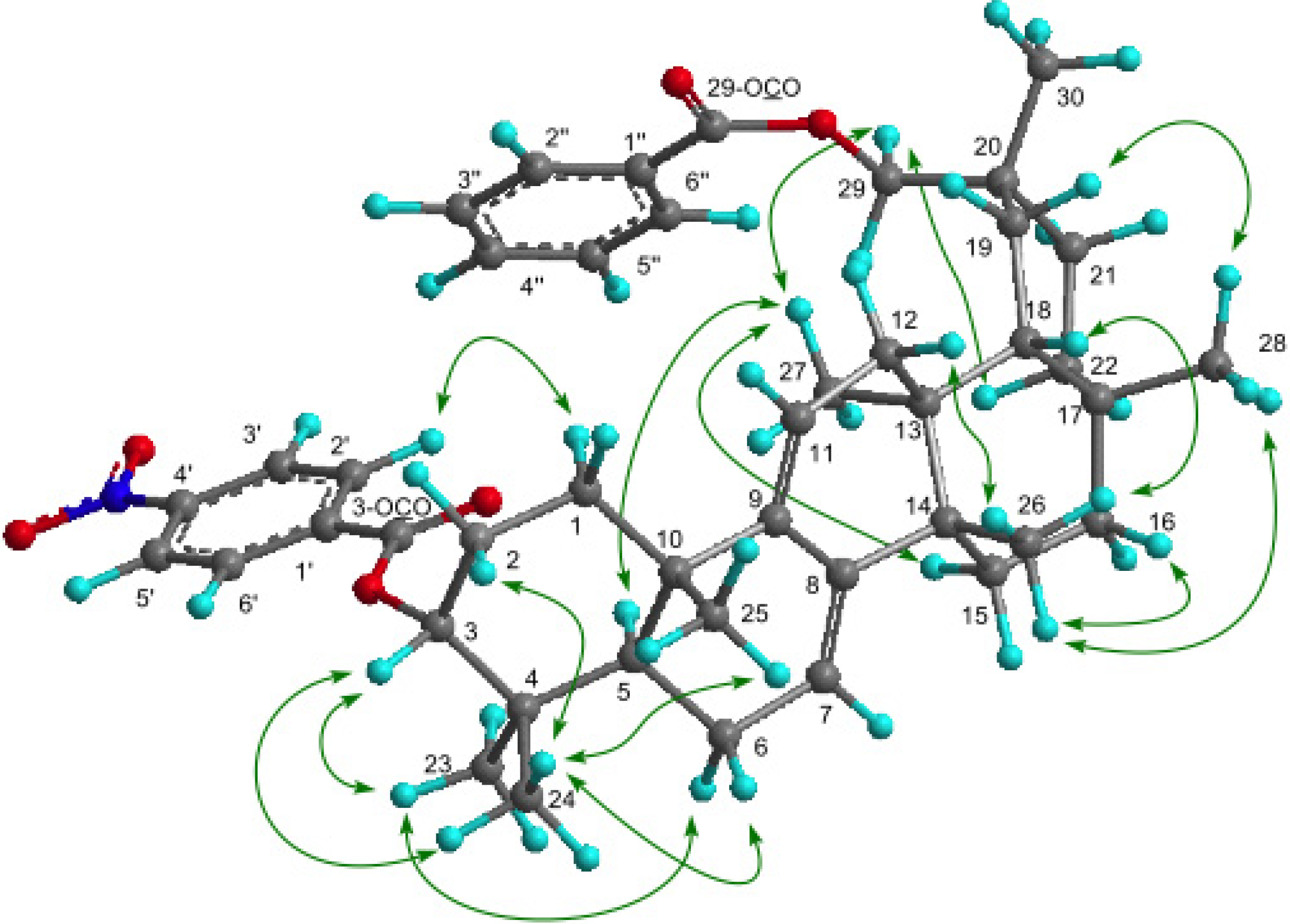
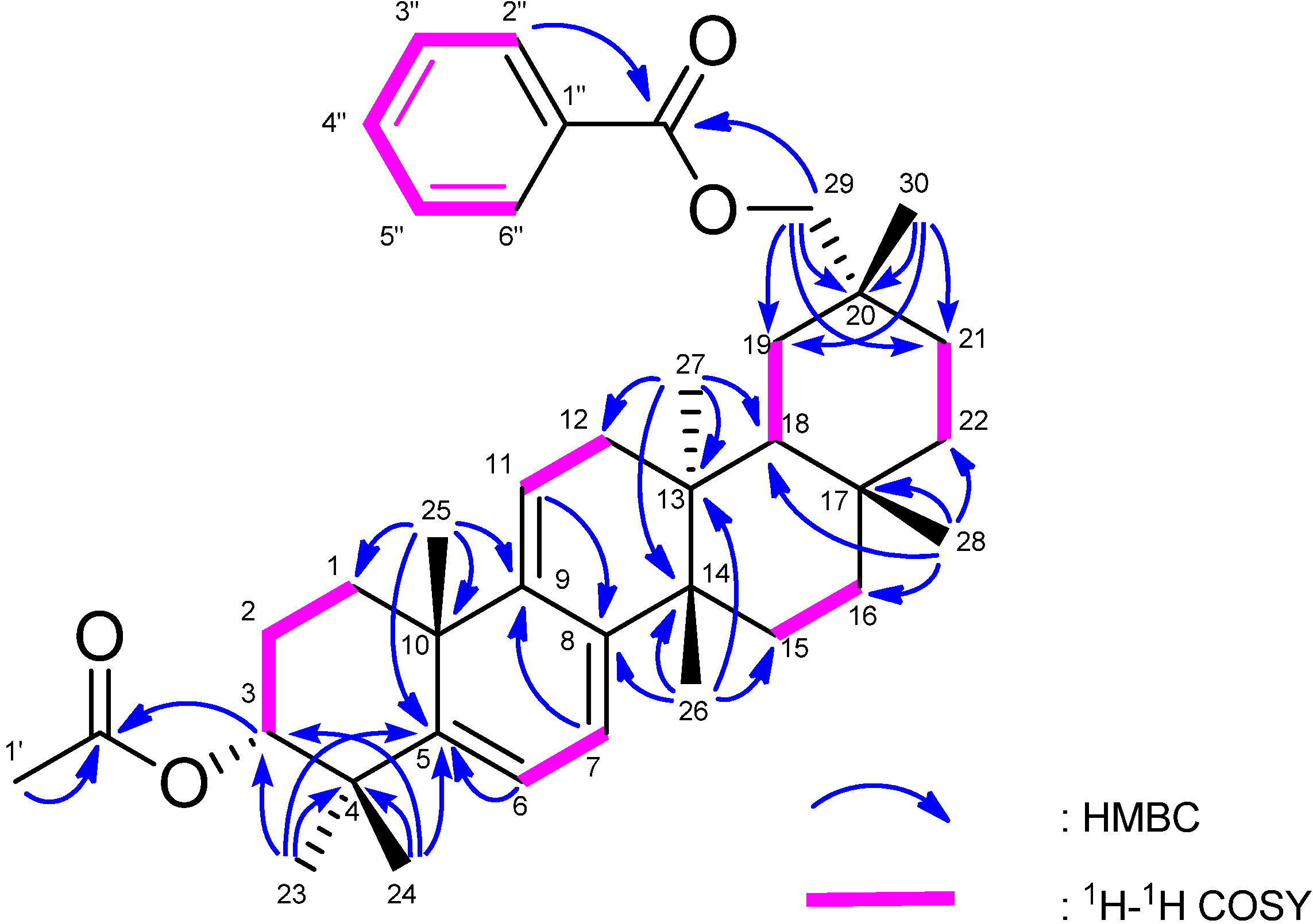
A Novel 3a-p-Nitrobenzoylmultiflora-7:9(11)-diene-29-benzoate and Two New Triterpenoids from the Seeds of Zucchini (Cucurbita pepo L)

Laboratory of Medicinal Chemistry, Osaka University of Pharmaceutical Sciences, 4-20-1 Nasahara, Takatsuki, Osaka 569-1094, Japan
*
Author to whom correspondence should be addressed.
Molecules 2013, 18(7), 7448-7459; https://doi.org/10.3390/molecules18077448
Submission received: 6 June 2013 / Revised: 21 June 2013 / Accepted: 21 June 2013 / Published: 26 June 2013
(This article belongs to the Section Natural Products Chemistry)

Abstract

:
Three novel multiflorane-type triterpenoids, 3α-p-nitrobenzoylmultiflora-7:9(11)-diene-29-benzoate (1), 3α-acetoxymultiflora-7:9(11)-diene-29-benzoate (2), and 3α-acetoxymultiflora-5(6):7:9(11)-triene-29-benzoate (3), along with two known related compounds 4 and 5 were isolated from the seeds of zucchini (Cucurbita pepo L). Their structures were determined on the basis of 1D and 2D NMR spectroscopy and HREIMS. Triterpenoids possessing a nitro group were not isolated previously.

1. Introduction

The species Cucurbita pepo is a cultivated plant of the genus Cucurbita that includes varieties of squash, gourd, and pumpkin. Cucurbita pepo L (zucchini, also known as field pumpkin or summer squash) (Cucurbitaceae) are widely cultivated in America, Europe, and Asia. The zucchini is a hybrid of the cucumber, and has been a commercially important crop in many countries since the 1950–1960s. It is a highly nutritional low caloric food that requires relatively little effort to prepare. It is full of nutrients like vitamin A, vitamin C, potassium, folate and fiber—all of which support a healthy metabolism. Zucchini, grows well in warm climates. This readily available vegetable can also be an important part of weight loss efforts.
As to triterpenoids from Cucurbita pepo L, Appendino reported 3α-p-aminobenzoyl, 29-benzoylmultiflor-8-en-7β-ol, and 3α-p-aminobenzoylmultiflora-7:9(11)-dien-29-benzoate [1,2]. Barker reported large-scale isolation of bryonolic acid (3β-hydroxymultiflor-8-en-29-oic acid) [3].Wang reported cucurbitacin glycoside [4], hexanorcucurbitane glycosides [5], and purine-containing cucurbitane triterpenoids [6], extracted from Cucurbita pepo cv dayangua. Ding et al., reported cerebroside, 13(18)-oleanen-3-ol, β-daucosterol, β-sitosterol, stigmasterol, dotriacontyl stearate, and tritriacontane from Cucurbita pepo cv dayangua [7]. Shibuya reported the biosynthesis of sterols and triterpenes in higher plants: Panax ginseng, Olea europaea, Taraxacum officinale, Betula platyphylla, Glycyrrhiza glabra, Luffa cylindrica, Pisum sativum, Allium macrostemon, and Cucurbita pepo [8]. Shibuya et al., also reported that three oxidosqualene cyclas (OSC) cDNAs (CPX, CPQ, CPR) were cloned from seedlings of Cucurbita pepo by a homology-based PCR method [9]. Careful examination of the seeds of Cucurbita pepo L. has led to the isolation of three novel multiflorane triterpenoids 13 along with the known compounds 4 and 5. The structures of 13 were determined on the basis of NMR spectroscopy, including 1D and 2D (1H, 1H-COSY, NOESY, HSQC, HMBC) NMR, and EIMS.

2. Results and Discussion

The seeds of Cucurbita pepo L. were extracted with MeOH and the extract was partitioned Et2O and H2O. The Et2O soluble portion (216.1 g) was subjected to silica gel column chromatography, medium-pressure liquid chromatography (MPLC), and normal-phase high performance liquid chromatography (HPLC) to yield five triterpenoids 15 (Figure 1) Compounds 4 and 5 were identified as 3α-p-aminobenzoylmultiflora-7:9(11)-dien-29-benzoate [1,2] and 5α,8α-peroxymultiflora-6:9(11)-diene-3α,29-dibenzoate [10], respectively by comparison of their characterization data with literature data.
Figure 1. Structures for compounds 15.
Molecules 18 07448 g001
The molecular formula of 1 was determined as C44H55NO6 (M+; m/z 693.4024) based on HREIMS. The UV spectrum showed a heteroannular diene moiety (λmax 230, 237, 248 nm, log ε 3.85, 3.80, 3.63). The IR spectrum showed bands assignable to ester groups (νmax 1713, 1287 cm−1) and a nitro group (νmax 1527, 1341 cm-1). The 1H- and 13C-NMR spectra (Table 1) exhibited signals assignable to seven tertiary methyls, ten CH2 groups including an oxymethylene [δH 4.11 and 4.17 (each 1H, d)], three sp3 methine groups including an oxymethine [δH 4.97 (1H, t)], two trisubstituted olefin [δH 5.24 (1H, brd); 5.53 (1H, brs)], six sp3 quaternary carbons, a benzoyl group [δH 7.43 (2H, tt), 7.52 (1H, tt), 8.08 (2H, dd); δC 128.6 (C-3″, C-5″), 129.7 (C-2″, C-6″), 130.8 (C-1″), 133.0 (C-4″), 166.9 (OCO)] and a p-nitrobenzoyl group [δH 8.10 (2H, dt), 8.20 (2H, dt); δC 123.8 (C-3′, C-5′), 130.7 (C-2′, C-6′), 136.3 (C-1′), 150.6 (C-4′), 164.4, (OCO)]. In the HMBC spectrum (Figure 2), long-range correlations were observed between Me-23 and C-3 (δC 80.7), C-4, C-5, and C-24; between Me-24 and C-3, C-4, C-5, and C-23; between Me-25 and C-1, C-5, C-9 (δC 144.6), and C-10; between Me-26 and C-8 (δC 142.3), C-13, C-14, and C-15; between Me-27 and C-12, C-13, C-14, and C-18; between Me-28 and C-16, C-17, C-18, and C-22; between Me-30 and C-19, C-20, C-21, and C-29 (δC 73.0); between H2-29 and C-19, C-20, C-21, and C-30; between H-2′ and H-6′ [δH 8.10 (2H)] and C-4′ (δC 150.6), O-C=O (δC 164.4); and between H-2″ and H-6″ [δH 8.08 (2H)] and O-C=O (δC 166.9). In the 1H-1H COSY spectrum (H-7–H2-6; H-11–H2-12; H-3–H2-2; H-3′ and H-5′– H-2′and H-6′; H-3″and H-5″–H-2″, H-6″ and H-4″) of 1 revealed the partial structures shown in bold face in Figure 2. EIMS showed a molecular ion peak at m/z 693 which is 30 mass units bigger than that of 4. Furthermore, the same base ion peak was observed in compounds 1 and 4 at m/z 526 [M–p-nitrobenzoic acid in 1; M–p-aminobenzoic acid in 4]. On the basis of the above spectral data, 1 was established to be a novel 3α-p-nitrobenzoylmultiflora-7:9(11)-dien-29-benzoate. Selected NOESY correlations for 1 are shown in Figure 3. The configuration of the p-nitrobenzoyl group at C-3 was established as the α (axial)-orientation due to the NOE correlations between H-3 and Me-23 and Me-24, and the coupling constants of H-3 [δH 4.97 (t, J3β.2α;3β,2β = 2.7 Hz)]. The benzoyl group was at C-29 because the NOESY correlation was observed between H2-29 and H-22α and Me-27. Therefore, 1 was determined as 3α-p-nitrobenzoylmultiflora-7:9(11)-dien-29-benzoate. Although, natural products containing nitro groups have been isolated from plants [11], e.g., monocyclic aromatic compounds [12], multicyclic aromatic compounds [13], amino acids and peptides [14], carbohydrates [15], aliphatic compounds [16], and O-nitro and N-nitro compounds [17], compound 1 is the first example which has a nitro group in triterpenoids.
Table 1. 1H- (500 MHz) and 13C-NMR (125 MHz) spectroscopic data of compounds 13 (CDCl3) a.
Position123
δH (J in Hz)δCδH (J in Hz)δCδH (J in Hz)δC
1.78m30.8(t)1.67m30.2(t)1.76td (14.0, 4.3)30.3(t)
1.62m 1.48m 1.72m
1.92m23.3(t)1.73m23.0(t)1.82m22.8(t)
2.07m 1.92m 2.06m
34.97t (2.7)80.7(d)4.67t (2.7)78.2(d)4.75dd(3.5, 2.4)(3.0)77.4(d)
4 37.4(s) 36.1(s) 38.9(s)
51.77m43.8(d)1.63d (4.9)42.8(d) 148.9(s)
2.21m24.0(t)2.12brt (4.9)23.7(t)5.85d (6.4)118.1(d)
2.21m 2.02m
75.53brs118.3(d)5.49brs118.1(d)5.61d (6.4)114.4(d)
8 142.3(s) 141.8(s) 141.2(s)
9 144.6(s) 144.3(s) 144.6(s)
10 36.4(s) 36.1(s) 39.2(s)
115.24brd (4.5)114.3(d)5.20d (5.3)113.8(d)5.34dt (5.0, 2.1)118.4(d)
12α2.10m39.6(t)2.07m39.3(t)2.16dd (17.4, 6.2)40.1(t)
12β1.77dd (11.7, 4.8) 1.75m 1.83m
13 37.5(s) 37.3(s) 38.2(s)
14 40.3(s) 40.0(s) 40.0(s)
15α1.85m27.6(t)1.71m27.3(t)1.77m26.8(t)
15β1.42m 1.37m 1.36m
16α1.69m36.8(t)1.70m36.6(t)1.74m36.6(t)
16β1.52m 1.50t (3.8) 1.52dt (10.0, 3.1)
17 31.8(s) 31.5(s) 31.6(s)
181.65m44.8(d)1.65m44.6(d)1.7dd (9.2, 2.6)44.6(d)
19α1.81m28.6(t)1.82m28.3(t)1.84m27.9(t)
19β1.61m 1.54m 1.55m
20 31.9(s) 31.6(s) 31.6(s)
21α1.482H, m30.2(t)1.582H, m30.0(t)1.46m30.1(t)
21β 1.60m
22α1.79m34.4(t)1.80dd (10.1, 4.4)34.0(t)1.81m34.0(t)
22β0.94m 0.94m 0.95dt (13.8, 3.1)
230.92s27.7(q)0.84s22.0(q)1.08s26.8(q)
241.07s22.2(q)0.98s27.2(q)1.22s31.6(q)
250.99s20.7(q)0.93s20.4(q)1.17s30.7(q)
260.95s22.1(q)0.93s21.7(q)1.04s21.0(q)
270.91s19.7(q)0.88s19.6(q)0.83s19.8(q)
281.13s31.3(q)1.12s31.0(q)1.13s31.1(q)
29A4.11d (10.7)73.0(t)4.10d (10.7)72.8(t)4.08d (10.9)72.6(t)
29B4.17d (10.7) 4.15d (10.7) 4.16d (10.9)
301.12s30.6(q)1.11s30.5(q)1.11s30.7(q)
3-OCO 164.4(s) 170.9(s) 171.0(s)
1' 136.3(s)2.03s21.3(q)2.00s21.3(s)
2', 6'8.10dt (8.9, 2.1)130.7(d)
3', 5'8.20dt(8.9,2.1)123.8(d)
4' 150.6(s)
29-OCO 166.9(s) 166.7(s) 166.7(s)
1'' 130.8(s) 130.6(s) 130.6(s)
2'', 6''8.082H, dd (7.4,2.1)129.7(d)8.082H, dd (7.4,1.4)129.4(d)8.072H, dd (8.2, 1.2)129.5(d)
3'', 5''7.432H, tt (7.4,2.1)128.6(d)7.452H, tt (7.4,1.4)128.4(d)7.452H, tt (8.2,1.2)128.4(d)
4''7.52tt (7.4,2.1)133.0(d)7.58tt (7.4,1.4)132.8(d)7.56tt (8.2,1.2)132.8(d)
aAssignments were based on 1H-1H COSY, HMQC, HMBC and NOESY spectroscopic data.
Figure 2. Selected 1H-1H COSY and HMBC correlations for 1.
Molecules 18 07448 g002
Figure 3. Key NOE correlations for 1.
Molecules 18 07448 g003
Compound 2 was assigned the molecular formula C39H54O4 (M+; m/z 586.4016) based on HREIMS. The UV absorption band showed a heteroannular diene (λmax 222, 237 nm, log ε 3.93, 3.95). The IR spectrum showed the presence of ester groups (νmax 1743, 1718, 1271 cm-1). The 1H- and 13C-NMR spectra (Table 1) exhibited signals assignable to seven tertiary methyls, ten CH2 groups including an oxymethylene [δH 4.10, 4.15 (each 1H, d)], three sp3 methine groups including an oxymethine [δH 4.67 (1H, t)], Δ7,9(11)-diene (δH 5.20, 5.49), an acetyl group [δH 2.03 (3H, s)], and a benzoyl group [δH 7.45 (2H, tt), 7.58 (1H, tt), 8.08 (2H, dd); δC 128.4 (C-3″, C-5″), 129.4 (C-2″, C-6″), 130.6 (C-1″), 132.8 (C-4″), 166.7 (OCO)]. In the HMBC spectrum of 2 (Figure 4), long-range correlations were observed between Me-25 (δH 0.93) and C-9 (δC 144.3); between Me-26 (δH 0.93) and C-8 (δC 141.8); between Me-23 (δH 0.84) and Me-24 (δH 0.98) and C-3 (δC 78.2); and between Me-30 (δH 1.11) and C-29 [δC 72.8 (t)]. The spectral data indicated 2 to be a novel 3α-acetoxymultiflora-7:9(11)-diene-29-benzoate.
Figure 4. Selected 1H-1H COSY and HMBC correlations for 2.
Molecules 18 07448 g004
The molecular formula of 3 was determined as C39H52O4 (M+; m/z 584.3864) based on HREIMS. The UV spectrum showed a 5(6),7,9(11)-triene moiety (λmax 227, 304, 315, 334 nm, log ε 4.19, 3.98, 4.00, 3.72). The IR spectrum showed bands assignable to ester groups (νmax 1725, 1239 cm-1). The 1H- and 13C-NMR spectra (Table 1) exhibited signals due to seven tertiary methyls, nine CH2 groups including an oxymethylene [δH 4.08, 4.16 (each 1H, d)], three sp3 methine groups including an oxymethine [δH 4.75 (1H, t)], three trisubstituted olefin [δH 5.34 (1H, dt); 5.61 (1H, d); 5.85 (1H, d)], six sp3 quaternary carbons, an acetyl group [δH 2.00; δC 171.0 (s)], and a benzoyl group [δH 7.45 (2H, tt), 7.56 (1H, tt), 8.07 (2H, dd); δC 128.4 (C-3″, C-5″), 129.5 (C-2″, C-6″), 130.6 (C-1″), 132.8 (C-4″), 166.7 (OCO)]. In the HMBC spectrum (Figure 5), long-range correlations were observed between Me-23 (δ 1.08) and C-3 (δC 77.4), C-4, C-5 [δC 148.9 (s)], and C-24; between Me-24 (δH 1.22) and C-3, C-4, C-5, and C-23; and between Me-25 (δ 1.17) and C-1, C-5, C-9 [δC 144.6 (s)], and C-10. In the 1H-1H COSY spectrum, H-6 (δH 5.85) correlated with only H-7 (δH 5.61); H-11 (δH 5.34) correlated with H2-12 (δH 1.83, 2.16). EIMS showed a fragment ion peak at m/z 524 [M–AcOH]+ as a base ion peak. Based on the spectral data, the structure of 3 was established as 3α-acetoxymultiflora-5(6):7:9(11)-trien-29-benzoate.
Figure 5. Selected 1H-1H COSY and HMBC correlations for 3.
Molecules 18 07448 g005
Compounds 15 were evaluated for cytotoxic activity against HL-60 and P388 cells using MTT methods (Table 2) [18]. Although, 2 exhibited weak cytotoxic activity against HL-60 (IC50 25.7 μM) and P388 (IC50 75.1 μM), 1 and 35 showed no activity against either cell line. Compound 3 showed melanogenesis inhibitory activity with low cytotoxicity at 100 μM (melanin content 66.9%, cell viability 92.5%) (Table 3). Compound 2 exhibited strong melanogenesis inhibitory activity, although probably due to its cytotoxic action (cell viability 32.8%, 69.3%, and 87.6% at 100, 30, and 10 μM, respectively).
Table 2. Cytotoxic activity of multiflorane-type triterpenes from Cucurbita pepo seeds.
CompoundIC50 (μM)a
HL-60P388
(human leukemia)(murine leukemia)
1>100>100
225.7 ± 1.175.1 ± 0.8
3>100>100
4>100>100
5>100>100
5-fluorouracil b2.3 ± 0.21.9 ± 0.2
a HL-60 and P388 cell lines (each 1 × 10⁴ cells in 100 μL) were treated with test compounds for 72 h, and MTT solution was added to the wells. The grown cells were labeled with 5 mg/ml MTT in phosphate-buffered saline (PBS), and the absorbance of formazan dissolved with 20% sodium dodecyl sulfate (SDS) in 0.1 N HCl was measured at 550 nm using a microplate reader. Data are expressed as mean ± standard deviation (S.D.) (n = 3); b Reference compound.
Table 3. Melanogenesis inhibitory activities and cytotoxicities in B16 mouse melanoma cell line of multiflorane-type triterpenes isolated from Cucurbita pepo a.
CompoundMean ± S.D. (%) at 10 μMMean ± S.D. (%) at 30 μMMean ± S.D. (%) at 100 μM
Melanin contentCell viabilityMelanin contentCell viabilityMelanin contentCell viability
1103.7±5.291.1±4.499.4±3.782.3±4.392.7±3.176.4±0.8
273.6±1.087.6±0.269.9±4.469.3±1.331.4±2.832.8±2.8
397.3±0.999.4±4.093.5±2.599.4±3.866.9±5.092.5±4.3
497.4±2.1102.4±4.396.8±1.096.2±1.398.5±8.488.0±5.9
5102.0±9.2100.9±1.8101.1±6.999.2±9.692.4±4.797.6±6.6
arbutinb88.9±2.3100.0±2.772.3±3.194.4±1.255.3±1.089.9±0.3
a Melanin content (%) and cell viability (%) were determined based on the absorbances at 450 nm, and 550 nm, respectively, by comparison with those for DMSO (100%). Each value represents the mean ± S.D. of three determinations. Concentration of DMSO in the sample solution was 2 μL/mL; b Reference compound.

3. Experimental

3.1. General Procedures

Melting points were determined on a Yanagimoto micro-melting point apparatus and are uncorrected. Optical rotations were measured using a JASCO DIP-1000 digital polarimeter. IR spectra were recorded using a Perkin-Elmer 1720X FTIR spectrophotometer. 1H- and 13C-NMR spectra were obtained on a Varian INOVA 500 spectrometer with standard pulse sequences, operating at 500 and 125 MHz, respectively. CDCl3 was used as the solvent and TMS, as the internal standard. EIMS were recorded on a Hitachi 4000H double-focusing mass spectrometer (70 eV). Column chromatography was carried out over silica gel (70–230 mesh, Merck, Darmstadt, Germany) and MPLC was carried out with silica gel (230–400 mesh, Merck, Darmstadt, Germany). HPLC was run on a JASCO PU-1586 instrument equipped with a differential refractometer (RI 1531). Fractions obtained from column chromatography were monitored by TLC (silica gel 60 F254, Merck).

3.2. Plant Material

The seeds of Cucurbita pepo L. produced in USA (California), were purchased from JA (Japan Agricultural Co-opwration)-Takatsuki in May, 2011.

3.3. Isolation Procedure

Air-dried seeds (10 kg) were ground and extracted × 3 for 3 days each with MeOH (10 L) employing an automatic percolator. Removal of the MeOH under reduced pressure left a greenish residue which was partitioned between Et2O and H2O. Evaporation of the Et2O phase gave a yellowish residue (216.1 g) which was subjected to silica gel (3.5 kg) column chromatography. Elution of the column with CHCl3 gave residue A (Fr. No. 1–18, 39.5 g), B (Fr. No. 19–25, 14.9 g) and C (Fr. No. 26–30, 10.6 g). Elution of the column with CHCl3/EtOAc (10:1) afforded residues D (Fr. No. 31–33, 21.5 g) and E (Fr. No. 34–57, 13.4 g) and subsequent column chromatography with CHCl3/EtOAc (2:1) to give residues F (Fr. (Fr. No. 58–68, 2.0 g). Elution was continued with EtOAc and MeOH to give residues G (Fr. No. 69–74, 2.0 g) and H (Fr. No. 75–77, 4.5 g).
Residue B was rechromatographed on a silica gel (230–400 mesh, 500 g) column using n-hexane:EtOAc = 20:1~EtOAc to give residues B-1 (Fr. Nos. 28–29, 10.9 mg), B-2 (Fr. Nos. 30–33, 30.2 mg), B-3 (Fr. Nos. 34–39, 32.2 mg). Residue B-1 was separated by HPLC (Normal phase silica gel, n-hexane:EtOAc = 10:1) to give compounds 1 (2.5 mg), 2 (5.1 mg) and 3 (1.8 mg).
Residue C was rechromatographed on a silica gel (230–400 mesh, 200 g) column using n-hexane:EtOAc = 10:1~EtOAc to give residues C-1 (Fr. Nos. 1–20, 176.4 mg), C-2 (Fr. Nos. 21–39, 60.2 mg), C-3 (Fr. Nos. 40–47, 3.4 g). Residue C-1–C-3 was separated by HPLC (Normal phase silica gel, n-hexane:EtOAc = 10:1) to give compounds 4 (28.3 mg) and 5 (4.9 mg). Compound 4 was identified as 3α-p-aminobenzoylmultiflora-7:9(11)-dien-29-benzoate on the basis of published data [1,2], and 5 was identified as 5α,9α-peroxymultiflora-6,9(11)-diene-3α,29-dibenzoate on the basis of published data [10].

3.4. Compound 1

Colorless crystals; mp 172–174 °C (from MeOH-CHCl3); [α]D26 +10.9° (c 0.048, CHCl3); HREIMS m/z: 693.4024 [M]+ (C44H55NO6, calcd for 693.4029); UV (EtOH) λmax nm (log ε): 230 (3.85), 237 (3.80), 248 (3.63); IR (KBr) νmax cm−1; 2945, 1713 (O-C=O), 1542 (Ar), 1527 and 1341 (NO2), 1510, 1371, 1287; 1H- and 13C-NMR, see Table 1. EIMS m/z (rel. int.): 693 (100) [M]+), 526 (41) [M–p-nitrobenzoic acid]+, 389 (26), 253 (71), 227 (37), 211 (37).

3.5. Compound 2

Colorless crystals; mp 93–94 °C (from MeOH-CHCl3); [α]D26–44.0° (c 0.11, CHCl3); HREIMS m/z: 586.4016 [M]+ (C39H54O4, calcd for 586.4022); UV (EtOH) λmax nm (log ε): 222 (3.93), 237 (3.95); IR (KBr) νmax cm−1; 2974, 1743, 1718 and 1271 (O-C=O), 1559 (Ar), 1521, 1489, 1458, 1271, 1114; 1H- and 13C-NMR, see Table 1. EIMS m/z (rel. int.): 586 (62) [M]+, 526 (100) [M–HOAc]+, 511 (31), 389 (35), 253 (62).

3.6. Compound 3

Colorless crystals; mp 105–107 °C; [α]D26–291.6° (c 0.255, CHCl3); HREIMS m/z: 584.3864 [M]+ (C39H52O4, calcd for 584.3866); UV λmax (EtOH) nm (log ε): 227 (4.19), 304 (3.98), 315 (4.00), 334 (3.72); IR (KBr) νmax cm−1: 2949, 2881, 1725 and 1239 (O-C=O), 1540 (Ar), 1450, 1274, 1105, 992, 773; 1H- and 13C-NMR, see Table 1. EIMS m/z (rel. int.): 584 (33) [M]+, 524 (100) [M–HOAc]+, 509 (52), 457 (11), 387 (35), 295 (23), 285 (36), 251 (30), 225 (51).

3.7. Cytotoxicity Assay

The cytotoxicity assay was determined previously [18]. Briefly, the HL-60 and P388 cell lines (each 1 × 10⁴ cells in 100 μL) were treated with test compounds for 72 h, and MTT solution was added to the wells. The grown cells were labeled with 5 mg/mL MTT in phosphate-buffered saline (PBS), and the absorbance of formazan dissolved with 20% sodium dodecyl sulfate (SDS) in 0.1 N HCl was measured at 550 nm using a microplate reader (Model 450, BioRad, Richmond, CA).

3.8. Determination of Cell Proliferation

Cell proliferation was examined according to a method reported previously [19]. Briefly, B16 4A5 cells [obtained from Riken Cell Bank (Tsukuba, Ibaraki, Japan)] (3 × 10⁴ cells in 500 μL), preincubated for 24 h were treated for 48 h with test samples dissolved in dimethyl sulfoxide (DMSO) at a final concentration of 100, 30 or 10 μM, and MTT solution was added. After 3 h of incubation, 2-propanol containing 0.08 M HCl was added to dissolve the formazan produced in the cells. The absorbance of each well was read at 550 nm using a microplate reader.

3.9. Assay of Melanin Content

The assay of melanin content was performed as described previously [19]. B16 cells were pre-incubated as above in α-MSH (100 nM) containing medium. Test samples dissolved in DMSO were added to the medium and the cells were cultured for 48 h. The medium was removed and the cells were dissolved in 2 M NaOH containing 10% DMSO. The amount of melanin was determined spectrophotometrically by measuring absorbance at 450 nm using a microplate reader. The optical density of control cells was assumed to be 100%.

4. Conclusions

The structure of 1 was established as 3α-p-nitrobenzoylmultiflora-7:9(11)-dien-29-benzoate. This is the first report of a triterpenoid having a nitro group in the molecule. At this time we have no explanation for the presence of a p-nitrobenzoic moiety in a zucchini metabolite, and wish to consider the role of the nitro group in the plant body in the future.

Supplementary Materials

Supplementary materials can be accessed at: https://www.mdpi.com/1420-3049/18/7/7448/s1.

Acknowledgments

We thank Katsuhiko Minoura and Mihoyo Fujitake (this university) for NMR and MS measurements.

Conflicts of Interest

The authors declare no conflict of interest.

References

  1. Appendino, G.; Jakupovic, J.; Belloro, E.; Marchesini, A. Multiflorane triterpenoid esters from pumpkin. An unexpected extrafolic source of PABA. Phytochemistry 1999, 51, 1021–1026. [Google Scholar] [CrossRef]
  2. Appendino, G.; Jakupovic, J.; Belloro, E.; Marchesini, A. Triterpenoid p-aminobenzoate from the seeds of zucchini. Fitoterapia 2000, 71, 258–263. [Google Scholar] [CrossRef]
  3. Barker, E.C.; Gatbonton-Schwager, T.N.; Han, Y.; Clay, J.E.; Letterio, J.J.; Tochtrop, G.P. Brynolic acid: A large-scale isolation and evaluation of heme oxygenase 1 expression in activated macrophages. J. Nat. Prod. 2010, 73, 1064–1068. [Google Scholar] [CrossRef]
  4. Wang, D.; Ge, S.; Gao, H.; Cai, H.; Wu, B.; Li, D.; Wu, L.; Deng, X. Structure determination of a cucurbitacin glycoside extracted from Cucurbita pepo cv Dayangua by 2D NMR. Bopuxue Zazhi 2005, 22, 417–422. [Google Scholar]
  5. Wang, D.; Pan, H.-Y.; Deng, X.-M.; Xiang, H.; Gao, H.; Cai, H.; Wu, L. Cucurbitane and hexanorcucurbitane glycosides from the fruits of Cucurbita pepo cv dayangua. J. Asian Nat. Prod. Res. 2007, 9, 525–529. [Google Scholar] [CrossRef]
  6. Wang, D.; Xiang, H.; Li, D.; Gao, H.; Cai, H.; Wu, L.-J.; Deng, X.-M. Purine-containing cucurbitane triterpenoids from Cucurbita pepo cv dayangua. Phytochemistry 2008, 69, 1434–1438. [Google Scholar] [CrossRef]
  7. Ding, Y.; Deng, X.; Cai, Hui.; Wang, F.; Wang, X.; Zhang, Y.; Yang, J. Studies on chemical constituents of Cucurbita pepo cv dayangua. Zhongguo Yaoxue Zazhi 2002, 37, 659–661. [Google Scholar]
  8. Shibuya, M. Biosynthesis of sterols and triterpenes in higher plants. Nat. Med. 2001, 55, 1–6. [Google Scholar]
  9. Shibuya, M.; Adachi, S.; Ebizuka, Y. Cucurbitadienol synthase, the first commited enzyme for cucurbitacin biosynthesis, is a distinct enzyme from cycloartenol synthase for phytosterol biosynthesis. Tetrahedron 2004, 60, 6995–7003. [Google Scholar] [CrossRef]
  10. Ma, Y.-P.; Li, N.; Gao, J.; Fu, K.-L.; Qin, Y.; Li, G.-Y.; Wang, J.-H. A new peroxy-multiflorane triterpene ester from the processed seeds of Trichosanthes kirilowii. Helv. Chim. Acta 2011, 94, 1881–1887. [Google Scholar] [CrossRef]
  11. Parry, R.; Nishino, S.; Spain, J. Naturally-occurring nitro compounds. Nat. Prod. Rep. 2011, 28, 152–167. [Google Scholar]
  12. Al-Zereini, W.; Schumann, I.; Laatsch, H.; Helmke, E.; Anke, H. New aromatic nitro compounds from Salegentibacter sp. T436, an Arctic sea ice bacterium: Taxonomy, fermentation, isolation and biological activities. J. Antibiotics 2007, 60, 301–308. [Google Scholar]
  13. Ohmori, T.; Hagiwara, S.-I.; Ueda, A.; Minoda, Y.; Yamada, K. Production of pyoluteorin and its derivatives from n-paraffin by Pseudomonas aeruginosa S10B2. Agric. Biol. Chem. 1978, 42, 2031–2036. [Google Scholar] [CrossRef]
  14. Zlatopolskiy, B.D.; Loscha, K.; Alvermann, P.; Kozhushkov, S.I.; Nikolaev, S.V.; Zeeck, A.; de Meijere, A. Final elucidation of the absolute configuration of the signal metabolite hormaomycin. Chem. Eur. J. 2004, 10, 4708–4717. [Google Scholar] [CrossRef]
  15. Hoeksema, H.; Mizak, S.A.; Bacznskyj, L.; Pschigoda, L.M. Structure of rubradirin. J. Am. Chem. Soc. 1982, 104, 5173–5181. [Google Scholar] [CrossRef]
  16. Alston, T.A.; Mela, L.; Bright, H.J. 3-Nitropropionate, the toxic substance of Indigofera, is a suicide inactivator of succinate dehydrogenase. Proc. Natl. Acad. Sci. USA 1977, 74, 3767–3771. [Google Scholar] [CrossRef]
  17. Palermo, J.A.; Rodriguez Brasco, M.F.; Spagnuolo, C.; Seldes, A.M. Illudalane sesquiterpenoids from the soft coral Alcyonium paessleri: The first natural nitrate esters. J. Org. Chem. 2000, 65, 4482–4486. [Google Scholar] [CrossRef]
  18. Yamada, T.; Muroga, Y.; Jinno, M.; Kajimoto, T.; Usami, Y.; Numata, A.; Tanaka, R. New class azaphilone produced by a marine fish-derived Chaetomium globosum. The stereochemistry and biological activities. Bioorg. Med. Chem. 2011, 19, 4106–4113. [Google Scholar] [CrossRef]
  19. Akihisa, T.; Seino, K.; Kaneko, E.; Watanabe, K.; Tochizawa, S.; Fukatsu, M.; Banno, N.; Metori, K.; Kimura, Y. Melanogenesis inhibitory activities of iridoid-, hemiterpene-, and fatty acid-glycosides from the fruits of Morinda citrifolia (Noni). J. Oleo Sci. 2010, 59, 49–57. [Google Scholar] [CrossRef]
  • Sample Availability: Not available.

