Article

Biosynthesis of Panaxynol and Panaxydol in Panax ginseng

by
Nihat Knispel
1,†,
Elena Ostrozhenkova
1,†,
Nicholas Schramek
1,
Claudia Huber
1,
Luis M. Peña-Rodríguez
1,‡,
Mercedes Bonfill
2,
Javier Palazón
2,
Gesine Wischmann
3,
Rosa M. Cusidó
2,* and
Wolfgang Eisenreich
1,*
1
Lehrstuhl für Biochemie, Technische Universität München, Lichtenbergstrasse 4, 85747 Garching, Germany
2
Laboratorio de Fisiología Vegetal, Facultad de Farmacia, Universidad de Barcelona, 08028 Barcelona, Spain
3
FloraFarm, Bockhorn, 29664 Walsrode-Bockhorn, Germany
*
Authors to whom correspondence should be addressed.
These authors contributed equally to this work.
On sabbatical leave from Centro de Investigación Científica de Yucatán, Mérida, México
Molecules 2013, 18(7), 7686-7698; https://doi.org/10.3390/molecules18077686
Submission received: 6 May 2013 / Revised: 13 June 2013 / Accepted: 28 June 2013 / Published: 2 July 2013
(This article belongs to the Special Issue Phytochemicals: Analytical and Medicinal Chemistry)

Abstract

:
The natural formation of the bioactive C17-polyacetylenes (−)-(R)-panaxynol and panaxydol was analyzed by 13C-labeling experiments. For this purpose, plants of Panax ginseng were supplied with 13CO2 under field conditions or, alternatively, sterile root cultures of P. ginseng were supplemented with [U-13C6]glucose. The polyynes were isolated from the labeled roots or hairy root cultures, respectively, and analyzed by quantitative NMR spectroscopy. The same mixtures of eight doubly 13C-labeled isotopologues and one single labeled isotopologue were observed in the C17-polyacetylenes obtained from the two experiments. The polyketide-type labeling pattern is in line with the biosynthetic origin of the compounds via decarboxylation of fatty acids, probably of crepenynic acid. The 13C-study now provides experimental evidence for the biosynthesis of panaxynol and related polyacetylenes in P. ginseng under in planta conditions as well as in root cultures. The data also show that 13CO2 experiments under field conditions are useful to elucidate the biosynthetic pathways of metabolites, including those from roots.

1. Introduction

Extracts of ginseng (Panax ginseng C.A. Meyer) roots are used as health promoting drugs in traditional Oriental medicine. In recent times, however, ginseng has also gained importance in Western medicine as an anti-aging drug with an increasing market value [1]. Although the mechanisms of action of ginseng on human metabolism and health are not well understood, bioactivity is mainly assigned to the presence of ginsenosides, a group of secondary metabolites belonging to the triterpene saponins class [2,3,4]. However, additional bioactive natural products are present in the extracts of P. ginseng that contribute to the overall effect of ginseng. Among these bioactive metabolites, the C17-polyacetylenes, which include panaxynol (1, Figure 1) and its related epoxide panaxydol (2), have attracted remarkable interest mainly due to their biological activities [5]. Panaxynol was first isolated from roots of P. ginseng C.A. Meyer and described in 1964 [6]. To date, more than 16 polyacetylenes have been reported from P. ginseng [7] and other plants, mainly from the Araliaceae and Apiaceae families, including carrots, parsnip, parsley, fennel and celery [8,9].
Figure 1. Structures of (−)-(R)-panaxynol (1) and panaxydol (2).
Molecules 18 07686 g001
Panaxynol and related polyynes have shown cytotoxic activity against several human tumour cell lines in vitro [10,11,12,13,14,15]. In vivo studies have confirmed the high potential of these metabolites for antitumour treatment [13]. Panaxynol-type polyacetylenes also exhibit significant antimicrobial (e.g., antimycobacterial) [16], antifungal [17,18,19], antiplatelet and anti-inflammatory [20,21,22,23], neuroprotective [24,25], antimutagenic [26,27,28], antiproliferative [12,21,29,30], antitrypanosomal [4], allergenic and skin-irritating activities [31,32,33,34]. The broad bioactivity of these metabolites, in combination with their high potential to benefit human health, reflects the importance of these polyacetylenes and the need for more detailed studies of their biosynthetic route, as a prerequisite to perhaps produce them by biotechnological means (e.g., using modified plants or recombinant microbial cultures).
On the basis of their structural similarity to fatty acids and of early experiments with radiolabeled fatty acids, it is widely accepted that the linear C17 polyacetylenes are derived from C18 unsaturated fatty acids [35,36,37] (reviewed in [9]). It has also been proposed, without experimental validation, that 3-hydroxyoleic acid could serve as an intermediate in panaxynol biosynthesis [36] and that aryl polyacetylenes are derived from the shikimate pathway [9]. However, the experimental evidence for the fatty acid route leading to C17 polyacetylenes is rather weak due to low incorporation rates of the radiolabeled precursors into the final products and the question remains open whether the fatty acid route is the main and only biosynthetic pathway leading to these secondary metabolites. In this study, we have used 13CO2 and 13C-labeled glucose as tracers for in vivo isotope labeling of P. ginseng plants and root cultures, respectively, to elucidate the biosynthetic pathway of C17 polyacetylenes.

2. Results and Discussion

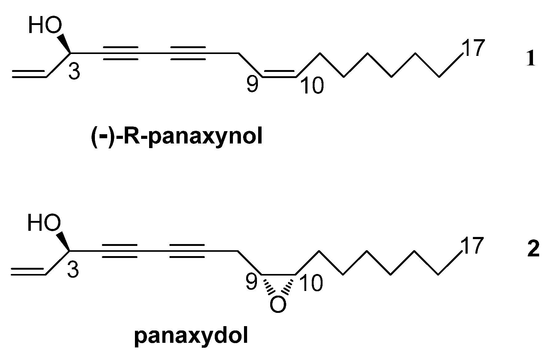
2.1. Isolation and Identification of Panaxynol (1) and Panaxydol (2)

Lyophilized roots from plants treated with 13CO2 or root cultures enriched with [U-13C6]glucose were extracted with hexane. Purification of the corresponding extracts using column chromatography yielded the less polar panaxynol (1) and more polar panaxydol (2) in pure form; both metabolites were identified by comparing their spectroscopic data (1H and 13C-NMR) to those reported in the literature [38,39,40,41]. However, in view of the conflicting reports on the structures of this type of polyacetylenes (e.g., their stereoconfigurations), it is important to emphasize the correct identification of the isolated panaxynol; this metabolite was originally reported by Takahashi from P. ginseng [6] and later reported with the names falcarinol from Falcaria vulgaris [42] and carotatoxin from Daucus carota [43]. The first attempt to establish the absolute stereochemistry at C-3 of the compound was carried out by Larsen et al. [44], who described falcarinol from Seseli gummiferum as having a 3-(R) chirality on the basis of chemical correlation studies. The second attempt was carried out by Shim et al. [45,46] who described panaxynol as having a 3-(S) chirality on the basis of CD measurements. More recently, modified Mosher’s methods have described falcarinol from Dendropanax arboreus as being dextrorotatory and having the 3-(S) chirality [47], whereas panaxynol from P. ginseng was reported as being levorotatory and having the 3-(R) chirality [40]. These later reports were confirmed by Zheng et al. [48] who carried out the enantiospecific synthesis of the two isomers of falcarinol/panaxynol and demonstrated that the 3-(R) and 3-(S) chiralities correspond to the levorotatory and dextrorotatory enantiomers, respectively. The negative value of the optical activity of panaxynol obtained in this study also indicated its 3-(R) chirality.

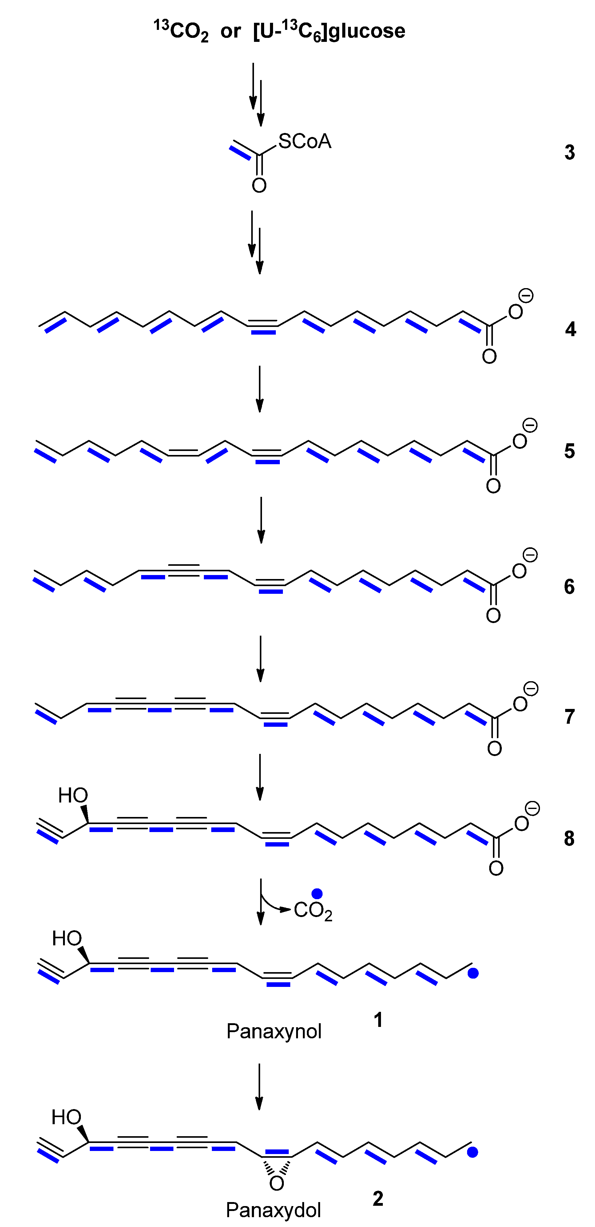
2.2. Biosynthesis of Panaxynol and Panaxydol in P. ginseng

2.2.1. In planta Experiments with 13CO2

Experiments with 13CO2 best resemble the physiological conditions for plants and the labeling profiles in the biosynthetic products represent quasi undisturbed in planta conditions. More specifically, the results obtained from these experiments are free from artifacts due to metabolic stress reactions (e.g., triggered by wounding in labeling experiments with cut plant organs) or due to the usage of non-physiological substrates in experiments with cell cultures. The strategic idea behind isotopologue profiling using 13CO2 is the photosynthetic generation of completely 13C-labeled metabolic intermediates (e.g., triose and pentose phosphates and products thereof) during an incubation period with 13CO2 (pulse period). During a subsequent chase period, the plants are allowed to grow under standard conditions (i.e., in a natural atmosphere with 12CO2) for several days in which unlabeled photosynthetic intermediates are generated (i.e., with 12C). These 13C- and 12C-intermediates from the pulse and the chase periods, respectively, are then taken by the plant as precursors for downstream biosynthetic processes. Consequently, the combination of these precursor units results in specific mixtures of 13C-isotopologues in the product. In other words, mixtures of unlabeled and multiple 13C-labeled isotopologues are generated as a consequence of the biosynthetic history of the metabolites under study. Using quantitative NMR spectroscopy, these isotopologue profiles can be assigned and attributed to biosynthetic pathways. Several recent examples have demonstrated the power of this experimental approach [49,50,51].
13CO2-labeling experiments of P. ginseng (Figure 2) were carried out using a portable 13CO2 unit [52]. To this aim, six-year-old plants of P. ginseng growing under field conditions were exposed to a 13CO2 atmosphere for 9.5 h and then allowed to grow for 19 days under natural conditions. Extraction of the roots yielded a mixture which, after purification, led to the isolation of labeled panaxynol (1) and panaxydol (2). The overall 13C-abundances (as determined from the 1H-NMR spectra of the compounds) were 1.5–2% for all carbon atoms. The relative intensities of the singlet signals due to 13C1-isotopologues in the 13C-NMR spectra of the labeled and unlabelled samples were identical. However, in the 13C-NMR spectra of labeled panaxynol (1) and panaxydol (2), all carbon signals, with the exception of the methyl carbon signals at 14.3 ppm, showed 13C-coupled satellite pairs (reflecting 13C2-isotopologues; Table 1, Table 2; Figure 3) at relative intensities of ca. 15% in the overall signal integrals for a given carbon atom. The same satellites from the unlabelled samples could display only 1% relative intensity in the global intensity of a given carbon due to the natural 13C-abundance of a 13C2-isotopologue (i.e., 0.01 mol%). Notably, with the given amounts of the samples (i.e., 2–3 mg, respectively), these natural abundance satellites could not be detected at all with the unlabelled compounds due to the low intrinsic sensitivity of 13C-NMR spectroscopy (for a review of quantitative 13C-NMR spectroscopy, see [53]). With the 13C-enriched samples, however, these satellites were detected and the analysis of the coupling constants for the 13C2-signals allowed the assignments of eight pairs of 13C2-labeled isotopologues, namely [1,2-13C2]-, [3,4-13C2]-, [5,6-13C2]-, [7,8-13C2]-, [9,10-13C2]-, [11,12-13C2]-, [13,14-13C2]-, [15,16-13C2]-1 and -2, respectively, at similar or identical abundances of ca. 0.2 mol-% (see also Figure 4 where these isotopologues are indicated by bold bars connecting 13C-atoms in the molecule). The observed pattern of adjacent 13C-pairs indicates a polyketide-type biosynthesis, starting from [1,2-13C2]-acetyl-CoA/malonyl-CoA via a mixture of [1,2-13C2]-, [3,4-13C2]-, [5,6-13C2]-, [7,8-13C2]-, [9,10-13C2]-, [11,12-13C2]-, [13,14-13C2]-, [15,16-13C2]-, and [17,18-13C2]-fatty acids that is finally converted into the isotopologue profile of 1 and 2.
On this basis, it can be concluded that decarboxylation of a putative C18-intermediate takes place at the site where the uncoupled methyl group is finally observed in panaxynol or panaxydol (cf. Figure 4). Furthermore, it is suggestive to propose oleic acid (4) and crepenynic acid (6) as potential intermediates, since the triple and double bonds are located at the same positions as in the C17-polyacetylenes after decarboxylation. Decarboxylation could then occur at the level of intermediate 8 resulting in panaxynol (1) (Scheme 1). Panaxydol (2) could be formed by oxygenation of the C9-C10 double bond in panaxynol. However, it is important to keep in mind that the decarboxlation step can also occur upstream, i.e., with 3-hydroxyoleic acid or 3-hydroxylinoleic acid as intermediates. However, in this scenario desaturases would unusually act on non-carboxylated substrates in order to introduce the required triple and double bonds in panaxynol and panaxydol. Theoretically, carboxylation of a labeled C16-fatty acid intermediate could also give rise of the detected labeling pattern. On the other hand, this hypothesis would be in contrast to earlier results that reported C18-precursors for C17-polyacetylenes [9,35,36,37].
Figure 2. The portable unit used in this study for controlled incubation of P. ginseng with 13CO2 under field conditions.
Molecules 18 07686 g002
Table 1. 1H- and 13C-NMR data of 13C-labelled panaxynol (1) (solvent, CDCl3; δ in ppm).
Atom1H (δ)JHH (Hz)Atom13C (δ)JCC (Hz)
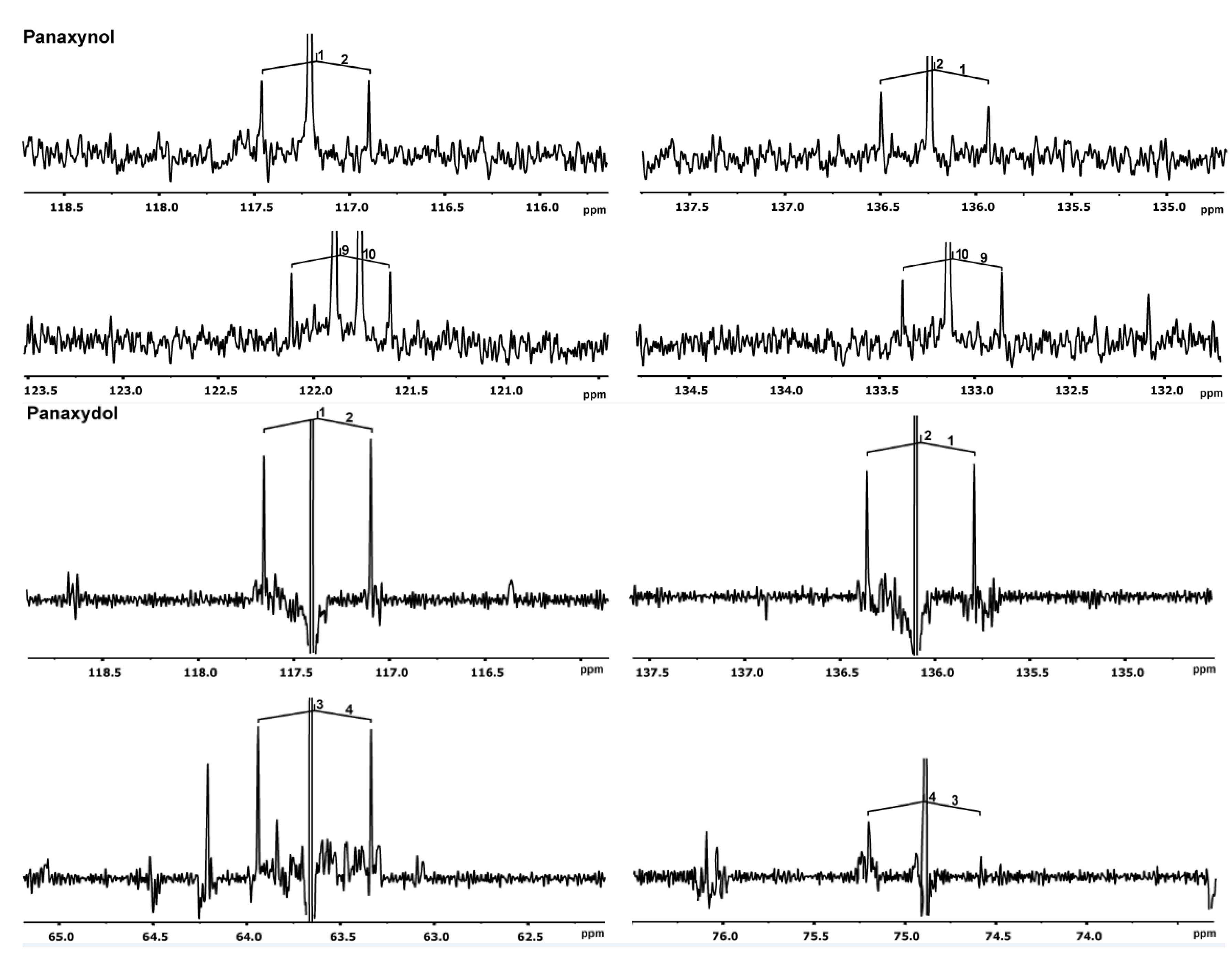
1a
1b
2
3
8a
8b
9
10
11
12
13
14
15
16
17
5.26
5.47
5.95
4.92
2.39
2.70
3.14
2.96
1.45–1.55
1.25–1.40
1.25–1.40
1.25–1.40
1.25–1.40
1.25–1.40
0.89
1H, ddd; 10.2, 1.5, 1.0
1H, ddd; 17.1, 1.5, 1.0
1H, ddd;16.8, 10.2, 5.4
1H, br d; 5.2
1H, ddd;17.7, 7.1, 0.9
1H, ddd; 17.7, 5.5, 0.9
1H, ddd; 7.1, 5.5, 4.2
1H, br td; 6.1, 4.1
2H, m
10H, m
10H, m
10H, m
10H, m
10H, m
3H, br t; 6.8
1
2
3
4
5
6
7
8
9
10
11
12
13
14
15
16
17
117.4
136.1
63.7
75.0
71.0
66.4
77.4
19.6
54.4
57.1
27.7
26.6
29.6
29.3
31.9
22.8
14.3
70.9
70.9
75.8
75.8
156.9
157.0
nd *
68.2
29.9
29.9
33.8
33.9
45.7
45.3
34.5
34.5
-
* nd, cannot be measured due to signal overlap.
Table 2. 1H- and 13C-NMR data of 13C-labelled panaxydol (2) (solvent, CDCl3; δ in ppm).
Atom1H (δ)JHH (Hz)Atom13C (δ)JCC (Hz)
1a
1b
2
3
8
9
10
11
12
13
14
15
16
17
5.47
5.24
5.94
4.91
3.03
5.39
5.52
2.03
1.24–1.39
1.24–1.39
1.24–1.39
1.24–1.39
1.24–1.39
0.88
1H, ddd; 17.1, 1.2
1H, ddd; 10.1, 1.2
1H, ddd; 17.0, 10.2, 5.4
3H, t; 5.9
2H, d; 6.9
1H, ddddd; 11.3, 6.1, 1.6
1H, ddddd; 9.8, 8.1, 1.7
2H, ddd; 10.7, 6.9
10H, m
10H, m
10H, m
10H, m
10H, m
3H, t; 6.9
1
2
3
4
5
6
7
8
9
10
11
12
13
14
15
16
17
117.2
136.3
63.7
74.3
71.5
64.1
80.5
17.8
122.0
133.3
27.4
29.3
29.3
29.3
31.7
22.8
14.3
71.0
70.7
76.0
76.0
156.6
nd *
68.1
67.8
71.4
71.3
34.0
34.0
34.6
34.6
34.5
34.5
-
* nd, cannot be measured due to signal overlap.
Figure 3. 13C-NMR signals of panaxynol (1) and panaxydol (2) from the 13CO2 experiment. Couplings between 13C-atoms are indicated. Notably, satellites due to couplings between three adjacent 13C-atoms are not observed in the upfield or downfield regions of the doublets.
Molecules 18 07686 g003
Scheme 1. Proposed biosynthetic pathway of panaxynol (1) and panaxydol (2). Adjacent 13C-atoms detected in experiments with 13CO2 or [U-13C6]glucose are indicated by blue bars.
Molecules 18 07686 g004

2.2.2. Experiments in Root Cultures with [U-13C6]glucose

Panaxynol (1), but not panaxydol (2), was obtained from the extract of P. ginseng hairy root cultures cultivated for four weeks in SH medium [54] containing a mixture of 88 mM sucrose and 4.4 mM [U-13C6]glucose. The carbon signals in the 13C-NMR spectrum of 1 showed the characteristic 13C-coupling satellites, with coupling constants identical to those described above for the panaxynol sample obtained from plants labeled with 13CO2 (Table 1). However, the relative sizes of the 13C-coupling satellites were higher (ca. 70% in the overall intensity for a given carbon) in the 13C spectrum of the panaxynol from the cultures than those from the field-grown plants. Not surprisingly, this reflects the high incorporation rates of [U-13C6]glucose in the stationary labeling experiment with the root cultures. It can be concluded that [U-13C6]glucose was efficiently incorporated into 1 via [U-13C2]acetyl-CoA following the same or closely related biosynthetic routes as described above for the plants growing under natural conditions. This shows that the pathways are obviously not affected by the supply of carbohydrates as carbon sources for root cultures.

3. Experimental

3.1. Chemicals

13CO2 and [U-13C6]glucose (99% 13C-abundance) and other compounds were obtained from Sigma-Aldrich (Steinheim, Germany).

3.2. Plants and Labeling Experiments with 13CO2

Labeling experiments with 13CO2 were carried out in August 2011 using six-year-old plants of Panax ginseng C.A. Meyer growing in the commercial field of FloraFarm (Walsrode, Germany). The plant was kept under a 13CO2 atmosphere (700 ppm) from 9 am for 9.5 h (pulse period; 13CO2 consumption: about 11 L) and left for 19 days (chase period) under natural field conditions before collecting the roots for extraction. The roots were washed, cut in pieces, frozen (liquid nitrogen) and lyophilized. Finally, the dry root pieces were ground using a mortar.

3.3. Root Cultures

Transformed roots of P. ginseng C.A. Meyer were induced from four-year-old rhizomes after infection with Agrobacterium rhizogenes A4 strain. Sterilized root discs were wounded with a sterile needle loaded with an A. rhizogenes suspension grown in liquid YEB medium [54] for 24 h at 28 ± 2 °C on a rotary shaker (Adolf Kühner AG, Birsfelden, Switzerland) at 100 rpm. The inoculated root discs were placed on Schenk and Hildebrandt’s medium (SH) [55] containing 3% (w/v) sucrose, 0.1% (w/v) myo-inositol and 0.27% (w/v) Phytagel (Sigma) at 26 °C. The medium was adjusted to pH 7.0 before autoclaving. After two days of co-cultivation, the explants were transferred to fresh medium containing cefotaxime (500 mg/L) in order to eliminate bacteria. After one or two months of cultivation, roots started to appear at the infection sites. In order to obtain the root lines, single roots were picked off and placed onto new media containing cefotaxime. Hairy roots free of bacterial contamination were cultured on hormone-free SH solid medium, in the dark at 26 °C. After six months of subculturing, the roots were cultured every two weeks on fresh solid medium and the selected root lines were transferred to SH liquid medium and kept in a rotary shaker at 100 rpm and 26 °C in the dark. The transformed nature of these root lines was confirmed by the presence of the TL-DNA rol C gene in the plant genome, detected by the pRiA4 by polymerase chain reaction analysis as described previously [56].

3.4. Labeling Experiments with [U-13C6]Glucose

The selected P. ginseng root line was cultured for four weeks in hormone-free SH liquid medium supplemented with 4.4 mM of [U-13C6]glucose. The root cultures were initiated from inocula of 2 ± 0.2 g of roots (fresh weight) maintained in 100-mL Erlenmeyer flasks, each containing 20 mL of SH medium. In this experiment, a total of 70 Erlenmeyer flasks were used, which corresponds to a total volume of 1.4 L. After four weeks, the roots (381 g, fresh weight) were harvested by filtration and freeze dried (21 g, dry weight).

3.5. Isolation of Panaxynol (1) and Panaxydol (2)

Dry-powdered roots or root cultures (about 20 g) were extracted by refluxing (70 °C) twice for 3 h with hexane (300 mL). The solvent was evaporated under reduced pressure and the resulting crude extract (209 mg) was purified by open column chromatography (3 × 25 cm) using a mixture of hexane/acetone/methanol (80:18:2; v/v) as the eluting solvent (fraction volume, 5 mL). Panaxynol (1, 2.3 mg) was obtained in pure form at a retention volume of 155 mL. Panaxydol (2, 2.0 mg) was obtained in pure form from the field-grown roots of P. ginseng at a retention volume of 205 mL. The identity of both metabolites was confirmed by comparing their spectroscopic data (1H- and 13C-NMR) with those reported in literature [38,39,40,41]. The chirality at the C-3 position of panaxynol (1) was established as (R) by comparing its optical activity value ([α]D −28.5°, c 0.17, CHCl3) with that reported in the literature ([α]D −31.5°, c 1.0, CHCl3) [57].

3.6. Chromatography

Thin-layer chromatography (TLC) was carried out using aluminum-backed silica gel (60 F254) plates (Merck, 0.2 mm thickness) and the spots on the TLC plates were visualized by using a solution of H2SO4/MeOH (1:10; v/v) followed by heating (95–100 °C). Column chromatography purifications were performed using silica gel 60 (0.063–0.200 mm; 70–230 mesh; ASTM) from Merck (Darmstadt, Germany).

3.7. NMR Spectroscopy and Optical Rotation

NMR spectra were recorded at 27 °C using DRX 500, Avance I 500 and Avance III 500 spectrometers (Bruker Instruments, Karlsruhe, Germany). 1H- and 13C-NMR spectra were measured in CDCl3. For the measurement of 13C-NMR spectra, a cryo-probe head (5 mm QNP, inner coil = 13C) was used. One-dimensional 1H-spectra and COSY, HSQC, and HMBC experiments were performed with an inverse probe head (5 mm SEI, inner coil = 1H). The resonance frequencies of 1H and 13C were 500.1 MHz and 125.8 MHz respectively. Data analysis was done with TOPSPIN 3.0 (Bruker) or MestReNova 7.0.0 (Mestrelab Research, Santiago de Compostela, Spain). The optical rotation was measured using a Perkin Elmer 241 MC polarimeter (Perkin Elmer, Waltham, MA, USA).

4. Conclusions

NMR-based isotopologue profiling of panaxydol and panaxynol confirmed their assumed origin from acetyl-CoA/malonyl-CoA via fatty acids with crepenynate as the putative intermediate. The decarboxylation site of the C18 intermediate(s) could now be clearly located to the methyl site of the product, panaxynol/panaxydol. The knowledge about the metabolite flux leading to the bioactive compounds and the factors for its control are useful to establish plants or root cultures of P. ginseng to produce C17-polyacetylenes at high yields. The study shows the feasibility of 13CO2-experiments to elucidate the biosynthetic origin of metabolites/products in field-grown plants. With the present study, it is demonstrated that the biosynthesis of root metabolites can be studied by pulse/chase experiments starting from the fixation of 13CO2 by the leaves.

Acknowledgments

We thank the Hans-Fischer-Gesellschaft e.V. (München) and the Deutsche Forschungsgemeinschaft (EI 384/8-1) for generous sponsoring of this research work. LMPR and WE thank the German Academic Exchange Service (DAAD, A/11/00471) and CONACYT-México (exp. No. 160813) for supporting the sabbatical stay of LMPR at the Technische Universität München. The research carried out at the UB has been supported by a grant from the Spanish MEC (BIO2011-29856-C02-01) and a grant from the Catalan Government (2009SGR1217).

Conflicts of Interest

The authors declare no conflict of interest.

References

  1. Jia, L.; Zhao, Y. Current evaluation of the millennium phytomedicine—ginseng (I): Etymology, Pharmacognosy, Phytochemistry, Market and regulations. Curr. Med. Chem. 2009, 16, 2475–2484. [Google Scholar] [CrossRef]
  2. Qi, L.-W.; Wang, C.-Z.; Yuan, C.-S. Ginsenosides from American ginseng: Chemical and pharmacological diversity. Phytochemistry 2011, 72, 689–699. [Google Scholar] [CrossRef]
  3. Baek, S.-H.; Bae, O.-N.; Park, J.H. Recent methodology in Ginseng analysis. J. Ginseng Res. 2012, 36, 119–134. [Google Scholar] [CrossRef]
  4. Herrmann, F.; Sporer, F.; Tahrani, A.; Wink, M. Antitrypanosomal properties of Panax ginseng C.A. Meyer: New possibilities for a remarkable traditional drug. Phytother. Res. 2013, 27, 86–98. [Google Scholar] [CrossRef]
  5. Christensen, L.P. Aliphatic C17-polyacetylenes of the falcarinol type as potential health promoting compounds in food plants of the Apiaceae family. Recent Pat. Food Nutr. Agric. 2011, 3, 64–77. [Google Scholar] [CrossRef]
  6. Takahashi, M.; Isoi, K.; Kimura, Y.; Yoshikura, M. Studies on the components of Panax ginseng C.A. Meyer. J. Pharm. Soc. Japan 1964, 84, 757–759. [Google Scholar]
  7. Hirakura, K.; Takagi, H.; Morita, M.; Nakajima, K.; Niitsu, K.; Sasaki, H.; Maruno, M.; Okada, M. Cytotoxic activity of acetylenic compounds from Panax ginseng. Nat. Med. 2000, 54, 342–345. [Google Scholar]
  8. Zidorn, C.; Jöhrer, K.; Ganzera, M.; Schubert, B.; Sigmund, E.M.; Mader, J.; Greil, R.; Ellmerer, E.P.; Stuppner, H. Polyacetylenes from the Apiaceae vegetables carrot, celery, fennel, parsley, and parsnip and their cytotoxic activities. J. Agric. Food Chem. 2005, 53, 2518–2523. [Google Scholar] [CrossRef]
  9. Minto, R.E.; Blacklock, B.J. Biosynthesis and function of polyacetylenes and allied natural products. Prog. Lipid Res. 2008, 47, 233–306. [Google Scholar] [CrossRef]
  10. Xu, L.-L.; Han, T.; Wu, J.-Z.; Zhang, Q.-Y.; Zhang, H.; Huang, B.-K.; Rahman, K.; Qin, L.-P. Comparative research of chemical constituents, antifungal and antitumor properties of ether extracts of Panax ginseng and its endophytic fungus. Phytomedicine 2009, 16, 609–616. [Google Scholar] [CrossRef]
  11. Hansen, S.L.; Purup, S.; Christensen, L.P. Bioactivity of falcarinol and the influence of processing and storage on its content in carrots (Daucus carota L). J. Sci. Food Agric. 2003, 83, 1010–1017. [Google Scholar] [CrossRef]
  12. Saita, T.; Katano, M.; Matsunaga, H.; Yamamoto, H.; Fujito, H.; Mori, M. The first specific antibody against cytotoxic polyacetylenic alcohol, panaxynol. Chem. Pharm. Bull. 1993, 41, 549–552. [Google Scholar] [CrossRef]
  13. Bernart, M.W.; Cardellina, J.H.; Balaschak, M.S.; Alexander, M.; Shoemaker, R.H.; Boyd, M.R. Cytotoxic falcarinol oxylipins from Dendropanax arboreus. J. Nat. Prod. 1996, 59, 748–753. [Google Scholar] [CrossRef]
  14. Matsunaga, H.; Katano, M.; Yamamoto, H.; Fujito, H.; Mori, M.; Takata, K. Cytotoxyc activity of polyacetylene compounds in Panax ginseng C.A. Meyer. Chem. Pharm. Bull. 1990, 38, 3480–3482. [Google Scholar] [CrossRef]
  15. Kuo, Y-C.; Lin, Y-L.; Huang, C-P.; Shu, J-W.; Tsai, W-J. A tumor cell growth inhibitor from Saposhnikovae divaricata. Cancer Invest. 2002, 20, 955–964. [Google Scholar] [CrossRef]
  16. Kobaisy, M.; Abramowski, Z.; Lermer, L.; Saxena, G.; Hancock, R.E.W.; Towers, G.H.N. Antimycobacterial polyynes of devil’s club (Oplopanax horridus), a North American native medicinal plant. J. Nat. Prod. 1997, 60, 1210–1213. [Google Scholar] [CrossRef]
  17. Kemp, M.S. Falcarindiol: An antifungal polyacetylene from Aegopodium podagraria. Phytochemistry 1978, 17, 1002. [Google Scholar] [CrossRef]
  18. Harding, V.K.; Heale, J.B. Isolation and identification of the antifungal compounds accumulating in the induced resistance response of carrot root slices to Botrytis cinerea. Physiol. Plant. Pathol. 1980, 17, 277–289. [Google Scholar]
  19. Hansen, L.; Boll, P.M. Polyacetylenes in Araliaceae: Their chemistry, biosynthesis and biological significance. Phytochemistry 1986, 25, 285–293. [Google Scholar] [CrossRef]
  20. Otsuka, H.; Komiya, T.; Fujioka, S.; Goto, M.; Hiramatsu, Y.; Fujimura, H. Studies on anti-inflammatory agents. IV. Anti-inflammatory constituents from roots of Panax ginseng C.A. Meyer. Yakugaku Zasshi 1981, 101, 1113. [Google Scholar]
  21. Baba, K.; Tabata, Y.; Kozawa, M.; Kimura, Y.; Arichi, S. Studies on Chinese traditional medicine Fang-feng (I). Structures and physiological activities of polyacetylene compounds from Saposhnikoviae radix. Shoyakugaku Zasshi 1987, 41, 189–194. [Google Scholar]
  22. Teng, C.-M.; Kuo, S.-C.; Ko, F.-N.; Lee, J.C.; Lee, L.-G.; Chen, S.-C.; Huang, T.-F. Antiplatelet actions of panaxynol and ginsenosides isolated from ginseng. Biochim. Biophys. Acta 1989, 990, 315–320. [Google Scholar] [CrossRef]
  23. Alanko, J.; Kurahashi, Y.; Yoshimoto, T.; Yamamoto, S.; Baba, K. Panaxynol, a polyacetylene compound isolated from oriental medicines, inhibits mammalian lipoxygenases. Biochem. Pharmacol. 1994, 48l, 1979–1981. [Google Scholar]
  24. Nie, B.M.; Jiang, X.Y.; Cai, J.X.; Fu, S.L.; Yang, L.M.; Lin, L.; Hang, Q.; Lu, P.L.; Lu, Y. Panaxydol and panaxynol protect cultured cortical neurons against Abeta25-35-induced toxicity. Neuropharmacology 2008, 54, 845–853. [Google Scholar] [CrossRef]
  25. Yang, Z.-H.; Sun, K.; Yan, Z.-H.; Suo, W.-H.; Fu, G.-H.; Lu, Y. Panaxynol protects cortical neurons from ischemia-like injury by up-regulation of HIF-1α expression and inhibition of apoptotic cascade. Chem. Biol. Interact. 2010, 183, 165–171. [Google Scholar] [CrossRef]
  26. Ahn, B.-Z.; Kim, S.-I. Beziehung zwischen Struktur und cytotoxischer Aktivität von Panaxydol-Analogen gegen L1210 Zellen. Arch. Pharm. 1988, 321, 61–63. [Google Scholar] [CrossRef]
  27. Matsunaga, H.; Katano, M.; Yamamoto, H.; Mori, M.; Takata, K. Studies on the panaxytriol of Panax ginseng C.A. Meyer. Isolation, Determination and antitumor activity. Chem. Pharm. Bull. 1989, 37, 1279–81. [Google Scholar] [CrossRef]
  28. Kobæk-Larsen, M.; Christensen, L.P.; Vach, W.; Ritskes-Hoitinga, J.; Brandt, K. Inhibitory effects of feeding with carrots or (−)-falcarinol on development of azoxymethane-induced preneoplastic lesions in the rat colon. J. Agric. Food Chem. 2005, 53, 1823–1827. [Google Scholar] [CrossRef]
  29. Ahn, B.Z.; Kim, S.I.; Lee, Y.H. Acetylpanaxydol und Panaxydolchlorhydrin, zwei neue, gegen L1210-Zellen cytotoxische Polyine aus Koreanischem Ginseng. Arch. Pharm. 1989, 322, 223. [Google Scholar] [CrossRef]
  30. Matsunaga, H.; Saita, T.; Nagamo, F.; Mori, M.; Katano, M. A possible mechanism for the cytotoxicity of a polyacetylenic alcohol, panaxytriol: inhibition of mitochondrial respiration. Cancer Chemother. Pharmacol. 1995, 35, 291–296. [Google Scholar] [CrossRef]
  31. Hansen, L.; Hammershøy, O.; Boll, P.M. Allergic contact dermatitis from falcarinol isolated from Schefflera arboricola. Contact Dermat. 1986, 14, 91–93. [Google Scholar] [CrossRef]
  32. Hausen, B.M.; Bröhan, J.; König, W.A.; Faasch, H.; Hahn, H.; Bruhn, G. Allergic and irritant contact dermatitis from falcarinol and didehydrofalcarinol in common ivy (Hedera helix L.). Contact Dermat. 1987, 17, 1–9. [Google Scholar] [CrossRef]
  33. Gafner, F.; Epstein, W.; Reynolds, G.; Rodriguez, E. Human maximization test of falcarinol, the principal contact allergen of English ivy and Algerian ivy (Hedera helix, H. canariensis). Contact Dermat. 1988, 19, 125–128. [Google Scholar] [CrossRef]
  34. Machado, S.; Silva, E.; Massa, A. Occupational allergic contact dermatitis from falcarinol. Contact Dermat. 2002, 47, 113–114. [Google Scholar]
  35. Barley, G.C.; Jones, E.H.R.; Thaller, V. Crepenynate as a precursor of falcarinol in carrot tissue culture. In Chemistry and Biology of Naturally-Occurring Acetylenes and Related Compounds; Lam, J., Breteler, H., Arnason, T., Hansen, L., Eds.; Elsevier: Amsterdam, The Netherlands, 1988; pp. 85–91. [Google Scholar]
  36. Bohlmann, F.; Burkhardt, T. Polyacetylenverbindungen. 166. Über die Biogenese von C17-Polyinen. Chem. Ber. 1969, 102, 1702–1706. [Google Scholar] [CrossRef]
  37. Bu’Lock, J.D.; Smalley, H.M. The biosynthesis of polyacetylenes. Part V. The role of malonate derivatives, and the common origin of fatty acids, polyacetylenes, and “acetate-derived” phenols. J. Chem. Soc. 1967, 332–336. [Google Scholar]
  38. Poplawski, J.; Wrobel, J.T.; Glinka, T. Panaxydol, a new polyacetylenic epoxide from Panax ginseng roots. Phytochemistry 1980, 19, 1539–1541. [Google Scholar]
  39. Hirakura, K.; Morita, M.; Nakajima, K.; Ikeya, Y.; Mitsuhashi, H. Polyacetylenes from the roots of Panax Ginseng. Phytochemistry 1991, 30, 3327–3333. [Google Scholar] [CrossRef]
  40. Kobayashi, M.; Mahmud, T.; Umezome, T.; Wang, W.; Murakami, N.; Kitagawa, I. The absolute stereostructures of the polyacetylenic constituents of Ginseng Radix Rubra. Tetrahedron 1997, 53, 15691–15700. [Google Scholar]
  41. Seger, C.; Godejohann, M.; Spraul, M.; Stuppner, H.; Hadacek, F. Reaction product analysis by high-performance liquid chromatography-solid-phase extraction-nuclear magnetic resonance. Application to the absolute configuration determination of naturally occurring polyyne alcohols. J. Chromatogr. A 1136, 82–88. [Google Scholar]
  42. Bohlmann, F.; Niedballa, U.; Rode, K.M. New polyines with a C17-chain. Chem. Ber. 1966, 99, 3552–3558. [Google Scholar] [CrossRef]
  43. Crosby, D.G.; Aharonson, N. The structure of carotatoxin, a natural toxicant from carrot. Tetrahedron 1967, 23, 465–472. [Google Scholar] [CrossRef]
  44. Larsen, P.K.; Nielsen, B.E.; Lemmich, J. The absolute configuration of falcarinol, an acetylenic compound from the roots of Seseli gummiferum Pall. Acta Chem. Scand. 1969, 23, 2552–2554. [Google Scholar] [CrossRef]
  45. Shim, S.C.; Koh, H.Y.; Chang, S. Determination of absolute stereochemistry of panaxynol. Tetrahedron Lett. 1985, 26, 5775–5776. [Google Scholar] [CrossRef]
  46. Shim, S.C.; Koh, H.Y.; Chang, S.; Moon, S.K.; Min, T.-J. Absolute configuration of p-substituted benzoates of panaxynol. Bull. Korean Chem. Soc. 1986, 7, 106–108. [Google Scholar]
  47. Bernart, M.W.; Hallock, Y.F.; Cardellina, J.H., II; Boyd, M.R. Stereochemistry of enynols—A caveat on the excition chirality method. Tetrahedron Lett. 1994, 35, 993–994. [Google Scholar] [CrossRef]
  48. Zheng, G.; Lu, W.; Aisa, H.A.; Cai, J. Absolute configuration of falcarinol, a potent antitumor agent commonly occurring in plants. Tetrahedron Lett. 1999, 40, 2181–2182. [Google Scholar] [CrossRef]
  49. Römisch-Margl, W.; Schramek, N.; Radykewicz, T.; Ettenhuber, C.; Eylert, E.; Huber, C.; Römisch-Margl, L.; Schwarz, C.; Dobner, M.; Demmel, N.; et al. 13CO2 as a universal metabolic tracer in isotopologue perturbation experiments. Phytochemistry 2007, 68, 2273–2289. [Google Scholar] [CrossRef]
  50. Ostrozhenkova, E.; Eylert, E.; Schramek, N.; Golan-Goldhirsh, A.; Bacher, A.; Eisenreich, W. Biosynthesis of the chromogen hermidin from Mercurialis annua L. Phytochemistry 2007, 68, 2816–2824. [Google Scholar] [CrossRef]
  51. Schramek, N.; Wang, H.; Römisch-Margl, W.; Keil, B.; Radykewicz, T.; Winzenhörlein, B.; Beerhues, L.; Bacher, A.; Rohdich, F.; Gershenzon, J.; et al. Artemisinin biosynthesis in growing plants of Artemisia annua. A 13CO2 study. Phytochemistry 2010, 71, 179–187. [Google Scholar] [CrossRef]
  52. Eisenreich, W.; Huber, C.; Kutzner, E.; Knispel, N.; Schramek, N. Isotopologue profiling—Toward a better understanding of metabolic pathways. In The Handbook of Plant Metabolomics, Metabolite Profiling and Networking; Weckwerth, W., Kahl, G., Eds.; Wiley: Weinheim, Germany, 2013; pp. 25–56. [Google Scholar]
  53. Eisenreich, W.; Bacher, A. Advances of high-resolution NMR techniques in the structural and metabolic analysis of plant biochemistry. Phytochemistry 2007, 68, 2799–2815. [Google Scholar] [CrossRef]
  54. Vervliet, G.; Holsters, M.; Teuchy, H.; Van, M.M.; Schell, J. Characterization of different plaque-forming and defective temperate phages in Agrobacterium. J. Gen. Virol. 1975, 26, 33–48. [Google Scholar] [CrossRef]
  55. Schenk, R.U.; Hildebrandt, A. Medium and techniques for induction and growth of monocotyledonous and dicotyledonous plant cell cultures. Can. J. Bot. 1972, 50, 199–204. [Google Scholar] [CrossRef]
  56. Mallol, A.; Cusidó, R.M.; Palazón, J.; Bonfill, M.; Morales, C.; Piñol, M.T. Ginsenoside production in different phenotypes of Panax ginseng transformed roots. Phytochemistry 2001, 57, 365–371. [Google Scholar] [CrossRef]
  57. McLaughlin, N.P.; Butler, E.; Evans, P.; Brunton, N.P.; Koidis, A.; Rai, D.K. A short synthesis of (+) and (−)-falcarinol. Tetrahedron 2010, 66, 9681–9687. [Google Scholar] [CrossRef]
  • Sample Availability: Samples of the compounds panaxynol and panaxydol are available from the authors.

