HPLC Analysis of Phenolics Compounds and Antioxidant Capacity of Leaves of Vitex megapotamica (Sprengel) Moldenke

Abstract
:
1. Introduction
2. Results and Discussion
2.1. Phenolics Contents, Total Flavonoids, Condensed Tannins and Free Radical-Scavenging Activities

| Extract/fraction | TP ± SD (mg GAE/g) | TF ± SD (mg RE/g) | T ± SD (mg CaE/g) | IC50 ± SD (μg/mL) |
|---|---|---|---|---|
| Crude extract | 309.1 b ± 0.88 | 198.09 b ± 0.33 | - | 20.44 b ± 0.37 |
| Dichloromethane | 113.1 d ± 1.30 | 148.44 c ± 0.55 | - | 37.63 a ± 0.34 |
| Ethyl acetate | 522.4 a ± 1.12 | 220.48 a ± 0.30 | 3.86 ± 0.53 | 16.21 c ± 0.38 |
| Butanolic | 216.8 c ± 0.42 | 151.02 c ± 0.46 | - | 14.17 c ± 0.21 |
| Ascorbic acid | - | - | - | 14.86 c ± 0.28 |
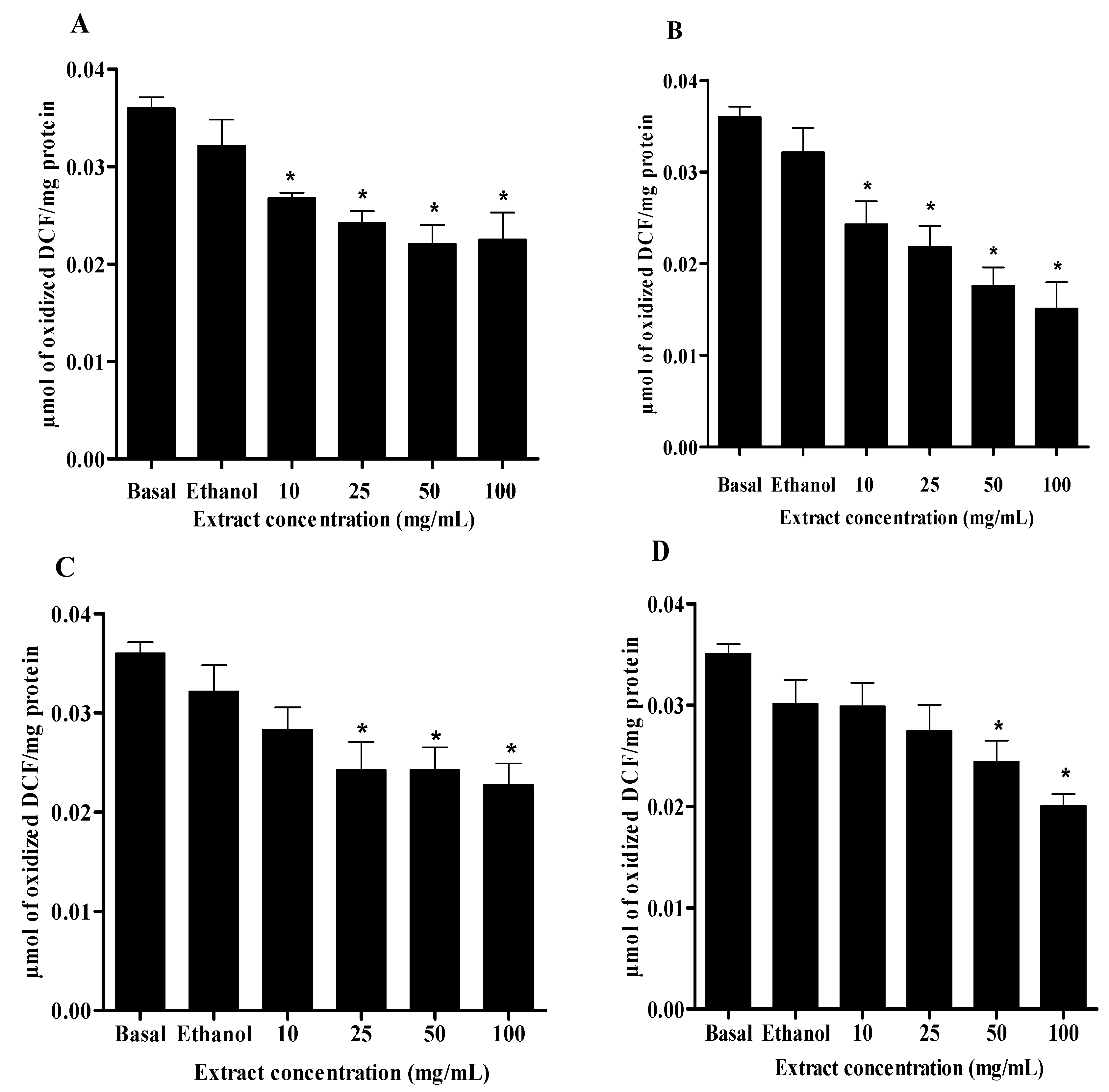
2.2. DCFH-DA Method

2.3. Lipid Peroxidation
| Crude extract/fractions | IC50 µg/mL(mean ± SD) |
|---|---|
| Crude extract | 20.22 ± 4.27 |
| Dichloromethane | 72.72 ± 7.22 |
| Ethyl acetate | 16.36 ± 5.09 |
| Butanolic | 32.78 ± 3.06 |

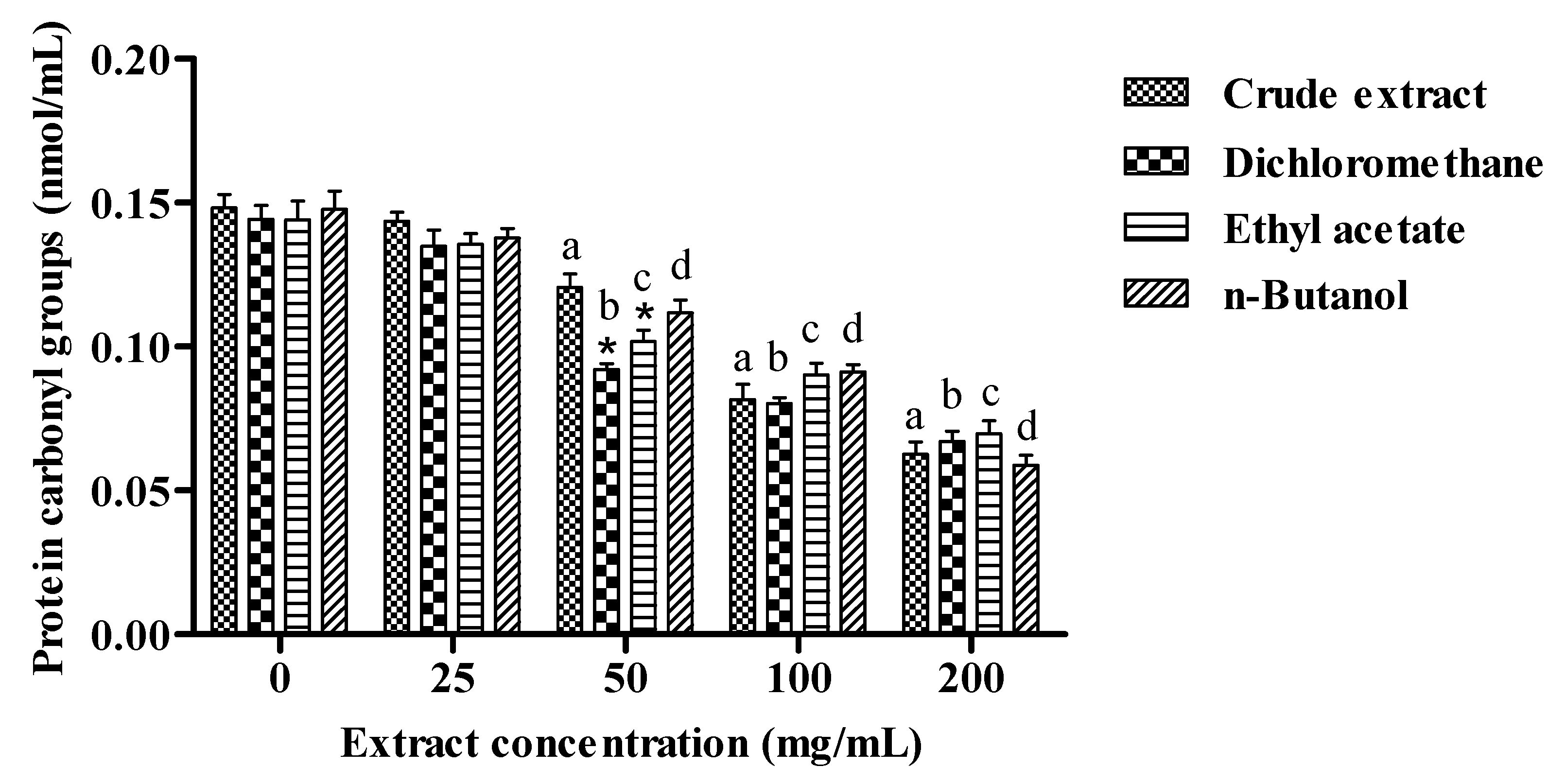
2.4. Protein Carbonyl

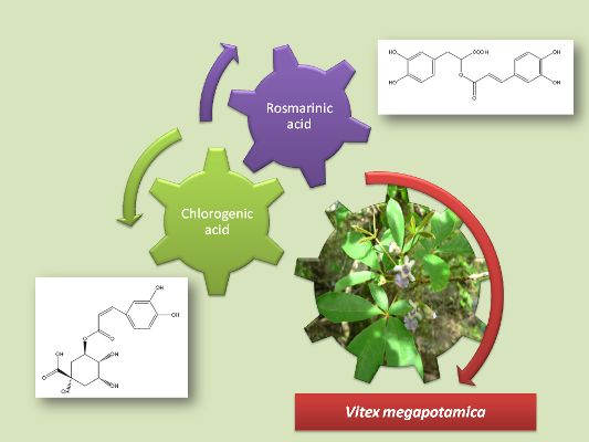
2.5. HPLC-DAD Quantitative Analysis of Rosmarinic and Chlorogenic Acids (RA and CLA)
3. Experimental
3.1. Chemicals
3.2. Animals
3.3. Plant Collection and Extractions
3.4. Determination of Total Phenolics
3.5. Determination of Total Flavonoids
3.6. Determination of Condensed Tannins
3.7. Radical—Scavenging Activity-DPPH Assay

3.8. DCFH-DA Method
3.9. In Vitro Fe (II)-Induced Lipid Peroxidation in Rat Brain
3.10. Protein Carbonyl Groups
3.11. HPLC-DAD Quantitative Analysis of Rosmarinic and Chlorogenic Acids
3.12. Statistical Analysis
4. Conclusions
Acknowledgments
Conflicts of Interest
References
- Boligon, A.A.; Pereira, R.P.; Feltrin, A.C.; Machado, M.M.; Janovik, V.; Rocha, J.B.T.; Athayde, M.L. Antioxidant activities of flavonol derivates from the leaves and stem bark of Scutia buxifolia Reiss. Bioresour. Technol. 2009, 100, 6592–6598. [Google Scholar] [CrossRef]
- Su, X.Y.; Wang, Z.Y.; Liu, J.R. In vitro and in vivo antioxidant activity of Pinus koraiensis seed extract containing phenolic compounds. Food Chem. 2009, 117, 681–686. [Google Scholar] [CrossRef]
- Kintzios, S.; Papageorgiou, K.; Yiakoumettis, I.; Baricevic, D.; Kusar, A. Evaluation of the antioxidants activities of four Slovene medicinal plant species by traditional and novel biosensory assays. J. Pharm. Biomed. Anal. 2010, 53, 773–776. [Google Scholar] [CrossRef]
- Zadra, M.; Piana, M.; Brum, T.F.; Boligon, A.A.; Freitas, R.B.; Machado, M.M.; Stefanello, S.T.; Soares, F.A.A.; Athayde, A.L. Antioxidant activity and phytochemical composition of the leaves of Solanum guaraniticum A. St.-Hil. Molecules 2012, 17, 12560–12574. [Google Scholar]
- Halliwell, B. Vitamin C and genomic stability. Mutat. Res. 2001, 45, 29–35. [Google Scholar]
- Mayne, S.T. Antioxidant nutrition and chronic disease: Use of biomarkers of exposure and oxidative stress status in epidemiological research. J. Nutr. 2003, 133, 933–940. [Google Scholar]
- Alice, C.B.; Mentz, L.; Siqueira, N.C.S.; Silva, G.A.A.B.; Jose, K.F. Plantas Medicinais De Uso Popular: Atlas Farmacognóstico; ULBRA: Canoas, RS, Brazil, 1995; pp. 185–187. [Google Scholar]
- Rimpler, H. Pterosteron, polypodin B and neues ecdysonartiges steroids (viticosteron E) aus Vitex megapotamica. Tetrahedron Lett. 1969, 5, 329–333. [Google Scholar] [CrossRef]
- Rimpler, H. Phytoecdysones and iridoids from Vitex megapotamica. Archiv. der Pharmazie 1972, 10, 746–751. [Google Scholar] [CrossRef]
- Zanatta, L.; Sousa, E.; Cazarolli, L.H.; Junior, A.C.; Pizzolatti, M.G.; Szpoganics, B.; Silva, F.R.M.B. Effect of crude extract and fractions from Vitex megapotamica leaves on hyperglycemia in alloxan-diabetic rats. J. Ethnopharmacol. 2007, 109, 151–155. [Google Scholar] [CrossRef]
- Brandt, A.P.; Oliveira, L.F.S.; Fernandes, F.B.; Alba, J. Avaliação in vivo do efeito hipocolesterolêmico e toxicológico preliminar do extrato bruto hidroalcoólico e decocção da Vitex megapotamica (Spreng) Moldenke (V. montevidensis Cham.). Rev. Bras. Farmacogn. 2009, 19, 388–393. [Google Scholar]
- Brum, T.F.; Zadra, M.; Froeder, A.L.F.; Boligon, A.A.; Frohlich, J.K.; Athayde, M.L. Análise fitoquímica preliminar das folhas de Vitex megapotamica (Sprengel) Moldenke. Rev. Saúde (Santa Maria) 2011, 37, 101–106. [Google Scholar]
- Brum, T.F.; Boligon, A.A.; Frohlich, J.K.; Schwanz, T.G.; Zadra, M.; Piana, M.; Froeder, A.L.F.; Athayde, M.L. Composition and antioxidant capacity of the essential oil of leaves of Vitex megapotamica (Sprengel) Moldenke. Nat. Prod. Res. 2012, 27, 767–770. [Google Scholar]
- Atoui, A.K.; Mansouri, A.; Boskou, G.; Kefalas, P. Tea and herbal infusions: their antioxidant activity and phenolic profile. Food Chem. 2005, 89, 27–36. [Google Scholar] [CrossRef]
- Boligon, A.A.; Brum, T.F.; Frohlich, J.K.; Froeder, A.L.F.; Athayde, M.L. HPLC/DAD profile and determination of total phenolics, flavonoids, tannins and alkaloids contents of Scutia buxifolia Reissek stem bark. Res. J. Phytochem. 2012, 6, 84–91. [Google Scholar] [CrossRef]
- Janovik, V.; Boligon, A.A.; Bandeira, R.V.; Athayde, M.L. HPLC/DAD analysis, determination of total phenolic and flavonoid contents and antioxidant activity from the leaves of Cariniana domestica (Mart) Miers. Res. J. Phytochem. 2011, 5, 209–215. [Google Scholar] [CrossRef]
- Schubert, A.; Pereira, D.F.; Zanin, F.F.; Alves, S.H.; Beck, R.C.; Athayde, M.L. Comparison of antioxidant activities and total polyphenolic and methylxanthine contents between the unripe fruit and leaves of Ilex paraguariensis A. St. Hil. Pharmazie 2007, 62, 876–880. [Google Scholar]
- Arnao, M.B.; Cano, A.; Acosta, M. A method to measure antioxidant activity in organic media: application to lipophilic vitamins. Redox Rep. 2000, 5, 365–370. [Google Scholar] [CrossRef]
- Turkmen, N.; Sari, F.; Velioglu, Y.S. Effects of extraction solvents on concentration and antioxidant activity of black and black mate tea polyphenols determined by ferrous tartrate and Folin–Ciocalteu methods. Food Chem. 2006, 99, 835–841. [Google Scholar] [CrossRef]
- Tung, Y.T.; Wu, J.H.; Huang, C.Y.; Kuo, Y.H.; Chang, S.T. Antioxidant activities and phytochemical characteristics of extracts from Acacia confuse bark. Bioresource Technol. 2009, 100, 509–514. [Google Scholar] [CrossRef]
- Fröhlich, J.K.; Froeder, A.L.F.; Janovik, V.; Venturini, T.P.; Pereira, R.P.; Boligon, A.A.; Brum, T.F.; Alves, S.H.; Rocha, J.B.T.; Athayde, M.L. Antioxidant capacity, antimicrobial activity and triterpenes isolated from Jatropha isabellei Müll Arg. Nat. Prod. Res. 2012, 27, 1049–1059. [Google Scholar]
- Mustafa, R.A.; Hamid, A.A.; Mohamed, S.; Bakar, F.A. Total Phenolic Compounds, Flavonoids, and Radical Scavenging Activity of 21 Selected Tropical Plants. J. Food Sci. 2010, 75, 28–30. [Google Scholar]
- Surveswaran, S.; Cai, Y.Z.; Corke, H.; Sun, M. Systematic evaluation of natural phenolic antioxidants from 133 Indian medicinal plants. Food Chem. 2007, 102, 938–53. [Google Scholar] [CrossRef]
- Kahkonen, M.P.; Hopia, A.I.; Vuorela, H.J.; Rauha, J.P.; Pihlaja, K.; Kujala, T.S.; Heinonen, M. Antioxidant activity of plant extracts containing phenolic compounds. J. Agric. Food Chem. 1999, 47, 3954–3962. [Google Scholar] [CrossRef]
- Yu, L.; Haley, S.; Perret, J.; Harris, M.; Wilson, J.; Qian, M. Free radical scavenging properties of wheat extracts. J. Agric. Food Chem. 2002, 50, 1619–1624. [Google Scholar] [CrossRef]
- Jayaprakasha, G.K.; Patil, B.S. In vitro evaluation of the antioxidant activities in fruit extracts from citron and blood orange. Food Chem. 2007, 101, 410–418. [Google Scholar] [CrossRef]
- Cho, E.J.; Yokozava, T.; Rhyu, D.Y.; Kim, S.C.; Shibahara, N.; Park, J.C. Study on the inhibitory effects of Korean medicinal plants and their main compounds on the DPPH radical. Phytomedicine 2003, 10, 544–551. [Google Scholar] [CrossRef]
- Tsimogiannis, D.I.; Oreopoulou, V. The contribution of flavonoids C-ring on the DPPH free radical scavenger efficiency. A kinetic approach for the 30,40-hydroxy substituted members. Food Sci. Emerging Technol. 2006, 7, 140–146. [Google Scholar]
- Salminen, J.; Ossipov, V.; Haukioja, E.; Pihlaja, K. Seasonal variation in the content of hydrolysable tannins in leaves of Betula pubescens. Phytochemistry 2001, 57, 15–22. [Google Scholar]
- Wang, H.; Joseph, J.A. Quantifying cellular oxidative stress by dichlorofluorescein assay using microplate reader. Free Radic. Biol. Med. 1999, 27, 612–616. [Google Scholar] [CrossRef]
- Vanden Hoek, T.L.V.; Becker, L.B.; Shao, Z.; Li, C.; Schumacker, P.T. Reactive oxygen species released from mitochondria during brief hypoxia induce preconditioning in cardiomyocytes. J. Biol. Chem. 1998, 273, 18092–18104. [Google Scholar]
- Esterbauer, H.; Schaur, R.J.; Zollner, H. Chemistry and biochemistry of 4-hydroxynonenal, malonaldehyde and related aldehydes. Free Radic. Biol. Med. 1991, 11, 81–128. [Google Scholar] [CrossRef]
- Bostanci, M.O.; Bagirici, F. Neuroprotective effect of aminoguanidine on iron induced neurotoxicity. Brain Res. Bull. 2008, 76, 57–62. [Google Scholar] [CrossRef]
- Bastianetto, S.; Quirion, S. Natural extracts as possible protective agents of brain aging. Neurobiol. Aging 2002, 23, 891–897. [Google Scholar]
- Patel, R.; Garg, R.; Erand, S.; Maru, G.B. Chemopreventive herbal anti-oxidant: current status and future perspectives. J. Clin. Bochem. Nutr. 2007, 40, 82–91. [Google Scholar] [CrossRef]
- Dalle-Done, I.; Rossi, R.; Giustarini, D.; Milzani, A.; Colombo, R. Protein carbonyl groups as biomarkers of oxidative stress. Clin. Chim. Acta 2003, 329, 23–38. [Google Scholar] [CrossRef]
- Morabito, F.; Cristani, M.; Saija, A.; Stelitano, C.; Callea, V.; Tomaino, A.; Minciullo, P.L.; Gangemi, S. Lipid peroxidation and protein oxidation in patients affected by Hodgkin’s lymphoma. Mediat. Inflamm. 2004, 13, 381–383. [Google Scholar] [CrossRef]
- Bahramikia, S.; Ardestani, A.; Yazdanparast, R. Protective effects of four Iranian medicinal plants against free radical-mediated protein oxidation. Food Chem. 2009, 115, 37–42. [Google Scholar] [CrossRef]
- Ardestani, A.; Yazdanparast, R. Antioxidant and free radical scavenging potential of Achillea santolina extracts. Food Chem. 2007, 104, 21–29. [Google Scholar] [CrossRef]
- Petersen, M.; Simmonds, M.S.J. Rosmarinic acid. Phytochemistry 2003, 62, 121–125. [Google Scholar] [CrossRef]
- Cao, H.; Cheng, W.; Li, C.; Pan, X.; Xie, X.; Li, T. DFT study on the antioxidant activity of rosmarinic acid. J. Mol. Struc. 2005, 719, 177–183. [Google Scholar] [CrossRef]
- Fadel, O.; El Kirat, K.; Morandat, S. The natural antioxidant rosmarinic acid spontaneously penetrates membranes to inhibit lipid peroxidation in situ. Biochim. Biophys. Acta 2011, 1808, 2973–2980. [Google Scholar] [CrossRef]
- Ohnishi, M.; Morishita, H.; Iwahashi, H.; Toda, S.; Shirataki, Y.; Kimura, M.; Kido, R. Inhibitory effects of chlorogenic acids on linoleic acid peroxidation and haemolysis. Phytochemistry 1993, 36, 579–583. [Google Scholar]
- Chen, J.H.; Ho, C.T. Antioxidant activities of caffeic acid and its related hydroxycinnamic acid. J. Agric. Food Chem. 1997, 45, 2374–2378. [Google Scholar] [CrossRef]
- Renzulli, C.; Galvano, F.; Pierdomenico, L.; Speroni, E.; Guerra, M.C. Effects of rosmarinic acid against Aflatoxin B1 and ochratoxin-A-induced cell damage in a human hepatoma cell line (Hep G2). J. Appl. Toxicol. 2004, 24, 289–296. [Google Scholar] [CrossRef]
- Rice-Evans, C.; Miller, N.J.; Paganga, G. Structure-antioxidant activity relationship of flavonoids and phenolic. Free Radic. Biol. Med. 1996, 20, 933–956. [Google Scholar] [CrossRef]
- Benavente-García, O.; Castillo, J.; Marín, F.R.; Ortuño, A.; Del Rio, J.A. Uses and properties of citrus flavonoids. J. Agric. Food Chem. 1997, 45, 4505–4515. [Google Scholar] [CrossRef]
- Hynes, M.J.; O’Coinceanainn, M. The kinetics and mechanisms of reactions of iron (II) with caffeic acid, Chlorogenic acid, Sinapic acid, Ferulic acid and naringin. J. Inorg. Biochem. 2004, 98, 1457–1464. [Google Scholar] [CrossRef]
- Chen, Z.; Peng, C.; Jiao, R.; Wong, Y.M.; Yang, N.; Huang, Y. Antihypertensive Nutraceuticals and Functional Foods. J. Agric. Food Chem. 2009, 57, 4485–4499. [Google Scholar] [CrossRef]
- Karthikesan, K.; Pari, L.; Menon, V.P. Combined treatment of tetrahydrocurcumin and Chlorogenic acid exerts potential antihyperglycemic effect on streptozotocin-nicotinamide-induced diabetic rats. Gen. Physiol. Biophys 2010, 29, 23–30. [Google Scholar] [CrossRef]
- Granado-Serrano, A.B.; Martín, M.A.; Izquierdo-Pulido, M.; Goya, L.; Ramos, S. Molecular mechanisms of (−)-epicathecin and chlorogenic acid on the regulation of the apoptotic and survival/proliferation pathways in a human hepatoma cell line. J. Agric. Food Chem. 2007, 55, 2020–2027. [Google Scholar]
- Zhang, X.; Huang, H.; Yang, T.; Ye, Y.; Shan, J.; Yin, Z.; Luo, L. Chlorogenic acid protects mice against lipopolysaccharide-induced acute lung injury. Injury 2010, 41, 943–949. [Google Scholar] [CrossRef]
- Marrassini, C.; Acevedo, C.; Miño, J.; Gorzalczany, S. Evaluation of antinoceptive, anti-inflammatory activities and phytochemical analysis of aerial parts of Urtica urens L. Phytother. Res. 2010, 12, 1807–1812. [Google Scholar]
- Woisky, R.G.; Salatino, A. Analysis of propolis: some parameters and procedures for chemical quality control. J. Apicult. Res. 1998, 37, 99–105. [Google Scholar]
- Morrison, I.M.; Asiedu, E.A.; Stuchbury, T.; Powell, A.A. Determination of lignin and tannin contents of Cowpea seed coats. Ann. Bot. London 1995, 76, 287–290. [Google Scholar] [CrossRef]
- Choi, C.W.; Kim, S.C.; Hwang, S.S.; Choi, B.K.; Ahn, H.J.; Lee, M.Y.; Park, S.H.; Kim, S.K. Antioxidant activity and free radical scavenging capacity between Korean medicinal plants and flavonoids by assay-guided comparison. Plant Sci. 2002, 163, 1161–1168. [Google Scholar] [CrossRef]
- Myrhe, O.; Andersen, J.M.; Aarnes, H.; Fonnum, F. Evaluation of the probes 2′,7′-dichlorofluorescin diacetate, luminol, and lucigenin as indicators of reactive species formation. Biochem. Pharmacol. 2003, 65, 1575–1582. [Google Scholar] [CrossRef]
- Lowry, O.H.; Rosebrough, N.J.; Farr, A.L.; Randall, R.J. Protein measurement with the Folin phenol reagent. Biol. Chem. 1951, 193, 265–275. [Google Scholar]
- Ohkawa, H.; Ohishi, N.; Yagi, K. Assay for lipid peroxides in animal tissues by thiobarbituric acid reaction. Anal. Biochem. 1979, 95, 351–358. [Google Scholar] [CrossRef]
- Levine, R.L.; Garland, D.; Oliver, C.N.; Amici, A.; Climent, I.; Lenz, Anke, G.; Ahn, B.W.; Shaltiel, S.; Standtman, E. Determination of carbonyl content in oxidatively modified proteins. Methods Enzymol. 1990, 186, 464–478. [Google Scholar] [CrossRef]
- Evaristo, I.M.; Leitão, M.C. Identificação e quantificação por DAD-HPLC, da fracção fenólica contida em folhas de Quercus suber L. Silva Lusitana 2001, 9, 135–141. [Google Scholar]
- Sample Availability: Samples of the compounds are available from the authors.
© 2013 by the authors; licensee MDPI, Basel, Switzerland. This article is an open access article distributed under the terms and conditions of the Creative Commons Attribution license (http://creativecommons.org/licenses/by/3.0/).
Share and Cite
De Brum, T.F.; Zadra, M.; Piana, M.; Boligon, A.A.; Fröhlich, J.K.; De Freitas, R.B.; Stefanello, S.T.; Froeder, A.L.F.; Belke, B.V.; Nunes, L.T.; et al. HPLC Analysis of Phenolics Compounds and Antioxidant Capacity of Leaves of Vitex megapotamica (Sprengel) Moldenke. Molecules 2013, 18, 8342-8357. https://doi.org/10.3390/molecules18078342
De Brum TF, Zadra M, Piana M, Boligon AA, Fröhlich JK, De Freitas RB, Stefanello ST, Froeder ALF, Belke BV, Nunes LT, et al. HPLC Analysis of Phenolics Compounds and Antioxidant Capacity of Leaves of Vitex megapotamica (Sprengel) Moldenke. Molecules. 2013; 18(7):8342-8357. https://doi.org/10.3390/molecules18078342
Chicago/Turabian StyleDe Brum, Thiele Faccim, Marina Zadra, Mariana Piana, Aline Augusti Boligon, Janaina Kieling Fröhlich, Robson Borba De Freitas, Sílvio Terra Stefanello, Amanda Luana Forbrig Froeder, Bianca Vargas Belke, Letícia Teixeira Nunes, and et al. 2013. "HPLC Analysis of Phenolics Compounds and Antioxidant Capacity of Leaves of Vitex megapotamica (Sprengel) Moldenke" Molecules 18, no. 7: 8342-8357. https://doi.org/10.3390/molecules18078342