Article

Synthesis and Relaxivity Studies of a DOTA-Based Nanomolecular Chelator Assembly Supported by an Icosahedral Closo-B122− -Core for MRI: A Click Chemistry Approach

by
Lalit N. Goswami
,
Lixin Ma
,
Peter J. Kueffer
,
Satish S. Jalisatgi
and
M. Frederick Hawthorne
*
International Institute of Nano and Molecular Medicine, School of Medicine, University of Missouri, Columbia, MO 65211-3450, USA
*
Author to whom correspondence should be addressed.
Molecules 2013, 18(8), 9034-9048; https://doi.org/10.3390/molecules18089034
Submission received: 1 July 2013 / Revised: 23 July 2013 / Accepted: 23 July 2013 / Published: 29 July 2013
(This article belongs to the Collection Advances in Click Chemistry)

Abstract

:
An icosahedral closo-B122− scaffold based nano-sized assembly capable of carrying a high payload of Gd3+-chelates in a sterically crowded configuration is developed by employing the azide-alkyne click reaction. The twelve copies of DO3A-t-Bu-ester ligands were covalently attached to an icosahedral closo-B122− core via suitable linkers through click reaction. This nanomolecular structure supporting a high payload of Gd3+-chelate is a new member of the closomer MRI contrast agents that we are currently developing in our laboratory. The per Gd ion relaxivity (r1) of the newly synthesized MRI contrast agent was obtained in PBS, 2% tween/PBS and bovine calf serum using a 7 Tesla micro MRI instrument and was found to be slightly higher (r1 = 4.7 in PBS at 25 °C) compared to the clinically used MRI contrast agents Omniscan (r1 = 4.2 in PBS at 25 °C) and ProHance (r1 = 3.1 in PBS at 25 °C).

Graphical Abstract

1. Introduction

Magnetic Resonance Imaging (MRI) is one of the most successful non-invasive diagnostic imaging techniques used in medicine [1,2]. Image contrast in MRI depends principally on differences in the relaxation times and proton density provided by water among neighboring tissues. The contrast between adjacent tissues can be altered by the administration of an MRI contrast agent (CA). The effectiveness of a CA is measured by the relaxivity r1 [mM−1s−1], which represents the increase in the water proton relaxation rate R1 in presence of the CA. The CAs used in clinical MRI are species that usually contain a paramagnetic metal ion, such as gadolinium (Gd3+) chelated within a diethylenetriaminepentaacetic acid (DTPA) or 1,4,7,10-tetraazacyclododecane- 1,4,7,10-tetraacetic acid (DOTA) ligands [3]. The non-targeted MRI CAs commonly used in clinical settings are small molecular weight compounds and have some disadvantages, namely relatively low r1 values (~3–4 mM−1s−1) at higher magnetic field strength, nephrogenic systemic fibrosis (NSF) in patients with renal dysfunction, and extremely short intravascular half-life (~20 min) [4,5]. According to the Solomon-Bloembergen-Morgan (SBM) theory [6,7], the relaxivity of Gd3+-based CAs can be improved by reducing their rotational motion in solution, e.g., by immobilizing the gadolinium complexes onto macromolecules of different shapes and sizes (proteins, polylysine, dendrimers, polysaccharides, micelles, liposomes etc.). The macromolecular CAs carrying multiple copies of Gd3+ chelates has shown great promise for enhancing the contrast, sensitivity and diagnostic imaging time frame, as well as slow vascular diffusion and clearance rates due to the enhanced permeability and retention (EPR) effect [8,9,10,11,12,13,14,15]. We herein report the click chemistry assisted synthesis and relaxivity measurements of a monodisperse, nanomolecular polyfunctional MRI CA that contains twelve radial Gd3+-DOTA chelate arms in close proximity, linked to a central rigid closo-B122− core via a suitable linker (CA-1, Figure 1).
Figure 1. Schematic representation of click assisted synthesis of a [closo-B12]2− scaffold supporting 12 copies of a Gd3+-DOTA chelate.
Molecules 18 09034 g001

2. Results and Discussion

The icosahedral dodecahydro-closo-dodecaborate [closo-B12H12]2− ion contains 12 identical vertices, each of which can be attached to pre-determined identical or differing substituents to generate attractive molecular construction modules. The major breakthrough towards the 12-fold functionalization of [closo-B12H12]2− ion came with the discovery of the B-H hydroxylation reaction which led to a robust synthesis of [closo-B12(OH)12]2− (2) molecular scaffold that can be used to anchor up to 12 radial arms with desired pendant groups even at the generation zero [16]. The chemistry of the icosahedral [closo-B12(OH)12]2− ion is broadening into a nanoparticle-size molecular architecture useful for carrying large payloads of pharmaceuticals or imaging agents. This is possible due to the facile functionalization of the B-OH vertices in essentially the same fashion as for alcohols. Subsequently, 12-fold ester, carbamate and ether derivatives, described by us as “closomers” are reported [17,18,19]. This unique platform provides an unprecedented multi-centric core for the development of high payload delivery systems for the pharmaceuticals and imaging agents. The 12 vertices of [closo-B12(OH)12]2− can be further branched to generate dendritic closomers [20]. However, compared to the dendrimers of similar functionality, the closomer derivatives of [closo-B12(OH)12]2− are monodispersed, compact molecules having greater rigidity and symmetry. These unique characteristics of closomers are ideally suited for the development of high performance macromolecular CAs [21].
The Cu(I)- catalyzed [3+2] cycloaddition of an alkyne and an azide functionality to generate five membered 1,2,3- triazole rings, a hallmark reaction in “Click” chemistry has found immense applications in biomedical research and material science [22,23] due to its very efficient and high yielding product formation. One of the requirements for the development of closomer chemistry for drug delivery application is availability of a high-yielding, multi-vertex coupling reaction such as click reaction. Therefore, blending click and closomer chemistries offers immense opportunities towards formation of novel drug molecules and imaging entities. We recently reported, for the first time, the use of 12-fold click reaction on closomer surface for the construction of discrete nano-size molecules carrying multiple copies of diethylenetriaminetetraacetic acid (DTTA)-Gd3+ chelates for MRI applications [24]. However, the urgency to pursue work presented here using DOTA type ligands was based on the fact that eight coordinate Gd3+-DOTA chelates has shown higher thermodynamic stability (log KGdL~23) compared to the seven coordinate Gd3+-DTTA chelates (log KGdL~17–19). Despite of high relaxivity value, r1 = 13.8 in PBS at 25 °C [24], the Gd3+-DTTA chelates are unfit for safe human use due to their compromised thermodynamic stability [1].

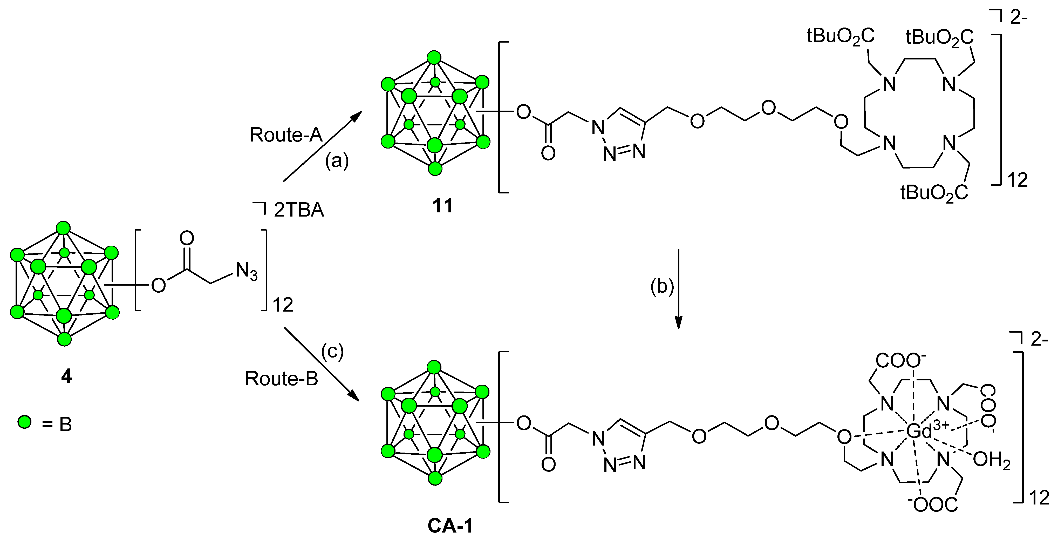
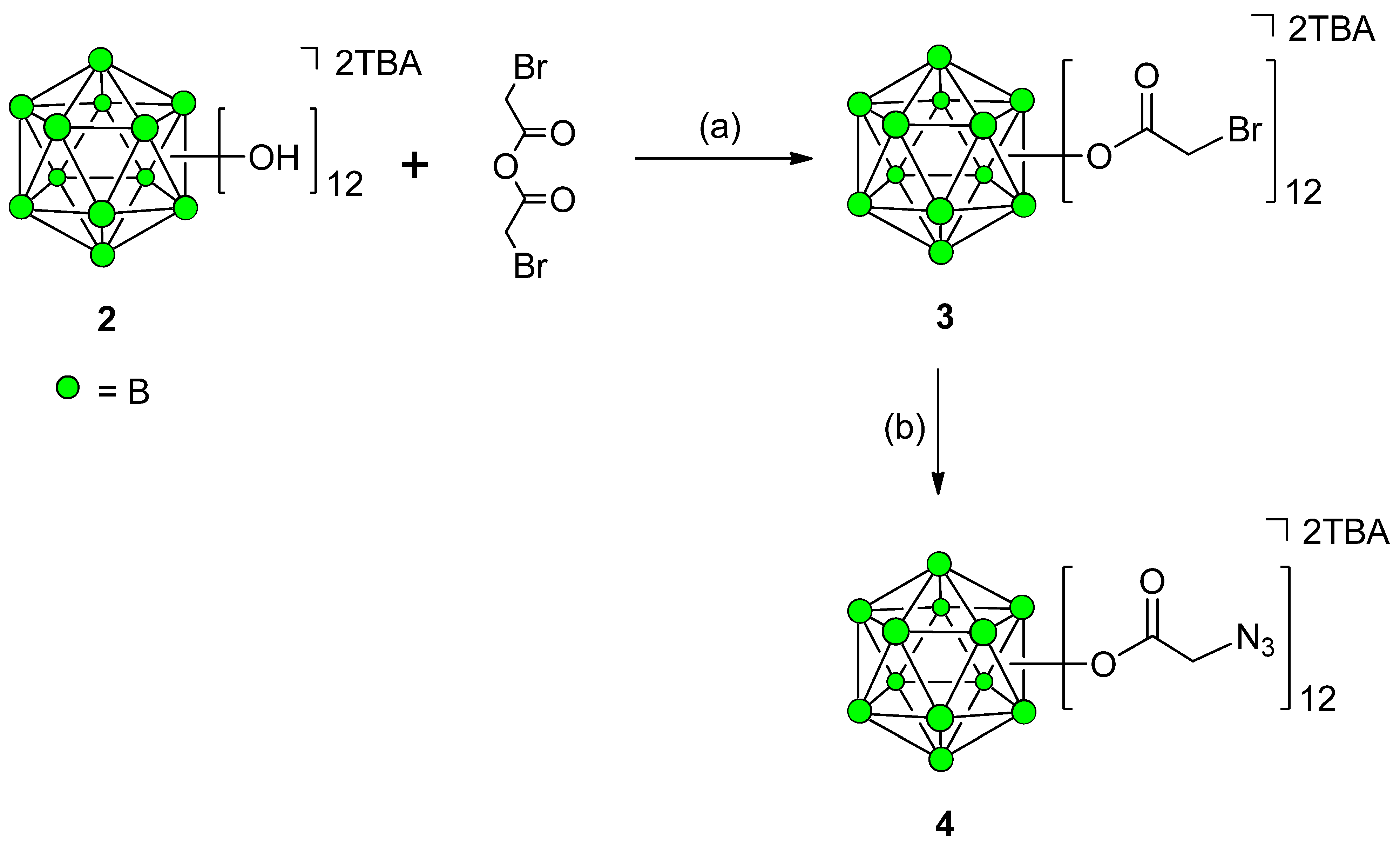
Synthesis of 12-Fold Azidoacetate Closomer 4

Previously, we have synthesized 12-fold azidoacetate closomer 4 via 12-fold esterification of TBA2-2 with chloroacetic anhydride followed by displacement of chloro functions with azido groups by reacting with NaN3 to form the 12-fold azidoacetate closomer 4. The presence of twelve azide groups on a single, compact closo-borane cage provides us with the opportunity to perform 12-fold click reactions with adequately functionalized terminal alkyne moieties, thus leading to the synthesis of novel 1,2,3-triazole ring attached polyfunctional closomers entities [19]. In this article, we describe an improved procedure for the synthesis of 12-azidoacetate closomers 4 via 12-fold bromoacetate closomer 3. Replacing the α-chloro with a more labile bromo leaving group provided more efficient substitution with azido group and a shorter reaction times. Under similar reaction conditions as those used to prepare 12-fold chloroacetate closomer [19], the TBA2-2 and bromoacetic anhydride (5.0 eq. per vertices) were heated at reflux temperature in acetonitrile (ACN) for 5 days resulting in the formation of 12-fold bromoacetate closomer 3 in 79% yield after purification using size-exclusion chromatography on a Lipophilic Sephadex LH-20 column (Scheme 1). The closomer 3 was reacted with a 10 folds excess of sodium azide in dimethylformamide (DMF) at room temperature (RT) and as expected, the reaction was completed in 2 days to give the 12-fold azidoacetate closomer 4 in 88% yield (Scheme 1).
Scheme 1. Synthesis of 12-fold azidoacetate closomer 4.
Molecules 18 09034 g006
Reagents and Conditions: (a) ACN, 5 days, reflux, 79%; (b) NaN3, DMF, 2 days, RT, 88%.

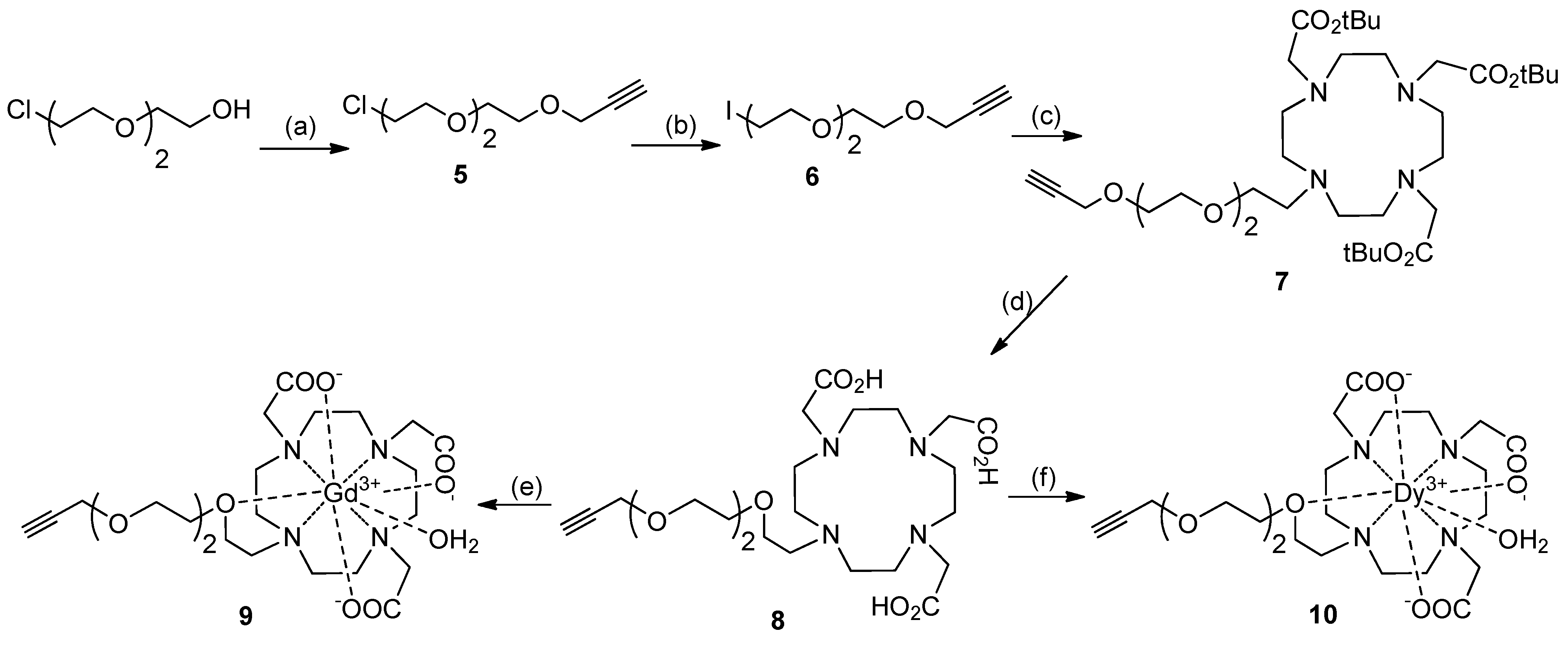
Synthesis of DO3A Ligands

We synthesized an alkyne terminated DOTA based ligand 7 from commercially available DO3A-t-Bu-ester (Scheme 2). First, a heterobifunctionalized alkyne-terminated short PEG linker, 6, that has an Iodo- group at the distal end, was synthesized from commercially available 2-[2-(2-chloroethoxy)ethoxy]ethanol [25]. The reaction of 6 with DO3A-t-Bu-ester afforded DO3A-t-Bu-ester ligand 7 in 94% yield. The tert-butyl groups on 7 were then deprotected using formic acid to obtain the alkyne terminated DO3A derivative, 8, in quantitative yield (95%). The Gd3+ complex 9 was synthesized by reacting 8 with Gd2O3 in water at 100 °C for 12 h. Similarly, the Dy3+-complex of the DO3A ligand, 10, was synthesized in 92% yield by reacting 8 with DyCl3·6H2O in pyridine (Scheme 2).

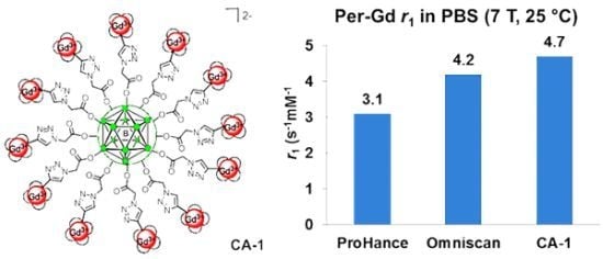
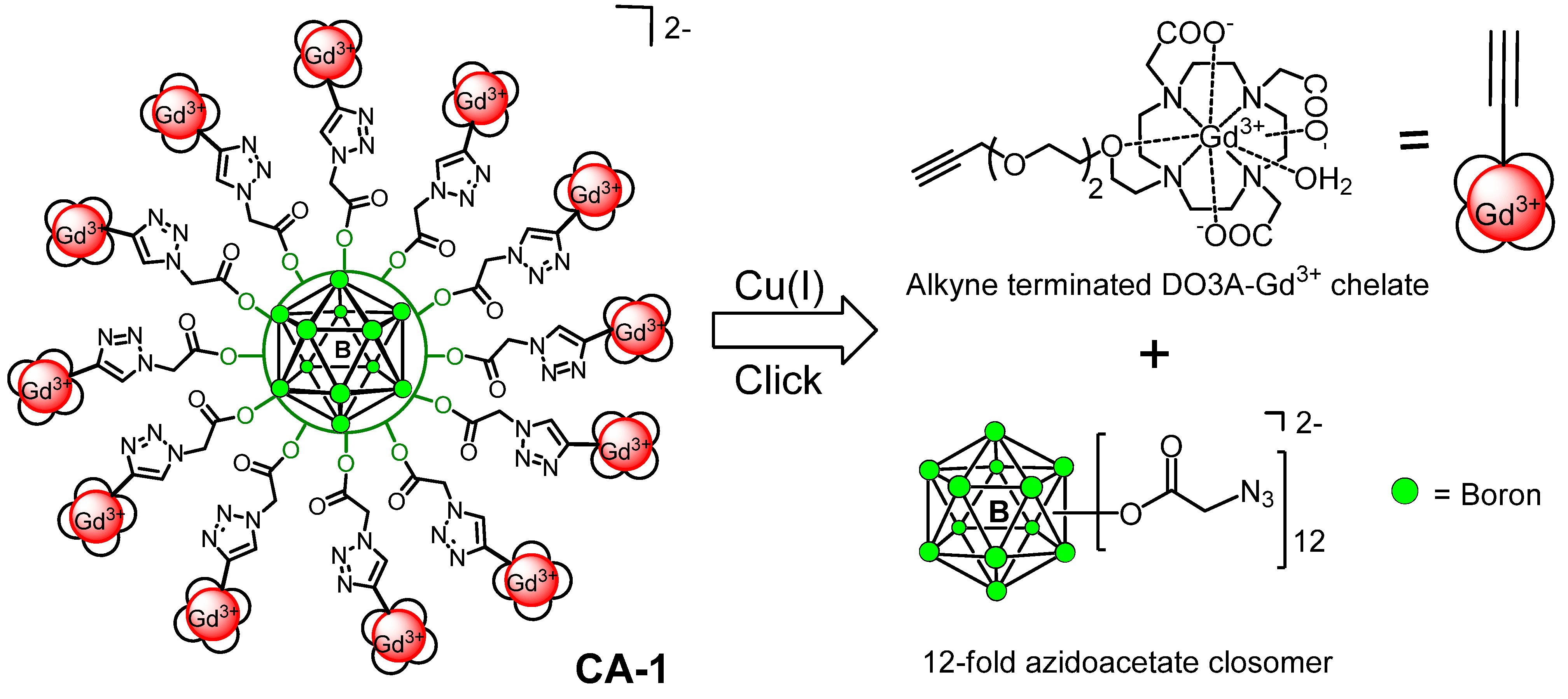
Synthesis of Closomer Contrast Agents CA-1

The closomer-DO3A conjugate 11 was synthesized in 67% yield by reacting 4 with the alkyne-terminated DO3A ligand 7 under Cu(I)-catalyzed 1,3-dipolar cycloaddition reaction conditions. The closomer 11 was purified by size-exclusion column chromatography using Lipophilic Sephadex LH-20 using ACN as the eluent and characterized by IR, NMR and HRMS spectroscopy. Closomer 11 exhibited a characteristic singlet at δ 7.74 ppm in the 1H-NMR spectrum that was assigned to the 12 alkene-CH protons of the 12 triazole rings. The IR spectrum of the product did not exhibit the characteristic peak at 2109 cm−1, which was attributed in closomer 4 to the asymmetric stretching of the azide group (Supplementary Information or SI). Next, closomer 11 was treated with 80% trifluoroacetic acid (TFA) in dichloromethane (DCM) to remove the tert-butyl ester groups; complete deprotection was confirmed by the absence of a large singlet at δ 1.46 ppm in the 1H-NMR spectrum originally attributed to the 36 tert-butyl ester groups of the 12 DO3A ligands attached to the closo-B122−-core in 11. Subsequently, the deprotected closomer 11 was reacted with GdCl3·6H2O in citrate buffer at pH ~7 to obtain CA-1 in 75% yield (Scheme 3, Route-A). The CA-1 was purified via exhaustive dialysis in ultrapure water and was characterized using IR and HRMS spectroscopy. The IR spectrum of CA-1 exhibited the characteristic shift of the carbonyl stretch from 1727 cm−1 to 1601 cm−1, which demonstrates the complexation of Gd3+ ions with carboxylic acid groups of the DO3A ligand (SI). The purity of CA-1 was tested by size-exclusion HPLC (SE-HPLC) analysis, and the gadolinium loading was determined using inductively coupled plasma optical emission spectroscopy (ICP-OES), which showed the formation of fully loaded chelates with an average of 12 Gd3+ ions per closomer. The presence of any free Gd3+ ions in closomer contrast agent CA-1 was tested by spectrophotometric method using xylenol orange [26], and found to contain no free Gd3+ ions. Alternatively, the synthesis of closomer contrast agent CA-1 was also attempted via a pre-complexation route as shown in Scheme 3, Route-B using Cu(I)-catalyzed 1,3-dipolar cycloaddition reaction between the azidoacetate closomer 4 and alkyne-terminated DO3A-Gd3+ complex 9. However, this route resulted in the lower yield (38%) of the CA-1 as compared to the Route-A (71%, overall yield from 4) shown in Scheme 3.
Scheme 2. Synthesis of alkyne-terminated DO3A ligands.
Molecules 18 09034 g007
Reagents and conditions: (a) NaH, THF, 0 °C-RT-60 °C, 24 h, 80%; (b) NaI, acetone, 60 °C, 12 h, 95%; (c) DO3A-t-Bu-ester, KHCO3, DMF, 12 h, 60 °C, 94%; (d) HCOOH, 12 h, 65 °C, 95%; (e) Gd2O3, H2O, 12 h, 100 °C, 91%; (f) DyCl3.6H2O, pyridine, 70 °C, 12 h, 92%.
Scheme 3. Synthesis of closomer contrast agent CA-1.
Molecules 18 09034 g008
Reagents and conditions: (a) DO3A ligand 7, CuI, DIPEA, ACN-THF, 5 days, RT, 67%; (b) (i) 80% TFA-DCM, 6 h, RT; (ii) GdCl3·6H2O, citrate buffer, pH 7, 20 h, RT, sonication; (iii) Dialysis, 1,000 MWCO, 48 h; (iv) Lyophilization, 75%; (c) 9, CuSO4·5H2O, sodium ascorbate, ACN-H2O, 3 days, 60 °C, size-exclusion chromatography, 38%. Tetrabutylammonium (TBA), N, N-Diisopropylethylamine (DIPEA); Molecular weight cut-off (MWCO)

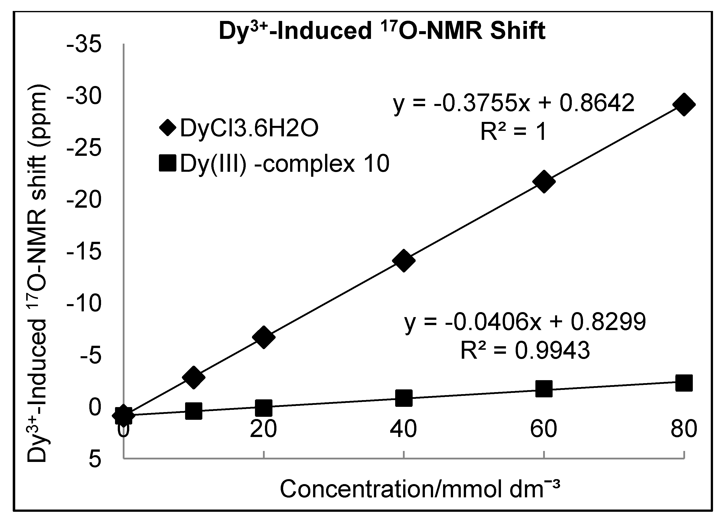
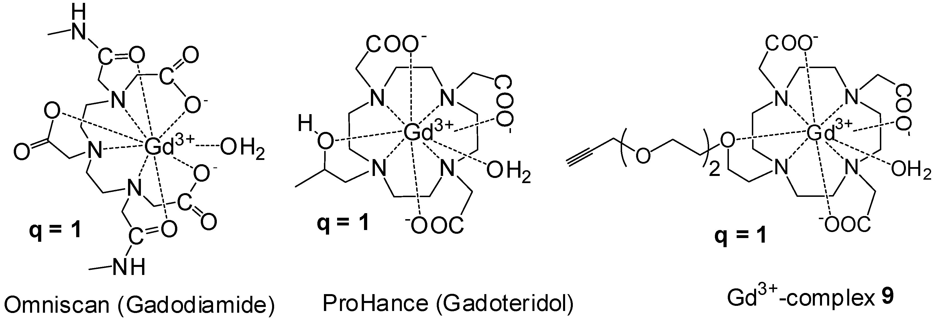
Determination of Hydration Number (q)

In general, the q value of the Gd3+ complex of DOTA is predicted to be 1; however, the PEG oxygen donor in the modified DO3A ligand can partially displace the water molecules coordinated to the DO3A-Gd3+, 9, consequently reducing the overall relaxivity of the CA [27]. In principle, the Gd3+-induced water 17O-NMR chemical shift can be used directly to determine the value of q, provided that the exchange between Gd3+ bound water and bulk water is fast on the 17O-NMR time scale. However, this situation exists at a temperature of about 80 °C. Furthermore, the severe line-broadening often makes it difficult to accurately measure q value on Gd3+ complexes and, therefore, measurements on the corresponding Dy3+ complex are preferred. Thus, the q value for the newly synthesized DO3A complex 9 was determined using the Dy3+-induced water 17O-NMR shifts (d.i.s.) method [28]. Various concentrations of Dy3+ complex 10 and DyCl3·6H2O over the range of 10-80 mmol dm−3 were prepared in 80% D2O-H2O and the d.i.s. (Δδ) was measured using a 400 MHz NMR instrument (Figure 2). The Δδ for a complex with the general formula Dy(ligand)n(H2O)q is given by the relation (Δδ) = qΔ[Dy(ligand)n(H2O)q]/[H2O]. The slope of a plot of the d.i.s. versus the Dy3+ concentration is proportional to the q value of the complex. As expected the d.i.s. method gave a q value of 1 for the complex 10 (SI).
Figure 2. Plot of the Dy3+-induced water 17O-NMR shift as a function of [Dy].
Molecules 18 09034 g002

Relaxivity Studies

The per-Gd relaxivity (r1) of closomer contrast agent CA-1, DO3A Gd3+-complex 9 and clinically used CA, Omniscan at 25 °C and 7 Tesla (T) were compared in three different formulations: PBS, 2% tween/PBS, and bovine calf serum. The relaxivity of each CA was relatively unaffected by formulation type, but the r1 values of CA-1 (4.7 mM−1s−1 in PBS) were slightly higher than Omniscan (4.2 mM−1s−1 in PBS) and the DO3A Gd3+-complex 9 across all formulations (SI). Furthermore, these relaxivity values are comparable to our previously reported results [21]. The limited increase in r1 at 7 T can be attributed to the fact that the contribution of the slow tumbling (rotational correlation time or τR) towards the enhancement of r1 is not observed at higher magnetic field strengths (4.7 T and above) [29].
Since Omniscan is a DTPA based ligand (acyclic polyaminocarboxylate core) that inherently has slightly higher r1 as compared to DOTA based ligands (cyclic polyaminocarboxylate core), we also compared the per-Gd r1 of DO3A Gd3+-complex 9 and closomer contrast agent CA-1 with structurally similar clinically used MRI CA, ProHance shown in Figure 3 [30]. The macromolecular CA-1 shows significantly higher r1 value (per Gd r1 4.7 mM−1s−1; molecular r1 56.5 mM−1s−1) as compared to ProHance (both per Gd and per molecular r1 3.1 mM−1s−1) at 7 T, 25 °C in PBS (Figure 4). Macromolecular CAs such as CA-1 are known to exhibit higher r1 values due to their larger size and the confinement of the Gd3+ ions in a sterically constrained space [8,9,10,11,12,13,14,15].
Figure 3. Structures of clinically used MRI CAs; Omniscan and ProHance and Gd3+-complex 9.
Molecules 18 09034 g003
Figure 4. Comparison of the per-Gd r1 values of CA-1, DO3A Gd3+-complex 9 with clinically used CAs; Omniscan and ProHance.
Molecules 18 09034 g004
The T1-weighted MRI images of CA-1 and Omniscan at 7 T at equimolar Gd3+ concentrations presented in Figure 5 shows greater contrast enhancement for CA-1 compared to Omniscan due to its slightly higher relaxivity value.
Figure 5. T1-weighted MRI images of CA-1 and Omniscan at 7 T and 25 °C in PBS at different Gd3+ concentrations.
Molecules 18 09034 g005
The possibility of aggregation in macromolecular CAs may affect their relaxivity values. To negate the possibility of aggregation of CA-1 in solution, a dynamic light scattering (DLS) analysis of CA-1 in PBS, 2% tween/PBS and bovine calf serum solution was performed. The average particle size of 11–13 nm was found in all formulations indicating no aggregation of the CA-1 particles (SI).

3. Experimental

3.1. General

Common reagents and chromatographic solvents were obtained from commercial suppliers (Sigma-Aldrich, Fisher Scientific) and used without any further purification. Lipophilic Sephadex LH-20 was obtained from GE Healthcare. DO3A-t-Bu-ester was purchased from Macrocyclics, Inc. NMR spectra were recorded on Bruker Avance 400 and 500 MHz spectrometers. The high-resolution mass spectrometry analysis was performed using Applied Biosystems Mariner ESI-TOF. IR spectra were recorded on Thermo Nicolet FT-IR spectrometer. The dynamic light scattering (DLS) analysis was performed on a Microtrac Zetatrac particle size analyzer. Gadolinium (Gd3+) concentrations of the samples used in MRI experiments were measured by inductively coupled plasma optical emission spectroscopy (ICP-OES) on a PerkinElmer OptimaTM 7,000 DV instrument.

3.2. Synthesis

Bis(tetrabutylammonium)-closo-dodecabromoacetoxydodecaborate (3). A solution of TBA2-2 (0.50 g, 0.61 mmol) and bromoacetic anhydride (9.54 g, 36.71 mmol) in dry ACN (50 mL) was refluxed for 5 days in an argon atmosphere with vigorous stirring. Progress of the reaction was monitored by 11B NMR. Completion of the reaction was indicated by the appearance of a singlet at −17 ppm. The reaction mixture was then concentrated to dryness and purified using a size-exclusion column on Lipophilic Sephadex LH-20 with ACN as the eluent. The product was obtained as light brown semi-solid. Yield: 1.1 g (79%). IR (KBr): 3053, 2986, 2305, 1753, 1422, 1330, 1264 and 1231 cm−1. 1H-NMR (400 MHz, CDCl3): δ 3.92 (s, 24H), 3.12 (m, 16H), 1.62 (m, 16H), 1.42 (m, 16H), 0.99 (t, J = 7.2 Hz, 24H). 13C-NMR (100.6 MHz, CDCl3): δ 165.4, 59.8, 31.3, 24.7, 20.5 and 14.5. 11B-NMR (128 MHz, CDCl3): δ -17.70. HRMS (m/z): calcd. for C24H24B12Br12O24 [M]2− 893.5956. Found: 893.5431 and calcd. for C24H24B12Br12O24+ C16H36N1− (M+TBA) 2027.4798. Found: 2027.4041.
Bis(tetrabutylammonium)-closo-dodecaazidoacetoxydodecaborate (4): In a 100 mL round bottom flask, bis(tetrabutylammonium)-dodecabromoacetoxydodecaborate 3 (0.20 g, 0.09 mmol) and sodium azide (0.69 g, 10.57 mmol) were mixed with dry DMF (10 mL). This mixture was vigorously stirred at RT for 2 days under an argon atmosphere. The progress of the reaction was monitored by 1H-NMR. After completion, the reaction mixture was filtered through a celite pad and the filtrate was concentrated to dryness, the residue was re-dissolved in ethyl acetate and filtered again through a celite pad. The filtrate was collected and evaporated to dryness. The product was purified using size-exclusion column chromatography on Lipophilic Sephadex LH-20 with ACN as eluent. The product was obtained as a light brown semi-solid. Yield: 0.14 g (88%). All the spectroscopic characterization data was in perfect agreement with the reported data [19].
3-(2-(2-(2-chloroethoxy)ethoxy)ethoxy)prop-1-yne (5): To a two neck oven dried 250 mL round bottom flask, THF (50 mL) was added and the flask was placed in an ice bath. NaH (60% dispersion in oil, 2.8 g, 71.1 mmol) was added to it and the contents were stirred under argon atmosphere. 2-(2-(2-chloro)ethoxy)ethanol (10.0 g, 59.3 mmol) dissolved in THF (20 mL) was added to the reaction mixture over 15 minutes at 0 °C. The contents were stirred at 0 °C for 45 minutes and then propargyl bromide (10.5 g, 88.9 mmol) was added to it over a period of 30 minutes at 0 °C. The contents were then stirred at 0 °C for 2 h. After 2 h, reaction mixture was slowly heated to 60 °C and stirred at this temperature for 22 h. After 24 h, reaction mixture was allowed to cool to RT and an ice-cold aqueous solution of 5% HCl (100 mL) was added to it. The product was then extracted using ether (3 × 100 mL), the ether layer was washed with brine, dried over Na2SO4 and concentrated. This crude reaction mixture was purified using flash chromatography over silica gel to obtain product as very viscous pale yellow colored oil. Yield: 9.80 g (80%). 1H-NMR (400 MHz, CDCl3): δ 4.12 (d, J = 2.4 Hz, 2H), 3.69 (t, J = 6.0 Hz, 2H), 3.63–3.54 (m, 10H), 2.41 (t, J = 2.4 Hz, 1H). 13C-NMR (100.6 MHz, CDCl3): δ 80.4, 75.4, 72.1, 71.3, 71.3, 71.2, 69.8, 59.1, 43.5. HRMS (m/z): Calcd for C9H15ClO3 [M+Na]+ 229.0602. Found: 228.9594.
3-(2-(2-(2-iodoethoxy)ethoxy)ethoxy)prop-1-yne (6): A mixture of 5 (2.70 g, 13.0 mmol) and sodium iodide (3.91 g, 26.1 mmol) in acetone (30 mL) was stirred at 60 °C for 12 h. The reaction mixture was then cooled to RT and filtered. Filtrate was concentrated, and the crude material was purified on a silica gel column to obtain pure product as pale yellow oil. Yield: 3.7 g (95%). 1H-NMR (400 MHz, CDCl3): δ 4.16 (d, J = 2.4 Hz, 2H), 3.71 (t, J = 6.8 Hz, 2H), 3.66–3.61 (m, 8H), 3.21 (t, J = 7.2 Hz, 2H), 2.42 (t, J = 2.4 Hz, 1H). 13C-NMR (100.6 MHz, CDCl3): δ 80.5, 75.4, 72.7, 71.4, 71.2, 70.9, 69.9, 59.2, 43.5. HRMS (m/z): Calcd for C9H15IO3 [M+Na]+ 320.9958. Found: 320.9684.
tri-tert-butyl2,2',2''-(10-(2-(2-(2-(prop-2-yn-1-yloxy)ethoxy)ethoxy)ethyl)-1,4,7,10 tetraazacyclododecane-1,4,7-triyl)triacetate (7): To a mixture of DO3A-t-Bu-ester (2.00 g, 3.88 mmol) and 6 (1.73 g, 5.83 mmol) in DMF (25 mL), KHCO3 (0.78 g, 7.77 mmol) was added, and the mixture was stirred at 60 °C for 12 h. The reaction mixture was cooled and DCM (100 mL) added. The mixture was washed with brine (100 mL), and the organic layer was separated, dried over Na2SO4, and concentrated. The pure product was obtained as viscous oil after column chromatography over alumina (IV). Yield: 2.5 g (94%). 1H-NMR (400 MHz, CDCl3): δ 4.15 (d, J = 2.4 Hz, 2H), 3.65–3.57 (m, 10H), 3.53–3.16 (m, 2H), 3.14–2.96 (m, 4H), 2.92–2.67 (m, 7H), 2.66–2.53 (m, 4H), 2.50–2.38 (m, 4H), 2.37–2.18 (m, 4H), 1.45 (m, 27H). 13C-NMR (100.6 MHz, CDCl3): δ 172.8, 172.4, 82.1, 82.1, 79.5, 74.6, 70.1, 69.7, 69.6, 68.9, 67.0, 58.3, 56.3, 55.6, 52.1, 50.5, 49.7, 28.1, 27.9, 27.8. HRMS (m/z): Calcd for C35H64N4O9 Calcd for C35H64N4O9 [M+Na]+ 707.4566. Found: 707.5989.
2,2',2''-(10-(2-(2-(2-(prop-2-yn-1-yloxy)ethoxy)ethoxy)ethyl)-1,4,7,10-tetraazacyclododecane-1,4,7-triyl)triacetic Acid (8): A solution of 7 (0.25 g, 0.36 mmol) in formic acid (10 mL, 90%) was stirred for 12 h at 65 °C and then concentrated under reduced pressure. The residue was dissolved in a minimal amount of MeOH, and the product was precipitated by adding ether. Yield: 0.18 g (95%). Mp: 165 °C. IR (KBr): 3423, 2862, 1634, 1401, 1353, 1088, 693 cm−1. 1H-NMR (500 MHz, CDCl3): δ 5.16 (brs, 3H), 4.34 (m, 2H), 3.9 (m, 2H), 3.80–3.62 (m, 14H), 3.51–3.15 (m, 18H), 3.06 (t, J = 2.3 Hz, 1H). 13C-NMR (125.7 MHz, CDCl3): δ 172.5, 167.5, 100. 6, 82.6, 79.6, 77.5, 75.1, 70.1, 69.9, 69.9, 69.9, 69.9, 69.8, 69.8, 69.0, 68.9, 67.6, 57.7, 56.7, 55.6, 52.5, 50.7, 50.5, 49.9. HRMS (m/z): Calcd for C23H40N4O9 [M+H]+ 517.2874. Found: 517.3040 and Calcd for C23H40N4O9 [M+Na]+ 539.2693. Found: 539.3009.
2,2',2''-(10-(2-(2-(2-(prop-2-yn-1-yloxy)ethoxy)ethoxy)ethyl)-1,4,7,10-tetraazacyclododecane-1,4,7-triyl)triacetic Acid Gd3+ Complex (9): A mixture of 8 (0.21 g, 0.40 mmol) and Gd2O3 (0.09 g, 0.24 mmol) in H2O (10 mL) was stirred for 12 h at 100 °C. The solvent was removed under reduced pressure and the residue was dissolved in a minimum amount of methanol and passed through a celite pad to remove any solid present. Ether was added to precipitate an off-white powder which was then filtered, washed with ether and dried to obtain the product as a white solid. Yield: 0.25 g (91%). MP: 240 °C (decomposed). IR (KBr): 3440, 2865, 1612, 1443, 1383, 1323, 1086, 941, 723 cm−1. HRMS (m/z): Calcd for C23H37GdN4O9 [M+Na]+ 694.1700. Found: 694.1610 and Calcd for C23H37GdN4O9 [3M+2Na]2+ 1029.7600. Found: 1029.7294 and C23H37GdN4O9 [2M+Na]+ 1364.3509. Found: 1364.4569.
2,2',2''-(10-(2-(2-(2-(prop-2-yn-1-yloxy)ethoxy)ethoxy)ethyl)-1,4,7,10-tetraazacyclododecane-1,4,7-triyl)triacetic Acid Dy3+ Complex (10): A mixture of 8 (0.37 g, 0.72 mmol) and DyCl3·6H2O (0.27 g, 0.72 mmol) in pyridine (10 mL) was stirred for 12 h at 70 °C. The solvent was removed under reduced pressure and the residue was dissolved in a minimum amount of methanol and passed through a celite pad to remove any solid present. Ether was added to precipitate an off-white powder which was then filtered, washed with ether and dried to obtain the product as a white solid. Yield: 0.45 g (92%). MP: 180 °C (decomposed). IR (KBr): 3425, 2869, 1611, 1444, 1383, 1321, 1087, 940, 723 cm−1. HRMS (m/z): Calcd for C23H37DyN4O9 [M+Na-H]+ 699.1672. Found: 699.0800 and Calcd for C23H37DyN4O9 [M+Na+Li-H]+ 706.1832. Found: 706.3500.
Synthesis of Closomer 11: A mixture of 12-fold azidoacetate closomer 4 (0.07 g, 0.04 mmol) and DO3A ligand 7 (1.26 g, 1.85 mmol) was dissolved in ACN-THF (20 mL, 50:50 mixture). To this mixture, DIPEA (0.60 g, 4.62 mmol) and copper (I) iodide (0.09 g, 0.46 mmol) were added and the resulting mixture was stirred for 5 days at RT under an argon atmosphere. The reaction mixture was then concentrated to dryness and the residue was dissolved in DCM and filtered. The filtrate was washed with an aqueous 2% ethylenediaminetetraacetic acid disodium salt (EDTA.2Na) solution to remove the trace of copper ions and the organic layer was separated, dried and concentrated. The closomer 11 was purified via size-exclusion chromatography over Lipophilic Sephadex LH-20 using ACN as eluent. Yield: 0.26 g (67%), obtained as viscous oil. IR (neat): 3441, 2975, 2933, 2868, 1727, 1457, 1386, 1310, 1227, 1161, 1112, 848, 732 cm−1. 1H-NMR (400 MHz, CD3CN): δ 7.74 (s, 12H), 4.96 (s, 24H), 4.58 (m, 24H), 3.84 9m, 24H), 3.59–3.26 (m, 200H), 3.03–2.98 (m, 52H), 2.80–2.29 (m, 172H), 1.46 (m, 331H), extra proton counts are due to the presence of H3O+ and TBA+ cations. 13C-NMR (100.6 MHz, CD3CN): δ 173.8, 173.6, 171.3, 171.1, 166.4, 145.1, 125.9, 82.7, 81.9, 81.8, 73.2, 71.0, 70.9, 70.7, 70.6, 70.3, 70.2, 70.0, 69.8, 68.1, 65.9, 64.6, 61.5, 58.7, 57.2, 57.0, 56.3, 55.7, 55.6, 53.7, 53.3, 53.1, 52.8, 51.2, 50.7, 48.4, 28.4, 28.3, extra carbon counts are due to the presence of TBA+ cations. 11B-NMR (128 MHz, CD3CN): δ −17.90. HRMS (m/z): Average mass calcd for C444H792B12N84O132 [M] 9549.2549. Found: 1689 [M6++4Na+4H], 1483 [M7++5Na+4H], 1247 [M8++2Na+8H], 1159 [M9++ 4Na+7H].

Synthesis of CA-1

Route A. Closomer 11 (0.20 g, 0.02 mmol) was treated with 80% TFA/DCM (10 mL) for 6 h at RT under an argon atmosphere. The reaction mixture was concentrated under reduced pressure and the resulting residue was dissolved in a minimum amount of MeOH and precipitated by adding ether. The solid was filtered, washed with ether and dried under high vacuum overnight. The solid was then dissolved in 1M citrate buffer (15 mL, pH-7) and added slowly to a solution of GdCl3.6H2O (0.74 g, 1.19 mmol) in 15 mL of 1M citrate buffer over 5 h at RT with vigorous stirring. The pH of the reaction mixture was maintained at approximately 7 using 0.3 N NaOH. The reaction mixture was stirred for additional 15 h at RT and was sonicated a few times during the course of the reaction. The reaction mixture was then dialyzed in deionized water for 2 days using 1,000 MWCO membrane tubes (Spectra/Por®). The product CA-1 was obtained as an off-white solid after lyophilization. Yield: 0.14 g (75%). MP: 260 °C (decomposed). IR (KBr): 3425, 2871, 1744, 1601, 1383, 1324, 1260, 1086, 718 cm−1. ICP-OES analysis: calcd average Gd per closomer: 12. Found: 12. HRMS-ESI-TOF (m/z): Calcd for B12C300H468Gd12N84O132 [M]2− 4692.2286. Found: 4692.8213.
Route B. Pre-complexation. A mixture of 12-fold azidoacetate closomer 4 (0.05 g, 0.03 mmol), CuSO4.5H2O (12.4 mg, 0.05 mmol), sodium ascorbate (20.0 mg, 0.10 mmol) and DO3A Gd3+ complex 9 (0.66 g, 1.00 mmol) was dissolved in ACN-H2O (30 mL, 50:50 mixture). This mixture was stirred for 3 days at 60 °C. The reaction mixture was then cooled, filtered and the filtrate was concentrated to dryness and the residue was purified via size-exclusion chromatography over Lipophilic Sephadex LH-20 using water as eluent. The product CA-1 was obtained as an off-white solid after lyophilization. Yield: 0.10 g (38%).

3.3. Relaxivity Determinations

Solutions of CA-1 and Omniscan were prepared in PBS, 2% Tween-80/PBS and bovine calf serum. T1 relaxation rates were determined for each of the three formulations of CA-1 at Gd3+ ion concentrations of 0.125, 0.25, 0.5 and 1 mM. The Gd3+ ion concentration of Omniscan was 1 mM. The measurements were repeated on two or more independently prepared samples to ensure consistency. A buffer matched blank sample (0 mM) was also used in the relaxivity measurements of each sample.
Measurements were performed using a 7 Tesla Varian Unity Inova MRI system (Varian Inc./Agilent Technologies) at 25 °C. A T1-weighted MRI pulse sequence was applied with TE = 15 ms, and TR = 500 ms, slice thickness = 1 mm, matrix = 256 × 256, and FOV = 30 × 30 mm. A series of inversion recovery (IR) spin-echo images were acquired using TR = 3 s, TE = 15 ms, and the following inversion delays: 0.082, 0.1, 0.12, 0.16, 0.24, 0.32, 0.64, 1.28, 2.56, 3.6 s. Water signal intensities were measured using VnmrJ2.1D software (Varian Inc./Agilent Technologies, 2005). Relaxation rates were calculated using a three-parameter exponential recovery fitting in Origin 8.5.0 (OriginLab Corporation, 2012). The final relaxivity values were obtained by linear fitting of relaxation rates against concentrations for each sample.

4. Conclusions

In summary, we report the synthesis of a novel multimeric MRI contrast agent (CA-1) covalently attached to a icosahedral closo-borane core via Cu(I) catalyzed azide-alkyne click chemistry approach. This unique macromolecular CA is highly water soluble and the preliminary relaxivity studies confirms that CA-1 exhibits higher relaxivity values (per Gd relaxivity: r1 = 4.7 mM−1s−1, and per molecule r1: = 56.5 mM−1s−1 at 7 T and 25 °C in PBS) as compared to the frequently used clinical MRI CAs. The detailed stability, toxicity and in vivo MRI assessments of CA-1 is currently in progress. We also investigated a more efficient route for the synthesis of the 12-fold azidoacetate closomer 4 from closo-B12(OH)122− through the intermediary of 12-fold bromoacetate closomer 3. The 12-fold azidoacetate closomer 4 is thus demonstrated to be a vital scaffold for the development of a wide variety of high-payload, receptor-specific diagnostic and therapeutic agents via click chemistry approach.

Supplementary Materials

Supplementary File 1

Acknowledgments

This research was funded by the National Cancer Institute (Grant R21-CA114090). Authors acknowledge the support provided by the Veterans’ Affairs Biomolecular Imaging Center at the Harry S. Truman VA Hospital and the University of Missouri-Columbia. Authors thank Brett Meers, Cecelia Urrutia, Melissa Luechtefeld and Sarah Higdon for technical assistance.

Conflicts of Interest

The authors declare no conflict of interest.

References

  1. Hermann, P.; Kotek, J.; Kubíček, V.; Lukeš, I. Gadolinium(III) complexes as MRI contrast agents: ligand design and properties of the complexes. Dalton Trans. 2008, 3027–3047. [Google Scholar]
  2. Caravan, P.; Ellison, J.J.; McMurry, T.J.; Lauffer, R.B. Gadolinium(III) Chelates as MRI Contrast Agents: Structure, Dynamics, and Applications. Chem. Rev. 1999, 99, 2293–2352. [Google Scholar] [CrossRef]
  3. Aime, S.; Botta, M.; Fasano, M.; Terreno, E. Lanthanide(III) chelates for NMR biomedical applications. Chem. Soc. Rev. 1998, 27, 19–29. [Google Scholar]
  4. Idée, J.M.; Port, M.; Medina, C.; Lancelot, E.; Fayoux, E.; Ballet, S.; Corot, C. Possible involvement of gadolinium chelates in the pathophysiology of nephrogenic systemic fibrosis: A critical review. Toxicology 2008, 248, 77–88. [Google Scholar] [CrossRef]
  5. Caravan, P. protein-targeted gadolinium-based magnetic resonance imaging (mri) contrast agents: Design and mechanism of action. Acc. Chem. Res. 2009, 42, 851–862. [Google Scholar] [CrossRef]
  6. Solomon, I. Relaxation processes in a system of two spins. Phys. Rev. 1955, 99, 559–565. [Google Scholar] [CrossRef]
  7. Bloembergen, N.J. Proton relaxation times in paramagnetic solutions. Chem. Phys. 1957, 27, 572–573. [Google Scholar]
  8. Botta, M.; Tei, L. Relaxivity enhancement in macromolecular and Nanosized GdIII-Based MRI contrast agents. Eur. J. Inorg. Chem. 2012, 2012, 1945–1960. [Google Scholar] [CrossRef]
  9. Langereis, S.; de Lussanet, Q.G.; van Genderen, M.H.P.; Backes, W.H.; Meijer, E.W. Multivalent contrast agents based on Gadolinium−Diethylenetriaminepentaacetic Acid-Terminated Poly(propylene imine) dendrimers for Magnetic Resonance Imaging. Macromolecules 2004, 37, 3084–3091. [Google Scholar] [CrossRef]
  10. Torchilin, V.; Babich, J.; Weissig, V. Liposomes and Micelles to Target the Blood Pool for Imaging Purposes. J. Liposome Res. 2000, 10, 483–499. [Google Scholar] [CrossRef]
  11. Floyd, W.C.; Klemm, P.J.; Smiles, D.E.; Kohlgruber, A.C.; Pierre, V.C.; Mynar, J.L.; Fréchet, J.M.J.; Raymond, K.N. Conjugation effects of various linkers on Gd(III) MRI contrast agents with dendrimers: Optimizing the hydroxypyridinonate (HOPO) ligands with nontoxic, Degradable Esteramide (EA) dendrimers for high relaxivity. J. Am. Chem. Soc. 2011, 133, 2390–2393. [Google Scholar] [CrossRef]
  12. De León-Rodríguez, L.M.; Lubag, A.; Udugamasooriya, D.G.; Proneth, B.; Brekken, R.A.; Sun, X.; Kodadek, T.; Sherry, A.D. MRI detection of VEGFR2 in vivo using a low molecular weight peptoid-(Gd)8-dendron for targeting. J. Am. Chem. Soc. 2010, 132, 12829–12831. [Google Scholar] [CrossRef]
  13. Bryson, J.M.; Chu, W.; Lee, J.; Reineke, T.M. A β-cyclodextrin “click cluster” decorated with seven paramagnetic chelates containing two water exchange sites. Bioconjugate Chem. 2008, 19, 1505–1509. [Google Scholar] [CrossRef]
  14. Song, Y.; Kohlmeir, E.K.; Meade, T.J. Synthesis of multimeric mr contrast agents for cellular imaging. J. Am. Chem. Soc. 2008, 130, 6662–6663. [Google Scholar] [CrossRef]
  15. Livramento, J.B.; Tóth, É.; Sour, A.; Borel, A.; Merbach, A.E.; Ruloff, R. High relaxivity confined to a small molecular space: A metallostar-based, Potential mri contrast agent. Angew. Chem. Intl. Ed. 2005, 14, 1480–1484. [Google Scholar]
  16. Bayer, M.J.; Hawthorne, M.F. An improved method for the synthesis of [closo-B12(OH)12]−2. Inorg. Chem. 2004, 43, 2018–2020. [Google Scholar] [CrossRef]
  17. Farha, O.K.; Julius, R.L.; Lee, M.W.; Huertas, R.E.; Knobler, C.B.; Hawthorne, M.F. Synthesis of stable Dodecaalkoxy derivatives of hypercloso-B12H12. J. Am. Chem. Soc. 2005, 127, 18243–18251. [Google Scholar]
  18. Jalisatgi, S.S.; Kulkarni, V.S.; Tang, B.; Houston, Z.H.; Lee, M.W.; Hawthorne, M.F. A convenient route to diversely substituted icosahedral closomer nanoscaffolds. J. Am. Chem. Soc. 2011, 133, 12382–12385. [Google Scholar] [CrossRef]
  19. Goswami, L.N.; Chakravarty, S.; Lee, M.W.; Jalisatgi, S.S.; Hawthorne, M.F. Extensions of the icosahedral closomer structure by using azide–alkyne click reactions. Angew. Chem. Int. Ed. 2011, 50, 4689–4691. [Google Scholar] [CrossRef]
  20. Pushechnikov, A.; Jalisatgi, S.S.; Hawthorne, M.F. Dendritic closomers: Novel spherical hybrid dendrimers. Chem. Comm. 2013, 49, 3579–3581. [Google Scholar] [CrossRef]
  21. Goswami, L.N.; Ma, L.; Cai, Q.; Sarma, S.J.; Jalisatgi, S.S.; Hawthorne, M.F. cRGD Peptide-conjugated Icosahedral closo-B122− core carrying multiple Gd3+-DOTA chelates for αvβ3 Integrin-Targeted Tumor Imaging (MRI). Inorg. Chem. 2013, 52, 1701–1709. [Google Scholar] [CrossRef]
  22. Rostovtsev, V.V.; Green, L.G.; Fokin, V.V.; Sharpless, K.B. A stepwise huisgen cycloaddition process: Copper(I)-catalyzed regioselective “Ligation” of Azides and Terminal Alkynes. Angew. Chem. Int. Ed. 2002, 41, 2596–2599. [Google Scholar] [CrossRef]
  23. Lutz, J.-F. 1,3-dipolar cycloadditions of azides and alkynes: A universal ligation tool in polymer and materials science. Angew. Chem. Int. Ed. 2007, 46, 1018–1025. [Google Scholar] [CrossRef]
  24. Goswami, L.N.; Ma, L.; Chakravarty, S.; Cai, Q.; Jalisatgi, S.S.; Hawthorne, M.F. Discrete Nanomolecular Polyhedral Borane Scaffold Supporting Multiple Gadolinium(III) Complexes as a High Performance MRI Contrast Agent. Inorg. Chem. 2013, 52, 1694–1700. [Google Scholar] [CrossRef]
  25. Piron, F.; Oprea, C.; Cismaş, C.; Terec, A.; Roncali, J.; Grosu, I. Synthesis of Podands with Cyanurate or Isocyanurate Cores and Terminal Triple Bonds. Synthesis 2010, 1639–1644. [Google Scholar]
  26. Barge, A.; Cravotto, G.; Gianolio, E.; Fedeli, F. How to determine free Gd and free ligand in solution of Gd chelates. A technical note. Contrast Med. Mol. Imaging 2006, 1, 184–188. [Google Scholar] [CrossRef]
  27. Doble, D.M.; Botta, M.; Wang, J.; Aime, S.; Barge, A.; Raymond, K.N. Optimization of the relaxivity of MRI contrast agents: Effect of poly(ethylene glycol) chains on the water-exchange rates of Gd(III) complexes. J. Am. Chem. Soc. 2001, 123, 10758–10759. [Google Scholar] [CrossRef]
  28. Djanashvili, K.; Peters, J.A. How to determine the number of inner-sphere water molecules in Lanthanide(III) complexes by 17O NMR spectroscopy. A technical note. Contrast Media Mol. Imaging 2007, 2, 67–71. [Google Scholar] [CrossRef]
  29. Caravan, P.; Farrar, C.T.; Frullano, L.; Uppal, R. Influence of molecular parameters and increasing magnetic field strength on relaxivity of gadolinium- and manganese-based T1 contrast agents. Contrast Media Mol. Imaging 2009, 4, 89–100. [Google Scholar] [CrossRef]
  30. Noebauer-Huhmann, I.M.; Kraff, O.; Juras, V.; Szomolanyi1, P.; Maderwald, S.; Mlynarik, V.; Thyesohn, J.M.; Ladd, S.C.; Ladd, M.E.; Trattnig, S. MR Contrast Media at 7 Tesla — Preliminary Study on Relaxivities. In Proceedings of 16th Annual Meeting of ISMRM, Toronto, Canada, 2008; p. 1457.
  • Sample Availability: Samples of the compounds are not available from the authors.

